© 2002 European Society for Medical Oncology
The impact of chip technology on cancer medicine
M. F. Fey
Institute of Medical Oncology, Inselspital, Bern, Switzerland
Introduction
Cancers are caused through gene mutations and other types of chromosomal or molecular abnormalities. The rare hereditary cancer predisposition syndromes have been given much atten-tion in recent years, because genes were found that account for the marked preponderance of particular neoplasms in such families. Individuals with hereditary cancer predisposition display germline mutations in such genes in their constitu-tional DNA [1]. The frequent sporadic cancers, i.e. cancers in individuals with a negative family history for cancer, carry somatic gene mutations acquired at mitosis. Genes implicated in cancers are mostly those involved in normal homeostasis of cellular proliferation, differentiation and death [2]. Cancer development usually requires that several different gene mutations accumulate in a cell of origin, and in its subclones during clonal evolution of malignant growth [3, 4]. Gene mutations in cancers invariably lead to alterations of gene expression patterns with respect to normal cellular counter-parts, including the mutated genes themselves and their down-stream targets. An overview of genes and their expression profiles possibly involved in cancer is essential to gain a detailed understanding of molecular carcinogenesis. Molec-ular data may be of clinical use to improve cancer diagnosis, to assess prognosis or (perhaps more importantly) to predict appropriate treatment selection. Until now most research efforts in this field have been directed at characterising single genes, which in one way or another contribute to the develop-ment of particular cancers. The human DNA sequence data bank, created by the Human Genome Project [5, 6], and associated technologies now provide new means to broaden this approach and to obtain more global views on cancer genes.
In molecular studies on cancer a single candidate gene is often chosen for further detailed analysis. The selection of such genes implies some sort of ‘educated guessing’. For example, a gene might be attractive because it is known to be involved in cellular differentiation, or because it is located in a genomic area which is targeted by chromosomal aberrations in a particular tumour type. This approach towards finding ‘new’ roles for genes in cancer is deliberately biased, whilst a random screening of gene expression in tumours would perhaps open up new avenues of research.
The molecular diagnostics of cancers
Clinical use of molecular data on human cancers requires that in diagnostic material molecular markers can be detected in an easy and reproducible way. Increasingly, genes and gene products are being investigated for their diagnostic use, and they may complement time-honoured techniques such as chemical cell and tissue staining. Immunostaining of tissue sections with specific antibodies or sensitive polymerase-chain reaction (PCR or RT-PCR) techniques are applicable not only to fresh but also to archival tissue [7]. Examples are the immunohistochemical detection of estrogen receptors in breast cancer, or the CD20 antigen in B-cell lymphoma. Molecular diagnostics may nowadays explore an astonish-ingly wide variety of nucleic acid sources for tumour diagno-sis. DNA or RNA extracted from bone marrow can be used to detect specific chromosomal translocations in leukaemia by PCR [8]. Sensitive molecular techniques allow us to detect tumour cells in urine [9], in stool for colorectal cancer screen-ing [10] and in bronchial lavage material [11], provided that suitable and specific genetic tumour markers are available.
Since the Human Genome Project now maps thousands of genes and their sequences [5, 6], a wealth of genetic informa-tion has become available for potential diagnostic use. How-ever, many molecular methods may be too cumbersome to survey all relevant molecular markers in a tumour biopsy. New techniques may help to overcome this limitation. The tissue microarray technology (not to be mistaken for the DNA microarrays to be discussed later) permits high-throughput molecular profiling of tumour specimens without tissue culture [12, 13]. As many as 1000 individual small tumour tissue samples are taken from blocks of tumours and com-posed into a new ‘recipient’ block. Sections of this recipient block allow the parallel detection of multiple molecular DNA markers [for example, with fluorescent in situ hybridisation (FISH)], the measurement of mRNA expression or the tracing of protein targets by immunohistochemistry. Tissue micro-arrays are well suited to absorb the diagnostic work-load potentially created by the impending inflow of generous molecular information from the human genome databank.
Molecular methods exploiting extracted nucleic acids often require that the diagnostic material contains enriched tumour cells, with as little ‘contamination’ with non-neoplastic cells as possible. This is particularly important for studies
speci-fically looking at gene expression in tumour cells. Leukaemic cells may be enriched through density-gradient centrifugation of peripheral blood samples. In solid tumours, laser-directed microdissection now permits the preparation of enriched tumour samples suitable for molecular studies, and even single cancer cells may be studied [14–16].
Analyses of cancers with DNA microarrays
Most established molecular diagnostic techniques are inade-quate to permit the comprehensive screening of a tumour biopsy sample for all possible types of genetic markers. Hitherto, standard molecular diagnostics such as the Southern blot have depended on tagged specific DNA probes comple-mentary to the sequences of interest in a sample. PCR is based on the specific annealing of nucleic acid sequences (known as primers), to the left and to the right of a gene sequence of inter-est, which thus enable specific amplification of a defined stretch of DNA or RNA. Technological advances have now permitted these standard molecular detection methods to be miniaturised. DNA microarrays are also known as ‘chips’, biochips, or gene arrays, not to be confused with the tissue arrays discussed above. DNA microarrays typically consist of rows and rows of oligonucleotide sequence strands, or cDNA sequences immobilised and lined up in dots on a silicon chip or glass slide (Figure 1). Arrays can accommodate up to20 000 specific sequences on a single chip, either chosen randomly, or deliberately ‘biased’ to represent collections of genes typically expressed in a cell type of interest, for example, lymphoid B-cells. With further advances of the technology, it is likely that single chips will contain compre-hensive human cDNA or oligo sequence banks [17–19].
The major application of microchips falls into three categories (Table 1):
1. Gene expression profiling. RNA is extracted from tumour samples and hybridised to the microarray to assess simul-taneously and in a single experiment the expression of thousands of genes within the sample.
2. Genotyping. Genomic DNA from an individual is tested for hundreds or thousands of genetic markers [notably single nucleotide polymorphisms (SNPs) or ‘snips’, or microsatel-lite markers] in a single hybridisation. This will yield a genetic fingerprint which in turn may be linked to the risk of developing single gene disorders or particular common complex diseases.
3. DNA sequencing. Sequence variations of specific genes can be screened in a test DNA sample, thereby greatly increas-ing the scope for precise molecular diagnosis in sincreas-ingle gene disorders or complex genetic diseases.
In cancers, the diagnostic material usually consists of RNA samples extracted from tumours of interest which are labelled
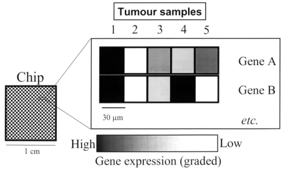
Figure 1. The principle of cDNA microarray gene expression analysis in tumours. Schematic representation of a DNA microchip. The chip (left panel)
consists of a siliconised or glass surface (an area of about 1 cm2) where up to 10 cDNAs or 250 oligonucleotide gene sequences are plotted in an orderly fashion. In the two-colour hybridisation scheme usually employed, labelled RNA from tumour samples 1–4 is hybridised to the chip simultaneously and in direct competition with labelled RNA from defined control samples. Different fluorescent dyes are used for sample and control RNA (shown here in black and white). The relative difference in gene expression between tumour and normal cells can be quantified through image analysis of the chip, and assessed as relative amounts of the two different fluorochrome signals arising from each defined quadrant of the chip (scale shown below the main panel). Tumours 1 and 2 show identical expression patterns of genes A and B, and may thus be grouped together by virtue of a clustered gene expression profile. In contrast, tumours 3–5 carry distinct molecular signatures with respect to the expression of these two genes.
for hybridisation on chips to study large-scale gene expression profiles. The use of RNA implies that freshly frozen intact tumour tissue must be used, but methods permitting the study of fixed tissue (where RNA is usually much degraded) are under active investigation. Many protocols foresee a com-parative and competitive hybridisation of tumour RNA samples on the chip against normal or reference RNA labelled with different colours (Figure 1). As with more traditional molecular diagnostic methods, the enrichment of tumour cells is important. This can be achieved by ‘virtual dissection’ of chip data in a computer where gene groups may be clustered and filtered out which are known to be derived from normal cells or from inflammatory infiltrates in a tumour. Laser-capture microdissection to isolate tumour cells mechanically may, however, still be necessary, although more laborious [20].
The ‘Lymphochip’ is a cDNA microarray collecting genes preferentially expressed in lymphoid cells [21]. Lymphoid malignancies studied with this chip exhibit an orderly picture of gene expression patterns, reflecting both B- or T-cell lineage characteristics, stage of maturation of lymphoid cells and proliferation signatures. Diffuse large B-cell lymphoma (a clinically heterogeneous group of lymphomas despite morphological similarity) can be split into subtypes exhibiting gene expression profiles either typical of germinal centre B-cells, or activated B-cells, perhaps with implications for prognosis.
On histology, one invasive-ductal breast cancer specimen may look deceptively similar to another one. Clinically the cases may be totally different, due to inherent biological dif-ferences elusive to morphological analysis. Gene expression patterns in human breast cancer specimens display distinct molecular portraits [22, 23]. Tumours may be clustered into subgroups by gene expression patterns, possibly representing distinct subtypes or entities of breast cancer.
In acute lymphoblastic and myeloid leukaemias [acute lymphoblastic leukaemia (ALL) and acute myelogenous leukaemia (AML), respectively] microchip analysis can define groups of genes, which neatly distinguish AML from ALL; their expression being high in one type of leukaemia and
low in the other [24]. Interestingly, genes most useful for AML versus ALL class prediction are not necessarily markers of haematopoietic lineage. Non-lineage restricted genes encoding cell cycle proteins, cell adhesion molecules and the like may also help to sort out AML from ALL.
Astrocytomas are heterogeneous glial neoplasms ranging from indolent astrocytomas to highly aggressive malignant glioblastoma multiforme. Microarrays may identify molecu-lar signatures distinguishing between these two tumour types which may be relevant for diagnosis and therapy [25].
Classification of human tumours according to their original anatomical site, i.e. their primary tumour, is important in the management of patients. Chips may identify subsets of genes whose expression is characteristic for each cancer class, e.g. breast cancer versus colorectal cancer versus bladder cancer, etc. [26]. Predictor genes include genes typically expressed in specific epithelial differentiation processes of the correspond-ing normal tissues. Thus microchips can be used to predict the tissue of origin of a carcinoma in the context of multiple cancer classes. This might be particularly helpful for further classification of metastatic cancer with unknown primary site. A very important potential of the chip technology will be the definition of new molecular factors predicting treatment suc-cess or failure. Targeting of therapy may improve treatment results, cut down on side effects and reduce costs potentially spent on a priori useless drugs. In breast cancer expression of estrogen receptors predicts response to hormone treatment. A high score of HER2/c-erbB2 expression is mandatory for the therapeutic success of monoclonal antibodies against this oncoprotein [27, 28]. CML hit the headlines when a new drug, STI571 or GlivecR, interfering specifically with the BCR-ABL
tyrosine kinase activity in this leukaemia was shown to be clinically active [29]. DNA microchips may identify genes or gene expression clusters in tumours, which may predict sens-itivity or resistance to particular drugs [30]. For example, cDNA microarray analyses of oesophageal tumours were able to predict the outcome of adjuvant chemotherapy, since par-ticular gene expression clusters (rather than expression of single genes) were correlated with sensitivity or resistance to cisplatin or 5-fluorouracil [31]. In brief, specific genetic
Table 1. Applications of DNA microarrays or ‘chips’ in oncology
• Global understanding of abnormal gene expression contributing to malignancy, i.e. snapshots of genes either up- or down-regulated in tumours
• Molecular classification of neoplasms by gene expression signatures, predicting the tissue of origin of a tumour in the context of multiple cancer classes
• Identification of novel molecular-based subclasses of tumours with clinical relevance • Discovery of new prognostic or predictive indicators and biomarkers of therapeutic response • Identification and validation of new molecular targets for drug development
• Prediction of drug side effects during preclinical development and toxicology studies • Identification of genes conferring drug resistance
• Prediction or selection of patients most likely to benefit from, or suffer from particular side effects of drugs (pharmacogenomics)
aberrations or gene expression profiles may become important in determining the appropriate choice of cancer treatment. The human genome sequence databank hosts all of these genes, many of which still await their discovery, characterisation and their being put to clinical use.
Problems and outlook
Molecular diagnostics with DNA microarrays do not displace time-honoured diagnostic tools such as morphology. The demands on bio-informatics to handle the impressive data flow generated by microchips are considerable, and costs still excessive. The DNA chip of microarray technology shares the problem of standardisation with other molecular diagnostic techniques. Considerable efforts still need to be invested in order to build up a system similar to the quality control networks established, for example, to monitor the quality of coagulation laboratory tests. The clinical relevance of the chip data in cancer specimens will have to be tested in appropriate clinical trials. To sort out clustered gene expression patterns, to define subgroups or ‘new’ entities of common cancers, to test ‘new’ cancer genes for their prognostic or predictive value, or to study the therapeutic value of new custom-designed drugs provides a formidable task, which will require new strategies from the clinical side.
The screening of tumour gene expression profiles with DNA microchips is a powerful strategy to discover new candid-ate genes with distinct roles in molecular cancer pathology. It possibly avoids the type of cumbersome mapping and cloning experiments hitherto required to define ‘new’ genes. How-ever, the fact that a gene cluster is over-expressed, or silent in a cancer cell, raises hypotheses on the role of the genes in carcinogenesis, but gives no final clues. Global gene expres-sion profiling of cancers with the DNA chip technology is now a reality, but the detailed characterisation of single genes elucidating their possible role in carcinogenesis is far from being outdated. Whilst DNA represents genomic hardware, and RNA a sort of genetic ‘interim’ software, the final pro-duct, protein, ultimately matters in the life of a cell. In the aftermath of the Human Genome Project, new strategies to characterise and detect human proteins in biological material (including clinical specimens) are now mandatory and indeed on the horizon. The new technology of proteomics is on track to provide a new wave of fascinating data with a great poten-tial for cancer medicine [32, 33].
Acknowledgements
The author’s experimental research is generously supported by the Swiss National Foundation, the Swiss and the Bernese Cancer League, the Marlies-Schwegler Foundation, the Hecht-Foundation and the Bernese Hecht-Foundation for Clinical Cancer Research.
References
1. Lindor NM, Greene MH and the Mayo Familial Cancer Program. The concise handbook of family cancer syndromes. J Natl Cancer Inst 1998; 90: 1039–1071.
2. Fey MF. Molecular biology of cancer. In Cavalli F, Hansen HH, Kaye SB (eds): Textbook of Medical Oncology, 2nd edition. London: Martin Dunitz 2000; 1–66.
3. Vogelstein B, Fearon ER, Hamilton SR et al. Genetic alterations during colorectal-tumor development. N Engl J Med 1988; 319: 525–532.
4. Wainscoat JS, Fey MF. Assessment of clonality in human tumors: a review. Cancer Res 1990; 50: 1355–1360.
5. International Human Genome Sequencing Consortium. Initial sequencing and analysis of the human genome. Nature 2001; 409: 860–921.
6. Venter JC, Adams MD, Myers EW et al. The sequence of the human genome. Science 2001; 291: 1304–1352.
7. Sidransky D. Nucleic acid-based methods for the detection of cancer. Science 1997; 278: 1054–1059.
8. Fey MF, Pilkington SP, Summers C, Wainscoat JS. Molecular diagnosis of haematological disorders using DNA from stored bone marrow slides. Br J Haematol 1987; 67: 489–492.
9. Steiner G, Schoenberg MP, Linn JF et al. Detection of bladder cancer recurrence by microsatellite analysis of urine. Nature Med 1997; 3: 621–624.
10. Dong SM, Traverso G, Johnson C et al. Detecting colorectal cancer in stool with the use of multiple genetic targets. J Natl Cancer Inst 2001; 93: 858–865.
11. Liloglou T, Maloney P, Xinarianos G et al. Cancer-specific genomic instability in bronchial lavage: a molecular tool for lung cancer detection. Cancer Res 2001; 61: 1624–1628.
12. Kononen J, Bubendorf L, Kallioniemi A et al. Tissue microarrays for high-throughput molecular profiling of tumor specimens. Nature Med 1998; 4: 844–847.
13. Bubendorf L, Kononen J, Koivisto P et al. Survey of gene ampli-fications during prostate cancer progression by high-throughput fluorescence in situ hybridisation on tissue microarrays. Cancer Res 1999; 59: 803–806.
14. Emmert-Buck MR, Bonner RF, Smith PD et al. Laser capture micro-dissection. Science 1996; 274: 998–1001.
15. Rubin MA. Use of laser capture microdissection, cDNA microarrays, and tissue microarrays in advancing our understanding of prostate cancer. J Pathol 2001; 195: 80–86.
16. Hasse U, Tinguely M, Oppliger Leibundgut E et al. Clonal loss of heterozygosity in Hodgkin/Reed–Sternberg cells. J Natl Cancer Inst 1999; 91: 1581–1583.
17. Liotta L, Petrocoin E. Molecular profiling of human cancer. Nature Rev 2000; 1: 48–56.
18. Ramsay G. DNA chips: state-of-the-art. Nature Biotech 1998; 16: 40–44.
19. Aitman TJ. DNA microarrays in medical practice. Br Med J 2001; 323: 611–615.
20. Alizadeh AA, Ross DT, Perou CM, van de Rijn M. Towards a novel classification of human malignancies based on gene expression patterns. J Pathol 2001; 195: 41–52.
21. Alizadeh AA, Eisen MB, Davis RE et al. Distinct types of diffuse large B-cell lymphoma identified by gene expression profiling. Nature 2000; 403: 503–511.
22. Perou CM, Sorlie T, Eisen MB et al. Molecular portraits of human breast tumours. Nature 2000; 406: 747–752.
23. West M, Blanchette C, Dresman H et al. Predicting the clinical status of human breast cancer by using gene expression profiles. Proc Natl Acad Sci USA 2001; 98: 11462–11467.
24. Golub TR, Slonim DK, Tamayo P et al. Molecular classification of cancer: class discovery and class prediction by gene expression monitoring. Science 1999; 286: 531–537.
25. Rickman DS, Bobek MP, Misek DE et al. Distinctive molecular pro-files of high-grade and low-grade gliomas based on oligonucleotide microarray analysis. Cancer Res 2001; 61: 6885–6891.
26. Su AI, Welsh JB, Sapinoso LM et al. Molecular classification of human carcinomas by use of gene expression signatures. Cancer Res 2001; 61: 7388–7393.
27. Slamon DJ, Leyland-Jones B, Shak S et al. Use of chemotherapy plus a monoclonal antibody against HER2 for metastatic breast cancer that overexpresses HER2. N Engl J Med 2001; 344: 783–792.
28. Thor AD, Berry DA, Budman DR et al. erbB2, p53, and efficacy of adjuvant therapy in lymph node positive breast cancer. J Natl Cancer Inst 1998; 90: 1346–1360.
29. Druker BJ, Talpaz M, Resta D et al. Efficacy and safety of a specific inhibitor of the BCR-ABL tyrosine kinase in chronic myeloid leukemia. N Engl J Med 2001; 344: 1031–1037.
30. Scherf U, Ross DT, Waltham M et al. A gene expression database for the molecular pharmacology of cancer. Nature Genet 2000; 24: 236–244.
31. Kihara C, Tsunoda T, Tanaka T et al. Prediction of sensitivity of esophageal tumours to adjuvant chemotherapy by cDNA microarray analysis of gene-expression profiles. Cancer Res 2001; 61: 6474– 6479.
32. Dua K, Williams TM, Beretta L. Translational control of the proteome: relevance to cancer. Proteomics 2001; 1: 1191–1199. 33. Martin DB, Nelson PS. From genomics to proteomics: techniques
and applications in cancer research. Trends Cell Biol 2001; 11: S60–S65.