Ischemic postconditioning protects remodeled myocardium via the
PI3K
–PKB/Akt reperfusion injury salvage kinase pathway
Min Zhu
a,1, Jianhua Feng
a,1, Eliana Lucchinetti
a, Gregor Fischer
b, Lin Xu
a,
Thierry Pedrazzini
c, Marcus C. Schaub
d, Michael Zaugg
a,⁎
aInstitute of Anesthesiology, University Hospital Zurich, and Zurich Center of Integrative Human Physiology (ZIHP),
University of Zurich, Zurich, Switzerland
bInstitute for Laboratory Animals, University of Zurich, Zurich, Switzerland cDivision of Hypertension, University of Lausanne Medical School, Lausanne, Switzerland
d
Institute of Pharmacology and Toxicology, University of Zurich, Zurich, Switzerland Received 23 April 2006; received in revised form 27 June 2006; accepted 28 June 2006
Available online 6 July 2006 Time for primary review 23 days
Abstract
Objective: We tested whether ischemic postconditioning (IPostC) is protective in remodeled myocardium.
Methods: Post-myocardial infarct (MI)-remodeled hearts after permanent coronary artery ligation and one kidney one clip (1K1C) hypertensive hearts of male Wistar rats were exposed to 40 min of ischemia followed by 90 min of reperfusion. IPostC was induced by six cycles of 10 s reperfusion interspersed by 10 s of no-flow ischemia. Activation of reperfusion injury salvage kinases was measured using Western blotting and in vitro kinase activity assays.
Results: IPostC prevented myocardial damage in both MI-remodeled and 1K1C hearts, as measured by decreased infarct size and lactate dehydrogenase release, and improved function. The reduction in infarct size and the recovery of left ventricular contractility achieved by IPostC was less in 1K1C hearts, but was unchanged in MI-remodeled hearts when compared to healthy hearts. In contrast, the recovery of inotropy was unaffected in 1K1C hearts, but was less in MI-remodeled hearts. Inhibition of the phosphatidylinositol 3-kinase (PI3K) pathway with LY294002 abolished the protective effects of IPostC on both disease models and healthy hearts. Western blot analysis in conjunction with in vitro kinase activity assays identified protein kinase B (PKB)/Akt but not p42/p44 extracellular-signal regulated kinase 1/2 (ERK1/2) as the predominant kinase in IPostC-mediated cardioprotection in remodeled hearts. IPostC increased phosphorylation of the PKB/Akt downstream targets eNOS, GSK3β, and p70S6K in remodeled hearts.
Conclusion: Our results offer evidence that IPostC mediates cardioprotection in the remodeled rat myocardium primarily via activation of the PI3K–PKB/Akt reperfusion injury salvage kinase pathway.
© 2006 European Society of Cardiology. Published by Elsevier B.V. All rights reserved.
Keywords: Cardiac remodeling; Ischemia–reperfusion injury; Postconditioning; Reperfusion injury salvage kinase; Cellular signaling
1. Introduction
Despite the powerful protective effects of precondition-ing, the clinical application of this phenomenon has been rather disappointing, mainly because preconditioning must be instituted before the ischemic event [1]. In contrast, a more promising approach to cardioprotection termed “ischemic postconditioning” (IPostC) has been described by Vinten–Johansen's group[2]. It consists of several cycles of coronary occlusion/reperfusion started immediately at the
⁎ Corresponding author. Cardiovascular Anesthesia Research Laboratory, Center of Clinical Research, Institute of Anesthesiology, University Hospital Zurich, Rämistrasse 100, CH-8091 Zurich, Switzerland. Tel.: +41 44 255 46 00; fax: +41 44 255 44 09.
E-mail address:[email protected](M. Zaugg).
1
Both authors equally contributed to this work.
0008-6363/$ - see front matter © 2006 European Society of Cardiology. Published by Elsevier B.V. All rights reserved. doi:10.1016/j.cardiores.2006.06.027
onset of restoration of the coronary flow after prolonged ischemia. Unlike preconditioning, postconditioning theoret-ically allows unrestricted application in the clinical settings, and thus has attracted much attention over the past years. Importantly, some pharmacological agents can afford comparable protection when applied during early reperfu-sion (“pharmacological postconditioning”)[3–5].
Recent studies revealed that IPostC exerts its protective effects through the recruitment of prosurvival kinases such as phosphatidylinositol 3-kinase (PI3K)–protein kinase B (PKB)/Akt and the p42/p44 extracellular signal-regulated kinase 1/2 (ERK1/2) pathways (also termed reperfusion injury salvage kinase or RISK pathway) at the time of reperfusion[6]. So far, nearly all experimental studies have evaluated IPostC in healthy juvenile hearts. However, this is far from clinical reality, where a high number of elderly patients with cardiovascular disease would benefit most from protection by IPostC. Hypertensive left ventricular trophy and post-myocardial infarct (MI)-remodeled hyper-trophy account for most of the clinically relevant cases of cardiac hypertrophy and remodeling. These hypertrophied and remodeled hearts, even during the compensated state, are at greater risk to suffer severe ischemic damage and may be less amenable to protection by postconditioning [7]. We therefore tested whether protection by IPostC would be diminished or lost in two highly controlled experimental models of markedly remodeled hypertrophic hearts.
The data provided in this study show for the first time that cardioprotection by IPostC is preserved in the remodeled myocardium after permanent coronary artery ligation and in one kidney one clip (1K1C) hypertensive rat hearts. Using in vitro kinase assays to directly measure PKB/Akt and ERK1/ 2 activities, we further identified PI3K–PKB/Akt as the predominant RISK pathway for IPostC-induced protection in both disease models.
2. Methods
This study was conducted in accordance with the guidelines of the Animal Care and Use Committee of the University of Zurich, Zurich, Switzerland. All experimental procedures conformed to the Guide for the Care and Use of Laboratory Animals published by the US National Institutes of Health (NIH publication 85-23, revised 1996).
2.1. Cardiac hypertrophy models
Myocardial infarction (∼35% of left ventricular mass) and subsequent hypertrophic remodeling was induced in male adult (180–200 g, 8–9 weeks old) Wistar rats by permanent ligation of the left anterior descending coronary artery (LAD) under anesthesia, as previously described in detail [8,9]. Sham-operated animals underwent the same procedure except that the suture was passed under the coronary artery without ligation. To induce hypertensive cardiac hypertrophy, rats were subjected to right
nephrecto-my and a silver clip (0.2 mm passage) was placed around the left renal artery under anesthesia (1K1C)[10]. Control sham rats (1K) had right nephrectomy without application of a clip. Rats of both disease models were sacrificed 6 weeks after surgery, and the body weight and heart weight were measured. Systolic blood pressure was measured in awake rats by means of the tail-cuff method.
2.2. Histology
Left ventricular tissue samples were fixed with 3.6% formaldehyde and embedded in paraffin. 3-μm sections at mid-ventricular level were subjected to Gomori's silver staining and nuclear fast red for visualization of individual myocytes in the viable left ventricular wall. The stained sections were analyzed by microscopy using image analysis (Zeiss KS 400, Germany).
2.3. Experimental protocol of isolated perfused rat hearts in the Langendorff apparatus
Six weeks after surgery, rats were heparinized (500 units i.p.) and 15 min later decapitated. Hearts were removed and perfused in a non-circulating Langendorff apparatus with Krebs–Henseleit buffer gassed with 95% O2and 5% CO2at
pH 7.4 and 37 °C [4]. Experimental conditions including coronary flow and temperature were carefully monitored throughout the protocols. After equilibration, perfusion pressure was set at 80 mm Hg and left ventricular end-diastolic pressure (LVEDP) at 10 mm Hg. Spontaneously beating hearts were exposed to 40 min of global no-flow ischemia (test ischemia) followed by 90 min of reperfusion (Fig. 1). IPostC was induced by six cycles of 10 s reperfusion interspersed by 10 s no-flow ischemia immediately after test ischemia. Five hearts were assigned to each of the following six groups: (1) CTL, time-matched perfusion without ischemia; (2) ISCH, test ischemia followed by 90 min reperfusion; (3) ISCH/LY, application of the PI3K inhibitor LY294002 (15μmol/l) during the first 15 min of reperfusion; (4) IPostC, immediately applied after prolonged test ischemia; (5) IPostC/LY, application of LY for 15 min starting immediately at reperfusion in postconditioned hearts; and (6) vehicle, application of 0.02% dimethyl sulfoxide (DMSO) vehicle without LY294002 during 15 min of reperfusion. The following parameters were recorded: left ventricular developed pressure (LVDP) and derivatives (± dp/dt), left ventricular end-diastolic pressure (LVEDP), epicardial ECG, coronary flow (CF), and perfusion pressure. Infarct size was determined by 1% 2,3,5-triphenyltetrazo-lium chloride staining after 90 min of reperfusion [4]. In addition, myocardial damage was estimated by measuring the release of lactate dehydrogenase (LDH) from necrotic tissue, as previously described[8]. Briefly, the perfusate was collected and LDH activity was determined by the Roche/ Hitachi 917 kit (sensitivity 6 U/l, intra- and interassay coefficients of varianceb1%). For Western blot analysis and
in vitro kinase assays, separate experiments (n = 4 in each group) were carried out and terminated after 15 min of reperfusion.
2.4. mRNA extraction and real-time quantitative polymerase chain reaction
Total RNA was prepared using RNeasy Mini Kit (Qiagen). First strand cDNA was synthesized from 1.0μg of total RNA using SuperScript II reverse transcriptase (Invitrogen) and
oligo-dT as primer. Real-time quantitative PCR was per-formed as previously described [8] using the following primers: atrial natriuretic peptide (ANP), 5′–ATCAC CAAGGGCTTCTTCCT–3′ (sense) and 5′–TGTTGGA
CACCGCACTGTAT–3′ (antisense); and α-skeletal actin
(α-skl-actin), 5′–CACGGCATTATCACCAACTG–3′
(sense) and 5′–CCGGAGGCATAGAGAGACAG–3′
(anti-sense).α-Tubulin was used for normalization with the sense and antisense primers 5′–CCATGCGTGAGTGTATCTCC– 3′ and 5′–GTGCCAGTGCGAACTTCATC–3′, respectively. 2.5. Western blot analysis
Western blot analysis was carried out as previously described[8]. The antibodies used were from the following sources: glycogen synthase kinase 3β (GSK3β), p70S6K, endothelial nitric oxide synthase (eNOS), ERK1/2, PKB/Akt (Ser473), GSK3β (Ser9), p70S6K (Thr389), eNOS (Ser1177), phospho-ERK1/2 (Thr202/Tyr204) (Cell Signaling Technology); monoclonal antibodies against pan-actin (Chemicon) and α-tubulin (Sigma); polyclonal antibody against ANP (Santa Cruz); polyclonal antibody against α-skl-actin was a gift from Dr. Christine Chaponnier (Department of Pathology, University of Geneva, Geneva, Switzerland); polyclonal anti-PKB/Akt antibody (Ab10) was a gift from Dr. Brian A. Hemmings (Friedrich Miescher Institute, Basel, Switzerland). 2.6. Immunoprecipitation and in vitro kinase assay
Left ventricular non-infarcted tissue (transmural) was extracted in ice-cold lysis buffer (25 mmol/l Tris–HCl, pH 7.4, 120 mmol/l NaCl, 2 mmol/l ethylenediamine tetraacetic acid, 1% Triton X-100, 0.5% Nonidet P-40, 1 mmol/ l phenylmethylsulfonyl fluoride, 1 mmol/l benzamidine, 0.1 mmol/l sodium orthovanadate, 10 mmol/l sodium fluoride, and 1 μmol/l microcystin-LR). Extracts were centrifuged (16 000×g, 30 min, 4 °C), and protein concen-trations determined using the Bradford assay. Tissue extracts (1 mg protein) were incubated at 4 °C for 2 h on a shaking
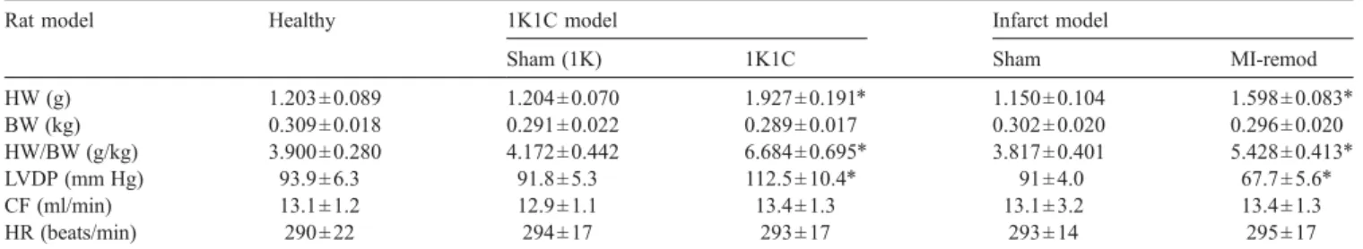
Table 1
Heart weight, body weight, and baseline cardiac function in the hypertrophic rat models
Rat model Healthy 1K1C model Infarct model
Sham (1K) 1K1C Sham MI-remod
HW (g) 1.203 ± 0.089 1.204 ± 0.070 1.927 ± 0.191⁎ 1.150 ± 0.104 1.598 ± 0.083⁎ BW (kg) 0.309 ± 0.018 0.291 ± 0.022 0.289 ± 0.017 0.302 ± 0.020 0.296 ± 0.020 HW/BW (g/kg) 3.900 ± 0.280 4.172 ± 0.442 6.684 ± 0.695⁎ 3.817 ± 0.401 5.428 ± 0.413⁎ LVDP (mm Hg) 93.9 ± 6.3 91.8 ± 5.3 112.5 ± 10.4⁎ 91 ± 4.0 67.7 ± 5.6⁎ CF (ml/min) 13.1 ± 1.2 12.9 ± 1.1 13.4 ± 1.3 13.1 ± 3.2 13.4 ± 1.3 HR (beats/min) 290 ± 22 294 ± 17 293 ± 17 293 ± 14 295 ± 17 HW: heart weight; BW: body weight; LVDP: left ventricular developed pressure; CF: coronary flow; HR: heart rate; healthy: age-matched rats (300 g) serving as healthy controls. 1K1C: one kidney one clip rat model; sham (1K), nephrectomy without applying a clip (one kidney); MI-remod: postinfarct-remodeled rat hearts (permanent coronary artery ligation); sham: sham-operated rats (a suture was passed under the coronary artery without ligation). Rats in the hypertrophic model groups were used 6 weeks after surgery. Data are mean ± S.D. (n = 20 for sham (1K) and sham, n = 30 for healthy, 1K1C, and MI-remod).
* pb0.001 vs. sham and healthy groups.
Fig. 1. Treatment protocols. ISCH: hearts exposed to 40 min of test ischemia followed by 90 min reperfusion. IPostC: ischemic postconditioning was induced by 6 cycles of 10 s reperfusion followed by 10 s no-flow ischemia right at the onset of reperfusion after 40 min of test ischemia. IPostC/LY: postconditioned hearts exposed to the PI3K inhibitor LY294002 (15μmol/l) dissolved in vehicle (0.02% dimethyl sulfoxide (DMSO)) during the first 15 min of reperfusion. CTL: time-matched perfusion. Infarct size was determined by 1% triphenyltetrazolium staining at the end of experiments (n = 5 hearts per experimental group). In separate experiments (n = 4 hearts per experimental group), phosphorylation (by Western blotting) and activity of PKB/Akt and extracellular-signal regulated kinase 1/2 (ERK1/2) were assayed after 15 min of reperfusion.
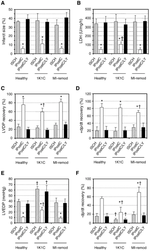
Fig. 2. Infarct size (panel A), lactate dehydrogenase release (panel B) and contractility parameters (panels C–F) of healthy, 1K1C, and MI-remodeled hearts. Left ventricular developed pressure (LVDP) (panel C), + dp/dt (panel D), left ventricular end-diastolic pressure (LVEDP) (panel E), and−dp/dt (panel F) were determined at 90 min of reperfusion and are indicated as percentage of baseline or mm Hg (LVEDP). ISCH: hearts exposed to 40 min of test ischemia followed by 90 min reperfusion. IPostC: ischemic postconditioning. IPostC/LY: postconditioned hearts exposed to the PI3K inhibitor LY294002 (15μmol/l) during first 15 min of reperfusion. CTL: time-matched perfusion. Data are mean ± S.D. (n = 5 per group). *pb0.05 vs. ISCH and†pb0.05 vs. same treatment in healthy
platform with 2 μg of either polyclonal anti-PKB (Ab10) antibody coupled to 10μl protein A-sepharose (Amersham Biosciences) or monoclonal anti-ERK1/2 (Zymed, clone ERK-7D8) coupled to 10μl protein G-sepharose (Amersham Biosciences). The immune-complexes were then washed 4 times with lysis buffer, once with kinase buffer (25 mmol/ l Tris–HCl, pH 7.4, 10 mmol/l MgCl2, 1 mmol/l
dithiothrei-tol, 1μmol/l PKI, and 1 μmol/l microcystin-LR), and finally resuspended in 20μl of kinase buffer. For PKB kinase assay,
the reaction was started with 60 μmol/l of Crosstide
(GRPRTSSFAEG) and 20 μmol/l of [γ-32P] ATP
(Amer-sham Biosciences) in a final volume of 30μl, incubated at
30 °C for 60 min, and stopped by addition of trichloroacetic acid (TCA) to a final concentration of 10%. After brief centrifugation, 20μl aliquots were removed and spotted onto P81 phosphocellulose paper. The paper was washed immediately for 10 min with 75 mmol/l phosphoric acid (4 times), dried, and 32P incorporation determined by Cerenkov counting. For ERK1/2 kinase assay, the reaction was started with 4μg of myelin basic protein and 20 μmol/ l of [γ-32
P] ATP in a final volume of 30μl and incubated at 30 °C for 30 min. The reaction was terminated by addition of SDS-sample buffer and boiled at 95 °C for 5 min. Aliquots were subjected to SDS–PAGE (15%) followed by
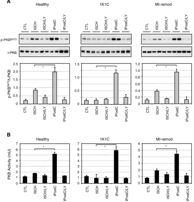
Fig. 3. Phosphorylation status (panel A) and kinase activity (panel B) of protein kinase B/Akt (PKB). Representative Western blots and average density ratio of p-PKBS473/total-PKB for each group. Data are mean ± S.D. (n = 4 per group). *pb0.05 vs. ISCH.
autoradiography or32P determination in excised gel slices. One unit of activity was defined as the amount of enzyme that transferred 1 pmol phosphate/min to the substrate at 30 °C.
2.7. Statistics
Data are presented as mean ± S.D. For the cardiac functional data, repeated-measures analysis of variance was
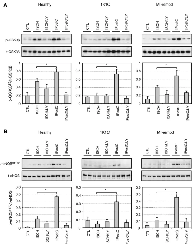
Fig. 4. Phosphorylation status of glycogen synthase kinase3β (GSK3β) (panel A). Representative Western blots and average density ratio of p-GSK3βS9/
total-GSK3β. Phosphorylation status of endothelial nitric oxide synthase (eNOS) (panel B). Representative Western blots and average density ratio of p-eNOSS1177/
used to evaluate differences over time between groups. Unpaired t-test was used to compare groups at identical time points, and paired t-test to compare within groups over time. p values were multiplied by the number of comparisons (Bonferroni correction). Tukey's post-hoc test was applied for multiple comparisons of the one-way analysis of variance for all other data. pb0.05 was considered as significant. SigmaStat (version 2.0; SPSS Science, Chicago, IL) was used for the analyses.
3. Results
3.1. Characterization of remodeling and hypertrophy in diseased hearts
Heart- (HW) and body- (BW) weight as well as their ratio (HW/BW) are summarized in Table 1 for the two disease models. HW and HW/BW were significantly higher in the 1K1C and the MI-remodeled (permanent coronary artery ligation) groups, as compared to the healthy and sham groups indicating cardiac hypertrophy in both disease models.Table 1also shows LVDP, CF, and heart rate (HR) ex vivo at equilibration on the Langendorff apparatus. LVDP was significantly higher in the 1K1C and lower in the MI-remodeled groups than in healthy or sham controls, while CF and HR were unchanged. The higher LVDP observed at the Langendorff apparatus corresponds to the significantly (pb0.001) higher systolic blood pressure measured in vivo in 1K1C (186 ± 18 mm Hg, n = 30)
compared to sham (1K) (123 ± 10 mm Hg, n = 20) and healthy age-matched animals (120 ± 9 mm Hg, n = 10). The post-infarct remodeling in MI-remodeled hearts has been characterized in detail in our previous study[8]. The 1K1C hearts displayed an increase in mRNA of about 10-fold for ANP and 4-fold forα-skl-actin, which was accompanied by an increase in protein levels as well (Supplementary Fig. S1A and B). Micrographs of cross-sections of the left ventricular free wall at the mid-ventricular level of 1K1C and MI-remodeled hearts displayed enlarged myocytes in comparison to sham controls (Supplementary Fig. S1C). 3.2. Cardioprotection by ischemic postconditioning is preserved in remodeled myocardium
IPostC significantly improved functional recovery and decreased infarct size in both the 1K1C as well as the MI-remodeled hearts, when compared to unprotected MI-remodeled hearts (Fig. 2A–F and Supplementary Tables 1–3). IPostC achieved over 80% reduction in infarct size and LDH release in MI-remodeled and healthy hearts, while in 1K1C hearts the reduction of both parameters was somewhat lower (69%) (Fig. 2A and B). With regard to hemodynamics, the recovery of LVDP was lower in 1K1C hearts as compared to MI-remodeled and healthy controls after application of IPostC (Fig. 2C). On the other hand, the rate of pressure rise (+ dp/dt, inotropy) was fully preserved in 1K1C, while it was lower by 38% in MI-remodeled hearts (Fig. 2D). The baseline values of LVDP and inotropy were significantly lower in MI-remodeled
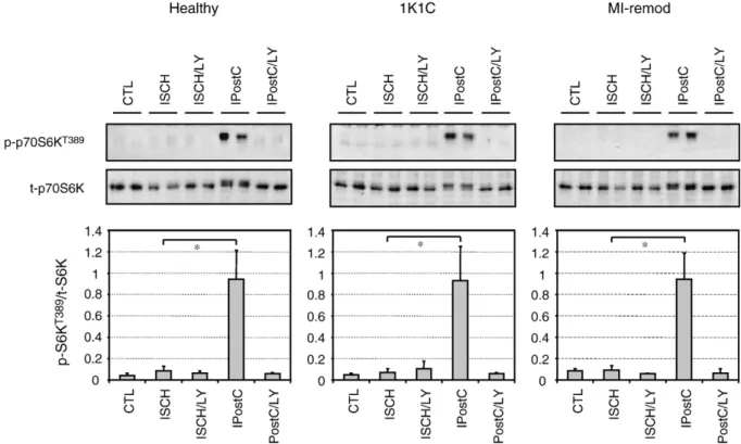
Fig. 5. Phosphorylation status of ribosomal S6 kinase (p70S6K). Representative Western blots and average density ratio of p-p70S6KT389/total-p70S6K. Data are
hearts, indicating impaired basal cardiac function (Supple-mentary Table 3). During reperfusion, LVEDP was increased and lusitropy was decreased in 1K1C hearts compared to healthy hearts, indicating impaired postischemic diastolic function in 1K1C hearts. The protection by IPostC was completely abolished by co-administration of the PI3K inhibitor LY294002 (Fig. 2A–F, and Supplementary Tables 1–3). LY294002 or the solvent dimethyl sulfoxide alone administered during reperfusion did not affect functional recovery or infarct size (data not shown). These results provide strong evidence that PI3K-dependent protection by IPostC is preserved and operative in both disease models.
3.3. Ischemic postconditioning in remodeled myocardium activates PKB/Akt and its downstream targets GSK3β, eNOS, and p70S6K in a PI3K-dependent manner
Activation of the pro-survival PI3K–PKB/Akt signaling pathway has been shown to be important for cardioprotection in several healthy animal models[4,5,11–14]. Here we show that IPostC significantly increased phosphorylation of PKB/ Akt (Fig. 3A) and its downstream targets GSK3β, eNOS,
and p70S6K in remodeled hearts (Figs. 4 and 5). IPostC-induced phosphorylation of PKB/Akt and its downstream substrates GSK3β, eNOS, and p70S6K was suppressed by
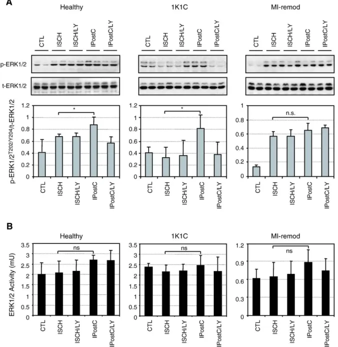
Fig. 6. Phosphorylation status (panel A) and kinase activity (panel B) of extracellular-signal regulated kinase 1/2 (ERK1/2). Representative Western blots and averaged density ratio of p-ERK1/2T202/Y204/total-ERK1/2 for each group. Data are mean ± S.D. (n = 4 per group). *pb0.05 vs. ISCH.
LY294002. Dimethyl sulfoxide alone had no effect on the phosphorylation of PKB/Akt when compared to the ischemia group (data not shown). To further characterize the activation status of PKB/Akt, an in vitro kinase assay was carried out. Consistent with the results from Western blots, the activity of PKB/Akt was significantly elevated by IPostC compared to the ischemic control and completely abolished by LY294002 (Fig. 3B). Phosphorylation and enzyme activity of PKB/Akt were partially increased by ischemia alone compared to the time-matched controls in both healthy and MI-remodeled hearts. However, this was not observed in the 1K1C hearts (Fig. 3A and B). Taken together, the data underscore the unique role of PKB/Akt signaling in the IPostC-mediated protection of the remodeled myocardium.
3.4. Cardioprotection by ischemic postconditioning in the remodeled myocardium does not primarily depend on ERK1/2 signaling
Beside PI3K–PKB/Akt, ERK1/2 signaling presents
another component in the RISK pathways that has also been suggested to contribute to the reduction of infarct size with postconditioning[13]. We therefore examined whether the protection by IPostC would be associated with activation of the ERK1/2 pathway in the remodeled hearts. In healthy hearts, phosphorylation of ERK1/2 was moderately increased (p = 0.033) by ischemia alone, and was further enhanced by IPostC (Fig. 6A). In 1K1C hearts, phosphorylation of ERK1/2 was not increased by ischemia alone but was increased by IPostC (Fig. 6A). The elevated phosphorylation of ERK1/2 by IPostC was reversed by LY294002 suggesting a PI3K-dependent phosphorylation of ERK1/2. In contrast, phosphorylation of ERK1/2 in the MI-remodeled hearts was strongly increased by ischemia alone, but was resistant to LY294002 and not further increased by IPostC, indicating that a PI3K-independent mechanism was responsible for the ischemia-induced ERK1/2 phosphorylation in the MI-remodeled hearts. These results based on the phosphorylation status of ERK1/2 suggest that the ERK1/2 pathway is differentially regulated in the two types of remodeling. In parallel, the activities of ERK1/2 were also determined by an in vitro kinase assay. Surprisingly, there was no increase in ERK1/2 activities by IPostC in both remodeling models as well as in healthy hearts (Fig. 6B). Moreover, the activities of ERK1/2 in the MI-remodeled hearts were 3-fold lower than that in healthy and 1K1C hypertrophied hearts, suggesting that subtle differences in the regulation of RISK pathways may exist between the models. Collectively, our results based on direct enzyme activity measurements indicate that the ERK1/2 pathway is not primarily involved in IPostC-mediated protection in both healthy and diseased hearts. 4. Discussion
Here we show for the first time that protection by IPostC is preserved in two rat models of myocardial remodeling.
IPostC was previously reported to be ineffective in limiting infarct size in rabbits with hypercholesterolemia and atherosclerosis[15]. However, our results are in accordance with a most recent clinical trial by Staat and colleagues[16], who postconditioned hearts of patients undergoing percuta-neous coronary interventions with four episodes of 1-min balloon inflations starting within 1 min of reflow. Using creatine kinase release as a surrogate marker of infarct size, these authors found that myocardial damage could be reduced by 36%.
Myocardial remodeling is a short-term adaptive but long-term maladaptive process to a variety of hemodynamic conditions associated with increased cardiac work. Character-istic structural changes[17], alterations in metabolism[18], and cellular signaling[19–21]put the remodeled myocardium at particular risk for further ischemic damage. Since most individuals who experience acute ischemic heart disease have underlying myocardial remodeling, we addressed the impor-tant question whether the diseased remodeled myocardium is still receptive to protection by IPostC using two experimental models of remodeling. While 1K1C remodeled hearts develop eccentric hypertrophy by volume overload [10], MI-remo-deled hearts display a far more complex architectural re-arrangement due to (i) loss of viable tissue and scar formation (ii) volume overload by scar expansion (iii) pressure overload induced by increasing volume overload [20]. Nonetheless, hypertrophy is a hallmark of both types of remodeling. Since these structural changes are associated with alterations in neurohormonal activation and cellular signaling, we hypoth-esized that remodeling may abolish innate protective mechan-isms and render the heart more susceptible to ischemia.
Previous studies identified the survival kinases PI3K– PKB/Akt and ERK1/2 as key players in the protection afforded by IPostC in healthy myocardium[6]. However, the relative importance of the two kinases in mediating the protection remains controversial. A study investigating pharmacological postconditioning by 5′-(N-ethylcarboxa-mido) adenosine and bradykinin suggests that PKB/Akt is upstream of ERK1/2[5], while results from a more recent study stress the pivotal role of ERK1/2 but not PI3K–PKB/ Akt in IPostC-mediated protection[13]. Moreover, Schwartz et al.[14]reported that IPostC activates both PKB/Akt and ERK1/2 but yet failed to protect against ischemic injury in pigs. In the present study, we show that PKB/Akt is activated and that its downstream targets GSK3β, eNOS, and p70S6K are markedly phosphorylated by IPostC not only in the healthy but also in the remodeled hearts. This phosphoryla-tion is commensurate with funcphosphoryla-tional and structural protec-tion and is sensitive to inhibiprotec-tion by LY294002. Importantly, our results on PKB/Akt activation are not solely based on Western blot analysis using phosphor-specific antibodies, but further rely on in vitro kinase assays, which directly measure catalytic activity. Although we observed an increase in ERK1/2 phosphorylation by IPostC in healthy and 1K1C hearts, the directly measured catalytic activity of ERK1/2 was not elevated by IPostC in healthy and diseased hearts.
Obviously, the observed phosphorylation of ERK1/2 by IPostC was not sufficient to protect. Hence our data suggest that PI3K–PKB/Akt but not ERK1/2 is the predominant mediator of IPostC-induced protection in both healthy and diseased hearts. Of note, our study is the first to measure ERK1/2 activation in IPostC by assessing the phosphoryla-tion status in conjuncphosphoryla-tion with in vitro activity measurements. Earlier studies on ERK1/2 activity in pre- and postcondition-ing exclusively relied on Western blottpostcondition-ing, which may, at least in part, explain the controversial role of ERK1/2 in IPostC. Nonetheless, our results cannot rule out the possibility of some cross talk between ERK1/2 and PKB/Akt, and the two pathways may also follow different spatio-temporal routes at the onset of reperfusion in the heart. Therefore, additional time-course and blocker experiments will be required to ultimately delineate the role of PKB/Akt and ERK1/2 in IPostC-mediated protection in remodeled myocardium.
We found noteworthy alterations in PKB and ERK1/2 signaling in the different disease models. First, although the maximal activation of PKB/Akt can be achieved by IPostC in all three models, partial activation of PKB/Akt by ischemia alone was lost in 1K1C hearts. This observation may explain the observed reduced protection by IPostC in 1K1C rats. Second, the profiles of ERK1/2 phosphorylation in both types of remodeling and the basal kinase activity of ERK1/2 in MI-remodeled hearts were different from those observed in healthy hearts. These alterations suggest that key players of the RISK pathway may be differentially regulated by different types of cardiac remodeling. Finally, we found that the recovery of inotropy achieved by IPostC was im-paired in MI-remodeled hearts, while both recovery of LVDP and infarct size reduction were unaffected. The reason for these differences is unclear. A recent study using a transgenic mouse model of hypertrophy with cardiac-specific expres-sion of myristoylated PKB/Akt demonstrated that activation of PKB/Akt alone is not sufficient for protection[22]. This implies that additional PI3K-dependent but PKB/Akt-independent signaling components may be required for full cardioprotection. Thus, it is possible that such alternative mechanisms are impaired in MI-remodeled hearts.
Ischemic preconditioning has been shown to be ineffec-tive in MI-remodeled rabbit hearts [23], but proved its efficacy in several hypertrophy models [24–27]. Despite these partly promising results, the concept of precondition-ing could not be successfully established in the clinical setting. Therefore, recent interventions aimed at modifying reperfusion, but IPostC may further jeopardize the diseased heart. This could be avoided by utilizing pharmacological agents mimicking the biological process of IPostC. Hence, the understanding of the molecular mechanisms underlying IPostC is of paramount importance and should be exploited in additional studies. Importantly, future studies should particularly investigate the effects of the disease-related alterations in signaling on cardioprotection.
In summary, using a highly controlled experimental setting, we show in our study that remodeled rat hearts with
hypertrophy induced by infarction (permanent coronary artery ligation) and 1K1C hypertension are still receptive to protection by IPostC. Furthermore, we identified in these models the PI3K–PKB/Akt signaling pathway as predom-inant mediator of IPostC-induced cardioprotection.
Acknowledgments
The authors thank Christine Perregaux for her expert technical assistance. This work was supported by grants from the Swiss National Science Foundation (Grant #32-00B0-103980/1 to Dr. Zaugg and Grant 32-00B0-102154/1 to Dr. Pedrazzini), the Swiss University Conference (Swiss Cardiovascular Research and Training Network), the Olga Mayenfisch Foundation, the Novartis Foundation, and the 5th Frontiers in Anesthesia Research Award from the International Anesthesia Research Society.
Appendix A. Supplementary data
Supplementary data associated with this article can be found, in the online version, atdoi:10.1016/j.cardiores.2006.06.027. References
[1] Kloner RA, Rezkalla SH. Preconditioning, postconditioning and their application to clinical cardiology. Cardiovasc Res 2006;70:297–307. [2] Zhao ZQ, Corvera JS, Halkos ME, Kerendi F, Wang NP, Guyton RA, et
al. Inhibition of myocardial injury by ischemic postconditioning during reperfusion: comparison with ischemic preconditioning. Am J Physiol Heart Circ Physiol 2003;285:H579–88.
[3] Bose AK, Mocanu MM, Carr RD, Yellon DM. Glucagon like peptide-1 is protective against myocardial ischemia/reperfusion injury when given either as a preconditioning mimetic or at reperfusion in an isolated rat heart model. Cardiovasc Drugs Ther 2005;19:9–11. [4] Feng J, Lucchinetti E, Ahuja P, Pasch T, Perriard JC, Zaugg M.
Isoflurane postconditioning prevents opening of the mitochondrial permeability transition pore through inhibition of glycogen synthase kinase 3β. Anesthesiology 2005;103:987–95.
[5] Yang XM, Krieg T, Cui L, Downey JM, Cohen MV. NECA and bradykinin at reperfusion reduce infarction in rabbit hearts by signaling through PI3K, ERK, and NO. J Mol Cell Cardiol 2004;36:411–21. [6] Hausenloy DJ, Tsang A, Yellon DM. The reperfusion injury salvage
kinase pathway: a common target for both ischemic preconditioning and postconditioning. Trends Cardiovasc Med 2005;15:69–75. [7] Ferdinandy P, Szilvassy Z, Baxter GF. Adaptation to myocardial stress
in disease states: is preconditioning a healthy heart phenomenon? Trends Pharmacol Sci 1998;19:223–9.
[8] Feng J, Fischer G, Lucchinetti E, Zhu M, Bestmann L, Jegger D, et al. Infarct-remodeled myocardium is receptive to protection by isoflurane postconditioning– role of protein-kinase-B/Akt signaling. Anesthesi-ology 2006;104:1004–14.
[9] Pfeffer MA, Pfeffer JM, Fishbein MC, Fletcher PJ, Spadaro J, Kloner RA, et al. Myocardial infarct size and ventricular function in rats. Circ Res 1979;44:503–12.
[10] Wiesel P, Mazzolai L, Nussberger J, Pedrazzini T. Two-kidney, one clip and one-kidney, one clip hypertension in mice. Hypertension 1997;29:1025–30.
[11] Yang XM, Proctor JB, Cui L, Krieg T, Downey JM, Cohen MV. Multiple, brief coronary occlusions during early reperfusion protect rabbit hearts by targeting cell signaling pathways. J Am Coll Cardiol 2004;44:1103–10.
[12] Yang XM, Philipp S, Downey JM, Cohen MV. Postconditioning's protection is not dependent on circulating blood factors or cells but involves adenosine receptors and requires PI3-kinase and guanylyl cyclase activation. Basic Res Cardiol 2005;100:57–63.
[13] Darling CE, Jiang R, Maynard M, Whittaker P, Vinten-Johansen J, Przyklenk K. Postconditioning via stuttering reperfusion limits myocardial infarct size in rabbit hearts: role of ERK1/2. Am J Physiol Heart Circ Physiol 2005;289:H1618–26.
[14] Schwartz LM, Lagranha CJ. Ischemic postconditioning during reperfusion activates Akt and ERK without protecting against lethal myocardial ischemia–reperfusion injury in pigs. Am J Physiol Heart Circ Physiol 2006;290:H1011–8.
[15] Iliodromitis EK, Zoga A, Vrettou A, Andreadou I, Paraskevaidis IA, Kaklamanis L, et al. The effectiveness of postconditioning and preconditioning on infarct size in hypercholesterolemic and normal anesthetized rabbits. Atherosclerosis 2005 [Electronic publication ahead of print Dec 22].
[16] Staat P, Rioufol G, Piot C, Cottin Y, Cung TT, L'Huillier I, et al. Postconditioning the human heart. Circulation 2005;112:2143–8. [17] Schaper J, Froede R, Hein S, Buck A, Hashizume H, Speiser B, et al.
Impairment of the myocardial ultrastructure and changes of the cytoskeleton in dilated cardiomyopathy. Circulation 1991;83:504–14. [18] Neubauer S, Horn M, Naumann A, Tian R, Hu K, Laser M, et al. Impairment of energy metabolism in intact residual myocardium of rat hearts with chronic myocardial infarction. J Clin Invest 1995;95: 1092–100.
[19] Dorn II GW, Force T. Protein kinase cascades in the regulation of cardiac hypertrophy. J Clin Invest 2005;115:527–37.
[20] Opie LH, Commerford PJ, Gersh BJ, Pfeffer MA. Controversies in ventricular remodelling. Lancet 2006;367:356–67.
[21] Miyamoto T, Takeishi Y, Takahashi H, Shishido T, Arimoto T, Tomoike H, et al. Activation of distinct signal transduction pathways in hypertrophied hearts by pressure and volume overload. Basic Res Cardiol 2004;99:328–37.
[22] Nagoshi T, Matsui T, Aoyama T, Leri A, Anversa P, Li L, et al. PI3K rescues the detrimental effects of chronic Akt activation in the heart during ischemia/reperfusion injury. J Clin Invest 2005;115:2128–38. [23] Miki T, Miura T, Tsuchida A, Nakano A, Hasegawa T, Fukuma T, et al.
Cardioprotective mechanism of ischemic preconditioning is impaired by postinfarct ventricular remodeling through angiotensin II type 1 receptor activation. Circulation 2000;102:458–63.
[24] Pantos CI, Davos CH, Carageorgiou HC, Varonos DV, Cokkinos DV. Ischaemic preconditioning protects against myocardial dysfunction caused by ischaemia in isolated hypertrophied rat hearts. Basic Res Cardiol 1996;91:444–9.
[25] Boutros A, Wang J. Ischemic preconditioning, adenosine and bethanechol protect spontaneously hypertensive isolated rat hearts. J Pharmacol Exp Ther 1995;275:1148–56.
[26] Randall MD, Gardiner SM, Bennett T. Enhanced cardiac precondition-ing in the isolated heart of the transgenic ((mREN-2) 27) hypertensive rat. Cardiovasc Res 1997;33:400–9.
[27] Speechly-Dick ME, Baxter GF, Yellon DM. Ischaemic preconditioning protects hypertrophied myocardium. Cardiovasc Res 1994;28:1025–9.