HAL Id: hal-01586174
https://hal-univ-rennes1.archives-ouvertes.fr/hal-01586174
Submitted on 28 Nov 2017HAL is a multi-disciplinary open access archive for the deposit and dissemination of sci-entific research documents, whether they are pub-lished or not. The documents may come from teaching and research institutions in France or abroad, or from public or private research centers.
L’archive ouverte pluridisciplinaire HAL, est destinée au dépôt et à la diffusion de documents scientifiques de niveau recherche, publiés ou non, émanant des établissements d’enseignement et de recherche français ou étrangers, des laboratoires publics ou privés.
Regulation of the unfolded protein response by
noncoding RNA
Mari Mcmahon, Afshin Samali, Eric Chevet
To cite this version:
Mari Mcmahon, Afshin Samali, Eric Chevet. Regulation of the unfolded protein response by noncoding RNA. American Journal of Physiology - Cell Physiology, American Physiological Society, 2017, 313 (3), pp.C243-C254. �10.1152/ajpcell.00293.2016�. �hal-01586174�
Regulation of the Unfolded Protein Response by non-coding RNA
12
Mari McMahon1,2,3, Afshin Samali3 and Eric Chevet1,2,* 3
4
1INSERM U1242 “Chemistry, Oncogenesis, Stress, Signalling”, Université de
5
Rennes 1, Rennes, France. 2Centre de Lutte Contre le Cancer Eugène Marquis, 6
Rennes, France. 3Apoptosis Research Centre, School of Natural Sciences, NUI 7
Galway, Galway, Ireland. 8 9 10 11 12 13 14
*Correspondance to EC. CLCC Eugène Marquis, Avenue de la bataille Flandres 15
Dunkerque, 35042 Rennes, France. Email: eric.chevet@inserm.fr 16
2
Abstract
18
Cells are exposed to various intrinsic and extrinsic stresses in both physiological and 19
pathological conditions. To adapt to those conditions, cells have evolved various 20
mechanisms to cope with the disturbances in protein demand, largely through the 21
Unfolded Protein Response (UPR) in the Endoplasmic Reticulum (ER), but also 22
through the Integrated Stress Response (ISR). Both responses initiate downstream 23
signaling to transcription factors that, in turn, trigger adaptive programs and/or in the 24
case of prolonged stress, cell death mechanisms. Recently, non-coding RNAs, 25
including microRNA and long non-coding RNA, have emerged as key players in the 26
stress responses. These non-coding RNAs act as both regulators and effectors of 27
the UPR, and fine-tune the output of the stress signaling pathways. Although much is 28
known about the UPR and the cross-talk that exists between pathways, the 29
contribution of small non-coding RNA has not been fully assessed. Herein we bring 30
together and review the current known functions of non-coding RNA in regulating 31
adaptive pathways in both physiological and pathophysiological conditions, 32
illustrating how they operate within the known UPR functions and contribute to 33
diverse cellular outcomes. 34
Introduction
36
As the starting point for production and modification of secretory and transmembrane 37
proteins which account for about 20-30% of all proteins produced in the cell, the 38
endoplasmic reticulum (ER), represents a fundamental part of the protein 39
homeostasis network (11). The latter integrates cellular processes that govern the 40
proteome through protein synthesis, degradation, folding, modification and 41
localization (4). Within the ER, the involvement of molecular machines ensuring 42
protein folding, quality control, export and clearance ensure the correct balance 43
between the cellular demand for secretory and transmembrane protein production 44
and the ER capacity to cope with it (36). When this balance is altered, a so-called ER 45
stress is triggered and the unfolded protein response (UPR) is activated to primarily 46
attenuate protein translation, resolve the presence of misfolded/unfolded proteins 47
and induce production of chaperone proteins (44). If the stress cannot be resolved, 48
the UPR signaling outputs will result in the activation of cell death pathways. 49
Although not entirely localized to the ER, other cellular processes such as nonsense-50
mediated decay (NMD) and the integrated stress response (ISR) also have an 51
impact on ER protein homeostasis. In recent years, numerous studies have 52
uncovered the existence of an additional layer of regulation in these process that is 53
achieved by miRNA and lncRNA. Cell stress pathways are areas of active 54
investigation and the addition of regulatory RNA pathways enhance our 55
understanding of these mechanisms. The current release of miRbase, version 21, 56
lists over 28,000 known miRNAs (55) and given that these numbers are expected to 57
increase, it raises the possibility that there are multiple undiscovered miRNAs that 58
may also regulate cellular stress pathways. This review presents the currently 59
identified non-coding RNA elements of the UPR and ISR. 60
61
The Unfolded Protein Response (UPR) and the Integrated Stress Response
62
(ISR)
63
The UPR is orchestrated by three ER transmembrane sensors; activating 64
transcription factor 6 (ATF6), protein kinase R-like ER kinase (PERK) and inositol-65
requiring enzyme 1 (IRE1). The three UPR sensors are maintained inactive through 66
the binding of GRP78/BiP to their ER luminal domains. Accumulation of misfolded 67
proteins in the ER promotes GRP78/BiP dissociation from the sensors thereby 68
leading to their activation and that of their downstream signaling (Figure 1). These 69
signaling events aim at restoring ER proteostasis through cellular reprogramming 70
4 mechanisms but if this fails, cell death will be triggered. The three UPR sensors are 71
activated through different mechanisms and their activation yields specific biological 72
outcomes. 73
ATF6 is a transmembrane transcription factor that contains a cytosolic bZIP 74
domain. The ATF6 luminal domain contains Golgi localization sequences that are 75
uncovered upon GRP78/BiP dissociation (87), along with conserved cysteine 76
residues which allow for export of ATF6 from the ER to the Golgi apparatus by COPII 77
vesicles (75). The protein disulfide isomerase 5 (PDIA5) is essential for ATF6 78
activation and transport to the Golgi through modulation of ATF6 disulfide bonds 79
(45). In the Golgi, ATF6 is cleaved by site-1 and -2 proteases on both sides of the 80
membrane thus releasing the cytosolic ATF6 fragment (ATF6f) that acts as an active 81
transcription factor (19). This transcription factor triggers the expression of ER 82
chaperone genes such as GRP78, GRP94 as well as the unspliced form of XBP1 83
and, in a heterodimer with XBP1, expression of genes associated with endoplasmic 84
reticulum-associated degradation (ERAD) (102). 85
IRE1 is a transmembrane protein with both cytosolic kinase and RNase 86
activities. Two isoforms of IRE1 are found, namely IRE1α and IRE1β, with IRE1α 87
(hereafter referred to as IRE1) ubiquitously expressed and IRE1β restricted to lung 88
and intestine epithelia (68). As with ATF6, IRE1 is under the control of the GRP78 89
chaperone. Indeed, upon GRP78/BiP dissociation, IRE1 90
homodimerizes/oligomerizes and is subjected to transautophosphorlyation. This 91
leads to a conformational change of the endoribonuclease domain promoting its 92
activation (82). In addition to GRP78/BiP, PDIA6 (protein disulfide isomerase 6) also 93
regulates IRE1 through the reduction of cysteine residues in the IRE1 luminal 94
domain (31), either stabilizing IRE1 oligomers (35) or selectively attenuating RIDD 95
activity (30). In mammals, IRE1 excises a twenty-six nucleotide fragment from XBP1 96
mRNA and the resulting 5’ and 3’ ends are ligated by RtcB ligase (64). The spliced 97
transcript, named XBP1s, encodes a transcription factor that triggers the expression 98
of genes coding for ER chaperones and ERAD associated proteins (1). IRE1 RNase 99
also downregulates other RNAs through targeted degradation, in a process known 100
as RIDD (regulated IRE1 dependent decay). RIDD targets mRNAs such as SPARC, 101
PER1 and BLOC1S1, along with individual miRNAs and miRNA families (12, 26, 43, 102
59, 84, 92, 94). 103
PERK, a transmembrane ER-resident kinase, is also activated upon 104
dissociation from GRP78/BiP, which in turn triggers homodimerization and trans-105
autophosphorylation. PDIA6 regulates PERK in a similar manner to IRE1, 106
attenuating PERK activity during ER stress (30, 31). Activated PERK phosphorylates 107
the eukaryotic translation initiation factor 2 alpha (eiF2α), the transcription factors 108
Nrf2 (nuclear factor, erythroid 2-like 2 transcription factor), and FOXO1 (forkhead 109
box O1). It also acts as a lipid kinase by phosphorylating diacylglycerol (DAG) to 110
produce phosphatidic acid (15). EiF2α phosphorylation attenuates general protein 111
translation, but allows the translation of a subset of mRNAs with short open reading 112
frames in the 5’-untranslated region. The ATF4 transcription factor contains such an 113
ORF so is translated, inducing the transcription of the growth arrest and DNA 114
damage-inducible protein coding gene GADD153 (also known as C/EBP 115
homologous protein - CHOP). CHOP increases transcription of apoptotic genes and 116
leads to cell death (40) on the one hand but also triggers the expression of GADD34 117
(67). GADD34 encodes a subunit of the phosphatase PP1c, which targets the 118
dephosphorylation of eiF2α (65), resulting in a negative feedback loop. NRF2, 119
another PERK substrate, is phosphorylated to release it from sequestration in the 120
cytoplasm by KEAP1, allowing NRF2 to enter the nucleus and regulate expression of 121
genes involved in redox metabolism (23). 122
Beyond its activation upon accumulation of misfolded proteins, the UPR can also be 123
triggered by stress-independent mechanisms. For instance the IRE1-XBP1 axis is 124
activated by Toll-like receptor 4 (TLR4) in macrophages (69) and activation of all 125
three arms of the UPR by VEGF is observed in endothelial cells (51). Other signaling 126
pathways that activate the UPR include the estrogen receptor (3), and epidermal 127
growth factor (105) in a process known as anticipatory UPR activation, where the 128
UPR is activated pre-emptively before the accumulation of improperly folded 129
proteins. 130
The ISR is a stress response that overlaps with the UPR through the 131
PERK/eiF2arm. In addition to PERK, three other kinases, activated upon sensing 132
specific alterations, lead to the phosphorylation of eiF2α and activation of 133
downstream signals. These three kinases are respectively, general control 134
nonderepressible 2 (GCN2), heme-regulated inhibitor (HRI) and protein kinase R 135
(PKR) (80). GCN2 responds to cellular stress induced by amino acid depravation by 136
binding to uncharged tRNA and phosphorylating eiF2α (17). HRI is activated by 137
cellular stress in the form of heme deficiency and also from oxidative stress, resulting 138
in activation by auto-phosphorylation, enabling subsequent phosphorylation of eiF2α 139
(63). PKR is activated by binding of double-stranded RNA resulting from viral 140
6 infection of the cell; it then undergoes a conformational change that allows it to 141
phosphorylate eiF2α on the same serine residue (serine 51) as the other kinases 142
(33) (Figure 1). 143
144
Stress responses and control of RNA expression levels
145
RNA metabolism, through the control of RNA turnover and translation, influences the 146
UPR, as mRNA levels at ER-bound ribosomes dictate the need for increased or 147
decreased folding capacity of the ER, as well as the effects of short RNA species on 148
components of the UPR. One of the cellular mechanisms involved in RNA 149
homeostasis is the nonsense-mediated decay (NMD) pathway. The NMD pathway 150
can degrade the mRNA of several elements of the UPR which leads to attenuation of 151
UPR signaling. The UPR can in turn downregulate NMD, possibly through translation 152
inhibition by phosphorylation of eiF2α (52). NMD also impacts the integrated stress 153
response, as NMD is decreased in hypoxic cells in a phoshpo-eiF2α dependent-154
manner. NMD also targets the mRNA of components of the ISR such as ATF3, ATF4 155
and CHOP (34). Another mechanism for regulating RNA expression/turnover or 156
translation is mediated by micro-RNA (miRNA). These are a subset of small non-157
coding RNA species that arise from RNA transcripts that fold into short hairpin 158
structures (6). Their biogenesis is first mediated by RNA Polymerase II, that 159
produces a long strand of primary miRNA (pri-miRNA) containing stem loop 160
structures adjacent to the miRNA sequence (58). This product is then processed into 161
precursor miRNA (pre-miRNA) by the microprocessor complex, a multi-subunit 162
complex containing the ribonuclease III protein Drosha and a DGCR8 dimer (76). 163
Pre-miRNAs are then exported from the nucleus via exportin-5-RAN-GTP (104) and 164
processed into mature miRNAs in the cytoplasm by the ribonuclease Dicer (54). 165
Mature miRNAs associate with the RNA-induced silencing complex (RISC). The 166
nucleoporin Nup358 assists in the targeting of mRNA to the RISC through 167
association with Argonaute proteins (86) that eject the passenger strand of miRNA 168
and facilitate miRNA-mediated regulation (39). Interestingly NMD and miRNA 169
networks also intersect as the NMD is regulated by miRNA. For example, NMD 170
components UPF1 and MLN51 are regulated by miR-128 (14), SMG1 by miR-125a 171
and -125b isoforms (93) and SMG5 by miR-433 (50). Finally, another player in RNA 172
homeostasis is the IRE1 homologue RNase L, which degrades RNA in response to 173
viral infection of the cell. RNase L was recently shown to indirectly disrupt the miRNA 174
machinery, which may be an ER-stress independent parallel pathway to RIDD, as 175
RNase L targets include the mRNA of proteins destined for the ER, i.e., membrane 176
and secreted proteins (85). RNase L itself is regulated by the miR-29 family in a 177
tumor-suppressive manner, consistent with miR-29’s role in other cancers (57) 178
(Table 4). 179
180
Connecting the UPR and miRNA-mediated signaling in disease
181
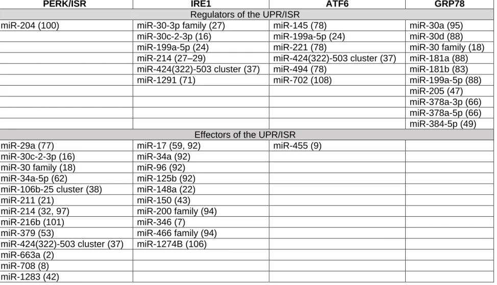
Several examples of the interactions between UPR signaling pathways and miRNAs 182
are now available in the literature (Tables 1&2). First, although PERK is regulated by 183
only one miRNA; miR-204 (100), its downstream signaling elements are highly 184
regulated by a large number of miRNAs and contribute to diverse processes from 185
apoptosis to cell survival, depending on the context. Moreover, miRNAs modulate 186
and fine-tune the IRE1-XBP1 axis, which in turn is a regulator of miRNA through 187
XBP1s transcription factor activity and the endoribonuclease activity of IRE1. IRE1 188
itself is regulated by several miRNAs, and can also cleave miRNA through RIDD 189
(miRIDD; Figure 2). The IRE1-miRNA network operates across a variety of cellular 190
processes, displaying context-specific effects and crosstalk between UPR sensor 191
pathways. The miRNA-ATF6 network has not been as extensively researched as 192
that of the other two UPR sensors, with only a handful of miRNAs characterized. 193
ATF6 has thus far only one identified effector miRNA; miR-455, which targets 194
calreticulin, providing an additional insight into the UPR interplay with the protein 195
folding machinery (9). As the knowledge of ATF6 biology in general improves, so too 196
should the knowledge of the ATF6-miRNA network. Finally, similar to what is 197
observed at the protein level, there is also evidence of UPR arms cross-talk at the 198
miRNA level, for instance miR-216b, which is regulated by both CHOP and IRE1 199
(101). To date, miRNA as either effectors or regulators of the UPR (UPR miRNAs) 200
were shown to be associated with numerous physiological and pathological 201
processes that are illustrated below. 202
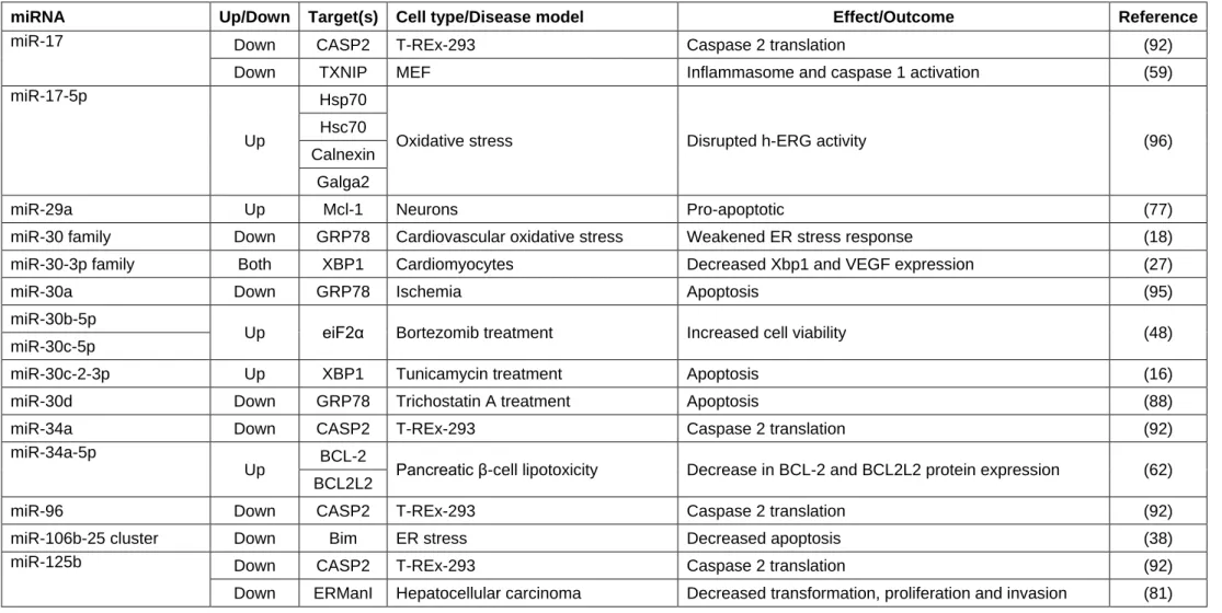
UPR miRNAs involved in Diabetes/pancreatic function - PERK activity is 203
instrumental in pancreas development and function (107), therefore it is not 204
surprising to see that many of the miRNAs implicated in pancreatic function and 205
diabetes are also associated with the PERK pathway. In pancreatic beta cells, PERK 206
was shown to be under the control of miR-204, with overexpression of the miRNA 207
enhancing beta cell apoptosis under ER stress conditions. The same miRNA also 208
targets the transcription factor MafA, resulting in decreased insulin production, 209
suggesting that one miRNA can operate at multiple levels to alleviate ER stress 210
8 (100). In a β-cell lipotoxicity model, miR-34a-5p was shown to be induced by PERK 211
through a p53-dependent mechanism and to target anti-apoptotic BCL2 and BCL2L2 212
(62). CHOP upregulates the expression of miR-379 in diabetic nephropathy, which 213
targets the ER mannosidase-like EDEM3 mRNA, possibly together with miR-200 214
family members, thus contributing to the ER stress and clinical features in this 215
disease (53). PERK is not the only arm of the UPR involved in diabetes, as the miR-216
200 and miR-466 families are degraded by the RIDD function of IRE1 at the pre-217
miRNA stage in diabetic bone marrow-derived progenitor cells. Defective IRE1 218
signaling causes the upregulation of these miRNAs and therefore reduced 219
expression of angiopoietin 1, linking RIDD to angiogenesis, a phenomenon altered in 220
diabetes (94). 221
UPR-associated miRNAs in cardio-vascular functions – UPR/ISR-associated 222
miRNAs were described in many instances in cardiovascular pathophysiology. 223
Indeed in a mouse cardiomyocyte model, ATF6 activation significantly alters the 224
expression levels of thirteen miRNAs. For instance, miR-455, which is down-225
regulated by ATF6, in turn negatively regulates the expression of the calcium-226
binding/chaperone protein calreticulin, which is increased in ischemic hearts and is 227
associated with a cytoprotective response (9). While ATF6 controls the expression of 228
miRNAs, it is also a target of miRNA. As such in mouse cardiomyocytes, mirtronic 229
mmu-miR-702 is down-regulated after isoproterenol treatment and targets ATF6. 230
Artificial over-expression of miR-702 through the use of miRNA mimics showed that 231
miR-702 has anti-apoptotic effects in vitro after isoproterenol treatment (108). A rat 232
model of heart failure revealed that XBP1 mRNA is a target of miR-214 and miR-30-233
3p family members in cardiomyocytes, and that these miRNAs are responsible for 234
the dynamic expression of XBP1s protein, which reaches a peak in the early stage of 235
heart failure and decreases afterwards (27). Another study by the same group 236
observed increased miR-214 levels in both serum and cardiac tissue of patients with 237
chronic heart failure, which had an anti-angiogenic effect in a corresponding mouse 238
model. miR-214 controls angiogenesis through suppression of XBP1 mRNA in 239
endothelial cells, as the XBP1 transcription factor target VEGF is also suppressed 240
(29). ATF4 mRNA is targeted by miR-1283 in vascular endothelial cells. Introduction 241
of a miRNA mimic decreased vascular endothelial injury markers, whereas miR-1283 242
inhibition led to increased levels of apoptosis and cardiac tissue damage (42). In 243
vascular smooth muscle cells (SMC), XBP1s downregulates the expression of 244
calponin 1 through increased transcription of miR-1274B, thereby allowing SMC 245
proliferation. In addition, miR-1274B is also released extracellularly, a possible 246
mechanism for paracrine regulation of proliferation in neighboring SMCs (106). The 247
miR-30 family, which is comprised of six miRNAs with identical seed sequences, is 248
down-regulated during cardiovascular oxidative stress, inhibits GRP78 and is at least 249
partially negatively regulated by CHOP (18). More specifically, the miR-30 family 250
member, miR-30a, which was found to be decreased in primary cortical neurons 251
after ischemia was also shown to target GRP78 mRNA. Transfection with miRNA 252
mimics and inhibitors modulated GRP78 protein levels, while the inhibitor displayed 253
a neuroprotective effect in vivo (95). In an ischemic stroke model, GRP78 mRNA is 254
target of two members of the miR-181 family; miR-181a and miR-181b, which 255
repress GRP78 mRNA through translational repression. Modulation of miR-181 with 256
mimics and inhibitors regulated both GRP78 expression and cell death, while 257
overexpression of the inhibitor diminishes neural damage in an in vivo mouse model 258
(79) ). Consistent with this result, another study also showed that inhibition of miR-259
181b demonstrated a cytoprotective effect, through blocking the targeting of the 260
GRP78 and ubiquitin hydrolase UCHL1 mRNAs (83). Both strands of the miR-378a 261
hairpin; miR-378a-5p and miR-378a-3p are highly expressed in cardiac cells. To 262
identify potential targets of these miRNAs, a proteomic screen was performed and 263
revealed that both miRNAs target GRP78 and calumenin mRNA, as well as other 264
mRNAs involved in glycolysis and the cytoskeleton. The protein homeostasis-265
relevant protein cyclophilin A is also a target of miR-378a-5p (66). As well as 266
targeting components of the UPR, miRNAs also affect other aspects of the 267
proteostasis machinery, such as chaperone proteins and enzymes involved in 268
protein folding. Under oxidative stress conditions, chaperone proteins including 269
Hsp70, Hsc70, calnexin and Galga2 were found to be the target of AP1-regulated 270
miR-17-5p, which led to reduced h-ERG export through the ER and Golgi, a feature 271
of cardiac diseases (96). 272
UPR-associated miRNAs involved in hepatic functions - Much of the research 273
in ER-related miRNAs in the liver has been performed in hepatocellular carcinoma 274
(HCC) models, thereby introducing a significant bias in all the results obtained. For 275
instance, miR-214, a miRNA down-regulated in HCC through possible NF-κB-276
dependent mechanisms, was shown to target XBP1 mRNA and act in a tumor-277
suppressive manner, as over-expression of miR-214 mimics reduced tumor volume 278
and weight in a mouse model (28). In HCC, bortezomib, the protease inhibitor used 279
in the treatment of multiple myeloma, induced the expression 30b-5p and miR-280
10 30c-5p late in the UPR, suggesting that their regulation is mediated by one of the 281
UPR sensors. These miRNAs target eiF2α mRNA and increase cell viability by 282
preventing the apoptotic ATF4-CHOP signaling downstream of eiF2α(48). In addition 283
to cleavage of miRNAs by IRE1 ribonuclease activity, IRE1 is also directly regulated 284
by miRNA. IRE1 mRNA is targeted in the 5’ UTR by miR-1291 in HCC, leading to 285
downregulation of IRE1 expression and the subsequent stabilization of the RIDD 286
substrate glypican-3 mRNA (71). In another HCC study, ER stress induction with the 287
N-glycosylation inhibitor tunicamycin increased the level of miR-663 and treatment 288
with miRNA mimics increased cell proliferation. miR-663 targets TGFB1 mRNA and 289
has an impact on ER-stress induced apoptosis by acting in a cytoprotective manner 290
(46). Along with direct regulation of UPR components by miRNAs, other 291
proteostasis-relevant targets of miRNAs have been found in HCC. The Golgi-292
resident protein ER mannosidase I (ERManI) that contributes to glycoprotein quality 293
control, is up-regulated in HCC and is targeted by miR-125b. miR-125b is down-294
regulated in HCC, so its target ERManI can maintain efficient cancer cell proliferation 295
and transformation phenotypes (81). UPR-miRNA interactions have also been 296
reported in non-HCC studies. For instance, IRE1 RIDD activity has been reported in 297
a liver fibrosis model; pre-miR-150, which in its mature form is stabilized by XBP1s 298
(109), was found to be cleaved by IRE1, reducing the repression of the 299
transcriptional activator c-Myb and enhancing liver fibrosis through alpha smooth 300
muscle actin (α-SMA) expression (43). In Dicer-deficient hepatocytes under 301
thapsigargin or bile acid (deoxycholic acid)-induced stress, ATF6, IRE1 and the 302
GRP78 chaperone are regulated by miR-199a-5p. The AP-1 transcription factor 303
controls the expression of miR-199-5p, which dampens the ER stress response and 304
prevents cell death through inhibition of IRE1 (24). In a model of nonalcoholic fatty 305
liver disease, miR-615-3p is downregulated under palmitate and tunicamycin-306
induced stress conditions, leading to an increase of its target CHOP and an 307
accompanying increase in cell death through lipoapoptosis. However the exact 308
mechanisms regulating the miRNA expression are as yet unknown (72). 309
UPR miRNAs involved in Respiratory functions – In human airway epithelial 310
cells, the active XBP1s transcription factor induces the expression of miR-346, which 311
in turn represses antigen peptide transporter 1 (TAP1) mRNA. This impacts on 312
immunity by reducing MHC I-associated antigen presentation during ER stress (7). In 313
bronchial brushings from cystic fibrosis (CF) patients, miRNAs -145, -221 and -494 314
expression levels were elevated and were predicted to target the 3' UTR of the UPR 315
sensor ATF6. In a corresponding CF mouse model, increased expression of these 316
miRNAs was also observed, and regulation of ATF6 by miR-221 was confirmed. The 317
effects of ATF6 regulation by miRNA were not examined in this study, but could have 318
an impact on the unfolded protein response in cystic fibrosis (78). As well as 319
targeting UPR components in the liver, other ER-relevant targets of miR-199a have 320
also been described. In monocytes from patients with α1-antitrypsin deficiency-321
related chronic obstructive pulmonary disease (COPD), miR-199a-5p levels are 322
increased in α1-antitrypsin deficiency and decreased in COPD. This is due to 323
hypermethylation at the miR-199a promoter, which modulates the UPR through its 324
targets including the NF-κB subunits p50 and p65 (41). 325
UPR miRNAs in nervous system functions – UPR-mediated regulation of 326
miRNA has been observed in many diseases associated with the nervous system 327
including cancers and degenerative diseases. As such, miR-29a is induced by ER 328
stress, possibly by ATF4 and contributes to ER-stress induced apoptosis in 329
neuroblastoma cells and murine primary cortical neurons by repressing the anti-330
apoptotic BCL-2 family member, MCL-1 (77). miR-210 regulates the ER chaperone 331
protein prolyl 4-hydroxylase, beta polypeptide (P4HB) in glioblastoma. P4HB 332
promotes resistance to the DNA alkylating agent temozolomide and in both cell lines 333
and clinical samples, P4HB mRNA and miR-210 show reciprocal expression levels. 334
In clinical samples, miRNA and mRNA expression levels correlate with disease 335
grade, as the worst grade; Grade IV, has the lowest levels of miR-210 and highest of 336
P4HB. Artificial overexpression of miR-210 in cell lines improved responsiveness to 337
temozolomide treatment (56). In irradiation-resistant glioma stem cells, reduced 338
capability to form tumor spheres and increased irradiation tolerance were associated 339
with decreased expression of GRP78 by miR-205. Treatment of the stem cells with 340
pterostilbene increased the level of miR-205 and to recapitulate this increase, 341
treatment with a miR-205 mimic decreased the expression of GRP78/BiP, along with 342
c-Myc, β-catenin and vimentin, reducing stemness and resistance to radiation 343
treatment (47). GRP78 was also found to be under the control of another miRNA; 344
miR-384-5p. In an in vitro Parkinson's disease model, where rotenone was used to 345
induce cell death, miR-384-5p levels increased and targeted the 3' UTR of GRP78 346
mRNA. The results were also validated in primary neurons, with miR-384-5p also 347
showing an increase after rotenone treatment and decreased cell viability upon 348
treatment with miRNA mimics (49). In a mouse model of Alzheimer’s disease, levels 349
of miR-200c correlated with ER stress levels, as miR-200c expression increased 350
12 upon ER stress. However, the exact mechanism of its upregulation is unknown. miR-351
200c, along with other members of the miR-200 family, regulate the tumor 352
suppressor PTEN, and displayed a cytoprotective effect during early ER stress while 353
contributing to apoptosis under later, irremediable ER stress (99). 354
355
UPR-associated miRNA and other cellular functions
356
UPR-associated miRNAs and apoptosis/cell survival - All three arms of the 357
UPR are known to influence these processes (44), however the majority of miRNA 358
regulation regarding apoptosis is focused on the PERK signaling pathway. The 359
processes by which these signals lead to apoptosis include a diverse range of 360
biological pathways, so examining the miRNA component(s) can help to clarify their 361
mechanisms and fine tune the balance between the adaptive and terminal UPR. The 362
miR-106b-25 cluster, which is comprised of the miRNAs miR-106b, -93 and -25, is 363
located in an intron of the MCM7 gene and is therefore under the same regulation 364
i.e., downregulated by the PERK substrates NRF2 and ATF4. Repression of this 365
miRNA cluster is required for ER-stress induced apoptosis, as it normally inhibits the 366
translation of pro-apoptotic Bim mRNA (38). Transcription of miR-211 is induced by 367
the PERK pathway in an ATF4/phospho-eiF2α dependent manner, which leads to 368
repression of CHOP by histone medication and ribosome stalling at the promoter 369
and resulting in increased cell survival (21). In addition to regulation by miRNA, 370
CHOP also regulates miRNA expression through its transcription factor activity, with 371
an increase in miR-216b levels after ER stress. In a convergence of two UPR 372
pathways, IRE1 indirectly represses transcription of miR-216b, possibly through 373
CHOP regulation. Upon ER stress activation, miR-216b contributes to the 374
downregulation of c-JUN and modulates ER-stress-induced apoptosis (101). 375
Overexpression of the miR-23a~27a~24-2 cluster induces apoptosis through ER 376
stress-dependent mechanisms by upregulating components of the UPR such as 377
PERK, ATF4 and CHOP. The cluster also acts on calcium signaling by increasing 378
the amount of cytoplasmic Ca2+, which may also contribute to apoptosis by affecting 379
mitochondrial membrane permeability and the release of cytochrome c and AIF (20). 380
GRP78 mRNA is regulated by the coordinated action of three miRNAs; miR-30d, 381
miR-181a and miR-199a-5p. Transfection of cells with all three of these miRNAs led 382
to reduced GRP78 expression, abrogating its cytoprotective effect and increasing 383
sensitivity to the histone deacetylase inhibitor trichostatin A, as well as reduced 384
tumor growth in an in vivo mouse model (88). In addition to regulating GRP78, miR-385
30 family members also regulate XBP1. miR-30c-2-3p contributes to the pro/anti-386
apoptotic balance of the UPR, as inhibition of the miRNA leads to a decrease in cell 387
death while also demonstrating the cross-talk between the three arms of the UPR as 388
the miRNA itself is regulated a PERK/NF-κB pathway (16). IRE1 is involved in the 389
regulation of miRNA through its kinase domain, as it cleaves miRNAs -17, 34a, 96 390
and 125b. This event leads to translation of the miR-17 target thioredoxin Interacting 391
Protein (TXNIP) and in the case of all four miRNAs, reduces the repression of 392
caspase-2 mRNA (59, 92). In a more detailed study of miR-34a in an acute myeloid 393
leukemia model, treatment with IRE1 RNase and miR-34a inhibitors slightly 394
decreased cell growth, demonstrating that targeting the UPR at multiple levels, i.e. at 395
the enzymatic and miRNA level, could be beneficial in cancer treatment (90). 396
Other-associated UPR miRNAs - ER/stress-responsive miRNA signaling is 397
involved in a diverse range of cellular processes from the cell cycle to developmental 398
processes (70). The intronic miRNA, miR-708 and the gene in which it is located, 399
Odz4, are regulated by the CHOP transcription factor during ER stress miR-708 is 400
highly expressed in the brain and eye, and represses the vision-relevant target 401
rhodopsin, which may impact on ER protein homeostasis as rhodopsin protein 402
requires processing in the ER (8). In addition to targeting XBP1 in cardiovascular 403
systems, miR-214 also has other UPR-relevant targets. In a screen to identify 404
miRNA regulators of bone development, high expression of miR-214 was found to 405
inhibit bone formation through targeting ATF4 mRNA (97). As well as participating in 406
the process of bone formation, miR-214 targets ATF4 in erythroid cells. miR-214, 407
under the transcriptional control of the PERK substrate NRF2, acts in a 408
cytoprotective manner after oxidative stress. miR-214 levels decrease, allowing for 409
expression of ATF4, while another miR-214 target, the histone modifier EZH2, 410
represses pro-apoptotic Bim, contributing to the cytoprotective effect of miR-214 411
(32). Dai et al profiled the expression of different miRNAs in response to 412
thapsigargin-induced ER stress and found increased expression of miR-221-3p and 413
miR-452-5p, as well as a decrease in miR-423-5p. CDKN1B was then identified as a 414
target of miR-452-5p and miR-221-3p, whereas miR-423-5p targets CDKN1A, 415
potentially illustrating cross-talk between the UPR and other cellular processes such 416
as the cell cycle (25). Also under thapsigargin-induced ER stress, the PERK branch 417
of the UPR controls the increased expression of miR-663a through unknown 418
mechanisms. miR-663a targets PLOD3 mRNA, and negatively impacts type IV 419
collagen secretion through repression of lysyl hydroxylase 3 activity at the mRNA 420
14 level (2). In tubular renal cells, miR-205 expression is decreased by ER stress, thus 421
increasing levels of its target mRNA, the hypoxia-inducible factor prolyl hydroxylase 422
EGLN2. This sensitizes cells to stress by negatively regulating the EGLN2 targets 423
ATF4, HIF1α and HIF2α (73). XBP1s transcription factor activity unrelated to the 424
canonical UPR signaling pathways has been reported in developmental processes. 425
In the process of adipogenesis, XBP1s has been shown to suppress WNT10B by up-426
regulating transcription of its inhibitory miRNA; miR-148a (22). In the context of 427
myofibre development and differentiation, miR-181a-5p was increased upon ER 428
stress and was again shown to target GRP78 mRNA, promoting apoptosis and 429
decreasing cell viability, along with indirectly regulating the expression of myogenic 430
related genes (98). The miR-424(322)-503 cluster is repressed by the PERK 431
pathway during ER stress and negatively regulates the transcription of ATF6 through 432
miR-424 binding to the 3' UTR of ATF6 mRNA. The miRNA cluster also positively 433
regulates IRE1 RIDD activity through indirect mechanisms (37). miR-322 expression 434
is lowered after thapsigargin-induced ER calcium depletion, however it is unclear if 435
this is through RIDD activity by IRE1 or by the action of IRE1 downstream factors. 436
The repression of miR-322 allows for the expression of its target PDIA6, which then 437
regulates IRE1 and PERK activity at a protein level (35). 438
439
Long non-coding RNA and ER homeostasis
440
Long non-coding RNAs (lncRNA), molecules over 200 base pairs long, are also 441
involved in regulation of protein homeostasis. LncRNAs are genomically abundant 442
regulatory RNA molecules involved in processes as diverse as post-transcriptional, 443
translational and post-translational regulation (35). The processes by which lncRNAs 444
are involved in protein homeostasis are somewhat analogous to miRNA, in that their 445
levels can increase or decrease upon ER stress, depending on their function, to 446
influence cell fate under stressed conditions (Table 3). For instance, the lncRNA 447
gadd7 is increased following induction of oxidative stress in CHO cells, acts a 448
general regulator of oxidative stress and is essential for palmitate-induced ER stress 449
(13). In addition to regulation by miR-455, the ER protein calreticulin is also under 450
the regulation of a lncRNA that shares a bidirectional promoter with the RB1 gene; 451
ncRNA-RB1. When ncRNA-RB1 is depleted, calreticulin levels also decrease. Cell 452
surface localized calreticulin functions as a signal in immunogenic cell death, so 453
decreased levels of ncRNA-RB1 results in reduced uptake by macrophages, 454
providing an insight into how cancer cells can escape this death mechanism (74). 455
Another lncRNA, lincRNA-p21, functions as a tumor suppressor in hepatocellular 456
carcinoma through activation of ER stress pathways (103), while the lncRNA Malat1 457
showed increased expression upon flavivirus infection through PERK-dependent 458
transcriptional activity. Interestingly, Malat1 itself is subject to miRNA regulation, 459
demonstrating the complexity of RNA regulation, particularly under ER stress (10). 460
The lncRNA TUG1 was identified through RNASeq as a lncRNA that may reduce 461
apoptosis through inhibition of ER stress in cold-induced liver cell damage, as 462
overexpression of TUG1 reduced expression of UPR components such as GRP78, 463
PERK and CHOP (89). The CHOP regulated miRNA-379 is part of a megacluster of 464
miRNAs present within a lncRNA transcript, lnc-MGC, and provides a link between 465
lncRNA and miRNA in the context of ER stress. The cluster of approximately forty 466
miRNAs display the same regulatory pattern by CHOP and together target genes 467
involved in ERAD and protein synthesis (53). This indicates that the extent of RNA-468
mediated regulation of the UPR is far more complex than anticipated and involves 469
mRNA, miRNA, lncRNA. A systematic analysis of the interconnectivity of these RNA 470
signaling networks will require extensive work in various experimental models before 471
being able to propose an integrated view of the underlying mechanisms and their 472
biological outcomes. 473
474
Conclusions and perspectives
475
It is clear from the evidence presented in this review that the contributions of miRNA 476
and lncRNA to the cellular stress responses represent an important regulatory role in 477
addition to the already recognized signaling pathways. These small non-coding 478
RNAs affect a diverse range of normal cellular processes, and when deregulated can 479
result in disease states. Even in the context of miRNA which is active under cellular 480
stress, the effects of these miRNAs are not limited to pathways related to the stress 481
response, but can affect processes as diverse as cell cycle to adipogenesis 482
regulation (Table 2). An important point to note is that many of the studies reviewed 483
in this article used animal models, which therefore need to be validated in the human 484
context, as certain miRNAs and miRNA families diverge between humans and other 485
species. The majority of the miRNAs reviewed here were examined in response to 486
cellular stress, such as the chemical stressors tunicamycin or thapsigargin, but also 487
physiological stresses such as hypoxia and nutrient deprivation, which more closely 488
mimic the natural environment. It has been suggested that miRNAs act as buffers to 489
regulate mRNA expression during stress and particularly affect transcription factors 490
16 and signaling molecules (60). During stress responses where global translation is 491
shut down through eiF2α phosphorylation, this buffering function may be more 492
important than in normal cellular metabolism. All of the signaling pathways 493
discussed; nonsense-mediated decay, RNase L mediated decay, the UPR and the 494
ISR are all under the regulation of non-coding RNA and in the case of the UPR and 495
ISR, the transcription factors which these processes use to mediate their effects are 496
also found to be under the control of non-coding RNA. The endoplasmic reticulum 497
itself is an important step in the miRNA: mRNA interaction, as a recent report from 498
Barman and Bhattacharyya identified ER-bound polysomes as the site where mRNA 499
associates with Ago2 proteins and miRNA, ultimately resulting in translational 500
repression (5). Much of the research reviewed here examines the role of non-coding 501
RNA in cancer. In the context of cancer, miRNAs can be classified as tumor-502
suppressive or oncogenic (91), however the cell type and context must be taken into 503
consideration. This holds true for ER protein homeostasis too, as the different 504
miRNAs involved in regulation show different effects, such as miR-199a-5p, which 505
shows a cytoprotective effect when IRE1 is inhibited, yet an apoptotic effect when 506
GRP78 or ATF6 is inhibited (24). As well as the different roles of various miRNA in 507
cancer, mutations in the miRNA biogenesis machinery such as those in Drosha or 508
Dicer, also alter the miRNA expression profiles and therefore gene and protein 509
expression (61). 510
This article reviews the complex, inter-connected non-coding RNA network 511
involved in the regulation of the UPR. Much of the work to date has been 512
concentrated on the well-established UPR, but other players, such as nonsense-513
mediated decay are emerging as ER protein homeostasis regulators, as well as the 514
molecular machinery itself. Non-coding RNA regulation of ER protein homeostasis 515
has been discovered in many cellular processes, both physiological and pathological 516
and future work can now concentrate on integrating the role of the identified miRNAs 517
and lncRNAs, as well as those yet to be identified, into the established networks as a 518
definite additional layer of regulation. 519
Acknowledgements
521
The research in EC’s lab is supported by grants from Institut National du Cancer 522
(INCa_5869, INCA_7981 and PLBIO: 2015-111) and la Ligue Contre le Cancer. 523
Research in AS’s lab is supported by funding from Breast Cancer Campaign grant 524
(2010NovPR13), Health Research Board (grant number HRA-POR-2014-643), 525
Belgium Grant (IAP 7/32), A Science Foundation Ireland (SFI) grant co-funded under 526
the European Regional Development Fund (grant Number 13/RC/2073) and EU 527
H2020 MSCA ITN-675448 (TRAINERS). MM is supported by an ARED PhD 528
scholarship from Région Bretagne. 529
18
Bibliography
531 532
1. Ali MMU, Bagratuni T, Davenport EL, Nowak PR, Silva-Santisteban MC,
533
Hardcastle A, McAndrews C, Rowlands MG, Morgan GJ, Aherne W,
534
Collins I, Davies FE, Pearl LH. Structure of the Ire1 autophosphorylation
535
complex and implications for the unfolded protein response. EMBO J 30: 894– 536
905, 2011. 537
2. Amodio G, Sasso E, D’Ambrosio C, Scaloni A, Moltedo O, Franceschelli 538
S, Zambrano N, Remondelli P. Identification of a microRNA (miR-663a)
539
induced by ER stress and its target gene PLOD3 by a combined microRNome 540
and proteome approach. Cell Biol. Toxicol. (2016). doi: 10.1007/s10565-016-541
9335-z. 542
3. Andruska N, Zheng X, Yang X, Helferich WG, Shapiro DJ. Anticipatory
543
estrogen activation of the unfolded protein response is linked to cell 544
proliferation and poor survival in estrogen receptor [alpha]-positive breast 545
cancer. Oncogene 34: 3760–3769, 2015. 546
4. Balch WE, Morimoto RI, Dillin A, Kelly JW. Adapting Proteostasis for
547
Disease Intervention. Science (80- ) 319: 916 LP-919, 2008. 548
5. Barman B, Bhattacharyya SN. mRNA Targeting to Endoplasmic Reticulum
549
Precedes Ago Protein Interaction and MicroRNA (miRNA)-mediated 550
Translation Repression in Mammalian Cells. J Biol Chem 290: 24650–24656, 551
2015. 552
6. Bartel DP. MicroRNAs: target recognition and regulatory functions. Cell 136:
553
215–33, 2009. 554
7. Bartoszewski R, Brewer JW, Rab A, Crossman DK, Bartoszewska S,
555
Kapoor N, Fuller C, Collawn JF, Bebok Z. The Unfolded Protein Response
556
(UPR)-activated Transcription Factor X-box-binding Protein 1 (XBP1) Induces 557
MicroRNA-346 Expression That Targets the Human Antigen Peptide 558
Transporter 1 (TAP1) mRNA and Governs Immune Regulatory Genes. J Biol 559
Chem 286: 41862–41870, 2011. 560
8. Behrman S, Acosta-Alvear D, Walter P. A CHOP-regulated microRNA
561
controls rhodopsin expression. J Cell Biol 192: 919–927, 2011. 562
9. Belmont PJ, Chen WJ, Thuerauf DJ, Glembotski CC. Regulation of
563
microRNA Expression in the Heart by the ATF6 Branch of the ER Stress 564
Response. J Mol Cell Cardiol 52: 1176–1182, 2012. 565
10. Bhattacharyya S, Vrati S. The Malat1 long non-coding RNA is upregulated by
566
signalling through the PERK axis of unfolded protein response during flavivirus 567
infection. Sci Rep 5: 17794, 2015. 568
11. Braakman I, Bulleid NJ. Protein Folding and Modification in the Mammalian
569
Endoplasmic Reticulum. Annu Rev Biochem 80: 71–99, 2011. 570
12. Bright MD, Itzhak DN, Wardell CP, Morgan GJ, Davies FE. Cleavage of
571
BLOC1S1 mRNA by IRE1 Is Sequence Specific, Temporally Separate from 572
XBP1 Splicing, and Dispensable for Cell Viability under Acute Endoplasmic 573
Reticulum Stress. Mol Cell Biol 35: 2186–2202, 2015. 574
13. Brookheart RT, Michel CI, Listenberger LL, Ory DS, Schaffer JE. The
Non-575
coding RNA gadd7 Is a Regulator of Lipid-induced Oxidative and Endoplasmic 576
Reticulum Stress. J Biol Chem 284: 7446–7454, 2009. 577
14. Bruno IG, Karam R, Huang L, Bhardwaj A, Lou CH, Shum EY, Song H-W,
578
Corbett MA, Gifford WD, Gecz J, Pfaff SL, Wilkinson MF. Identification of a
579
microRNA that activates gene expression by repressing nonsense-mediated 580
RNA decay. Mol Cell 42: 500–510, 2011. 581
15. Bu Y, Diehl JA. PERK Integrates Oncogenic Signaling and Cell Survival
During Cancer Development. J. Cell. Physiol. (2016). doi: 10.1002/jcp.25336. 583
16. Byrd AE, Aragon I V, Brewer JW. MicroRNA-30c-2* limits expression of
584
proadaptive factor XBP1 in the unfolded protein response. J Cell Biol 196: 585
689–698, 2012. 586
17. Castilho BA, Shanmugam R, Silva RC, Ramesh R, Himme BM, Sattlegger
587
E. Keeping the eIF2 alpha kinase Gcn2 in check. Biochim Biophys Acta 1843:
588
1948–68, 2014. 589
18. Chen M, Ma G, Yue Y, Wei Y, Li Q, Tong Z, Zhang L, Miao G, Zhang J.
590
Downregulation of the miR-30 family microRNAs contributes to endoplasmic 591
reticulum stress in cardiac muscle and vascular smooth muscle cells. Int J 592
Cardiol 173: 65–73, 2014. 593
19. Chen X, Shen J, Prywes R. The Luminal Domain of ATF6 Senses
594
Endoplasmic Reticulum (ER) Stress and Causes Translocation of ATF6 from 595
the ER to the Golgi. J Biol Chem 277: 13045–13052, 2002. 596
20. Chhabra R, Dubey R, Saini N. Gene expression profiling indicate role of ER
597
stress in miR-23a~27a~24-2 cluster induced apoptosis in HEK293T cells. RNA 598
Biol 8: 648–664, 2011. 599
21. Chitnis N, Pytel D, Bobrovnikova-Marjon E, Pant D, Zheng H, Maas N,
600
Frederick B, Kushner JA, Chodosh LA, Koumenis C, Fuchs SY, Diehl JA.
601
miR-211 is a pro-survival micro-RNA that regulates chop expression in a 602
PERK-dependent manner. Mol. Cell 48: 353–364, 2012. 603
22. Cho YM, Kim T-M, Hun Kim D, Hee Kim D, Jeong S-W, Kwon O-J.
miR-604
148a is a downstream effector of X-box-binding protein 1 that silences Wnt10b 605
during adipogenesis of 3T3-L1 cells. Exp Mol Med 48 KSBMB.: e226, 2016. 606
23. Cullinan SB, Zhang D, Hannink M, Arvisais E, Kaufman RJ, Diehl JA. Nrf2
607
Is a Direct PERK Substrate and Effector of PERK-Dependent Cell Survival. 608
Mol Cell Biol 23: 7198–7209, 2003. 609
24. Dai B-H, Geng L, Wang Y, Sui C-J, Xie F, Shen R-X, Shen W-F, Yang J-M.
610
microRNA-199a-5p protects hepatocytes from bile acid-induced sustained 611
endoplasmic reticulum stress. Cell Death Dis 4: e604, 2013. 612
25. Dai L, Huang C, Chen L, Shan G, Li Z. Altered expression of microRNAs in
613
the response to ER stress. Sci Bull 60: 202–209, 2015. 614
26. Dejeans N, Pluquet O, Lhomond S, Grise F, Bouchecareilh M, Juin A,
615
Meynard-Cadars M, Bidaud-Meynard A, Gentil C, Moreau V, Saltel F,
616
Chevet E. Autocrine control of glioma cells adhesion and migration through
617
IRE1α-mediated cleavage of SPARC mRNA. J Cell Sci 125: 4278–4287, 2012. 618
27. Duan Q, Chen C, Yang L, Li N, Gong W, Li S, Wang DW. MicroRNA
619
regulation of unfolded protein response transcription factor XBP1 in the 620
progression of cardiac hypertrophy and heart failure in vivo. J Transl Med 13: 621
1–11, 2015. 622
28. Duan Q, Wang X, Gong W, Ni L, Chen C, He X, Chen F, Yang L, Wang P,
623
Wang DW. ER Stress Negatively Modulates the Expression of the
miR-624
199a/214 Cluster to Regulates Tumor Survival and Progression in Human 625
Hepatocellular Cancer. PLoS One 7: 1–10, 2012. 626
29. Duan Q, Yang L, Gong W, chaugai S, Wang F, Chen C, Wang P, Zou M-H,
627
Wang DW. MicroRNA-214 Is Upregulated in Heart Failure Patients and
628
Suppresses XBP1-Mediated Endothelial Cells Angiogenesis. J Cell Physiol 629
230: 1964–1973, 2015. 630
30. Eletto D, Eletto D, Boyle S, Argon Y. PDIA6 regulates insulin secretion by
631
selectively inhibiting the RIDD activity of IRE1. FASEB J 30: 653–665, 2016. 632
31. Eletto D, Eletto D, Dersh D, Gidalevitz T, Argon Y. Protein Disulfide
633
Isomerase A6 controls the decay of IRE1α signaling via disulfide-dependent 634
20 association. Mol Cell 53: 562–576, 2014.
635
32. Gao M, Liu Y, Chen Y, Yin C, Chen J-J, Liu S. miR-214 protects erythroid 636
cells against oxidative stress by targeting ATF4 and EZH2. Free Radic Biol 637
Med 92: 39–49, 2016. 638
33. García MA, Gil J, Ventoso I, Guerra S, Domingo E, Rivas C, Esteban M. 639
Impact of Protein Kinase PKR in Cell Biology: from Antiviral to Antiproliferative 640
Action. Microbiol Mol Biol Rev 70: 1032–1060, 2006. 641
34. Gardner LB. Hypoxic Inhibition of Nonsense-Mediated RNA Decay Regulates 642
Gene Expression and the Integrated Stress Response. Mol Cell Biol 28: 643
3729–3741, 2008. 644
35. Groenendyk J, Peng Z, Dudek E, Fan X, Mizianty MJ, Dufey E, Urra H, 645
Sepulveda D, Rojas-Rivera D, Lim Y, Kim DH, Baretta K, Srikanth S,
646
Gwack Y, Ahnn J, Kaufman RJ, Lee S-K, Hetz C, Kurgan L, Michalak M.
647
Interplay Between the Oxidoreductase PDIA6 and microRNA-322 Controls the 648
Response to Disrupted Endoplasmic Reticulum Calcium Homeostasis. Sci 649
Signal 7: ra54-ra54, 2014. 650
36. Guerriero CJ, Brodsky JL. The Delicate Balance Between Secreted Protein 651
Folding and Endoplasmic Reticulum-Associated Degradation in Human 652
Physiology. Physiol Rev 92: 537 LP-576, 2012. 653
37. Gupta A, Hossain MM, Read DE, Hetz C, Samali A, Gupta S. PERK 654
regulated miR-424(322)-503 cluster fine-tunes activation of IRE1 and ATF6 655
during Unfolded Protein Response. Sci Rep 5: 18304, 2015. 656
38. Gupta S, Read DE, Deepti A, Cawley K, Gupta A, Oommen D, Verfaillie T, 657
Matus S, Smith MA, Mott JL, Agostinis P, Hetz C, Samali A.
Perk-658
dependent repression of miR-106b-25 cluster is required for ER stress-induced 659
apoptosis. Cell Death Dis. 3: e333-, 2012. 660
39. Ha M, Kim VN. Regulation of microRNA biogenesis. Nat Rev Mol Cell Biol 15: 661
509–524, 2014. 662
40. Han J, Back SH, Hur J, Lin Y-H, Gildersleeve R, Shan J, Yuan CL, 663
Krokowski D, Wang S, Hatzoglou M, Kilberg MS, Sartor MA, Kaufman RJ.
664
ER-stress-induced transcriptional regulation increases protein synthesis 665
leading to cell death. Nat Cell Biol 15: 481–490, 2013. 666
41. Hassan T, Carroll TP, Buckley PG, Cummins R, O’Neill SJ, McElvaney 667
NG, Greene CM. miR-199a-5p Silencing Regulates the Unfolded Protein
668
Response in Chronic Obstructive Pulmonary Disease and α1-Antitrypsin 669
Deficiency. Am J Respir Crit Care Med 189: 263–273, 2013. 670
42. He L, Yuan J, Xu Q, Chen R, Chen L, Fang M. miRNA-1283 Regulates the 671
PERK/ATF4 Pathway in Vascular Injury by Targeting ATF4. PLoS One 11: 672
e0159171, 2016. 673
43. Heindryckx F, Binet F, Ponticos M, Rombouts K, Lau J, Kreuger J, 674
Gerwins P. Endoplasmic reticulum stress enhances fibrosis through
675
IRE1alpha-mediated degradation of miR-150 and XBP-1 splicing. EMBO Mol 676
Med 8: 729–744, 2016. 677
44. Hetz C, Chevet E, Oakes SA. Proteostasis control by the unfolded protein 678
response. Nat Cell Biol 17: 829–838, 2015. 679
45. Higa A, Taouji S, Lhomond S, Jensen D, Fernandez-Zapico ME, Simpson 680
JC, Pasquet J-M, Schekman R, Chevet E. Endoplasmic Reticulum
Stress-681
Activated Transcription Factor ATF6α Requires the Disulfide Isomerase PDIA5 682
To Modulate Chemoresistance. Mol Cell Biol 34: 1839–1849, 2014. 683
46. Huang Y, Liu J, Fan L, Wang F, Yu H, Wei W, Sun G. miR-663 684
overexpression induced by endoplasmic reticulum stress modulates 685
hepatocellular carcinoma cell apoptosis via transforming growth factor beta 1. 686
Onco. Targets. Ther. 9: 1623–1633, 2016. 687
47. Huynh T-T, Lin C-M, Lee W-H, Wu ATH, Lin Y-K, Lin Y-F, Yeh C-T, Wang 688
L-S. Pterostilbene suppressed irradiation-resistant glioma stem cells by
689
modulating GRP78/miR-205 axis. J Nutr Biochem 26: 466–475, 2015. 690
48. Jiang L, Zang D, Yi S, Li X, Yang C, Dong X, Zhao C, Lan X, Chen X, Liu S, 691
Liu N, Huang H, Shi X, Wang X, Liu J. A microRNA-mediated decrease in
692
eukaryotic initiation factor 2alpha promotes cell survival during PS-341 693
treatment. Sci Rep 6: 21565, 2016. 694
49. Jiang M, Yun Q, Shi F, Niu G, Gao Y, Xie S, Yu S. Downregulation of miR-695
384-5p attenuates rotenone-induced neurotoxicity in dopaminergic SH-SY5Y 696
cells through inhibiting endoplasmic reticulum stress. Am J Physiol - Cell 697
Physiol 310: C755–C763, 2016. 698
50. Jin Y, Zhang F, Ma Z, Ren Z. MicroRNA 433 regulates nonsense-mediated 699
mRNA decay by targeting SMG5 mRNA. BMC Mol Biol 17: 17, 2016. 700
51. Karali E, Bellou S, Stellas D, Klinakis A, Murphy C, Fotsis T. VEGF Signals 701
through ATF6 and PERK to Promote Endothelial Cell Survival and 702
Angiogenesis in the Absence of ER Stress. Mol Cell 54: 559–572, 2014. 703
52. Karam R, Lou C, Kroeger H, Huang L, Lin JH, Wilkinson MF. The unfolded 704
protein response is shaped by the NMD pathway. EMBO Rep 16: 599 LP-609, 705
2015. 706
53. Kato M, Wang M, Chen Z, Bhatt K, Oh HJ, Lanting L, Deshpande S, Jia Y, 707
Lai JYC, O/’Connor CL, Wu Y, Hodgin JB, Nelson RG, Bitzer M, Natarajan
708
R. An endoplasmic reticulum stress-regulated lncRNA hosting a microRNA
709
megacluster induces early features of diabetic nephropathy. Nat Commun 7, 710
2016. 711
54. Ketting RF, Fischer SEJ, Bernstein E, Sijen T, Hannon GJ, Plasterk RHA. 712
Dicer functions in RNA interference and in synthesis of small RNA involved in 713
developmental timing in C. elegans . Genes Dev 15: 2654–2659, 2001. 714
55. Kozomara A, Griffiths-Jones S. miRBase: annotating high confidence 715
microRNAs using deep sequencing data. Nucleic Acids Res 42: D68–D73, 716
2013. 717
56. Lee D, Sun S, Zhang XQ, Zhang P De, Ho ASW, Kiang KMY, Fung CF, Lui 718
WM, Leung GKK. MicroRNA-210 and Endoplasmic Reticulum Chaperones in
719
the Regulation of Chemoresistance in Glioblastoma. J Cancer 6: 227–232, 720
2015. 721
57. Lee TY, Ezelle HJ, Venkataraman T, Lapidus RG, Scheibner KA, Hassel 722
BA. Regulation of human RNase-L by the miR-29 family reveals a novel
723
oncogenic role in chronic myelogenous leukemia. J Interferon Cytokine Res 724
33: 34–42, 2013. 725
58. Lee Y, Jeon K, Lee J, Kim S, Kim VN. MicroRNA maturation: stepwise 726
processing and subcellular localization. EMBO J 21: 4663–4670, 2002. 727
59. Lerner AG, Upton J-P, Praveen PVK, Ghosh R, Nakagawa Y, Igbaria A, 728
Shen S, Nguyen V, Backes BJ, Heiman M, Heintz N, Greengard P, Hui S,
729
Tang Q, Trusina A, Oakes SA, Papa FR. IRE1alpha induces
thioredoxin-730
interacting protein to activate the NLRP3 inflammasome and promote 731
programmed cell death under irremediable ER stress. Cell Metab 16: 250–264, 732
2012. 733
60. Leung AKL, Sharp PA. MicroRNA Functions in Stress Responses. Mol Cell 734
40: 205–215, 2010. 735
61. Lin S, Gregory RI. MicroRNA biogenesis pathways in cancer. Nat Rev Cancer 736
15: 321–333, 2015. 737
62. Lu H, Hao L, Li S, Lin S, Lv L, Chen Y, Cui H, Zi T, Chu X, Na L, Sun C. 738
22 Elevated circulating stearic acid leads to a major lipotoxic effect on mouse 739
pancreatic beta cells in hyperlipidaemia via a miR-34a-5p-mediated 740
PERK/p53-dependent pathway. Diabetologia 59: 1247–1257, 2016. 741
63. Lu L, Han A-P, Chen J-J. Translation Initiation Control by Heme-Regulated 742
Eukaryotic Initiation Factor 2α Kinase in Erythroid Cells under Cytoplasmic 743
Stresses. Mol Cell Biol 21: 7971–7980, 2001. 744
64. Lu Y, Liang F-X, Wang X. A synthetic biology approach identifies the 745
mammalian UPR RNA ligase RtcB. Mol Cell 55: 758–70, 2014. 746
65. Ma Y, Hendershot LM. Delineation of a Negative Feedback Regulatory Loop 747
That Controls Protein Translation during Endoplasmic Reticulum Stress. J Biol 748
Chem 278: 34864–34873, 2003. 749
66. Mallat Y, Tritsch E, Ladouce R, Winter DL, Friguet B, Li Z, Mericskay M. 750
Proteome modulation in H9c2 cardiac cells by microRNAs 378 and miR-751
378. Mol Cell Proteomics 13: 18–29, 2014. 752
67. Marciniak SJ, Yun CY, Oyadomari S, Novoa I, Zhang Y, Jungreis R, 753
Nagata K, Harding HP, Ron D. CHOP induces death by promoting protein
754
synthesis and oxidation in the stressed endoplasmic reticulum. Genes Dev 18: 755
3066–3077, 2004. 756
68. Martino MB, Jones L, Brighton B, Ehre C, Abdulah L, Davis CW, Ron D, 757
O’Neal WK, Ribeiro CMP. The ER stress transducer IRE1[beta] is required for
758
airway epithelial mucin production. Mucosal Immunol 6: 639–654, 2013. 759
69. Martinon F, Chen X, Lee AH, Glimcher LH. TLR activation of the 760
transcription factor XBP1 regulates innate immune responses in macrophages. 761
Nat Immunol 11, 2010. 762
70. Maurel M, Chevet E. Endoplasmic reticulum stress signaling: the microRNA 763
connection. Am J Physiol - Cell Physiol 304: C1117 LP-C1126, 2013. 764
71. Maurel M, Dejeans N, Taouji S, Chevet E, Grosset CF. MicroRNA-1291-765
mediated silencing of IRE1α enhances Glypican-3 expression. RNA 19: 778– 766
788, 2013. 767
72. Miyamoto Y, Mauer AS, Kumar S, Mott JL, Malhi H. Mmu-miR-615-3p 768
regulates lipoapoptosis by inhibiting C/EBP homologous protein. PLoS One 9: 769
e109637, 2014. 770
73. Muratsu-Ikeda S, Nangaku M, Ikeda Y, Tanaka T, Wada T, Inagi R. 771
Downregulation of miR-205 Modulates Cell Susceptibility to Oxidative and 772
Endoplasmic Reticulum Stresses in Renal Tubular Cells. PLoS One 7: e41462, 773
2012. 774
74. Musahl A-S, Huang X, Rusakiewicz S, Ntini E, Marsico A, Kroemer G, 775
Kepp O, Orom UA. A long non-coding RNA links calreticulin-mediated
776
immunogenic cell removal to RB1 transcription. Oncogene 34: 5046–5054, 777
2015. 778
75. Nadanaka S, Okada T, Yoshida H, Mori K. Role of Disulfide Bridges Formed 779
in the Luminal Domain of ATF6 in Sensing Endoplasmic Reticulum Stress. Mol 780
Cell Biol 27: 1027–1043, 2007. 781
76. Nguyen TA, Jo MH, Choi Y-G, Park J, Kwon SC, Hohng S, Kim VN, Woo J-782
S. Functional Anatomy of the Human Microprocessor. Cell 161: 1374–87,
783
2015. 784
77. Nolan K, Walter F, Tuffy LP, Poeschel S, Gallagher R, Haunsberger S, 785
Bray I, Stallings RL, Concannon CG, Prehn JHM. Endoplasmic reticulum
786
stress-mediated upregulation of miR-29a enhances sensitivity to neuronal 787
apoptosis. Eur J Neurosci 43: 640–652, 2016. 788
78. Oglesby IK, Agrawal R, Mall MA, McElvaney NG, Greene CM. miRNA-221 789
is elevated in cystic fibrosis airway epithelial cells and regulates expression of 790