HAL Id: inserm-01332606
https://www.hal.inserm.fr/inserm-01332606
Submitted on 16 Jun 2016
HAL is a multi-disciplinary open access archive for the deposit and dissemination of sci-entific research documents, whether they are pub-lished or not. The documents may come from teaching and research institutions in France or abroad, or from public or private research centers.
L’archive ouverte pluridisciplinaire HAL, est destinée au dépôt et à la diffusion de documents scientifiques de niveau recherche, publiés ou non, émanant des établissements d’enseignement et de recherche français ou étrangers, des laboratoires publics ou privés.
regulation by epithelial polarity
Peter Coopman, Alexandre Djiane
To cite this version:
Peter Coopman, Alexandre Djiane. Adherens Junction and E-Cadherin complexes regulation by epithelial polarity: Adherens junctions regulation by polarity. Cellular and Molecular Life Sciences, Springer Verlag, 2016, �10.1007/s00018-016-2260-8�. �inserm-01332606�
Adherens Junction and E-Cadherin complexes regulation by epithelial polarity
Peter Coopman1,2,3,4
and Alexandre Djiane1,2,3,4, *
1
IRCM, Institut de Recherche en Cancérologie de Montpellier, Montpellier, F-34298, France.
2
INSERM, U1194, Montpellier, F-34298, France.
3
Université de Montpellier, Montpellier, F-34090, France.
4
Institut régional du Cancer de Montpellier, Montpellier, F-34298, France.
*Author for correspondence: IRCM, Inserm U1194 208, rue des Apothicaires
34298 Montpellier, Cedex, France
tel: +33 (0) 467 612 441
email: alexandre.djiane@inserm.fr
Running title: Adherens junctions regulation by polarity word count: 12679 words
# refs: 165
ABSTRACT
E-Cadherin-based Adherens Junctions (AJs) are a defining feature of all epithelial sheets. Through the homophilic association of E-Cadherin molecules expressed on neighboring cells, they ensure intercellular adhesion amongst epithelial cells, and regulate many key aspects of epithelial biology. While their adhesive role requires these structures to remain stable, AJs are also extremely plastic. This plasticity allows for the adaptation of the cell to its changing environment: changes in neighbors after cell division, cell death, or cell movement, and changes in cell shape during differentiation. In this review we focus on the recent advances highlighting the critical role of the apico-basal polarity machinery, and in particular of the Par3/Bazooka scaffold, in the regulation and remodeling of AJs. We propose that by regulating key phosphorylation events on the core E-Cadherin complexes components, Par3 and epithelial polarity promote meta-stable protein complex governing the correct formation, localization, and functioning of AJ.
KEYWORDS
E-Cadherin; Adherens Junctions; Remodeling; Epithelial polarity; Par3; Magi scaffolds
INTRODUCTION
In multicellular organisms, cells contact their neighbors to generate tissues and organs of very stereotypical shapes and forms. One of the most ubiquitous cell types is the epithelial cell. Epithelial cells organize as mono-layered or pseudo-stratified epithelial sheets that serve to create boundaries between different environments, for instance between the outside and inside of an organism, where the exchanges and fluxes of macromolecules, nutrients, metabolites, have to pass through and are therefore tightly controlled [1]. As such, maintaining the integrity of the epithelial sheets is crucial to their function.
Epithelial cells are polarized along their apico-basal (A/B) axis where the apical side faces the exterior of the organism or the lumen of the epithelial tube. This A/B polarity is established and maintained by the asymmetric segregation of evolutionarily conserved protein complexes which defines several lateral membrane domains (reviewed in [2–4]). Along these different lateral domains, different sets of intercellular junctions mediate cell-cell adhesion, integrity of the epithelial sheets, and the tightness of the barrier [5–7].
Adherens Junctions (AJs) are a defining feature of all epithelial sheets and constitute apical adhesive structures where the close membrane apposition between neighboring epithelial cells is mediated and strengthened by the homophilic interactions of single-pass transmembrane E-Cadherin (E-Cad) molecules. These structures are stabilized by the accumulation of a dense actin filaments-based cortical network, and in particular by the molecular links anchoring E-Cad clusters to the inner cytoskeleton (reviewed in [8–11]).
While stable adhesion is critical for epithelial sheet integrity and function, numerous studies also highligh the incredible plasticity of AJs, allowing for the destruction of ‘old’ cellular contacts and the creation of new ones [11–15]. This remodeling of the E-Cad complexes and AJs is intimately associated with changes in cell size, cell shape, and relative cell movements that are the basis of the global morphogenetic processes producing stereotypical tissues and organs fulfilling their functions during embryonic development and adaptive processes of adult tissues [8,10,16]. The study of AJ remodeling and dynamics is a very active field of research combining different model organisms and a very broad range of approaches from biochemical studies to monitor protein complexes, to bio-physics and mathematical modeling, to refined microscopic techniques in the living tissues or organisms, allowing a description of this process from the molecular and cellular scale, up to the tissular scale and its implication during morphogenesis. While different organisms might use slightly different mechanisms to regulate AJs, the molecular conservation of the key players involved, as well the ubiquity of AJ remodeling throughout the animal kingdom (see Table 1 for homology between human and Drosophila genes involved in AJ establishment and regulation), strongly suggest that common evolutionarily-rooted themes exist and that combining information gathered using different organisms and levels of analysis should stimulate the emergence of new hypotheses.
The current review will first introduce the basic organization of E-Cad-based complexes found at AJs, and some key morphogenetic events that require AJ remodeling. Integrating information from different organisms, mainly the genetically tractable model organism Drosophila melanogaster and its powerful cellular and in
vivo microscopic approaches, and mammalian tissue culture and its refined
then describe the events that direct E-Cad complex formation, before focusing on the role of apical cortical scaffolds, in particular of Par3, and how they control the reversible assembly/disassembly of the E-Cad-based complexes to modulate the localization, size, and strength of AJs.
1. Core E-Cadherin complex composition and regulation
1.1. E-Cadherin
Epithelial cadherin (E-Cad, CDH1 in humans, shotgun in Drosophila; Table 1) is a member of the classical cadherins, together with the neural cadherin (N-Cad), placental cadherin (P-Cad), and the vascular endothelial cadherin (VE-Cad). E-Cad is an evolutionarily conserved large single-pass transmembrane glycoprotein involved in Ca2+
-dependent cell-cell adhesion (reviewed in [17]). E-Cad, like all cadherins, harbor extracellular cadherin repeats with Ca2+
binding sites, which mediate homophilic interactions between cadherin molecules expressed on neighboring cells (trans-engagement). The highly conserved intracellular tail of E-Cad associates with many different cytoplasmic proteins, which are mainly composed of the α-, β- and p120-catenins (for reviews see [8,9]; Fig. 1). These different E-Cad binding partners mediate and regulate the activity of E-Cad, and in particular its association with the actin-myosin cytoskeleton, its transport and recycling, and its interactions with the different A/B and planar polarity machineries at play in epithelial cells (reviewed in [5,11,15]). They represent therefore key players in the remodeling of E-Cad-based adhesion.
The present review focuses on E-Cad, but a lot of E-Cad binding partners and regulators also control other classical cadherins, and the change of cadherin flavor (E-Cad, N-(E-Cad, P-Cad), a.k.a. cadherin switch, occurs during normal development or during pathogenesis such as in certain cancers.
1.2. Catenins
The two main interactors of the E-Cad cytoplasmic domain are the β-catenin (β-Cat) and the p120-catenin (p120-Ctn). These two proteins are highly conserved throughout evolution and consist of repeats of the armadillo domain (β-Cat is also known as armadillo in Drosophila; Table 1).
The link to the actin cytoskeleton is mainly mediated by β-Cat via its association with
α-Catenin (α-Cat; Fig. 1). It has been proposed that a complex composed of E-Cad, β-Cat, α-Cat, and actin is responsible for the association between E-Cad molecules and the actin/myosin network. Indeed, both in mammalian cell line studies and in
Drosophila, direct linkage between α-Cat and E-Cad by the generation of
E-Cad/α-Cat fusions can rescue most of E-Cad loss-of-function phenotypes, including remodeling [18,19]. This model fits well with the observation that α-Cat plays a critical role in the transduction of mechanical tension to the AJ and E-Cad clusters [20,21]. This model is further supported by the recent observation that under force, E-Cad/Catenin complexes bind directly F-actin [22]. Besides its interaction with β-Cat, Cat can interact with many actin-binding proteins such as formin, vinculin, α-actinin, ZO-1, AF6/afadin, or EPLIN [23] (reviewed in [24,25]), that could act as elements of an extra bridge between β-Cat and actin. These different proteins represent different possible levels of regulation and plasticity of the AJ in its relationship with the actin/myosin network (Fig. 1).
Many studies in mammalian systems have highlighted the key role of p120-Ctn in the regulation of AJs [26]. p120-Ctn has been shown to interact with various microtubule regulating proteins such as CLASP2 [27] or kinesin [28], but also with actin
local Rho/Rac activity [29,30], E-Cad endocytosis and turn-over [31,32], and microtubule control of AJs. More specifically, p120-Ctn dynamically regulates Rho-GTPase activity at the Cadherin complex through transient interaction with several of its up- and downstream effectors, including ROCK1 [33]. These studies also demonstrate that p120-Ctn contributes to the maintenance of cell-cell adhesion by regulating E-Cadherin stability in epithelial cells. Surprisingly, p120-Ctn function is dispensable in Drosophila, suggesting a rather supportive role in the fly [34,35]. However, a recent study in Drosophila demonstrated that p120-Ctn facilitates the endocytosis and recycling of the dynamic E-Cadherin-Par3 subcomplex (Par3 is known as Bazooka in Drosophila; Table 1), whereas its absence stabilizes this subcomplex at the membrane [36].
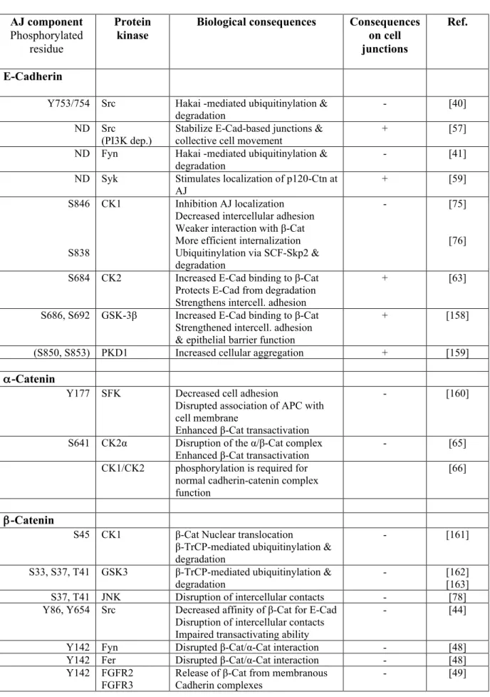
E-Cad and the Catenins are the substrates of key AJ regulating kinases and phosphatases that modulate their protein/protein interactions, thereby regulating the strength of interaction between E-Cad and the Catenin complex and their levels at the membrane. These are key steps in modulating adhesive strength and AJ remodeling. The vast majority of these biochemical studies have been performed in human (and other mammalian) cell lines, thus the residues and mechanisms described below (1.3, 1.4, and 1.5) primarily apply to the human E-Cad/Catenin complexes (more details in Table 2).
1.3. Regulation of E-Cad/Catenin association by phosphorylation
In mammalian cells, the fine-tuned regulation of the E-Cad/Catenin complex and AJ stability by phosphorylation is being well studied and its complexity is only gradually
well documented and AJ dynamics and remodeling rely more on cellular and genetic approaches. Consequently, the below-mentioned phosphorylation-based E-Cad complex regulation studies mainly focus on mammalian cells, but when appropriate studies in Drosophila are also mentioned. Early observations evidenced that a short core region of the intracellular E-Cad part is highly phosphorylated on serine [37] and tyrosine residues [38,39] and essential for Catenin binding (Table 2).
The Src kinase and other Src-family kinases (SFK) are critical protein tyrosine kinases for the regulation of E-Cad and Catenin interactions (Fig. 1 and Table 2). In mammalian cells, the Src kinase phosphorylates E-Cad on two consecutive tyrosine residues (Y753/754), thereby creating an interaction domain with the E3-ubiquitin ligase Hakai to promote ubiquitinylation and degradation of the E-Cad complex and disruption of the cell-cell contacts [40]. Other SFK members, such as Fyn, are also involved in Hakai-mediated E-Cad degradation [41], and chemical inhibition of SFKs can restore E-Cad mediated cell adhesion and reduce cancer metastasis in human cancer cell lines [42].
SFKs also phosphorylate β-Cat (Fig. 1 and Table 2) resulting in a reduced association to E-Cad and α-Cat, and subsequent decreased cell-cell adhesion [43]. C-src-mediated β-Cat phosphorylation on Y654 reduces its affinity for E-Cad leading to AJ disruption [44]. The RET receptor kinase in epithelial cells also promotes the phosphorylation of the Y654 residue of β-Cat and likewise impairs its interaction with E-Cad at AJs [45].
The SFK kinase Fyn phosphorylates p120-Ctn on the Y112 residue of its N-terminal regulatory domain, and inhibits its interaction with RhoA [46] thus potentially destabilizing AJs (see later; Fig. 1). In Drosophila, a similar negative role for
p120-increased Src activity, the associated AJ destabilization is suppressed when p120-Ctn is mutated [47] even though there is no direct proof of Src mediated phosphorylation of p120-Ctn in Drosophila. Once phosphorylated by Fyn, p120-Ctn increases its affinity for E-Cad and, consequently, promotes the association of Fyn with the AJ complex [48]. This appears however as a destabilizing event since the p120-Ctn-mediated recruitment of Fyn leads to the phosphorylation of β-Cat on the Y142 residue to prevent the association with α-Cat. Other tyrosine kinases (e.g. FGFR2, FGFR3, EGFR and TRKA; Table 2) also directly phosphorylate β-Cat at Y142, releasing Cat from membranous Cadherin complexes, increasing the cytoplasmic β-Cat concentration, and ultimately activating the canonical WNT pathway signaling [49].
In Drosophila, there are only two Src kinases, Src42A and Src64B. When overexpressed, they induce a destabilization of E-Cad based AJs [50]. Src42A is found in a complex with E-Cad and with Arm, the Drosophila β-Cat orthologue (Table 1), and is able to promote the tyrosine phosphorylation of Arm [51]. It is therefore tempting to propose that this Y-phosphorylation of β-Cat/Arm is responsible for E-Cad/Catenin complexes destabilization as observed in mammals [43,44]. However, it remains unclear whether Drosophila Src directly phosphorylates Arm, or whether this is mediated by another kinase [51], such as the Abelson kinase [52] (Fig. 1). Indeed, the E-Cad and AJ destabilization observed after increased Src activity in
Drosophila, are suppressed when Abl levels are impaired suggesting that Abl
mediates, at least in part the effect of Src [53]. Abl-mediated phosphorylation of β-Cat/Arm is further supported by studies in Drosophila embryos, where asymmetrically localized Abl kinase directs planar polarized junctional remodeling
conserved Y667 residue, resulting in β-Cat/Arm accumulation in stable junctions parallel to the embryo antero-posterior axis, while Abl is found in shrinking junctions [52]. However, this effect has yet to be linked to Src kinases. Like its mammalian counterparts, the overall AJ destabilizing effect of Src in Drosophila is further supported by the observation that mutations for Csk, the C-terminal Src kinase, a kinase that inhibits Src by phosphorylating its C-terminal tail [54], promotes AJ destabilization and cell delamination [47,55].
While overall SFK activity appears to promote E-Cad/Catenin complex destabilization and AJ destruction, the actual picture is much more dynamic, and SFK can also promote E-Cad/Catenin stability (see Table 2). For instance, Src signaling supports E-Cad signaling to PI3-kinase [56] and elevated SFK activity may also stabilize E-Cad-based junctions and collective cell movement [57], even though the unequivocal involvement of Src in a direct E-Cad phosphorylation has not been evidenced in these studies. Tyrosine phosphorylation of α-Cat enhances its translocation to the plasma membrane and its interaction with β-Cat, leading to enhanced actin polymerization and stabilization of AJs [58]. E-Cad and α-Cat are also phosphorylated by the Syk tyrosine kinase, which behaves as a tumor suppressor in epithelial cells. Their phosphorylation by Syk supports the proper localization of p120-Ctn at AJs [59], and the formation of cell-cell contacts [60].
SFKs also phosphorylate directly p120-Ctn, an essential regulator of Cadherin complexes in mammals, which binds and stabilizes E-Cad to promote its adhesive and
tumor suppressing function [26]. The Src-mediated phosphorylation pattern of p120-Ctn is complex and encompasses 8 major tyrosine sites [61] and conversely to its
phosphorylation of p120-Ctn was not observed in v-Src-transformed cells expressing
E-Cad mutants deleted of their cytoplasmic part, indicating that tyrosine phosphorylation of p120-Ctn depends on its complex formation with E-Cad and membrane localization [62]. Unlike the SFK Fyn, Src-mediated p120-Ctn phosphorylation (on Y217 and Y228) promotes increased affinity towards RhoA [46] and thus potentially AJ stabilization.
Another key kinase in the modulation of E-Cad/Catenin complexes is Casein kinase 2 (CK2; Fig. 1 and Table 2). CK2 phosphorylates E-Cad on the S684, which probably in combination with the effect of glycogen synthase kinase-3β (GSK-3β), results in increased β-Cat binding and stronger cell-cell adhesion [63]. Accordingly, decreased E-Cad phosphorylation by CK2 is associated with the disruption of AJs [64]. While CK2 appears to stabilize junctions, EGFR-ERK mediated activation of CK2α promoting the phosphorylation of α-Cat on the S641 residue, results in the disruption of the α/β-Cat complex and weaker adhesion and promotion of the β-Cat transactivation [65]. Recently, several phosphorylation sites have been identified within mammalian and Drosophila α-Cat that is sequentially modified at CK2 and CK1 consensus sites. In mammalian cells, non-phosphorylatable forms of α-Cat showed defects in intercellular adhesion suggesting that these CK1 and CK2 sites are required for normal Cadherin-Catenin complex function [66].
CK1ε, not only phosphorylates α-Cat, it can also phosphorylate p120-Ctn [67]. In response to Wnt signaling, p120-catenin is phosphorylated at S268 and S269, disrupting its interaction with E-Cadherin [68] and therefore facilitating the activation of Rac1 signaling. Strikingly, this later effect is inhibited by p120-Ctn tyrosine phosphorylation by Src or Fyn [69].
Finally, different PKC isoforms regulate p120-Ctn association with the E-Cad/Cat complexes. There are eight PKC-dependent Ser/Thr phosphorylation sites in mammalian p120-Ctn [70], and signaling events that activate PKC induce rapid phosphorylation of p120-Ctn, suggesting that p120-Ctn activity is regulated, in part, by one or more PKC isoforms. For instance, physiologic activation of several receptor tyrosine kinases such as PDGFR, induce rapid and robust p120-Ctn phosphorylation at S879, an effect mediated by PKCα, a conventional PKC isoform shown to be implicated in AJ disruption [71]. p120-Ctn is also phosphorylated at S268 in a strictly PKCɛ-dependent manner and is a key effector of the Ras-PKCɛ oncogenic signaling axis [72].
In conclusion, regulation of the E-Cad/Catenin complexes by phosphorylation is coordinated by an increasingly diverse series of phosphorylation events. Even though reviewed here, they are strongly dependent on cell type and stimulus, and might not all occur at the same time in the same cell. This largely accounts for the current apparent contradictory effects (strengthening versus disrupting AJ) (see Table 2). It is also possible that contradictory events occur simultaneously, to maintain the plasticity of the AJs, allowing rapid remodeling. The relative dynamics of many of these destruction/creation of protein/protein contacts will ultimately tilt the balance towards strengthening or disrupting of AJs. It is therefore impossible in this review to present a complete and clear-cut/unambiguous unifying model on these dynamic regulations. We did, however, opt not to simplify but realistically represent the current provisional view with the many questions remaining.
1.4. Regulation of E-Cad/Catenin levels by phosphorylations
Cadherins associate with β-Cat in the endoplasmic reticulum shortly after their synthesis, and uncomplexed cadherins are degraded [73,74], highlighting one of the mechanisms by which phosphorylations can regulate AJ strength. Alternatively, rather than this indirect effect on protein stability, key phosphorylation events may directly control E-Cad and Catenins degradation.
CK1 colocalizes with E-Cad and phosphorylates its cytoplasmic domain on the highly conserved S846 residue (Fig. 1). Constitutively phosphorylated E-Cad on S846 is unable to localize at cell-cell contacts, has a decreased intercellular adhesive activity, and binds weakly with β-Cat [75]. Indeed CK1-mediated E-Cad phosphorylation triggers its ubiquitinylation by the SCF-Skp2 E3 ubiquitin ligase complex, and its subsequent degradation [76].
Both in mammals and Drosophila, CK1 also mediates the phosphorylation and degradation of β-Cat, even though this process has been mainly documented in the regulation of the Wnt signaling-dedicated cytoplasmic pool of β-Cat (reviewed in [77]). Alternatively the membrane-associated pool of β-Cat could also be controlled by phsphorylation-primed degradation. Lee and collaborators reported that the c-Jun amino-terminal kinase (JNK) binds to the E-Cad/β-Cat complex and phosphorylates β-Cat at S37 and T41, sites that are shared with GSK-3β, disrupting intercellular contacts [78].
AJs are dynamic structures that need to adapt and are subject to constant stabilization, destruction, and renewal. The phosphorylations described above are therefore counterbalanced by dephosphorylation events. For instance, the protein Ser/Thr phosphatase-6 catalytic subunit (PP6c) accumulates at AJs, associates directly with E-Cad, and opposes CK1 to maintain cell surface localization of E-Cad [79]. Similarly, PP2A activity is required for cell-cell adhesion [80].
Several phosphatases mediate β-Cat dephosphorylation, such as PP1cγ (on the T41 and S45 residues, previously phosphorylated by GSK3 and CK1 respectively [81]), the protein-tyrosine phosphatase LAR (leukocyte common antigen related) [82], or the receptor-like protein-tyrosine phosphatase PCP-2 [83] to stabilize AJs. Similarly, dephosphorylation of p120-Ctn by phosphatases (for instance by the CD148 transmembrane tyrosine phosphatase [84]) increase the homophilic binding affinity of E-Cad, thus directly demonstrating that cell surface E-Cad is allosterically regulated by p120-Ctn [85].
2. When are Adherens Junctions remodeled?
The primary function of E-Cad based AJs is to mediate intercellular adhesion between neighboring cells, ensuring the integrity of the epithelium. It is therefore essential that AJs are maintained as stable structures to prevent weakening and ultimately rupture of the epithelial sheet, with its potential damaging effects on the homeostasis of the tissue and survival of the organism. AJs also represent anchoring points with respect to the A/B axis of epithelial cells, and as such prevent the mixing between the apical and basal determinants, the Crumbs/Patj/Pals1/Par-6/aPKC and Scrib/Dlg/Lgl/Par-1 complexes respectively [2–4,86]. The antagonistic activities of these apical and basal determinants, together with their interactions with AJs components help position and stabilize the exact position of the AJs, defining therefore the relative sizes of the apical and baso-lateral membranes, and as a consequence contribute to the accurate positioning of the other asymmetrically distributed compartments, organelles and traffic in epithelial cells.
Despite the need for stability, AJs also exhibit a tremendous plasticity and are highly remodeled during development and morphogenesis. AJs shrink and expand, change in size and strength, are dissociated and reformed, to accommodate for the changes in cell shape and to mediate relative cell movements, as epithelial cells adapt to their environment and follow stereotypical developmental morphogenesis. While not exhaustive, AJ remodeling occurs for instance in the following morphogenetic events:
- When epithelial cells move with respect to each other, such as during the convergent and extension movements of the Drosophila embryo ectodermal cells. During this morphogenetic process, global extrinsic and intrinsic forces command epithelial cell intercalation along a dorso-ventral axis generating a global antero-posterior extension.
In order to achieve this, planar polarized AJs are destroyed and created whilst cells "slide" with respect to each other (reviewed in [8,11,13,87]).
- When epithelial cells divide and undergo mitosis, new AJ material accumulates at the newly created interface between the neighboring cells that partly involve a redistribution of the cortical material [88–90].
- When epithelial cells change shape, such as during apical constriction of columnar epithelial cells of vertebrate sensory placodes or during neural tube closure. Apical constriction reduces the total apical circumference of epithelial cells and hence is accompanied by a shrinking of AJs around the apex (reviewed in [16,87]).
- When epithelia change their overall epithelial type to become either squamous or columnar. While all the previous examples implicated a remodeling of AJs along the plane of the epithelium, AJs can also be remodeled along the apico-basal axis of cells by changing their position and size, affecting the respective sizes of the apical and baso-lateral membranes. This is occurring when epithelial cells transition from a cuboidal "classical" epithelial structure, to a flatter squamous, or thicker columnar epithelium [16]. Such transitions are documented for instance when some of the cuboidal follicle cells surrounding the developing female Drosophila egg chamber become flatter and adopt a squamous type around the anterior nurse cells, while others located around the posterior oocyte become taller and columnar [91,92].
3. Establishment of E-Cadherin-based Adherens Junctions
The mechanisms and the sequence of events leading to the formation of AJs is a very active field of research and recent advances have been the subject of many excellent reviews [8,9,11,14,15]. Even though this topic is slightly outside of the primary focus of this review, notions about the mechanisms at play, and the current model describing how AJs form, are necessary to understand AJ remodeling. Indeed, in many instances, AJ remodeling appears as a partial re-use or modulation of AJ establishment.
While many details remain to be elucidated, E-Cad based AJs establishment and stabilization in metazoans, is controlled by a sequence of events involving local adhesion triggered by transmembrane adhesion molecules, the progressive recruitment of E-Cad/Catenin complexes to the membrane apposition sites, the cross-talks and dynamic interactions between the actin cytoskeleton and E-Cad clusters, and the shift from a dynamic Rac-controlled filamentous actin cytoskeleton to a more stable Rho-controlled bundled actin cytoskeleton [10,11].
Based on work in cell culture, a model emerges where AJ formation starts with the contact of actin based protrusions between two neighboring cells. This first contact is triggered by the homophilic interaction 'in trans' between E-Cad molecules initiating the formation of little clusters of E-Cad connected to the actin radial cytoskeleton [93]. These little clusters of E-Cad engagement activate Rac signaling, in particular through the activation of the RacGEF TIAM1, to promote a branched actin network and more protrusions, ultimately expanding the initial small clusters of E-Cad both by coalescence and by the diffusion of free E-Cad monomers that finally become engaged and retained on-site. This leads to the formation of discontinuous and very
dynamic spot AJs. In parallel to activating Arp2/3 filamentous actin nucleation, Rac signaling also recruits p190RhoGAP to the maturing AJs, inhibiting therefore Rho signaling at these early junctions. Alternatively, these initial contacts could be mediated or reinforced by the trans-engagement of immunoglobulin-like adhesion molecules of the Nectin family, to help recruit free E-Cad complexes to the apposition sites [11,15].
Once junctions grow, the scaffolds Par3 (PARD3 in humans, Bazooka in Drosophila; Table 1) and α-Cat are recruited to the expanding junctions and inhibit Rac signaling. The joint inhibitions of the RacGEF TIAM1 by Par3 (or its orthologue Sif by Baz in
Drosophila, see later for the role of Par3 in AJ regulation; Fig. 1) [94,95] and of
Arp2/3 and p190RhoGAP by α-Cat [96,97] result in the activation of Rho signaling and the formation of stable actin bundles that run parallel to the cell-cell contact surface and stabilize mature AJs. This ultimately results in the formation of a subcortical actin ring at the apex of epithelial cells supporting a continuous ring of E-Cad based AJs.
While this model accounts primarily for AJ formation at the interface between connecting mammalian cells, most of it can be extrapolated to other systems. There are, however, many little variations depending on the epithelial cell type, or the nature of the initial contact. For instance two main differences can be highlighted in the extensively studied Drosophila paradigms of AJ remodeling.
First, Drosophila does not appear to have a clear Nectin orthologue, even though the IgCAM molecule Echinoid (Ed) is taking over some of its Ca2+
-independent adhesive role in parallel to the action of E-Cad complexes (Table 1). For instance the
between Ed and Canoe. The link between Ed and Baz (orthologue of Par3; Table 1) will be discussed later.
Second, in the developing Drosophila embryo, the first AJs are formed during lateral membrane creation, isolating little patches of cytoplasm around individual nuclei out of the initial syncytial embryo. In this intensely studied model, there is no initial apposition of membrane from two isolated cells, and E-Cad accumulation appears primarily mediated by targeted traffic of E-Cad/Catenin complexes (reviewed in [8]). Most epithelia of the Drosophila embryo, larva, and adult, are inherited from this initial 'epithelization' and AJ creation. It is noteworthy that in the developing mammalian organism, AJs are also inherited and extended between dividing cells. The situation is therefore different from the meeting of isolated cells seen in culture conditions, suggesting that the mechanisms describing AJ initiation after apposition of membrane might be more specific than general.
Despite little differences particular for each system, a few key features of AJs formation with respect to their necessary remodeling can be highlighted:
- The formation of AJs is a multi-step process that is reversible allowing for the remodeling of E-Cad based AJs.
- The many components of E-Cad complexes and accessory proteins and the complex interactions they mutually engage in, offer many points of regulation, in particular through post-translational modifications (Fig. 1 and Table 2).
- AJ formation and evolution is driven by the cross-talks and feed-backs between E-Cad clusters and the underlying actin cytoskeleton. In particular, the switch between
Rac (flexible), and Rho (stable) signaling is critical for the transition from more mobile spot-like AJs to more stable belt-like AJs.
- A few protein scaffolds, and in particular afadin/AF-6 [93] and Par3/Baz control locally this switch between Rac and Rho signaling, and offer key entry points to regulate AJ remodeling.
4. Regulation of Adherens Junctions by the Par3/Baz scaffolds
The regulation of E-Cad-based AJs is highly dependent on the interactions of AJs with the cytoskeleton and the many factors that can regulate the formation of dynamic protein complexes centered on the E-Cad C-terminal-tail (see 1.3 & 1.4). From studies with mammalian cell culture and in vivo models such as the developmental morphogenesis in Drosophila, the scaffold Par3 has emerged as a critical node of AJ regulation. Par3 was first identified in the worm Caenorhabditis elegans as one of the critical factors controlling the initial polarization of the worm embryo. Worms mutant for PAR-3 show a partition defective phenotype during the first cell division, hence the name PAR-3 [98]. PAR-3 orthologues are found in Drosophila (Bazooka, Baz, [99]) and vertebrates (known as PARD3 in human) and form a family of evolutionarily conserved protein scaffolds, possessing multiple protein/protein interaction domains (for reviews see [3,4,100]; Table 1). These different domains and the many protein partners they interact with regulate Par3 localization and activity, and through their effect on the actin cytoskeleton and on protein kinases and/or phosphatases, they mediate the action of Par3 on AJ dynamics.
4.1. Par3 as a localization clue for Adherens Junctions
Studies performed in the developing Drosophila embryo have suggested a pivotal role of Par3/Baz in the formation and regulation of AJs [8]. Indeed, in the cellularizing early embryo, small clusters of E-Cad/Catenin complexes co-localize apically with clusters of Par3/Baz protein, but their initial localizations appear independent [101]. However, in the absence of Par3/Baz these small initial E-Cad clusters fail to grow
least to expand, even though the exact mechanisms remain unclear but likely involve redundant mechanisms affecting the cytoskeleton and trafficking [102]. As development proceeds (gastrulating embryo and later stages) these early spot AJs mature to more robust and stable belt AJs (zonulae adherens) encircling the whole circumference of the epithelial cell apex [8]. Strikingly, at this stage, E-Cad-based AJs accumulate where the Par3/Baz protein is enriched [103,104]. This correlation is also seen in the gastrulating Drosophila embryo during germ-band extension when Par3/Baz, excluded from the shrinking myosin II-rich membranes by a direct phosphorylation by Rho-kinase ROCK (Rok in Drosophila; Fig. 2), is planar polarized and accumulates along the expanding cell-cell junctions running parallel to the antero-posterior axis of the embryo, and co-localizes with an enrichment of the AJ component Arm [105–107]. The current evidence supports therefore an important role for Par3/Baz as a landmark for AJs localization, regulation and remodeling in these different Drosophila epithelia [103,108] (Fig. 2).
In mammalian cells, the role of Par3 has been primarily associated with the formation and stabilization of ZO-1-containing tight junctions (TJs Fig. 2; [94]). However, TJs and E-Cad-based AJs are in very close proximity in mammalian cells [7] and photon microscopy is insufficient to unmistakably separate them. Moreover, TJs and AJs share common mechanisms of formation and most likely influence and stabilize each other. For instance, in MDCK cells, Par3 depletion results in both AJ (marked with E-Cad) and TJ (marked with ZO-1) defects [109] suggesting that beyond its role on TJ, a role for Par3 in AJs might have been overlooked. Indeed, mouse primary mammary epithelial cells depleted of Par3 in the context of Notch or Ras oncogenic activation, exhibit increased tumor formation and cell invasiveness, and a dramatic
direct evidence of the conserved role of Par3 in mammalian AJs stabilization was recently obtained in MCF-10A immortalized human mammary cells stably transfected with the ErbB2 tyrosine kinase receptor. In these cells, Par3 knock-down by shRNA triggered decreased cell-cell adhesion, increased E-Cad membrane mobility and less stable AJs [111].
4.2. Mechanisms of Adherens Junction regulation by Par3
Even though Par3 regulates AJ formation and stability at multiple steps, the exact mechanisms by which it exerts its actions require further studies. Through its different binding partners Par3 has been shown to regulate E-Cad/Catenin complex turnover and traffic, the dynamics of the actin cytoskeleton, and the linkage between E-Cad and the underlying actin network. It is noteworthy that these different processes are all very sensitive to protein complex formation and to the phosphorylation status of the proteins involved, and in particular E-Cad and the Catenins (Fig. 1 and Table 2). The link between Par3 and the local activity of kinases and phosphatases represents therefore a promising avenue for future studies.
In multiple organisms, from invertebrates to humans, Par3 has been shown to interact with Par6 and atypical PKC (PKCζ/aPKC; Table 1), to form the so-called Par complex [3,4,100,112,113]. This complex is required to establish and maintain apico-basal polarity by defining the identity of the apical side. In Drosophila, despite a physical interaction, Par3/Baz is actually segregated from Par-6/aPKC in mature epithelial cells, and accumulates at the level of AJs (Fig. 2). Par-6 and aPKC, together with the small GTPase cdc42 but independently of Par3/Baz, control E-Cad
suggested that Par3 could have an inhibitory effect on PKCζ/aPKC [112,113], it is tantalizing to propose that the accumulation of Par3/Baz at the level of AJs prevents cdc42/Par-6/aPKC mediated E-Cad endocytosis to stabilize E-Cad complexes at the AJ level, even though this has not yet been formally demonstrated. In mammalian cells, Par3 has been observed both at AJs and the more apical TJs suggesting that Par3 might not actually segregate from the Par6/PKCζ territory (Fig. 2). However, being confined in the same broad domain does not exclude mutual antagonisms in more discrete sub-domains.
In mammalian cells, Par3 has been shown to interact with the RacGEF Tiam1 [94,117,118]. Functional assays in MDCK cultured epithelial cells show that Par3 inhibits Tiam1 activity leading to an inhibition of Rac1 signaling to stabilize TJ formation [94]. Similar observations have been made using the Drosophila pupal notum columnar epithelia where Par3/Baz inhibits the activity of Sif, the Drosophila orthologue of Tiam1 [115], supporting a model where Par3/Baz through the inhibition of Tiam1, inhibits Rac1 signaling to the actin cytoskeleton and its protrusive activity to promote the switch towards Rho signaling and its effect on actin cable formation and consequent stabilization of AJs (Reviewed in [11]; Fig. 1).
Par3 also binds to the FERM domain containing protein Nf2 (a.k.a. Merlin), which serves as a linker with α-Cat. Nf2 is required for Par3 and E-Cad stabilization at AJs in mouse keratinocytes [119] suggesting a model where Nf2 and Par3 through the recruitment of α-Cat stabilize E-Cad complexes at AJs. However, in Drosophila, the two Nf2 orthologues, Expanded and Merlin, act redundantly on membrane protein trafficking, and opposite to what could be expected from the before-mentioned study,
expanded and merlin mutant clones [120], indicating that more studies are required to
evaluate whether this role of Nf2 is limited to mouse keratinocytes or more general.
4.3. Par3 apico-basal restriction
Par3 localization is one of the critical factors that regulate AJs. Stabilizing/recruiting Par3 to certain sub-domains of the plasma membrane will govern local stabilization of the E-Cad complex clustering, controlling where AJs are stable. Remodeling patterns of AJs correlate with Par3 membrane association. Par3/Baz accumulates at the interface of the apical and baso-lateral membrane domains where AJs form [4,121]. In
Drosophila and in mammals, it appears to be achieved by a conjunction of exclusion
mechanisms from both the most apical and baso-lateral domains (Fig. 2).
In Drosophila, apical Par3/Baz is excluded by the conjunction of two mechanisms (Fig. 2). First, following its physical binding to the apical Par-6/aPKC complex, Par3/Baz is phosphorylated by aPKC on S980 which further prevents its association with aPKC, resulting in the release of Par3/Baz from the Par-6/aPKC apical complex [108,122]. However, exclusion from the most apical part of the cell is also achieved by a second mechanism involving the large transmembrane protein and apical marker Crumbs (Crb). Par-6 can bind, via its PDZ domain, to both Baz and the Crb intracellular cytoplasmic C-tail. These interactions have been shown to be mutually exclusive, and the presence of Crb at the most apical part of epithelial cells excludes Baz by engaging Par-6, [108,122]. The system is further refined as other apical factors such as Stardust (Sdt), the Drosophila orthologue of the mammalian Pals1, can also bind to Par3/Baz, and Crb or Par-6, to modulate complex formation [123].
Pals1/Sdt that matures in epithelial cells to ultimately resolve with the apical exclusion of Par3/Baz, appears to correspond to the re-wiring of the initial network responsible for setting up the apico-basal polarity [3,4,100]. Evolving from this initial network to the mature epithelial network likely involves subtle modifications in some of its parameters such as relative abundance of the different factors, strength of the different protein/protein interactions, and presence or absence of additional factors regulating protein complex formation such as accessory scaffolds, and phosphatases that could control the balance between phosphorylated and un-phosphorylated S980.
In Drosophila epithelial cells, Par3/Baz is also excluded by the activity of the baso-lateral resident kinase Par-1 (also known as EMK1/MARK2 in humans; Fig. 2). Par-1 phosphorylates Par3/Baz on S151 and S1085 generating 14-3-3 protein binding sites preventing Par3/Baz oligomerization and its binding to aPKC [124]. This results in the exclusion of Par3/Baz from the baso-lateral membranes, even though the mechanisms are not entirely clear, but might involve a recently described Par3/Baz-centrosome pathway where the authors speculate that 14-3-3-bound Par3/Baz could be targeted to the microtubules [125].
This double exclusion mechanism of Par3/Baz from the most apical and basal part is generally conserved in mammalian epithelial cells. Par3 is phosphorylated by the Ser/Thr kinases PKCζ/ι (atypical protein kinase C zeta/iota; Table 1) on S827 preventing its binding to PKCζ, and promoting the spreading of Par3 to the junctional part of the cell. Par3 is also a substrate of Par1 (EMK1/MARK2) creating 14-3-3 binding sites excluding Par3 from the most basal part of epithelial cells [126–128]. The Ser/Thr protein phosphatase 1 (PP1) is required for the dephosphorylation of
Par3 at several key serine residues, thereby regulating its association with 14-3-3 proteins and PKCζ [129].
This dual exclusion mechanism stabilizes Par3/Baz at the interface of the apical and basal domains, where AJs will form (Fig. 2). Nevertheless, since apical and baso-lateral domains antagonize each other's activity (Fig. 2), the dynamic equilibrium between them will ultimately define their relative sizes [4,86], and thus the size and localization of the domain competent to accumulate Par3/Baz and form AJs. Movements and remodeling of AJs along the apico-basal axis, or apical fattening of AJs seen during epithelial type transitions (squamous - cuboidal - columnar, see chapter 2) or during morphogenesis (such as apical constriction, see chapter 2) are ultimately controlled by these antagonistic interactions and their consequences on Par3/Baz localization [16]. It would be interesting to re-examine the morphogenesis and AJ remodeling models to study whether patterns of AJ stability could be correlated with subtle changes in the relative sizes of these lateral Par3 exclusion domains.
4.4. Par3 retention
Baz/Par3 is not only excluded from both the apical and basal sides of epithelial cells, it is also specifically retained at the level of AJs (Fig. 3).
Par3 and Baz bind to phospho-inositides, and in particular PIP2 and PIP3 that are found along the apical and basal membranes, and the PIP2/PIP3-binding domain of Par3/Baz is critical for its membrane localization [104,130]. Recently, studies in
phospho-inositide for Baz membrane recruitment and AJ formation in the follicular epithelial cells [131]. Interestingly, in both mammals and Drosophila, Par3/Baz binds the PTEN phosphatase [132,133], which converts PIP3 to PIP2. PTEN was recently shown to control morphogenesis in the Drosophila wing epithelia, at least in part through AJ remodeling and myosin II localization [134]. It is tempting to propose that the localized PIP2/PIP3 balance controlled by PTEN, impacts subtle changes in the ability of Baz to associate with the membrane, and affects AJ remodeling and dynamics, even though this remains yet to be proven.
Par3 binds several AJ resident proteins involved in its retention at the interface between apical and basal domains. In Drosophila, the PDZ domains of Par3/Baz bind both to the C-termini of the adhesion molecule Echinoid (Ed) and the β-Cat orthologue Arm [135] (Fig. 3). It remains, however, controversial whether these two interactions are critical, or if they are required in different epithelial tissues since AJs form normally when these interactions are impaired [102,136]. More subtle effects, and redundant action between Arm and Ed, or with an as yet un-identified other AJ resident factor cannot be excluded. It is noteworthy that Par3 binds to the C-terminus of the Nectin AJ regulators in mammalian cells, weak orthologues of Ed [109,137]. This evolutionarily conserved interaction suggests a key role for the Nectin-Par3 interaction in AJ regulation, even though this still remains unclear in Drosophila.
4.5. A new apical complex regulating Par3 apical localization
with the most apical part of the AJs [138,139] (Fig. 4). More importantly, ASPP2 binds to Par3 and is required for the correct Par3 apical localization. TJs are strongly delayed in ASPP2 knock-down cells, as well as in cells overexpressing the ASPP2 destabilizing enzyme Siah2 (an E3 ubiquitin-protein ligase) [138–140]. ASPP2 was also shown to interact with β-Cat at AJs (Fig. 4) in human mammary cells MCF-10A, where it was proposed to inhibit β-Cat accumulation and transport to the nucleus, thus preventing ZEB1 expression and E-Cad gene repression [141]. It emerges that ASPP2 is an apical junction promoter, and in particular AJs, via the binding to both Par3 and β-Cat. In Drosophila, the unique ASPP orthologue is required for E-Cad based AJ integrity at least in part through its activation of Csk activity (an inhibitor of the Src kinase [55], see 1.3). Furthermore, in the developing Drosophila pupal eye ASPP is required both for Par3/Baz recruitment at the membrane at AJ level, and for the integrity of the E-Cad belt found around the cortex of inter-ommatidial epithelial cells [142]. No direct binding between ASPP and Par3/Baz has been documented in the fly, so the actual mechanism by which ASPP brings Par3/Baz to the membrane in
Drosophila remains elusive.
A remaining key question is what brings ASPP to the cortex and at the level of future AJs? The situation in mammalian cells is complex: while ASPP2 is required for correct localization of Par3 at junction sites, reciprocally Par3 is required for ASPP2 localization [138,139,143]. This could reflect the early role of Par3 in setting up the apico-basal axis, rather than its more dedicated role in junction specification. In the
Drosophila pupal eye, ASPP localization is not dramatically affected in Par3/Baz
mutant cells, ruling out Par3/Baz as the primary anchor of ASPP in the fly [142]. However, ASPP correct AJ localization is mediated by its physical interaction with
pattern of interrupted AJs [144]. We have recently shown that in Drosophila the scaffold Magi is the upstream anchor, and that through its physical interaction with RASSF8, Magi controls ASPP and Par3/Baz localization at AJs and regulates the integrity of E-Cad junctions [142] (Fig. 4). The genetic requirement for Magi and
RASSF8 are, however, limited to a small time window during pupal eye development
suggesting that the Magi-RASSF8-ASPP axis of Par3/Baz localization at AJs is redundant with other mechanisms [142], such as interaction with β-Cat [135], or with the double exclusion machineries described before (Fig. 2 and Fig. 3). In the nematode C. elegans, MAGI-1 interacts genetically with the Cadherin/Catenins complex to define junction domains [145]. In mammalian cells, there are three Magi orthologues, MAGI1, 2, and 3. They have been shown to regulate TJ and AJ formation and invasiveness (reviewed in [146]). Of note, this AJ stabilizing effect of MAGI scaffolds has been proposed to be mediated by physical interactions with β-Cat [147,148], with PTEN [149,150], or with regulators of the junction remodeling small GTPase Rap1 [151,152]. A role for MAGI scaffolds on the actin cytoskeleton is therefore suggested by this link with Rap1 signaling [153], but also by the physical interactions of MAGI1 with the actin binding proteins alpha-actinin-4 and synaptopodin in MDCK cells [154]. Interestingly, in biochemical studies, MAGI1 was shown to bind directly to ASPP2 [155], suggesting that a common ASPP-MAGI axis regulating AJs, likely through the localization of Par3, could be conserved from
Drosophila to human (Fig. 4). Whether mammalian orthologues of RASSF8 are also
involved in the complex remains to be explored, but a role of the RASSF7-10 scaffold family (the RASSF with the Ras association domain in the N-terminal) is supported by the observation that RASSF8 is found at the level of AJs co-localizing with E-Cad
AJ destabilization [156], reminiscent of the role proposed for its Drosophila counterpart.
Many open questions regarding this emerging MAGI-RASSF8-ASSP complex remain, including the evolutionary conservation of some of the protein interactions in the complex, and the potential cross regulations with the other Par3 localization and AJ regulation machineries.
CONCLUSIONS
E-Cad based AJs are a crucial structure of epithelial cells. Even though their primary role is to mediate intercellular adhesion, they participate in the overall polarization of epithelial cells to segregate apical and basal membrane compartments, and to polarize organelles and intracellular traffic. They engage in complex feedback interactions with the actin-myosin cytoskeleton network and are hotspots to integrate tissue force sensing, and to assemble cell signaling platforms. Despite these key cellular functions, AJs that seem stable on a short time-scale, appear very plastic on a longer time-scale, a feature that is critical for tissue morphogenesis and cell shape changes. In this review, we have discussed the recent findings regarding the ever more elaborate mechanisms ensuring the correct localization and remodeling of AJs by the epithelial polarity machineries and in particular the Par3 apical scaffold. The correct localization and stability of Par3 scaffolds is under the tight control of many other scaffolds, and ultimately regulated by numerous phosphorylation/de-phosphorylation events such as by the apico-basal polarized aPKC/PKCζ and PAR1 kinases, or the planar polarized ROCK kinase, in Drosophila and mammals. The recent identification in Drosophila of a Magi/RASSF8/ASPP complex regulating both Par3/Baz recruitment and the activity of Csk, a negative regulator of SFK, opens the exciting prospect that SFK activity could be regulating Par3 localization and/or activity as was suggested by studies in human cell lines, where Par3 is phosphorylated on tyrosine by the SFK members c-Src and c-Yes, releasing the LIM kinase 2 (LIMK2) to promote junction formation [157]. The integration of Par3/Baz, and other key apical scaffolds, with the reversible phosphorylation events controlling the stability and strength of the
controlling AJ stability and remodeling during normal epithelial development and pathology.
ACKNOWLEDGEMENTS
The authors would like to thank the members of the PC and AD labs, and S. Zaessinger for critical reading. The PC lab is supported by grants from the “Plan Cancer/INCa” (ASC14021FSA) and the “Fondation ARC pour la recherche sur le cancer” (SL220110603480). The AD lab is supported by grants from the "Fondation ARC pour la recherche sur le cancer", "Marie Curie CIG", and "ATIP/Avenir programme".
FIGURE LEGENDS
Figure 1. Core E-Cadherin/Catenin complexes at Adherens Junctions and their regulation by phosphorylation.
Shown is a simplified comparison of the Drosophila (left) and mammalian (right) adherens junction (AJ) complexes and key kinases (in bold and in blue) regulating the association and stability between E-Cadherin and the different catenins. Arrows represent phosphorylations with an activating function (e.g. stabilization of complexes and AJs); bars represent inhibitory phosphorylations.
Figure 2. Apical restriction of Par3/Baz localization by inhibitory kinases.
Structures of epithelial cells in Drosophila (left) and in mammals (right), with the different protein orthologues implicated in Par3/Baz restriction, are presented. AJs, (shown in red) are localized apically at the interface between the apical and basal membranous domains characterized by antagonizing protein complexes: the apical Par6/PKCζ (green), and the baso-lateral Dlg/Lgl/Scrib/Par1 (blue). Par3 accumulates at the level of AJs due to a double restriction by the apical kinase PKCζ and by the basal kinase Par1. The domains of these two kinases stay segregated due to the mutual exclusion of the apical (green) and baso-lateral (blue) protein complex networks. Par3 levels are further modulated along the circumference of the AJ domain by the antagonizing Rho Kinase (ROCK).
Figure 3. Adherens Junction retention of Par3/Baz.
Comparison between the Drosophila (left) and mammalian (right) mechanisms to anchor Par3/Baz at AJs. Lines (black, red and green) indicate physical interaction.
PTEN promotes the production of PIP2 phospholipids, a major component of the plasma membrane, anchoring Par3/Baz at the membrane.
Figure 4. A MAGI-ASPP network to localize Par3/Baz at the Adherens Junction. Comparison between the Drosophila (left) and mammalian (right) emerging role of the MAGI-ASPP network in AJ regulation. Lines (black, red and green) indicate physical interaction. Further work on this network will allow to clarify the exact localizations and interactions between the different members of the complexes, and between the MAGI-ASPP network and the other factors regulating E-Cad complexes and Par3 localization presented in Figures 1, 2, and 3.
REFERENCES
[1] Bryant DM, Mostov KE. From cells to organs: building polarized tissue. Nat Rev Mol Cell Biol 2008;9:887–901. doi:10.1038/nrm2523.
[2] Goldstein B, Macara IG. The PAR proteins: fundamental players in animal cell polarization. Dev Cell 2007;13:609–22. doi:10.1016/j.devcel.2007.10.007. [3] St Johnston D, Ahringer J. Cell polarity in eggs and epithelia: parallels and
diversity. Cell 2010;141:757–74. doi:10.1016/j.cell.2010.05.011.
[4] Tepass U. The apical polarity protein network in Drosophila epithelial cells: regulation of polarity, junctions, morphogenesis, cell growth, and survival. Annu Rev Cell Dev Biol 2012;28:655–85.
doi:10.1146/annurev-cellbio-092910-154033.
[5] Nelson WJ, Dickinson DJ, Weis WI. Roles of cadherins and catenins in cell-cell adhesion and epithelial cell polarity. Prog Mol Biol Transl Sci 2013;116:3–23. doi:10.1016/B978-0-12-394311-8.00001-7.
[6] Wei Q, Huang H. Insights into the role of cell-cell junctions in physiology and disease. Int Rev Cell Mol Biol 2013;306:187–221. doi:10.1016/B978-0-12-407694-5.00005-5.
[7] Harder JL, Margolis B. SnapShot: tight and adherens junction signaling. Cell 2008;133:1118, 1118.e1–2. doi:10.1016/j.cell.2008.06.002.
[8] Harris TJC. Adherens junction assembly and function in the Drosophila embryo. Int Rev Cell Mol Biol 2012;293:45–83.
doi:10.1016/B978-0-12-394304-0.00007-5.
[9] Baum B, Georgiou M. Dynamics of adherens junctions in epithelial
establishment, maintenance, and remodeling. J Cell Biol 2011;192:907–17. doi:10.1083/jcb.201009141.
[10] Röper K. Integration of cell-cell adhesion and contractile actomyosin activity during morphogenesis. Curr Top Dev Biol 2015;112:103–27.
doi:10.1016/bs.ctdb.2014.11.017.
[11] Collinet C, Lecuit T. Stability and dynamics of cell-cell junctions. Prog Mol Biol Transl Sci 2013;116:25–47. doi:10.1016/B978-0-12-394311-8.00002-9.
[12] de Beco S, Gueudry C, Amblard F, Coscoy S. Endocytosis is required for E-cadherin redistribution at mature adherens junctions. Proc Natl Acad Sci U S A 2009;106:7010–5. doi:10.1073/pnas.0811253106.
[13] Lecuit T, Yap AS. E-cadherin junctions as active mechanical integrators in tissue dynamics. Nat Cell Biol 2015;17:533–9. doi:10.1038/ncb3136.
[14] Yap AS, Crampton MS, Hardin J. Making and breaking contacts: the cellular biology of cadherin regulation. Curr Opin Cell Biol 2007;19:508–14.
doi:10.1016/j.ceb.2007.09.008.
[15] Ivanov AI, Naydenov NG. Dynamics and regulation of epithelial adherens junctions: recent discoveries and controversies. Int Rev Cell Mol Biol 2013;303:27–99. doi:10.1016/B978-0-12-407697-6.00002-7.
[16] St Johnston D, Sanson B. Epithelial polarity and morphogenesis. Curr Opin Cell Biol 2011;23:540–6. doi:10.1016/j.ceb.2011.07.005.
[17] Gumbiner BM. Regulation of cadherin-mediated adhesion in morphogenesis. Nat Rev Mol Cell Biol 2005;6:622–34. doi:10.1038/nrm1699.
[18] Desai R, Sarpal R, Ishiyama N, Pellikka M, Ikura M, Tepass U. Monomeric α-catenin links cadherin to the actin cytoskeleton. Nat Cell Biol 2013;15:261–73. doi:10.1038/ncb2685.
[19] Nagafuchi A, Ishihara S, Tsukita S. The roles of catenins in the cadherin-mediated cell adhesion: functional analysis of E-cadherin-alpha catenin fusion molecules. J Cell Biol 1994;127:235–45.
[20] Cavey M, Rauzi M, Lenne P-F, Lecuit T. A two-tiered mechanism for stabilization and immobilization of E-cadherin. Nature 2008;453:751–6. doi:10.1038/nature06953.
[21] Yonemura S, Wada Y, Watanabe T, Nagafuchi A, Shibata M. alpha-Catenin as a tension transducer that induces adherens junction development. Nat Cell Biol 2010;12:533–42. doi:10.1038/ncb2055.
[22] Buckley CD, Tan J, Anderson KL, Hanein D, Volkmann N, Weis WI, et al. Cell adhesion. The minimal cadherin-catenin complex binds to actin filaments under force. Science 2014;346:1254211. doi:10.1126/science.1254211.
[23] Abe K, Takeichi M. EPLIN mediates linkage of the cadherin catenin complex to F-actin and stabilizes the circumferential actin belt. Proc Natl Acad Sci U S A 2008;105:13–9. doi:10.1073/pnas.0710504105.
[24] Bershadsky A. Magic touch: how does cell-cell adhesion trigger actin assembly? Trends Cell Biol 2004;14:589–93. doi:10.1016/j.tcb.2004.09.009.
[25] Yonemura S. Cadherin-actin interactions at adherens junctions. Curr Opin Cell Biol 2011;23:515–22. doi:10.1016/j.ceb.2011.07.001.
[26] Kourtidis A, Ngok SP, Anastasiadis PZ. p120 catenin: an essential regulator of cadherin stability, adhesion-induced signaling, and cancer progression. Prog Mol Biol Transl Sci 2013;116:409–32. doi:10.1016/B978-0-12-394311-8.00018-2. [27] Shahbazi MN, Megias D, Epifano C, Akhmanova A, Gundersen GG, Fuchs E, et
al. CLASP2 interacts with p120-catenin and governs microtubule dynamics at adherens junctions. J Cell Biol 2013;203:1043–61. doi:10.1083/jcb.201306019. [28] Chen X, Kojima S, Borisy GG, Green KJ. p120 catenin associates with kinesin
and facilitates the transport of cadherin-catenin complexes to intercellular junctions. J Cell Biol 2003;163:547–57. doi:10.1083/jcb.200305137.
[29] Wildenberg GA, Dohn MR, Carnahan RH, Davis MA, Lobdell NA, Settleman J, et al. p120-catenin and p190RhoGAP regulate cell-cell adhesion by coordinating antagonism between Rac and Rho. Cell 2006;127:1027–39.
doi:10.1016/j.cell.2006.09.046.
[30] Noren NK, Liu BP, Burridge K, Kreft B. p120 catenin regulates the actin cytoskeleton via Rho family GTPases. J Cell Biol 2000;150:567–80. [31] Davis MA, Ireton RC, Reynolds AB. A core function for p120-catenin in
cadherin turnover. J Cell Biol 2003;163:525–34. doi:10.1083/jcb.200307111. [32] Hoshino T, Sakisaka T, Baba T, Yamada T, Kimura T, Takai Y. Regulation of
E-cadherin endocytosis by nectin through afadin, Rap1, and p120ctn. J Biol Chem 2005;280:24095–103. doi:10.1074/jbc.M414447200.
[33] Smith AL, Dohn MR, Brown MV, Reynolds AB. Association of Rho-associated protein kinase 1 with E-cadherin complexes is mediated by p120-catenin. Mol Biol Cell 2012;23:99–110. doi:10.1091/mbc.E11-06-0497.
[34] Myster SH, Cavallo R, Anderson CT, Fox DT, Peifer M. Drosophila p120catenin plays a supporting role in cell adhesion but is not an essential adherens junction component. J Cell Biol 2003;160:433–49.
doi:10.1083/jcb.200211083.
[35] Pacquelet A, Lin L, Rorth P. Binding site for p120/delta-catenin is not required for Drosophila E-cadherin function in vivo. J Cell Biol 2003;160:313–9.
[36] Bulgakova NA, Brown NH. Drosophila p120-catenin is crucial for endocytosis of the dynamic E-cadherin-Bazooka complex. J Cell Sci 2016;129:477–82. doi:10.1242/jcs.177527.
[37] Stappert J, Kemler R. A short core region of E-cadherin is essential for catenin binding and is highly phosphorylated. Cell Adhes Commun 1994;2:319–27. [38] Behrens J, Vakaet L, Friis R, Winterhager E, Van Roy F, Mareel MM, et al.
Loss of epithelial differentiation and gain of invasiveness correlates with tyrosine phosphorylation of the E-cadherin/beta-catenin complex in cells transformed with a temperature-sensitive v-SRC gene. J Cell Biol
1993;120:757–66.
[39] Matsuyoshi N, Hamaguchi M, Taniguchi S, Nagafuchi A, Tsukita S, Takeichi M. Cadherin-mediated cell-cell adhesion is perturbed by v-src tyrosine
phosphorylation in metastatic fibroblasts. J Cell Biol 1992;118:703–14. [40] Fujita Y, Krause G, Scheffner M, Zechner D, Leddy HEM, Behrens J, et al.
Hakai, a c-Cbl-like protein, ubiquitinates and induces endocytosis of the E-cadherin complex. Nat Cell Biol 2002;4:222–31. doi:10.1038/ncb758.
[41] Smyth D, Leung G, Fernando M, McKay DM. Reduced surface expression of epithelial E-cadherin evoked by interferon-gamma is Fyn kinase-dependent. PloS One 2012;7:e38441. doi:10.1371/journal.pone.0038441.
[42] Nam J-S, Ino Y, Sakamoto M, Hirohashi S. Src family kinase inhibitor PP2 restores the E-cadherin/catenin cell adhesion system in human cancer cells and reduces cancer metastasis. Clin Cancer Res Off J Am Assoc Cancer Res 2002;8:2430–6.
[43] Hu P, O’Keefe EJ, Rubenstein DS. Tyrosine phosphorylation of human keratinocyte beta-catenin and plakoglobin reversibly regulates their binding to E-cadherin and alpha-catenin. J Invest Dermatol 2001;117:1059–67.
doi:10.1046/j.0022-202x.2001.01523.x.
[44] Roura S, Miravet S, Piedra J, García de Herreros A, Duñach M. Regulation of E-cadherin/Catenin association by tyrosine phosphorylation. J Biol Chem
1999;274:36734–40.
[45] Fernández-Sánchez ME, Barbier S, Whitehead J, Béalle G, Michel A, Latorre-Ossa H, et al. Mechanical induction of the tumorigenic β-catenin pathway by tumour growth pressure. Nature 2015;523:92–5. doi:10.1038/nature14329. [46] Castaño J, Solanas G, Casagolda D, Raurell I, Villagrasa P, Bustelo XR, et al.
Specific phosphorylation of p120-catenin regulatory domain differently modulates its binding to RhoA. Mol Cell Biol 2007;27:1745–57. doi:10.1128/MCB.01974-06.
[47] Vidal M, Larson DE, Cagan RL. Csk-deficient boundary cells are eliminated from normal Drosophila epithelia by exclusion, migration, and apoptosis. Dev Cell 2006;10:33–44. doi:10.1016/j.devcel.2005.11.007.
[48] Piedra J, Miravet S, Castaño J, Pálmer HG, Heisterkamp N, García de Herreros A, et al. p120 Catenin-associated Fer and Fyn tyrosine kinases regulate beta-catenin Tyr-142 phosphorylation and beta-beta-catenin-alpha-beta-catenin Interaction. Mol Cell Biol 2003;23:2287–97.
[49] Krejci P, Aklian A, Kaucka M, Sevcikova E, Prochazkova J, Masek JK, et al. Receptor tyrosine kinases activate canonical WNT/β-catenin signaling via MAP kinase/LRP6 pathway and direct β-catenin phosphorylation. PloS One
[50] Shindo M, Wada H, Kaido M, Tateno M, Aigaki T, Tsuda L, et al. Dual function of Src in the maintenance of adherens junctions during tracheal epithelial
morphogenesis. Dev Camb Engl 2008;135:1355–64. doi:10.1242/dev.015982. [51] Takahashi M, Takahashi F, Ui-Tei K, Kojima T, Saigo K. Requirements of
genetic interactions between Src42A, armadillo and shotgun, a gene encoding E-cadherin, for normal development in Drosophila. Dev Camb Engl
2005;132:2547–59. doi:10.1242/dev.01850.
[52] Tamada M, Farrell DL, Zallen JA. Abl regulates planar polarized junctional dynamics through β-catenin tyrosine phosphorylation. Dev Cell 2012;22:309– 19. doi:10.1016/j.devcel.2011.12.025.
[53] Singh J, Aaronson SA, Mlodzik M. Drosophila Abelson kinase mediates cell invasion and proliferation through two distinct MAPK pathways. Oncogene 2010;29:4033–45. doi:10.1038/onc.2010.155.
[54] Pedraza LG, Stewart RA, Li D-M, Xu T. Drosophila Src-family kinases function with Csk to regulate cell proliferation and apoptosis. Oncogene 2004;23:4754– 62. doi:10.1038/sj.onc.1207635.
[55] Langton PF, Colombani J, Aerne BL, Tapon N. Drosophila ASPP regulates C-terminal Src kinase activity. Dev Cell 2007;13:773–82.
doi:10.1016/j.devcel.2007.11.005.
[56] Pang J-H, Kraemer A, Stehbens SJ, Frame MC, Yap AS. Recruitment of phosphoinositide 3-kinase defines a positive contribution of tyrosine kinase signaling to E-cadherin function. J Biol Chem 2005;280:3043–50.
doi:10.1074/jbc.M412148200.
[57] Veracini L, Grall D, Schaub S, Beghelli-de la Forest Divonne S, Etienne-Grimaldi M-C, Milano G, et al. Elevated Src family kinase activity stabilizes E-cadherin-based junctions and collective movement of head and neck squamous cell carcinomas. Oncotarget 2015;6:7570–83. doi:10.18632/oncotarget.3071. [58] Burks J, Agazie YM. Modulation of alpha-catenin Tyr phosphorylation by SHP2
positively effects cell transformation induced by the constitutively active FGFR3. Oncogene 2006;25:7166–79. doi:10.1038/sj.onc.1209728. [59] Larive RM, Urbach S, Poncet J, Jouin P, Mascré G, Sahuquet A, et al.
Phosphoproteomic analysis of Syk kinase signaling in human cancer cells reveals its role in cell-cell adhesion. Oncogene 2009;28:2337–47.
doi:10.1038/onc.2009.99.
[60] Zhang X, Shrikhande U, Alicie BM, Zhou Q, Geahlen RL. Role of the protein tyrosine kinase Syk in regulating cell-cell adhesion and motility in breast cancer cells. Mol Cancer Res MCR 2009;7:634–44. doi:10.1158/1541-7786.MCR-08-0371.
[61] Mariner DJ, Anastasiadis P, Keilhack H, Böhmer FD, Wang J, Reynolds AB. Identification of Src phosphorylation sites in the catenin p120ctn. J Biol Chem 2001;276:28006–13. doi:10.1074/jbc.M102443200.
[62] Ozawa M, Ohkubo T. Tyrosine phosphorylation of p120(ctn) in v-Src transfected L cells depends on its association with E-cadherin and reduces adhesion activity. J Cell Sci 2001;114:503–12.
[63] Lickert H, Bauer A, Kemler R, Stappert J. Casein kinase II phosphorylation of E-cadherin increases E-cadherin/beta-catenin interaction and strengthens cell-cell adhesion. J Biol Chem 2000;275:5090–5.