HAL Id: inserm-00869705
https://www.hal.inserm.fr/inserm-00869705
Submitted on 4 Oct 2013
HAL is a multi-disciplinary open access
archive for the deposit and dissemination of
sci-entific research documents, whether they are
pub-lished or not. The documents may come from
teaching and research institutions in France or
abroad, or from public or private research centers.
L’archive ouverte pluridisciplinaire HAL, est
destinée au dépôt et à la diffusion de documents
scientifiques de niveau recherche, publiés ou non,
émanant des établissements d’enseignement et de
recherche français ou étrangers, des laboratoires
publics ou privés.
Mélanie Bousquenaud, Fatiha Maskali, Sylvain Poussier, Jennifer Zangrando,
Pierre-Yves Marie, Henri Boutley, Renaud Fay, Gilles Karcher, Daniel
Wagner, Yvan Devaux
To cite this version:
Mélanie Bousquenaud, Fatiha Maskali, Sylvain Poussier, Jennifer Zangrando, Pierre-Yves Marie, et
al.. Cardioprotective effects of adenosine within the border and remote areas of myocardial infarction..
EJNMMI Research, SpringerOpen, 2013, 3 (1), pp.65. �10.1186/2191-219X-3-65�. �inserm-00869705�
O R I G I N A L R E S E A R C H
Open Access
Cardioprotective effects of adenosine within the
border and remote areas of myocardial infarction
Mélanie Bousquenaud
1, Fatiha Maskali
2, Sylvain Poussier
2, Jennifer Zangrando
1, Pierre-Yves Marie
2,3, Henri Boutley
2,
Renaud Fay
4, Gilles Karcher
2, Daniel R Wagner
1,5and Yvan Devaux
1*Abstract
Background: Adenosine may have beneficial effects on left ventricular function after myocardial infarction (MI), but the magnitude of this effect on remote and MI areas is controversial. We assessed the long-term effects of
adenosine after MI using electrocardiogram-triggered18F-fluorodeoxyglucose positron emission tomography. Methods: Wistar rats were subjected to coronary ligation and randomized into three groups treated daily for 2 months by NaCl (control; n = 7), 2-chloroadenosine (CADO; n = 8) or CADO with 8-sulfophenyltheophilline, an antagonist of adenosine receptors (8-SPT; n = 8).
Results: After 2 months, control rats exhibited left ventricular remodelling, with increased end-diastolic volume and decreased ejection fraction. Left ventricular remodelling was not significantly inhibited by CADO. Segmental contractility, as assessed by the change in myocardial thickening after 2 months, was improved in CADO rats compared to control rats (+1.6% ± 0.8% vs.−2.3% ± 0.8%, p < 0.001). This improvement was significant in border (+5.6% ± 0.8% vs. +1.5% ± 0.8%, p < 0.001) and remote (−4.0% ± 1.0% vs. −10.4% ± 1.3%, p < 0.001) segments, but absent in MI segments. Histological analyses revealed that CADO reduced fibrosis, cardiomyocyte hypertrophy and apoptosis. Protective effects of CADO were blunted by 8-SPT.
Conclusion: Long-term administration of adenosine protects the left ventricle from contractile dysfunction following MI.
Keywords: Adenosine; Myocardial infarction; Contractility; Left ventricular remodelling; Gated cardiac positron emission tomography
Background
Despite reperfusion therapies, left ventricular (LV) re-modelling occurs in many patients after acute myocar-dial infarction (MI), leading to a risk of subsequent heart failure [1]. This has stimulated efforts to develop new pharmacological strategies to prevent or reverse LV remod-elling. LV remodelling is defined as a complex sequence of changes in left ventricle geometry and function [2,3] involving progressive LV dilatation, cardiomyocyte hypertrophy and fibrosis, ultimately leading to the loss of contractile function and LV dysfunction. The long-term outcome of acute MI patients largely depends on the amount of the initial injury which conditions subsequent
LV remodelling. Thus, therapeutic strategies aiming at reducing infarct size and improving LV remodelling are of major interest.
Adenosine is a ubiquitous endogenous nucleoside that modulates physiological functions in various organs and plays important roles within the cardiovascular system. It can signal through four sub-types of G protein-coupled receptors, all of which are expressed in the heart [4]. Cardioprotective properties of adenosine have been characterized, particularly during ischemic pre-conditioning. However, the long-term effect of adenosine after MI has received less attention.
Disappointing results were documented when adeno-sine was administrated during the acute phase of reperfused MI in humans [5-7]. However, further experi-mental data suggest that beneficial effects could be
* Correspondence:yvan.devaux@crp-sante.lu
1
Laboratory of Cardiovascular Research, Centre de Recherche Public de la Santé, 84 Val Fleuri, Luxembourg L1526, Luxembourg
Full list of author information is available at the end of the article
© 2013 Bousquenaud et al.; licensee Springer. This is an Open Access article distributed under the terms of the Creative Commons Attribution License (http://creativecommons.org/licenses/by/2.0), which permits unrestricted use, distribution, and reproduction in any medium, provided the original work is properly cited.
Bousquenaudet al. EJNMMI Research 2013, 3:65 http://www.ejnmmires.com/content/3/1/65
long-term administration of A2B adenosine receptor agonist in a rat infarct model improved global LV function assessed by echocardiography [8]. A recent study showed that infarct size could be reduced by increasing adenosine levels in transgenic swine through over-expression of ectonucleoside triphosphate diphosphohydrolase-1 (CD39) [9].
While adenosine appears to have certain beneficial ef-fects after MI, the mechanisms and sites of these efef-fects, as well as the consequences on global LV remodelling, are poorly documented. Therefore, the aim of the current study was to investigate the effects of long-term administration of adenosine on LV function and remod-elling after MI by serial non-invasive imaging with elec-trocardiogram (ECG)-triggered 18F-fluorodeoxyglucose positron emission tomography (FDG-PET). FDG-PET has been reported as an imaging technique for assessing the severity and the location of MI and is also capable of quantifying global and segmental LV function [10,11]. Thus, FDG-PET allows for a separate analysis of the contractile function of MI, remote and border segments.
Methods
This study was performed in accordance with the regula-tions of the Animal Welfare Act of the National Institutes of Health Guide for the Care and Use of Laboratory Animals (NIH Publication No. 85-23, revised 1996), and protocols were approved by the Regional Veterinary Department (‘Direction Départementale de la Protection des Populations’), agreements RAR1A03516811825 and 54–100.
Animal model and experimental design
Twenty-three adult male Wistar rats (282- to 335-g body weight at the beginning of the study; Charles Rivers Laboratories, Wilmington, MA, USA) were enrolled. All animals were housed on a 12-h light-dark cycle in a room with temperature and humidity control, and with ad libitum access to tap water and standard rodent food. Permanent occlusion of the left anterior descending (LAD) coronary artery was performed in 23 rats as pre-viously described [10,12]. The choice of this model ra-ther than a model of ischemia/reperfusion was based on preliminary experiments suggesting that adenosine may be more protective in large infarcts than small infarcts (not shown). Rats were anaesthetized by inhalation of an isoflurane/oxygen mixture (2% to 3%:1.5% v/v) and were intubated and ventilated with a rodent ventilator (Harvard Apparatus, Holliston, MA, USA). The heart was exposed through a left lateral thoracotomy of the fifth intercostal space. After pericardial incision, the proximal part of the LAD (2 to 3 mm from the top of the left atrium) was ligated with a 7-0 Prolene suture (Ethicon, Somerville, NJ, USA). Perioperative lidocaine (10 mg/kg, Aguettant, Lyon, France) was used to provide
a local anaesthesia and to reduce the incidence of ven-tricular tachycardia and fibrillation. Finally, the chest was closed in layers with a 2-0 Vicryl suture (Ethicon, Somerville, NJ, USA), the lungs were reinflated using positive-end expiratory pressure, and the endotracheal tube was removed once spontaneous breathing had resumed. Amoxicillin was injected intramuscularly (60 mg/kg, GlaxoSmithKline, Marly-le-Roi, France) to avoid bacterial infections. The mortality of the proced-ure was <20%.
Two days after surgery, rats were assessed by ECG-triggered 18F-FDG-PET. This first PET exam allowed for randomizing the animals into three treatment groups according to infarct size. This randomization was crucial to avoid any bias due to different infarct sizes in experi-mental groups. Infarct size is indeed a major determin-ant of LV remodelling. A three-point classification was used to determine MI size, as previously described by our team [10]: small MI (no more than 3 among the 17 LV segments), moderate MI (4 to 5 segments) and large MI (at least 6 MI segments). Groups were constituted in order to exhibit equivalent rates of rats with small, moder-ate and large MI. In each of the three groups, rats were assigned to treatment with NaCl (control group; n = 7) or 2-chloroadenosine (2 mg/kg/day), a stable analogue of ad-enosine, which was given alone (CADO group; n = 8) or with 8-sulfophenyltheophilline (10 mg/kg/day), an antag-onist of adenosine receptors (8-SPT group; n = 8). NaCl, CADO and 8-SPT (Sigma-Aldrich, Bornem, Belgium) were administered intraperitoneally twice daily for 2 months, starting 7 days after the date of LAD occlusion.
18
F-Fluorodeoxyglucose PET
Two days, 1 month and 2 months after surgery, LV func-tion and infarct size were assessed in vivo by FDG-PET. As already described in detail elsewhere [10,13], rats re-ceived an oral pre-medication of 50 mg/kg of acipimox, a potent nicotinic acid derivative yielding high-quality cardiac FDG-PET images. Around 74 MBq of 18F-FDG (IBA, Nancy, France) were injected intravenously under a transient anaesthesia with isoflurane. A 16-min record-ing was started 1 h later on a high-resolution dedicated small animal PET system (Inveon, Siemens, Knoxville, TN, USA) and under continuous anaesthesia by isoflurane. Im-ages were reconstructed in 16 cardiac intervals with a 3-D ordered subset expectation maximization algorithm lead-ing to a voxel size of 0.8 × 0.4 × 0.4 mm.
FDG uptake was determined on the set of collapsed short-axis slices in each segment from the 17-segment LV division from the American Heart Association [14] with the QGS software [15]. LV end-diastolic volume (EDV), LV end-systolic volume (ESV) and ejection frac-tion (EF) were obtained from the set of contiguous ECG-triggered short-axis slices with the QGS software
[15,16]. The accuracy of these volume measurements was previously demonstrated on phantoms from LV rats above the level of 100 μL, corresponding to the lower limit for the LV end-systolic volume in adult rats [13]. The QGS software was also used for assessing the con-tractility of each of the 17 LV segments according to the percentage of systolic increase in myocardial counts
[15]. This parameter is strongly linked to the percentage of myocardial thickening with both myocardial SPECT and TEP imaging techniques [17,18].
As previously described and validated [10], all seg-ments for which the average FDG uptake was very low (<50% of the maximal voxel value) could be considered as predominantly necrotic, and the percentage of such
Figure 1 Examples of FDG-PET images at 48 h, 1 month and 2 months in representative cases. Horizontal and vertical long-axis slices are shown at both end-diastole (ED) and end-systole (ES), and segmental contractility is assessed through the increase in myocardial counts between ED and ES. Counts are represented through a colour scale, which is displayed on the right side of the figure and where the maximal count level (100%) is represented in white, 50% is in blue and less than 10% is in black. The change in the colour of myocardial walls from ED to ES images, which reflects contractile function, was marked outside the MI area (white arrows) for the three rats at baseline. This change was unaffected at 2 months for CADO rats, but was clearly affected for control and 8-SPT rats. Global LV remodelling was similar in the three experimental groups, with expansions of infarct areas (red arrows) and increases in LV volumes.
Bousquenaudet al. EJNMMI Research 2013, 3:65 Page 3 of 10
segments was used to assess the extent of MI areas. For the segmental analysis of LV contractility, MI segments were defined as those showing a very low FDG uptake (<50% of the maximal voxel value) on the entire segment area, remote segments were defined as those which were not adjacent to any segment showing such a low uptake, and the remaining segments were considered to be within the border zone.
The average heart rate value during PET acquisition was extracted from the list mode recording data. Systolic blood pressure was calculated as the mean value of four recordings by the tail-cuff method during the PET acqui-sition (AD Instruments, Powerlab, Paris, France).
Histological analyses
Rats were sacrificed 1 to 2 days after the last 2 months of PET acquisition with an overdose of sodium pentobarbital. The hearts were excised, snap frozen in liquid nitrogen, fixed and embedded in optimal cutting media (VWR, Fontenay-sous-Bois, France). Contiguous sections (8μm), orientated along the vertical or horizontal long axis of the
LV (depending on infarct location), were obtained in a cryostat at−22°C and stored at −80°C until analysis.
The degree of fibrosis in heart sections was assessed by Masson's trichrome staining. The collagen volume frac-tion was measured while omitting fibrosis of the perivas-cular, epicardial and endocardial areas [19]. The fibrosis area in the border zone was measured in three random fields per heart section (×400 magnification) by dividing the area of collagen to the total area and using ImageJ ver-sion 1.42 (http://rsbweb.nih.gov/ij/index.html).
Haematoxylin and eosin staining was performed to as-sess the cross-sectional area of cardiomyocytes in heart sections. For each section, at least five cardiomyocytes sectioned transversely were randomly chosen, and their area was quantified using ImageJ software. The average area was calculated for each experimental group.
Immunohistochemical staining was performed for annexin-5 apoptosis marker using a rabbit polyclonal antibody (Abcam, Cambridge, UK). Alexa Fluor® 635-coupled goat anti-rabbit antibody was used as secondary antibody (Invitrogen, Merelbeke, Belgium). Technical controls without primary antibody were performed for each marker to ensure staining specificity. DAPI was used to reveal nuclei. Images were recorded on a confocal microscope (Zeiss Laser Scanning Microscope LSM 510, Carl Zeiss Microscopy, Oberkochen, Germany) with a ×
Table 1 Cardiac FDG-PET parameters Control (n = 7) CADO (n = 8) 8-SPT (n = 8) 48 h Extent of MI area (% of LV) 35 ± 6 32 ± 15 32 ± 21 EDV (μL) 412 ± 61 397 ± 57 441 ± 66 ESV (μL) 239 ± 48 230 ± 43 257 ± 74 EF (%) 42 ± 7 42 ± 6 42 ± 7 1 month Extent of MI area (% of LV) 32 ± 9 29 ± 14 33 ± 23 Change from 48 h −3 ± 7 −3 ± 4 1 ± 7 EDV (μL) 600 ± 95 576 ± 111 666 ± 175 Change from 48 h 188 ± 76 179 ± 120 225 ± 160 ESV (μL) 353 ± 67 324 ± 106 428 ± 185 Change from 48 h 114 ± 61 94 ± 112 171 ± 149 EF (%) 42 ± 2 45 ± 9 38 ± 12 Change from 48 h 0 ± 8 3 ± 10 −5 ± 7 2 months Extent of MI area (% of LV) 30 ± 11 26 ± 15 32 ± 27 Change from 48 h −5 ± 9 −7 ± 8 0 ± 9 EDV (μL) 692 ± 132 637 ± 151 780 ± 232 Change from 48 h 280 ± 113 241 ± 158 339 ± 212 ESV (μL) 432 ± 120 379 ± 149 530 ± 210 Change from 48 h 19 ± 99 149 ± 145 273 ± 174 EF (%) 38 ± 5 42 ± 8 34 ± 10 Change from 48 h −4 ± 6 0 ± 8 −9 ± 6
Table 2 Body weight, heart rate and blood pressure Control (n = 7) CADO (n = 8) 8-SPT (n = 8) 48 h Body weight (g) 312 ± 25 307 ± 35 307 ± 20 Systolic blood pressure (mmHg) 104 ± 3 107 ± 8 102 ± 7 Heart rate (bpm) 419 ± 14 403 ± 38 422 ± 21 1 month
Body weight (g) 353 ± 22 332 ± 30 332 ± 27 Change from 48 h 41 ± 15 25 ± 14 25 ± 22 Systolic blood pressure (mmHg) 106 ± 4 106 ± 4 104 ± 4 Change from 48 h 2 ± 4 −2 ± 8 2 ± 7 Heart rate (bpm) 374 ± 31 338 ± 35* 335 ± 48 Change from 48 h −45 ± 36 −66 ± 69 −87 ± 60 2 months Body weight (g) 402 ± 12 363 ± 26* 376 ± 45 Change from 48 h 90 ± 22 56 ± 29 69 ± 43 Systolic blood pressure (mmHg) 104 ± 4 104 ± 3 104 ± 5 Change from 48 h 0 ± 5 −3 ± 6 2 ± 10 Heart rate (bpm) 383 ± 22 338 ± 33* 353 ± 39
Change from 48 h −36 ± 27 −66 ± 44 −68 ± 36
400 magnification using the LSM 510 META software (Carl Zeiss Microscopy, Oberkochen, Germany).
Statistical analysis
Analyses were performed using the SAS R9.3 software (SAS Institute, Cary, NC, USA), and the two-tailed sig-nificance level was set to p < 0.05.
Pairwise comparisons of quantitative variables between the CADO or 8-SPT group and the control group were carried out using the Mann-Whitney test, and corre-sponding variables were expressed with mean ± standard error of the mean (SEM).
Segmental contractility was analysed over time using repeated measures mixed ANOVA: (1) with treatment
group and segment location as fixed factors and (2) with rat as a random factor nested within the ‘treat-ment’ variable (all segments from a single rat received the same treatment). The results of these analyses were expressed as adjusted means ± SEM. Validity conditions of the models were thoroughly checked (normality of residuals, homoscedasticity and absence of collinearity or interaction). These global analyses were conducted for assessing, first, the effect of CADO alone (CADO vs. control) and, thereafter, the effect of CADO associ-ated with 8-SPT (8-SPT vs. control). In order to pre-serve the overall 5% alpha error rate, the comparisons of CADO or 8-SPT vs. control were carried out at the 2.5% level.
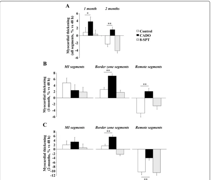
Figure 2 CADO treatment improves segmental contractile function in remote regions after MI. The evolution of segmental contractility in the three experimental groups is expressed as the change from 48 h to 1 and 2 months of the percentage of myocardial thickening. (A) Global evolution of segmental contractility (i.e. whole heart). (B, C) Evolution of segmental contractility after 1 and 2 months according to segment location (MI, border zone or remote area). Data are adjusted mean ± SEM. *p < 0.01; **p < 0.001.
Bousquenaudet al. EJNMMI Research 2013, 3:65 Page 5 of 10
Results
Early FDG-PET imaging and randomization
Representative FDG-PET images obtained at 48 h, 1 month and 2 months are shown in Figure 1. On the baseline FDG-PET scans recorded 2 days after coronary occlusion, nec-rotic segments were observed in all 23 animals. According to the three-point classification of MI size (small, moderate and large MI) [10], rats were subsequently randomized into three treatment groups: control (n = 7), CADO (n = 8) and 8-SPT (n = 8). Thus, the mean infarct size was equivalent between the three groups (Table 1). The mean baseline values of LV volumes, LV ejection fraction, heart rate, blood pressure and body weight were also similar between the three groups (Tables 1 and 2).
Evolution of body mass, hemodynamic parameters and global LV function
During the 2-month follow-up, all rats showed an in-crease in body weight, a dein-crease in heart rate and a stable level of systolic blood pressure. At 2 months, however, body weight and heart rate were slightly but significantly lower in CADO-treated rats than in control rats (Table 2).
As detailed in Table 1, control rats exhibited marked LV remodelling during the 2-month follow-up, as attested by increased EDV and ESV, and decreased EF. These changes were not significantly different in the
CADO group or in the 8-SPT group, even though there were trends toward a beneficial effect on LV volumes and EF in the CADO group and a detrimental effect on LV volumes and EF in the 8-SPT group (Table 1).
Evolution of segmental contractility
Seventeen segments were analysed in each of the 23 ani-mals, leading to a total of 391 analysed segments. On the 2-day FDG-PET exams, 43 segments were totally necrotic (MI segments), 109 were considered as remote segments and the 239 remaining segments were consid-ered to be within the border zone. Myocardial thicken-ing, assessed through the percentage of systolic increase in myocardial counts, exhibited a continuous decline be-tween remote (40% ± 9%), border zone (23% ± 13%) and MI (12% ± 4%) segments (p < 0.001).
Analyses of variance revealed that the evolution of myocardial thickening over 2 months was enhanced in CADO-treated rats, compared to control rats, with a sig-nificant interaction by segment location (remote, border zone or MI area) (p = 0.03). In contrast, no difference was observed when 8-SPT rats were compared to con-trol rats using the same model.
Changes in segmental contractility from baseline were also compared between the three groups at the specific time-points of 1 and 2 months, as detailed in Figure 2. The main observation was that, on average, the
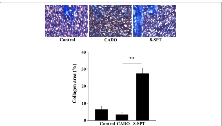
Figure 3 8-SPT treatment aggravates cardiac fibrosis. Upper panel: cardiac sections of non-infarcted areas of the left ventricle stained with Masson's trichrome. Representative pictures from the three experimental groups are shown. Magnification, ×400. Lower panel: quantitative analysis of collagen volume fraction in non-infarcted areas. Results are mean ± SEM. **p < 0.001.
contractile function of the overall segments from CADO rats was enhanced when compared to controls at 1 and 2 months (Figure 2A). After 1 month, the increase of myocardial thickening was +3.8% ± 0.7% in the CADO group compared to +0.9% ± 0.7% in the control group (p < 0.01). After 2 months, contractile function was im-paired in control rats, as attested by a decrease of myo-cardial thickening of −2.3% ± 0.8%, and preserved in CADO-treated rats (+1.6% ± 0.8%, p < 0.001). This effect of CADO was blunted by 8-SPT (Figure 2A).
Interestingly, the beneficial effect of CADO on con-tractile function varied according to segment location, both after 1 month (Figure 2B) and after 2 months (Figure 2C). In particular, at 2 months, this effect was obvious (1) in the border zone where CADO rats exhibited a significant increase in contractile function (+5.6% ± 0.8%), whereas control rats did not (+1.5% ± 0.8%, p < 0.001 vs. CADO), and (2) in remote segments where a decline in contractile function was docu-mented in all groups but to a lower extent in the CADO group (−4.0% ± 1.0%) than in the control group (−10.4% ± 1.3%, p < 0.001 vs. CADO). By contrast, the contractile function of MI segments was stable and
comparable in the two groups at 2 months (CADO +3.4% ± 2.0% vs. control +2.0% ± 1.8%, p = NS).
The beneficial effect of CADO on the contractile func-tion of the border and remote segments was lost for rats additionally treated with 8-SPT (Figure 2A,B).
Fibrosis, cardiomyocyte hypertrophy and apoptosis
We examined the extent of fibrosis outside the MI areas (Figure 3). CADO appeared to reduce fibrosis in remote areas, when compared to controls, although this differ-ence did not reach the level of statistical significance. However, this fibrosis was markedly increased by the ad-ministration of 8-SPT (eightfold increase compared to the CADO group, p < 0.001).
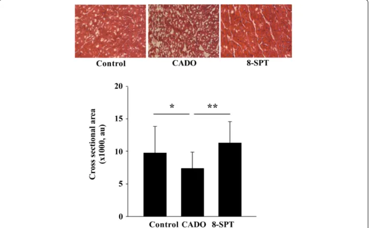
The cardiomyocyte cross-sectional area in healthy parts of the heart was lower in the CADO than in the control group, and this effect was blunted by 8-SPT (Figure 4).
Annexin-5 staining was reduced in CADO rats com-pared to control rats and was robustly increased by 8-SPT (Figure 5). These differences were evident in the remote and border zones.
Figure 4 CADO treatment limits cardiomyocyte hypertrophy. Upper panel: representative histological sections stained with haematoxylin and eosin in remote areas showing cardiomyocyte hypertrophy. Lower panel: quantitative analysis of the cross-sectional area of cardiomyocytes from remote areas. Cardiomyocyte hypertrophy was decreased in CADO-treated rats compared to control rats. Results are mean ± SEM. au, arbitrary units. *p < 0.005; **p < 0.001. Magnification, ×400.
Bousquenaudet al. EJNMMI Research 2013, 3:65 Page 7 of 10
Therefore, activation of adenosine receptors reduces MI-induced fibrosis, cardiomyocyte hypertrophy and apoptosis.
Discussion
Using FDG-PET in a rat infarct model, the present study shows that adenosine provides beneficial effects within the remote and border zones, but not within the MI area. However, these effects have a limited impact on global LV remodelling.
We have previously shown that the method of acipimox-enhanced FDG-PET provides cardiac images
of a very high quality in rats [13], as well as accurate as-sessments of LV function and of MI size and location in the experimental model of LAD occlusion [10]. In the present study, the FDG-PET exams, performed 48 h after surgery, allowed for randomizing the animals into different treatment groups according to initial infarct size. This size, which is highly variable in the rat model, constitutes a main determinant of subsequent LV re-modelling in the rat model [10]. Therefore, such a pre-therapeutic randomization was critical for providing an accurate comparison with the control group. This was not done in the previous echocardiographic study of long-term adenosine A2B receptor stimulation [8].
In addition, the FDG-PET technique allowed for a sep-arate survey of the contractile function of the MI, re-mote and border segments. These data showed that treatment with CADO was beneficial on the contractile function of the segments lying outside the MI area. CADO treatment inhibited the progressive decline in the contractile function of remote areas and enhanced the contractile function within the border zone. These effects were truly attributable to the stimulation of ad-enosine receptors since they were abolished when a pan-antagonist of adenosine receptors was added to CADO treatment (8-SPT group).
The observation of a protective effect in the border zone is clinically relevant since the border zone is con-sidered as a main therapeutic target after MI. For in-stance, injection of stem cells into the border zone has been shown to enhance vascular density and contractile performance in animals [20]. Similar results were ac-hieved by the co-expression of vascular endothelial growth factor and angiopoietin-1 in the infarct border zone [21]. In addition, we have previously shown that the perfusion of the infarct border zone was enhanced after the injection of stem cells within the infarct core [22]. However, these therapeutic approaches are not easy to apply, whereas the present study shows that a simple pharmacological treatment is able to improve myocardial contractility.
The exact mechanism by which the stimulation of ad-enosine receptors improves contractile function in the border and remote zones remains to be fully defined. We observed a mild lowering of heart rate after 2 months of CADO treatment, but this is unlikely to fully explain the cardioprotective effects of CADO. Previous in vitro experiments from our group have shown that adenosine regulates multiple pathways involved in LV remodelling, such as inflammation, angiogenesis and extracellular matrix turnover [23-26]. In the present in vivo study, we further extend the cardioprotective properties of adeno-sine to apoptosis, consistent with a previous report by Simonis et al. [27], fibrosis [8] and cardiomyocyte hyper-trophy [28]. All together, these results suggest that
Figure 5 CADO treatment reduces apoptosis. Representative pictures of annexin-5 immunostaining in the remote, border and infarct (MI) zones are shown. Annexin-5 staining appears in pink, and nuclei are coloured in blue by DAPI. CADO reduced apoptosis and 8-SPT exacerbated apoptosis, particularly in the border zone. Magnification, ×400.
therapeutic targeting of adenosine receptors may have multiple cardioprotective effects.
Angiotensin-converting enzyme (ACE) inhibitors and beta-blockers are currently used to limit LV remodelling after MI. Although their effects on global function and on the limitation of infarct expansion are well docu-mented [29], their effects in the border zone are still poorly known. Beta-blockers may only have a limited ef-fect in the border zone because of multiple deef-fects in the membrane beta-adrenergic receptor complex in the infarcted heart [30]. Our results, showing a preservation of contractile function in remote and border zones, but no effect on the infarct area, suggest that adenosine may target other signalling pathways than ACE inhibitors and beta-blockers. Whether adenosine exerts complementary cardioprotective effects to ACE inhibitors and beta-blockers could be the subject of further studies.
In our study, CADO was unable to significantly pro-tect the heart from the development of adverse LV re-modelling, as assessed by LV volumes and EF. These results differ from those of Wakeno and colleagues [8], where a selective adenosine A2B receptor agonist was reported to prevent LV remodelling after MI. In that study, LV function was assessed in only two dimensions by echocardiography and there was no documentation that the different experimental groups had equivalent in-farct size before treatment. A2B receptors play critical roles in the reduction of remodelling and in the revascu-larization of the infarcted heart induced by mesenchymal stem cells [31]. Activation of A2B receptors inhibits apoptosis after MI [27]. However, a specific blockade of A2B receptors has been also shown to limit LV remodel-ling after MI [32]. The cardioprotective properties of A2B receptors are therefore complex and have been re-cently reviewed [33].
The roles of A1 and A3 receptors are also multiple and complex in this setting. Initial studies by Matherne and colleagues indicated that activation of the A1 adeno-sine receptor protects from myocardial ischemia [34]. This observation was subsequently confirmed by other groups (reviewed in [35]). The failure of CADO, a pref-erential A1 receptor agonist, to protect from LV remod-elling in our study might be related to A3 activation, which has shown deleterious cardiac effects [36]. On the opposite, the A3 receptor has also cardioprotective prop-erties [37].
Finally, all four sub-types of adenosine receptors ap-pear to have the potential to exert cardioprotective ef-fects after ischemia. Adenosine has multiple efef-fects on several components of LV remodelling such as inflam-mation, fibrosis, angiogenesis, apoptosis, etc. These ef-fects rely on the sub-type of adenosine receptors present on the cell surface. It remains to be determined whether the activation or blockade of several sub-types of
adenosine receptors has superior anti-remodelling ef-fects than the preferential activation of one receptor. It will also be interesting in future studies to address the cardioprotective effects of adenosine in a model of ische-mia/reperfusion, which more closely resembles the clin-ical MI setting.
Conclusions
We have shown that, when administrated long-term after MI, adenosine exerts beneficial effects on the myo-cardial contractility of remote and border zones, but with limited impact on global LV remodelling. Further studies are required to determine the therapeutic poten-tial of this observation.
Competing interests
The authors declare that they have no competing interests. Authors' contributions
MB performed the animal experiments and participated in the data analysis and manuscript drafting. FM, SP and HB participated in the PET exams and image analysis. JZ participated in the ex vivo experiments. RF performed the statistical analysis. GK and DW participated in the study design and manuscript drafting. YD and PYM supervised the study and drafted the manuscript. All authors read and approved the final manuscript. Acknowledgements
This work was supported by the Society for Research on Cardiovascular Diseases and the Ministry of Culture, Higher Education and Research of Luxembourg. MB and JZ are recipients of fellowships from the National Fund for Research of Luxembourg (grants # PhD-AFR 08–024 and # 3972501, respectively).
Author details
1Laboratory of Cardiovascular Research, Centre de Recherche Public de la
Santé, 84 Val Fleuri, Luxembourg L1526, Luxembourg.2Nancyclotep Experimental Imaging Platform, 54 511, Nancy, France.3INSERM, U1116, 54
000, Nancy, France.4INSERM, Centre d'Investigation Clinique CIC-P 9501, Nancy 54000, France.5Division of Cardiology, Centre Hospitalier,
Luxembourg L-1210, Luxembourg.
Received: 17 June 2013 Accepted: 31 August 2013 Published: 12 September 2013
References
1. Wolk MJ, Scheidt S, Killip T: Heart failure complicating acute myocardial infarction. Circulation 1972, 45:1125–1138.
2. Jessup M, Brozena S: Heart failure. N Engl J Med 2003, 348:2007–2018. 3. Cohn JN, Ferrari R, Sharpe N: Cardiac remodeling—concepts and clinical
implications: a consensus paper from an international forum on cardiac remodeling: behalf of an international forum on cardiac remodeling. J Am Coll Cardiol 2000, 35:569–582.
4. Headrick JP, Peart JN, Reichelt ME, Haseler LJ: Adenosine and its receptors in the heart: regulation, retaliation and adaptation. Biochimica et Biophysica Acta (BBA) - Biomembranes 2011, 1808:1413–1428. 5. Kloner RA, Forman MB, Gibbons RJ, Ross AM, Alexander RW, Stone GW:
Impact of time to therapy and reperfusion modality on the efficacy of adenosine in acute myocardial infarction: the AMISTAD-2 trial. Eur Heart J 2006, 27:2400–2405.
6. Desmet W, Bogaert J, Dubois C, Sinnaeve P, Adriaenssens T, Pappas C, Ganame J, Dymarkowski S, Janssens S, Belmans A, Van de Werf F: High-dose intracoronary adenosine for myocardial salvage in patients with acute ST-segment elevation myocardial infarction. Eur Heart J 2011, 32:867–877.
7. Fokkema ML, Vlaar PJ, Vogelzang M, Gu YL, Kampinga MA, de Smet BJ, Jessurun GA, Anthonio RL, van den Heuvel AF, Tan ES, Zijlstra F: Effect of high-dose intracoronary adenosine administration during primary
Bousquenaudet al. EJNMMI Research 2013, 3:65 Page 9 of 10
percutaneous coronary intervention in acute myocardial infarction: a randomized controlled trial. Circ Cardiovasc Interv 2009, 2:323–329. 8. Wakeno M, Minamino T, Seguchi O, Okazaki H, Tsukamoto O, Okada K,
Hirata A, Fujita M, Asanuma H, Kim J, Komamura K, Takashima S, Mochizuki N, Kitakaze M: Long-term stimulation of adenosine A2b receptors begun after myocardial infarction prevents cardiac remodeling in rats. Circulation 2006, 114:1923–1932.
9. Wheeler DG, Joseph ME, Mahamud SD, Aurand WL, Mohler PJ, Pompili VJ, Dwyer KM, Nottle MB, Harrison SJ, d'Apice AJ, Robson SC, Cowan PJ, Gumina RJ: Transgenic swine: expression of human CD39 protects against myocardial injury. J Mol Cell Cardiol 2012, 52:958–961. 10. Bousquenaud M, Maskali F, Poussier S, Marie PY, Boutley H, Karcher G,
Wagner DR, Devaux Y: Acipimox-enhanced (18)F-fluorodeoxyglucose positron emission tomography for characterizing and predicting early remodeling in the rat infarct model. Int J Cardiovasc Imaging 2012, 28:1407–1415.
11. Maskali F, Poussier S, Marie PY, Tran N, Antunes L, Olivier P, Plenat F, Maitrejean S, Zannad F, Karcher G: High-resolution simultaneous imaging of SPECT, PET, and MRI tracers on histologic sections of myocardial infarction. J Nucl Cardiol 2005, 12:229–230.
12. Pfeffer MA, Pfeffer JM, Fishbein MC, Fletcher PJ, Spadaro J, Kloner RA, Braunwald E: Myocardial infarct size and ventricular function in rats. Circ Res 1979, 44:503–512.
13. Poussier S, Maskali F, Tran N, Person C, Maureira P, Boutley H, Karcher G, Lacolley P, Regnault V, Fay R, Marie PY: ECG-triggered
(18)F-fluorodeoxyglucose positron emission tomography imaging of the rat heart is dramatically enhanced by acipimox. Eur J Nucl Med Mol Imaging 2010, 37:1745–1750.
14. Cerqueira MD, Weissman NJ, Dilsizian V, Jacobs AK, Kaul S, Laskey WK, Pennell DJ, Rumberger JA, Ryan T, Verani MS: Standardized myocardial segmentation and nomenclature for tomographic imaging of the heart: a statement for healthcare professionals from the Cardiac Imaging Committee of the Council on Clinical Cardiology of the American Heart Association. Circulation 2002, 105:539–542.
15. Germano G, Kiat H, Kavanagh PB, Moriel M, Mazzanti M, Su HT, Van Train KF, Berman DS: Automatic quantification of ejection fraction from gated myocardial perfusion SPECT. J Nucl Med 1995, 36:2138–2147. 16. Germano G, Kavanagh PB, Berman DS: An automatic approach to the
analysis, quantitation and review of perfusion and function from myocardial perfusion SPECT images. Int J Card Imaging 1997, 13:337–346. 17. Marie PY, Djaballah W, Franken PR, Vanhove C, Muller MA, Boutley H,
Poussier S, Olivier P, Karcher G, Bertrand A: OSEM reconstruction, associated with temporal fourier and depth-dependant resolution recovery filtering, enhances results from sestamibi and 201Tl 16-interval gated SPECT. J Nucl Med 2005, 46:1789–1795.
18. Slart RH, Bax JJ, van Veldhuisen DJ, van der Wall EE, Dierckx RA, de Boer J, Jager PL: Prediction of functional recovery after revascularization in patients with coronary artery disease and left ventricular dysfunction by gated FDG-PET. J Nucl Cardiol 2006, 13:210–219.
19. Soeki T, Kishimoto I, Okumura H, Tokudome T, Horio T, Mori K, Kangawa K: C-type natriuretic peptide, a novel antifibrotic and antihypertrophic agent, prevents cardiac remodeling after myocardial infarction. J Am Coll Cardiol 2005, 45:608–616.
20. Wang X, Jameel MN, Li Q, Mansoor A, Qiang X, Swingen C, Panetta C, Zhang J: Stem cells for myocardial repair with use of a transarterial catheter. Circulation 2009, 120:S238–246.
21. Tao Z, Chen B, Tan X, Zhao Y, Wang L, Zhu T, Cao K, Yang Z, Kan YW, Su H: Coexpression of VEGF and angiopoietin-1 promotes angiogenesis and cardiomyocyte proliferation reduces apoptosis in porcine myocardial infarction (MI) heart. Proc Natl Acad Sci U S A 2011, 108:2064–2069. 22. Tran N, Franken PR, Maskali F, Nloga J, Maureira P, Poussier S, Groubatch F,
Vanhove C, Villemot JP, Marie PY: Intramyocardial implantation of bone marrow-derived stem cells enhances perfusion in chronic myocardial infarction: dependency on initial perfusion depth and follow-up assessed by gated pinhole SPECT. J Nucl Med 2007, 48:405–412. 23. Ernens I, Rouy D, Velot E, Devaux Y, Wagner DR: Adenosine inhibits matrix
metalloproteinase-9 secretion by neutrophils: implication of A2A receptor and cAMP/PKA/Ca2+ pathway. Circ Res 2006, 99:590–597. 24. Velot E, Haas B, Leonard F, Ernens I, Rolland-Turner M, Schwartz C, Longrois D,
Devaux Y, Wagner DR: Activation of the adenosine-A3 receptor stimulates
matrix metalloproteinase-9 secretion by macrophages. Cardiovasc Res 2008, 80:246–254.
25. Leonard F, Devaux Y, Vausort M, Ernens I, Rolland-Turner M, Wagner DR: Adenosine modifies the balance between membrane and soluble forms of Flt-1. J Leukoc Biol 2011, 90:199–204.
26. Rolland-Turner M, Goretti E, Bousquenaud M, Leonard F, Nicolas C, Zhang L, Maskali F, Marie PY, Devaux Y, Wagner D: Adenosine stimulates the migration of human endothelial progenitor cells: role of CXCR4 and microRNA-150. PLoS ONE 2013, 8:e54135.
27. Simonis G, Wiedemann S, Joachim D, Weinbrenner C, Marquetant R, Strasser RH: Stimulation of adenosine A2b receptors blocks apoptosis in the non-infarcted myocardium even when administered after the onset of infarction. Mol Cell Biochem 2009, 328:119–126.
28. Fassett JT, Hu X, Xu X, Lu Z, Zhang P, Chen Y, Bache RJ: Adenosine kinase regulation of cardiomyocyte hypertrophy. Am J Physiol Heart Circ Physiol 2011, 300:H1722–H1732.
29. Ali SM, Brown EJ Jr, Nallapati SR, Alhaddad IA: Early angiotensin converting enzyme inhibitor therapy after experimental myocardial infarction prevents left ventricular dilation by reducing infarct expansion: a possible mechanism of clinical benefits. Coron Artery Dis 1998, 9:815–821. 30. Steinberg SF, Zhang H, Pak E, Pagnotta G, Boyden PA: Characteristics of
the beta-adrenergic receptor complex in the epicardial border zone of the 5-day infarcted canine heart. Circulation 1995, 91:2824–2833. 31. Ryzhov S, Zhang Q, Biaggioni I, Feoktistov I: Adenosine A2B receptors on
cardiac stem cell antigen (Sca)-1-positive stromal cells play a protective role in myocardial infarction. Am J Pathol 2013, 183:665–672.
32. Toldo S, Zhong H, Mezzaroma E, Van Tassell B, Kannan H, Zeng D, Belardinelli L, Voelkel N, Abbate A: GS-6201, a selective blocker of the A2B adenosine receptor, attenuates cardiac remodeling following acute myocardial infarction in the mouse. J Pharmacol Exp Ther 2012, 343:587–595.
33. Eltzschig HK, Bonney SK, Eckle T: Attenuating myocardial ischemia by targeting A2B adenosine receptors. Trends Mol Med 2013, 19:345–354. 34. Matherne GP, Linden J, Byford AM, Gauthier NS, Headrick JP: Transgenic A1
adenosine receptor overexpression increases myocardial resistance to ischemia. Proc Natl Acad Sci U S A 1997, 94:6541–6546.
35. Albrecht-Kupper BE, Leineweber K, Nell PG: Partial adenosine A1 receptor agonists for cardiovascular therapies. Purinergic Signal 2012, 8:91–99. 36. Lu Z, Fassett J, Xu X, Hu X, Zhu G, French J, Zhang P, Schnermann J, Bache RJ,
Chen Y: Adenosine A3 receptor deficiency exerts unanticipated protective effects on the pressure-overloaded left ventricle. Circulation 2008, 118:1713–1721.
37. Du L, Gao ZG, Nithipatikom K, Ijzerman AP, Veldhoven JP, Jacobson KA, Gross GJ, Auchampach JA: Protection from myocardial ischemia/ reperfusion injury by a positive allosteric modulator of the A(3) adenosine receptor. J Pharmacol Exp Ther 2012, 340:210–217.
doi:10.1186/2191-219X-3-65
Cite this article as: Bousquenaud et al.: Cardioprotective effects of adenosine within the border and remote areas of myocardial infarction. EJNMMI Research 2013 3:65.
Submit your manuscript to a
journal and benefi t from:
7 Convenient online submission 7 Rigorous peer review7 Immediate publication on acceptance 7 Open access: articles freely available online 7 High visibility within the fi eld
7 Retaining the copyright to your article