HAL Id: hal-01893823
https://hal.archives-ouvertes.fr/hal-01893823
Submitted on 6 May 2021
HAL is a multi-disciplinary open access
archive for the deposit and dissemination of
sci-entific research documents, whether they are
pub-lished or not. The documents may come from
teaching and research institutions in France or
abroad, or from public or private research centers.
L’archive ouverte pluridisciplinaire HAL, est
destinée au dépôt et à la diffusion de documents
scientifiques de niveau recherche, publiés ou non,
émanant des établissements d’enseignement et de
recherche français ou étrangers, des laboratoires
publics ou privés.
Distributed under a Creative Commons Attribution| 4.0 International License
appendicularian-ciliate interactions
Fabien Lombard, Damien Eloire, Angélique Gobet, Lars Stemmann, John
Dolan, Antoine Sciandra, Gabriel Gorsky
To cite this version:
Fabien Lombard, Damien Eloire, Angélique Gobet, Lars Stemmann, John Dolan, et al..
Exper-imental and modeling evidence of appendicularian-ciliate interactions.
Limnology and
Oceanog-raphy Bulletin, American Society of Limnology and OceanogOceanog-raphy, 2010, 55 (1), pp.77 - 90.
�10.4319/lo.2010.55.1.0077�. �hal-01893823�
Experimental and modeling evidence of appendicularian–ciliate interactions
Fabien Lombard,
1,*Damien Eloire, Angelique Gobet, Lars Stemmann, John R. Dolan,
Antoine Sciandra, and Gabriel Gorsky
Centre National de la Recherche Scientifique (CNRS), Universite´ Pierre et Marie Curie – Paris 6, UMR 7093, Laboratoire d’Oce´anographie de Villefranche-sur-Mer, Villefranche-sur-Mer, France
Abstract
Interactions between appendicularians and ciliates were observed over the life span of Oikopleura dioica in laboratory cultures and clarified with the use of mathematical modeling and microscopic observations. Complex interactions including competition, parasitism, predation, and histophagy occurred simultaneously, resulting in apparent mutualism. The large ciliate Strombidium sp. entered the inlet filters of appendicularian houses (larger than 500 mm body size) by distorting the mesh. Once inside, Strombidium fed on particles concentrated on the filters. When appendicularians were larger than 900 mm, both the high flow rate in the buccal tube and their esophagus width allowed the ‘‘host’’ appendicularian to capture and ingest ciliates. Thus, ciliates seem to be sequentially competitors, then parasites or commensal in appendicularian houses, and finally prey for appendicularians. Appendicularian rates of somatic growth and reproduction were enhanced when ciliates were ingested. This additional food supply could be essential in oligotrophic environments. Reciprocally, appendicularians support higher ciliate growth rates, allowing ciliates to survive and grow in food-limited environments. Appendicularians thus modify the size spectrum of the microbial food web both by removing small organisms (0.2–30 mm) and enhancing the growth of mid-sized and large ciliates.
Appendicularians are one of the most common members of the zooplankton community, often second only to copepods in the upper layers (Gorsky and Fenaux 1998) and second after large crustaceans in the mesopelagic layers (Stemmann et al. 2008). They play an important role in the marine food web through their consumption of small particles and as food for higher trophic levels (Zubkov and Lo`pez-Urrutia 2003; Purcell et al. 2004). Appendicularians use a mucopolysaccharid filter, termed ‘‘a house’’ to filter particles. These houses, once discarded, sink through the water column and can be a major component of the marine snow (Hansen et al. 1996; Alldredge 2004; Robison et al. 2005). With this extremely efficient filtration structure, appendicularians can consume particles from 0.2 to 30 mm (Flood and Deibel 1998) and therefore are usually considered to be both bacterivorous and herbivorous, although ciliates have been observed inside appendicularian houses (Davoll and Silver 1986; Vargas and Gonza´lez 2004; To¨nnesson et al. 2005), which suggested a possible interaction.
Up to 83 ciliates can colonize discarded houses, representing an enrichment factor of 110–2131 compared with ambient sea water (Davoll and Silver 1986). They might be attracted to the high concentration of food particles such as algae, bacteria, and organic matter retained by the house. Despite the observation that ciliates can be removed from the ambient seawater by appendicu-larians (Vargas and Gonza´lez 2004; To¨nnesson et al. 2005), the role of ciliates as a food source for the appendicularians
has not yet been demonstrated (Gorsky and Fenaux 1998). Nevertheless, in our laboratory cultures, ciliates frequently coexisted with appendicularians over long periods, sug-gesting that a commensal relationship could exist between them.
The aim of our study was to identify and quantify the interactions between appendicularians and ciliates. We conducted an experiment in a controlled environment inspired by the principle of a chemostat, wherein appendi-cularians and ciliates were allowed to interact and their dynamics monitored. Simple models were then used to test and quantify the processes and relationships suggested by the experimental results. Additional microscopic observa-tions were also performed to confirm hypotheses generated by the models and understand precisely how the observed interactions took place.
Methods
The culture protocol of the appendicularian Oikopleura dioica growing on the haptophyceaen alga Isochrysis galbana and the diatom Thalassiosira pseudonana is similar to those described by Lombard et al. (2005). When fresh sea water, used for the stock cultures of appendicularians, is sieved through a 20-mm mesh net, the occurrence of ciliates was frequent. Consequently, the ciliates commonly occurred within the appendicularian culture.
Trophic relationship experiment—The experiment was conducted at 15uC and under controlled conditions. The food comprised I. galbana and T. pseudonana and was maintained at a constant level with the use of a chemostat-like system (Fig. 1). Four experimental containers with different contents were monitored: one with ciliates only (C), one with appendicularians only (A), one with
*Corresponding author: [email protected]
1Present address: Technical University of Denmark, National
Institute of Aquatic Resources, Oceanography Section, Charlot-tenlund, Denmark
appendicularians and ciliates (A+C), and a control without organisms (T). Each setup comprised a 20-liter plastic beaker filled with 15 liters of sea water and stirred with plastic paddles at 10 rpm to ensure homogeneity. The food solution consisted of 0.2-mm filtered sea water and a mixture of I. galbana and T. pseudonana delivered to yield a final concentration of 10,000 cells mL21. The food solution
was continuously stirred and transferred to the experimen-tal beakers by perisexperimen-taltic pumps at a flow rate of 5.6 L d21.
A constant volume in each experimental beaker was maintained by a peristaltic pump with a higher flow rate than the incoming food solution. The outflow tube was placed at the surface of the beaker to remove water in excess of 15 liters. To avoid damage or evacuation of appendicularians, the overflow tube was equipped with a large surface support with a 50-mm Nitex net that decreased the suction pressure.
The experiment was initiated with 50 mature females and 25 mature males from a stock appendicularian culture. Each organism was photographed and forced to abandon its house. They were then rinsed three times in 0.2-mm filtered water and placed in a 2-liter beaker to spawn. The spawning beaker was gently stirred to stimulate spawning, and the time was recorded as t 5 0. The female body size and volume were measured from photographs, and the number of eggs from each female was calculated from gonad volume (Lombard et al. 2009a).
In parallel, a ciliate inoculum was prepared. A sample of discarded houses and water from the permanent culture containing ciliates was introduced into a 2-liter beaker. Simultaneously, the four experimental beakers were filled with the experimental solution (water and algae). The algal
concentration was estimated by triplicate counts with a particle counter (Multisizer model II, Coulter).
The experiment was started 24 h after fertilization (day 1). The product of the spawn was divided into two equal parts and placed in the experimental beakers A and A+C. The ciliate mixture was divided and introduced in experimental beakers C and A+C. From day 1 to 8, the four experimental beakers were sampled daily. Appendi-cularians present in a subsample randomly taken from experimental beakers A and A+C were counted for animal density and mortality rate estimations and photographed for size analysis. The sampling volume increased with the decrease of appendicularian density of up to 1 liter. On the last day, the entire 15-liter beaker was sampled. If mature appendicularians were present, they were removed and photographed. The algal concentration was estimated in triplicate with a Coulter counter. Every day, from C and A+C, 500 mL of water was sampled and fixed with 2% Lugol solution. A 50 mL subsample was placed in an Utermo¨hl settling chamber for 24 hours and the ciliates were counted using an inverted microscope. Ten inhabited appendicularian houses were randomly sampled in A+C, placed in an Utermo¨hl chamber and fixed with 2% Lugol solution. Once settled, ciliates present inside the houses were counted.
Appendicularian and ciliate models—To quantify inter-actions observed during the experiment, we used a physiological model previously published for appendicu-larian growth (Lombard et al. 2009b) and a model described below for ciliate growth. The models were used as analytical tools to identify and quantify the effect of appendicularian–ciliate interactions on the growth rates of each as observed during the experiment. Both models were calibrated in the absence of interaction (A and C) and afterward applied to A+C, in which both organisms interacted. For these models, the algal concentrations were converted from biovolumes to carbon concentration by using the conversion factor of Strathmann (1967). For the appendicularian model, the observed ciliate concentration was converted to carbon units by approximating their shape to spheres and using a 0.19 pg C mm23conversion
factor for Lugol-fixed oligotrichs (Putt and Stoecker 1989). Carbon concentrations of algae and ciliates (Strombidium sp.) were interpolated by fitting polynomial functions, which were used for growth simulations.
We used a physiological budget model simulating growth in carbon units of a single appendicularian as a function of the temperature and food concentration (Lombard et al. 2009b). Physiological processes (respira-tion, filtra(respira-tion, inges(respira-tion, assimila(respira-tion, houses pro-duction) are simulated throughout the life cycle of one appendicularian in relation to the environment, and growth was defined as the difference between inputs (feeding) and outputs (respiration, house secretion) of matter. This model has been calibrated and validated for various food concentrations and 3 temperatures (Lombard et al. 2009b).
For the ciliates, we simply considered the growth rate (m) of ciliates as a function of the food concentration
Fig. 1. Experimental setup used for studying appendicular-ian–ciliate interactions. Four 15-liter chemostat were filled with 0.2-mm filtered sea water enriched with algae; three were inoculated with ciliates (C), appendicularians (A), and the two organisms (A+C), whereas one was not inoculated and served as control (T). A close-up of the opening of the outflow tube was added indicating its position and nature.
(X ),
m~Fmax
X kzX{L
where L represents a term of mass loss (e.g., respiration and mortality), Fmax is the maximum net feeding rate, and k is
the half-saturation constant of the feeding rate in relation to the concentration of food (Montagnes 1996). The ciliate concentration (C, individuals [ind.] mL21) is then
calculat-ed for each time step from the differential equation dC
dt ~mC{D
where D represents the dilution rate of each experimental setup. Ciliate loss is calculated from D because their size is inferior to the mesh size of the evacuation tube and ciliates are continually removed by the overflow.
The model parameters were calibrated according to a least square minimization method (Nelder–Mead simplex method) in absence of appendicularians (C).
Microscopic observations—To examine the conceptual hypothesis of appendicularian-consuming ciliates inferred from the experiment, we conducted a series of different microscopic observations on appendicularians of different sizes directly sampled from the stock culture.
House volume: Appendicularians in their houses were gently removed from the stock culture with the use of a wide-bore pipette and placed in a petri dish filled with sea water and sepia ink. After a few seconds, the transparent house became easily visible because of filtration and accumulation of sepia ink particles in the filter and on the internal walls of the house chambers. The appendicu-larian was then removed, placed in a second petri dish filled with filtered sea water, oriented in profile view, and photographed. Both body size and house diameter were measured, and house volume was calculated assuming a spherical shape (Flood and Deibel 1998; Alldredge 2004). The relationship between appendicularian size and house volume was used to estimate the house volume for the rest of the chemostat experiment and then to determine the enrichment factor (EF) of ciliates inside houses, calculated as the ratio of ciliate concentration inside the house relative to ciliate concentration in the water.
Appendicularian morphology: Some details of the appendicularian morphology were examined in relation to appendicularian size. All photographs of appendicularians were taken in the lateral view and the appendicularian body size and mouth and esophagus width were measured. The point of measurement for the esophagus was located at its bent part near the stomach where the width is minimal (Fig. 2).
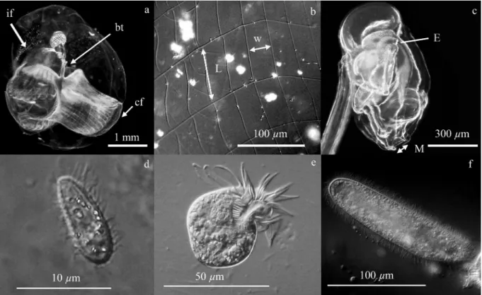
Fig. 2. Oikopleura dioica. (a) Mature female inside its house, which is colored with red china ink. (b) Inlet filter mesh. (c) Body of an appendicularian. The different parts of the house and measurements made are indicated: if, inlet filter; cf, concentrating filter; bt, buccal tube; L, inlet filter mesh length; w; inlet filter mesh width; M, mouth; and E, esophagus. Different kinds of ciliates were observed in the appendicularian cultures: (d) small bacterivorous scuticociliate ciliates, (e) medium-sized oligotrich ciliates Strombidium sp., and (f) large scuticociliate ciliates. Most of the ciliates observed during the experiment and in culture were Strombidium sp.
House inlet filter mesh size: Appendicularians of different sizes were sampled with their houses, placed in small dishes, and forced to abandon their houses. Appendicularians were photographed, and body size was estimated from images. Houses were colored with a 2% Lugol solution and mounted for microscopic observations. The inlet filter mesh was examined and photographed at 3100–200 magnification with the use of Nomarski differential interference contrast. For each house, length and width of the mesh were measured on 5–10 areas of the filter, depending on the quality of the photograph.
Appendicularian flow velocity in the house buccal tube and ciliate swimming speed: Appendicularians were sam-pled from the culture and placed in a small cylindrical microaquarium (depth 1 cm, diameter 3 cm) designed for photography and video observation. Before the recording, appendicularians were acclimated to the aquarium for a few minutes. A short duration of the sequences (maximum 5–7 min) was employed to avoid a heat effect. Appendicu-larians were filmed under a dark field with a Sony digital camcorder equipped with a Raynox 12X magnification macro-objective lens. The field of view was calibrated using a micrometric grid (1 pixel 5 3.84 mm). The video sequences were acquired on lateral views of the house, focused on the buccal tube. Short sequences of the appendicularian intake of particles from the concentrating filter through the buccal tube to the pharyngeal cavity were analyzed. Still images were extracted from the video frame,
and successive positions of natural food particles were calculated with the mouth as a reference point. The flow velocity was then calculated from successive particle positions and the camera acquisition speed. For selected video frames, the type and the number of ingested ciliates were noted. The swimming speed of the different ciliate types was measured from video frames without appendi-cularians.
Results
Algal concentration—During the experiment, the algal concentration in the food input solution was approximately 9800 6 600 cells mL21 (mean 6 SD). The algal
concentration in the control (T) remained stable until day 4 (Fig. 3a). Considerable bacterial growth was observed after day 5, leading to an aggregation of algal particles after day 7, explaining a large decrease of particle concentration recorded in T during the experiment. These features, however, were not observed in the other experimental setups. In T, the algal concentration was always higher than in the presence of predators (A, C, A+C), suggesting that the density of ciliates or appendicularians is sufficient to significantly reduce the algal concentration after day 2. Algal concentration in the presence of appendicularians alone (A) was generally higher than for ciliates alone (C), and the concentration was even lower when appendicular-ian and ciliates were present together (A+C). This suggests
Fig. 3. Results from the four experimental setups. (a) Algal concentration. (b) Appendicularian body size. (c) Free-living ciliate concentration measured and simulated with a simple model calibrated using C. (d) Ciliate concentration inside appendicularian houses. T, control; C, ciliates; A, appendicularians; A+C, appendicularians and ciliates.
that the two populations compete for the same resource. In these three experimental setups, the algal concentration decreased significantly after day 5 and reached a quasi-stable minimal concentration for the last 2 or 3 d of the experiment. The significant algal concentration decrease observed after day 5 was linked to the growth of the two populations.
Appendicularian and ciliate growth—Appendicularian body size increased exponentially during the whole life cycle in both setups A and A+C (Fig. 3b). Despite lower food concentration in A+C, no significant difference in appendicularian growth was observed between the two setups. Appendicularians in A+C seemed to have a slightly higher mortality rate (0.40 6 0.11 d21) than without
ciliates (0.34 6 0.19 d21). The difference, although not
significant, results in a clear distinction in the population size at the end of the experiment. Only 60 individuals remained at day 7 in A+C compared with 217 appendicu-larians in A, although they had balanced density at day 0. In the two conditions (A and A+C), the first mature appendicularians were observed on day 6, and all the remaining appendicularians were mature by the end of the experiment (day 8).
The ciliates observed during this experiment were for the most part oligotrich ciliates of the type Strombidium sp. with diameter range around 35 (6 9) mm (Fig. 2). Some small (# 10 mm) bacterivorous scuticociliate ciliates were also observed but at densities two orders of magnitude lower than Strombidium sp. (maximum density 0.16 ind. mL21). An exponential growth of the ciliate populations
(free-living ciliates) was observed in the two conditions (C and A+C; Fig. 3c). The growth in A+C, however, was greater from day 3 to 6 and led to higher ciliate density (20.16 ind. mL21at day 6) compared with C population
(17.96 ind. mL21at day 7). At the end of the experiment,
ciliate concentrations declined in both treatments, likely as a result of the low algal concentrations. This decrease began 1 d earlier in A+C than in C and resulted in a lower ciliate density at day 8 (7.08 ind. mL21). After day 5,
ciliates were observed inside inhabited appendicularian houses (Fig. 3d). The number of ciliates inside houses increased to 55.8 ind. house21at day 7 and decreased to 9.4
ind. house21 on the last day of the experiment. Varying
with house volume, the ciliate enrichment factor inside houses was about 1.5, 18, 35, and 10 on days 5, 6, 7, and 8, respectively.
Comparison between observed and modeled ciliates and appendicularian growth—The model used to simulate the ciliate growth as a function of algal concentration was calibrated by minimization on the observed growth of population C. Parameters for this model were identified as Fmax 5 3.6 d21, k 5 14.9 mg C L21, and L 5 2.08 d21.
These parameters allow exponential growth of the popu-lation and the subsequent decrease because of food depletion at the end of the experiment (Fig. 3c). However, the same model used on the A+C population did not reproduce the growth rates of free ciliates observed in the A+C incubation. The timing of the change from
exponen-tial growth to depleted conditions was correctly simulated, but the growth rate estimate was lower than that observed. Thus, the simulated population reached a maximum density of about 9.16 ind. mL21 instead of 20.16 ind.
mL21 (Fig. 3d), indicating that ciliate growth in A+C is
higher than theoretically allowed by food availability only. By comparing the observed and simulated growth rates in A+C, the free ciliate growth rate in the presence of appendicularians was enhanced by+0.27 (6 0.04) d21.
In the ciliate growth model, the difference between Fmax
and L can be compared with the maximal growth rate (mmax) estimated by Montagnes (1996) from several studies.
Our estimation of mmaxis within the higher range of growth
rates observed and corresponds to those estimated for several Strombidium species (1.28–2.71; Rivier et al. 1985; Fenchel and Jonsson 1988; Ohman and Snyder 1991). Similarly, the range of L can be compared with estimates of ciliate respiration. L seems to be larger than some estimates of respiration for mixotrophic species of Strombidium (1 d21, Stoecker and Michaels 1991; 1.2 d21; Crawford
and Stoecker 1996). However, these respiration measure-ments were conducted in the absence of food, and feeding can potentially significantly increase the respiration rate. L also includes a mortality rate that can reach 0.5 d21
(Crawford and Stoecker 1996). We can then conclude that, despite being higher than other respiration measurements, our L estimation is consistent with expectations.
Without any new calibration and with the use of observed algal concentration as an input (Fig. 4a), the appendicularian model correctly simulates the growth observed in A (Fig. 4c). In contrast, our model does not correctly simulate appendicularian growth in the A+C treatment (Fig. 4b); the algal concentration, as a unique food source, is insufficient to support the observed growth (Fig. 4d). Effectively, the model significantly deviates from observed growth after day 7, and the simulated appendi-cularians barely reach a 700-mm body size at day 8, whereas the observed population size is 1040 (6 250) mm. It is interesting to note that this difference between observed and simulated growth appears at the same time that ciliates were observed in appendicularian houses. We hypothesized that appendicularians used ciliates as a supplementary food source. To examine this hypothesis, we have used both algal and ciliates concentration as a total potential food concentration in the model (Fig. 4b). When this total food concentration is used, the model is able to correctly reproduce the A+C appendicularian growth (Fig. 4d), suggesting that appendicularians had effectively used ciliates as food. However, the observation of ciliates in appendicularian houses does not necessarily mean they can be ingested. Consequently, microscopic observations were performed to examine this question (Fig. 4b).
Microscopic observations—House and appendicularian morphology: We related house diameter, mesh size of the inlet filters, and mouth and esophagus width to appendi-cularian size (Fig. 5). The house diameter increased linearly with appendicularian size and was 6.04 times larger than appendicularian body size (Fig. 5a). The inlet filter mesh size also increased linearly with appendicularian body size
(Fig. 5b). The minimal and maximal observed inlet filter mesh widths were 3.81 (6 0.23) and 30.3 (6 2.8) mm for 175- and 1238-mm appendicularians, respectively. For the same individuals, the mesh length ranged between 16.1 (6 0.9) and 81.4 (6 5.8) mm. Generally, the ratio between length and width is 3.55 (6 0.74), and did not change significantly with appendicularian size. The appendicular-ian mouth width was generally larger than the mesh size, whereas the esophagus width had sizes slightly larger than mesh width (Fig. 5c).
Ciliates present in appendicularian cultures and houses: Because our appendicularian stock culture conditions were not always homogeneous and certainly differed from in situ conditions, our observations have only a qualitative value. Three kinds of ciliates were generally present in appendi-cularian stock cultures and in the houses (Fig. 2): small (# 10 mm) bacterivorous scuticociliate ciliates, intermediate-sized (30–50 mm) oligotrich ciliates of the genera Strombi-dium, and large ($ 100 mm) scuticociliate ciliates. Some other ciliate types, such as Euplotes sp. or Dileptus sp., were also occasionally observed but in low concentrations. Their concentrations were not recorded.
The small scuticociliate ciliates were observed both in the water and in houses for all sizes of appendicularians. They were present in all houses observed from the
appendicularian stock culture but did not occur in large numbers. Unfortunately, their small size did not allow detailed quantitative behavioral observations with our video device.
Strombidium sp., the dominant ciliates observed during the chemostat experiment and in stock cultures, were found in large numbers in both the water and in cultured appendicularian houses. These ciliates were generally observed in large houses (. 500 mm) in their concentrating filter or attached to the buccal tube (Fig. 6). For appendicularians of . 500 mm, the number of ciliates could be very high (up to 500 Strombidium sp.) but generally was , 50 individuals.
The large scuticociliate ciliates were generally observed in the surrounding water and, in some cases, inside houses. While in the house, the large scuticociliate ciliates were observed in low numbers (1 or 2 individuals), and their intense activity in the concentrating filter or around the trunk seemed to stress the appendicularian. In most cases, the appendicularian would rapidly discard its house.
Ciliate swimming speeds: The swimming speeds of Strombidium sp. and large scuticociliate ciliates were recorded in the water and, for the smaller ciliates, inside the buccal tube of a discarded house. Small scuticociliate
Fig. 4. Experimental and modeled growth of appendicularians. (a, b) Algal and ciliate concentrations observed during the experiment (dots) and polynomial fits used in our model to simulate the appendicularian growth. (c, d) Mean trunk size (including gonads) of the two appendicularian populations observed during the experiment and simulated by our model. The two conditions corresponds to experiments: A, appendicularians; A+C, appendicularians and ciliates.
ciliates and Strombidium sp. swimming patterns seemed to be mostly random, whereas the large ciliates had linear trajectories and changed direction mainly when touching a surface. Swimming speeds of ciliates increased with their size (Table 1), with low values (300 mm s21) for small
ciliates, intermediate values (1300 mm s21) for Strombidium
sp., and high values (3460 mm s21) for the large
scutico-ciliate scutico-ciliates. In all cases, these swimming speeds corresponded to approximately 30-fold their body length per second. They also showed an escape behavior with fast swimming speeds, 1.2–2.7 times faster than their mean swimming speed, but these fast movements were only observed for short periods that did not exceed three or four still images (0.08–0.12 s).
Appendicularian flow velocity in the house buccal tube: The flow velocity exerted by the appendicularian in its buccal tube increased with the size of the appendicularian from 928 to 1365 mm s21 for body sizes of 682 and
1350 mm, respectively (Table 2). The speed was higher at the center of the buccal tube and at the mouth area. Suction was strongly intermittent and occurred only when the appendicularian had an empty stomach. When the stomach was full, the appendicularian continued to filter water but did not ingest the filtered particles, which then remained on the concentrating filter. The speed of particles was also variable within a suction event: more rapid at the beginning and decreasing to a stable speed afterward.
Observations of ciliate ingestion by appendicularians: The small scuticociliate ciliates observed in the buccal tube of appendicularian houses were always ingested, even in small appendicularians (250 mm body size). The slow swimming speed of the small scuticociliates prevented them from escaping the appendicularian suction flow (see Web Appendix: http://www.aslo.org/lo/toc/vol_55/issue_1/ 0077a.html). For Strombidium sp. ciliates, ingestion de-pended on the size of the appendicularian. No case of ingestion was observed for appendicularian body sizes ranging from 500 to 900 mm. For this appendicularian size class, ciliates were frequently observed attached to the wall of the buccal tube. Ciliate swimming speed allowed them to sometimes escape the suction, but in the few cases in which a ciliate was ingested, the appendicularian did not succeed in swallowing the ciliate and, after few minutes, rejected it. The appendicularian in such cases reversed direction of the cilia beating in the spiracles, rejecting the ciliate into the house water exit cavity and expelling it from the house. When the appendicularian was larger than 900 mm, ingestion of the ciliate became more frequent, even if the ciliate tried to escape and the appendicularian was able to swallow it. However in some cases when the Strombidium sp. inside the appendicularian pharyngeal cavity resisted for a long time (e.g., . 100 seconds), the appendicularian could also reject it. When their body size exceeded 1000 mm, appendicularians had no difficulties ingesting and swallowing the Strombidium sp. ciliates. More than 10 ciliates can be ingested in 1 min (Fig. 6b, see Web Appendix). For the large scuticociliate ciliates, no true case of ingestion was observed. In only one case, one of these ciliates was sucked inside the pharyngeal cavity of one
Fig. 5. Oikopleura dioica house and digestive tract mor-phometry. (a) House diameter (H, mm) observed as a function of the appendicularian body size (Bs). The regression corresponds to the relationship H 5 6.04Bs (R250.97). (b) Length and width of
the house inlet filter mesh. The inlet filter length (L) and width (w) can be calculated from appendicularian body size by the respective relationships: L 5 0.043Bs+ 20.53 (R250.73) and w 5 0.0136Bs
+ 5.45 (R250.72). (c) Mouth width (M) and esophagus diameter
(E) compared with the length and width of the house inlet filter mesh (dashed lines). Regressions lines correspond to M 5 0.095Bs + 26.35 (R250.83) and E 5 0.018Bs+ 7.65 (R250.79) for the
appendicularian (body size 1350 mm), but the ciliate was able to escape the mouth by swimming against the suction flow (see Web Appendix).
Additional observation on large scuticociliate ciliates: The behavior of the large scuticociliate ciliates was further investigated. In the presence of anaesthetized, wounded, or dead appendicularians (after reproduction), these ciliates showed a remarkably positive chemotaxis behavior, and in ,10 minutes, all ciliates present in a 10-mL petri dish were grouped around the appendicularians (Fig. 7b, see Web Appendix). They penetrated the appendicularian through the pharyngeal cavity or anus and then penetrated farther into the tissues between the digestive tract and the oikoplastic layer, where they consumed the content of cells. When all of the content of the epithelium was consumed, ciliates entered massively in the gonad. The last part of the appendicularian to be consumed was the digestive tract. When ciliates were present in large numbers, all of the appendicularian was consumed in less than 30 min, and only the house rudiment, a cuticular layer, and the tail remained (Fig. 7d). Feeding ciliates increased their volume by a factor of 4.5 (Fig. 7e,f, calculated on an ellipsoidal equivalent). We succeeded in maintaining these ciliates in culture for 2 months by only feeding them with wounded appendicularians. After each feeding period, these ciliates remained nearly inactive inside discarded appendicularian houses, and, after 24–48 h, ciliates in the culture were composed of thin cells, likely all postdivision forms, and swam actively in the petri dish.
Discussion
To our knowledge, this study is the first in which a full set of experiments, model simulations, and microscopic
observations were conducted to evaluate the interactions between appendicularians and ciliates. On the one hand, the chemostat-like experiment combined with the models allowed us to identify and quantify the ciliate–appendicu-larian interactions. On the other hand, the microscopic observations allowed us to confirm and describe precisely the interactions observed and expand those observations to other ciliate types.
House and appendicularian morphology—The house morphology, especially the mesh size of its filters, determines the size of particles that appendicularians can efficiently filter and ingest. Appendicularians use three filters: the inlet filter located at the two entries of the house, the food-concentrating filter in the center of the house, and a pharyngeal filter continually secreted inside the pharyngeal cavity. During the filtration process, only the two filters located in the house have an effect on the size of particles retained: the food-concentrating filter, which has mesh sizes ,0.2 mm (Flood and Deibel 1998), allows retention of small particles such as phytoplankton (Acun˜a et al. 2002), bacteria (Zubkov and Lo`pez-Urrutia 2003), colloidal matter (Flood et al. 1992), and even dissolved organic matter (Urban-Rich et al. 2006). In contrast, the inlet filter retains the large particles outside of the house. Our measurements of the inlet filter mesh size is consistent with previous measurements (Kiefer and Acun˜a unpubl. in Ferna´ndez et al. 2004). Furthermore, and in accordance with the mesh width, particles . 7 mm for appendicularians of 120 mm and particles of 24.5 mm for appendicularians of 1400 mm cannot enter the house and thus are neither filtered nor ingested. It is generally thought that these filters prevent entry of large particles, potentially harmful to the concentrating filter (Fenaux 1986; Flood and Deibel 1998).
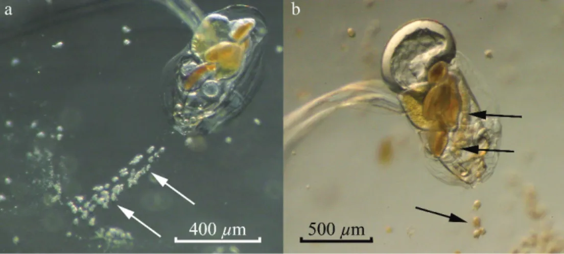
Fig. 6. Ciliates inside appendicularian houses. (a) Strombidium sp. ciliates attached to the buccal tube. (b) Ciliates inside the food-concentrating filter and ingested by a large appendicularian.
Table 1. Mean and maximum swimming speeds (6SD) of the different ciliate types.
Small politrich Strombidium sp. Large polytrich Swimming speed (mm s21) 299.75(6118.42) 1317.28(6426.86) 3460.36(6704.57)
This hypothesis remains plausible, but we can propose another. If the mouth is large enough to ingest large particles, the esophagus diameter is relatively narrow, and its size corresponds approximately to the inlet filter mesh width, then the other possible function of the inlet filter could be to prevent filtering large particles that cannot be swallowed by the appendicularian. In addition, the presence of these filters can be advantageous during algal blooms. When algal concentration is high, the house can clog (Tiselius et al. 2003), and in the case of large diatom blooms, the inlet filters can prevent clogging by restricting the size of algae filtered. Interestingly, Oikopleura long-icauda, a species usually considered to be adapted to oligotrophic environments (Lo`pez-Urrutia et al. 2004; Scheinberg and Landry 2004; Scheinberg et al. 2005), has no inlet filter. Without an inlet filter, O. longicauda could potentially retain all ingestible particles, including the large ones that can be essential for growth.
Observed interactions between appendicularians and cili-ates—All of our observations lead to the conclusion that small scuticociliate ciliates are the prey of appendicularians. Their small size allows them to enter the houses of even the smaller appendicularians, but their swimming speed is not sufficient to escape the suction in the buccal tube and they can pass through the esophagus. In contrast, a complex, size-specific interaction exists between appendicularians and the intermediate-sized Strombidium sp. ciliates (35 mm), which was the predominant ciliate in our experiment (Fig. 8). These ciliates cannot enter the house of small appendicularians (, 500 mm body size). Therefore, for small-sized appendicularians, the principal trophic interac-tion between appendicularians and Strombidium sp. is competition for the same food resource (Fig. 3a). However, we observed ciliate growth to be higher than the theoretical simulated growth rate if they were only to feed upon algae (Fig. 3c). This suggests that ciliates might have benefited by feeding directly on particles produced by appendicularians (discarded houses, fecal pellets) or the associated microbial populations.
For appendicularians of intermediate sizes (500– 900 mm), Strombidium sp. can enter the houses, but appendicularians are unable to ingest them. Consequently, the ciliates can feed intensively on the algae concentrated on the appendicularian house filter, resulting in an enhanced growth rate when compared with their theoretical growth rate based on algal concentration. However, the
presence of ciliates inside appendicularian houses could increase the mortality rate of appendicularians, potentially because of stress. In the stock culture, appendicularians with ciliates seemed to be more stressed and discarded their house more frequently under small perturbations (pipett-ing). This can potentially explain the increased mortality observed during the experiment. Consequently, the inter-action between ciliates and appendicularians of the 500– 900-mm body class size seems to be a parasitism relation-ship of ciliates on appendicularian houses rather than a symbiosis. However, because the difference in the mortality rate was not significantly different, we cannot exclude commensalism.
The entry of Strombidium sp. ciliates in the houses of the 500–900-mm class size of appendicularians seems to be counterintuitive. The mesh width of the inlet filter, 12 mm for a 500-mm appendicularian, should not allow ciliates to enter. However, the mesh shape of the inlet filter is rectangular with a 42-mm length that, if distorted into a circle, has a 34.3-mm-diameter aperture. Thus, these ciliates can pass the inlet filters by distorting the house inlet mesh. Our results suggest that as appendicularians become larger (, 900 mm body size), they feed on ciliates. This conclusion is based on the comparison of the appendicu-larian model with our experiment (Fig. 4d) and further confirmed by microscopic observations. The appendicular-ian flow velocity in the house buccal tube exceeds the ciliate swimming speed, and the directional escape behavior (Jakobsen 2001, 2002) is inefficient in the closed environ-ment of the buccal tube. Despite the small diameter of the esophagus (25 mm for a 500-mm appendicularian), ciliates can also be swallowed.
Finally, these shifts between competition, parasitism-commensalism, and predation between appendicularians and Strombidium sp. ciliates led to an apparent mutualism: the ciliate growth rate was higher than theoretically allowed by the algal concentration, whereas appendicularians used ciliates as a food to permit increased growth and reproduction during the last days of our experiment, despite the low algal concentration.
Considering the case of the large scuticociliate ciliates, they seem not to feed on appendicularian houses, and reciprocally, appendicularians are not able to ingest or swallow them. On the basis of our observations, these ciliates are likely histophagus and feed on moribund appendicularians. This feeding behavior is not surprising considering that appendicularians died after spawning.
Our results show that appendicularians, generally considered as bacterivorous or herbivorous filter feeders (Fortier et al. 1994; Legendre and Rivkin 2002), can feed efficiently on some ciliate types and therefore must be considered omnivorous. We found that appendicularians can feed on ciliates, and it is a size-related phenomenon. Interestingly, a previous study reported that some ciliate remains were observed in Oikopleura vanhoeffeni fecal pellets (Urban et al. 1992). Also, it has been shown that appendicularians can remove ciliates from the water (Vargas and Gonza´lez 2004; To¨nnesson et al. 2005), but ingestion of ciliates was not considered. Instead, the authors generally considered, on the basis of the inlet filter
Table 2. Mean and maximal flow velocity (6SD) inside the house buccal tube recorded for appendicularians of different body sizes.
Body size (mm)
Velocity (mm s21)
Flow Maximum flow 682 928.62(6352.85) 1837.72 911 976.7 (6485.64) 3330.48 1128 1381.45(6691.15) 3761.39 1350 1365.04(6734.48) 3905.42
mesh size, that ciliates were retained outside of the house (Vargas and Gonza´lez 2004). Our study suggests that ciliates could have entered the appendicularian houses in their studies and also might have served as food for appendicularians.
Study observations compared with in situ conditions— Concentrations of both algae and ciliates during the chemostat experiment were similar to in situ conditions. Initial concentrations were chosen to represent typical mesotrophic spring conditions observed in the nearshore Mediterranean Sea (1–8 ciliates mL21; Dolan and Marrase´
1995; Pe´rez et al. 2000), although they deviated significantly
during the experiment. By the end of the experiment, the algal concentration decreased to a level comparable to oligotrophic situations. Maximum ciliate concentrations observed in our study (15–20 cells mL21) are comparable to
concentrations observed under eutrophic conditions in nearshore ecosystems or upwelling (Sherr et al. 1986; Dolan and Coats 1991; Santoferrara and Alder 2009). Furthermore, they correspond to conditions in which ciliates were observed in situ inside appendicularian houses (1–18 ciliates mL21, corresponding to 6–84 ciliates
house21; Davoll and Silver 1986). Ciliate community
composition in the experiment, however, significantly differed from in situ assemblages, and only those able to
Fig. 7. Sequence of images showing the histophagous behavior of large scuticiliate ciliates. (a) Appendicularian freshly anaesthetized with MS-222. (b) Only 9 minutes after, ciliates had penetrated inside the oikoplastic epithelium of the appendicularian. (c) When the trunk of the appendicularian is empty, the ciliates attack the gonad. (d) Thirty-three minutes after, the appendicularian is almost fully consumed, and only the house rudiment, a cuticular layer, and the tail remain. (e, f ) Large scuticociliate ciliates before and after appendicularian consumption. The scale for panels a–d is indicated in panel a, and the scale for panels e–f is indicated in panel e.
survive under the appendicularian culture conditions were present. In our experiment, ciliates were essentially large (, 35 mm) oligotrich ciliates of the genus Strombidium, whereas in the field, large (. 20 mm) oligotrich ciliates correspond only to half of the ciliate assemblage (Pe´rez et al. 2000), in that very small (, 20 mm) ciliates often dominate total ciliate biomass (Sherr et al. 1986).
Possible effect on in situ populations—Because the ciliate densities in houses in our experiment are comparable to those observed in situ (Davoll and Silver 1986; Hansen et al. 1996), it is probable that the observed interactions also occur in the natural environment and have an effect on appendicularian and ciliate populations. In our experiment, appendicularians and ciliates coexisted and benefited reciprocally from their interactions. Moreover, similar observations in our stock culture, in which appendicularian cohorts are less synchronized, show that this coexistence can remain stable over several appendicularian generations, and no disappearance of either ciliates or appendicularians occurs. This can be explained by the fact that appendicu-larians are able to ingest ciliates only during the few last days of their life cycle, whereas ciliates benefit from the food concentrated by smaller appendicularians. A similar relationship might occur under in situ conditions.
In natural populations, appendicularians have mean body sizes around 650 mm and are generally not synchro-nized (Lo´pez-Urrutia et al. 2003b; Maar et al. 2004; Vargas and Gonza´lez 2004). As a result, only a small fraction of appendicularians likely feed on ciliates, and their predation pressure on ciliates should be low. Moreover, ciliates would benefit from the food contained within appendicularian
houses, whereas the house can represent a refuge where ciliates escape from the predation pressure of copepods. As a consequence, the presence of appendicularians could have an overall positive effect on ciliate populations, leading to increasing their density or serving as a refuge for ciliates. This hypothesis is supported by the high density of ciliates observed when appendicularians are present in high abundance (Davoll and Silver 1986). In addition, by feeding on small ciliates and harboring larger ones, appendicularians could modify the size spectra of ciliate populations. Then by supporting higher growth rates for ciliates and larger ciliates, appendicularians might also indirectly support other organisms, such as copepods and euphausiids, that feed preferentially on ciliates (Wiadnyana and Rassoulzadegan 1989; Jonsson and Tiselius 1990; Nakagawa et al. 2004). Finally, when the house is discarded, ciliates remain trapped inside until its degrada-tion, which could last 6 d (Davoll and Silver 1986). During these 6 d and because of its high sinking speed (Gorsky et al. 1984; Alldredge and Gotschalk 1988), the discarded house can transport ciliates to more than 400 m depth. By this phenomenon, appendicularians can also enhance the density of mesopelagic ciliate populations.
The ciliate effect on appendicularians is also complex: ciliates seem to increase the mortality rate of appendicular-ians, whereas this could be counterbalanced by a better growth rate and fertility of large (. 900 mm body size) appendicularians. Ciliates are generally considered to be a high-quality food resource for copepods and contain some important nutritional elements usually absent from micro-phytoplankton. Ciliates have a high nutritional quality with a high content of protein (C : N < 3.5; Stoecker and McDowell Capuzzo 1990) and contain amino acids, fatty acids, and sterols essential for zooplankton growth, survival, and reproduction (Phillips 1984; Stoecker and McDowell Capuzzo 1990). As a consequence, copepods, when feeding on ciliates, exhibit higher growth and egg productions rates than when fed on algae alone (Stoecker and Egloff 1987; Bonnet and Carlotti 2001). Thus, because appendicularians are able to feed on ciliates during maturation of their gonads, it is possible that this additional high-quality food supply could support a higher egg production rate. This higher fertility could permit appendicularian populations to survive despite the en-hanced mortality rate caused by ciliates. Moreover, under oligotrophic or offshore conditions, even if the ciliate concentration is lower than in the nearshore environment (, 1 mg C L21; Sherr et al. 1986), this high-energy
additional food supply might allow them to complete growth and reproduction despite the food-depleted envi-ronment. For example, in the case of O. dioica, the condition under which growth is severely limited is close to those observed in oligotrophic environments (, 30 mg C L21; Lo´pez-Urrutia et al. 2003a; Lombard et
al. 2009b), and any additional food supply can make a difference and might allow appendicularians to survive. Ciliate biomass in carbon units is generally closely linked to chlorophyll a (Chl a) with a C : Chl a ratio usually observed between 0.8–2.7 (Dolan and Marrase´ 1995). If we assume a C : Chl a of 50 when calculating a first-order estimate of
Fig. 8. Schematic representation of the interaction observed between appendicularians and Strombidium sp. ciliates and their respective benefit as a function of appendicularian size. The details of the interactions during the early stage of the appendicularian is not known, and the possible negative effect on middle-sized appendicularians through stress is speculative. Consequently, commensalism instead of parasitism cannot be excluded.
phytoplankton biomass, then ciliates could represent from 1.5% to 6% of the available food for appendicularians. However, because all ciliate sizes cannot be ingested by appendicularians and some ciliate species might have a different escape response, these proportions represent only an upper estimate of the potential bulk contribution of ciliates to appendicularian diet.
These interactions between ciliates and appendicularians likely depend on the appendicularian species considered. The O. dioica used in our study is relatively small compared with high-latitude or deep species. For example, O. vanhoeffeni can attain a body size of 6500 mm (Deibel 1998), Oikopleura labradorensis 6000 mm (Shiga 1976), Mesochordaeus erythrocephalus 7300 mm (Hopcroft and Robison 1999), and Bathochordaeus charon . 25,000 mm (Galt 1979; Hamner and Robison 1992). Moreover, and especially for mesopelagic species, a large number of organisms were observed inside their houses. For B. charon, up to 15,000 ciliates (Silver et al. 1998) and up to 500 copepods (Steinberg et al. 1994, 1997) were observed in only one house. If the relationship between body size and flow velocity in the house buccal tube that we observed with O. dioica is extrapolated to these species, then B. charon could have a flow velocity in its buccal tube of 20,000 mm s21, which is enough to ingest ciliates as well as
small copepods that have a lower swimming speed (Landry and Fagerness 1988; Mazzocchi and Paffenho¨fer 1999; van Duren and Videler 2003). Consequently, it is possible that tintinids, foraminifera, copepod eggs, nauplii, and the unidentified crustacean fragments found in their stomachs (Hopcroft and Robison 1999) might have been ingested alive and were not detrital, as suggested in that study. Given the large size of this species, the large number of organisms inside its houses, and the low food concentration and quality in the mesopelagic environment, this potential food supply could be essential for the growth of mesope-lagic species.
The interaction existing between appendicularians and ciliates might also influence the carbon cycle. On the one hand, appendicularians produce discarded houses enriched in all the constituents of the microbial loop, including ciliates. These sinking particles, highly loaded with microorganisms, could increase the transport of carbon to deep layers and serve as food for mesopelagic organisms. On the other hand, the presence of ciliates in discarded houses might also decrease the transport of organic carbon to deep layers. In the house microcosm, effectively all the constituents of the microbial loop are present at a high density, and the ciliates could feed, respire, grow, and reproduce intensively. By these actions, ciliates should decrease the carbon content of the houses and, by their mechanical action, increase the degradation of houses. It is then possible that the presence of ciliates could either increase or decrease the transport of carbon to deep layers. Further investigations are needed to better understand these processes.
Acknowledgments
We thank P. Nival, D. Deibel, J. L. Acun˜a, C. Poggiale, and F. Carlotti for constructive discussions. We thank the two
anony-mous reviewers whose remarks greatly improved the manuscript and J. Frost for her critical reading of the manuscript.
Financial support was provided by the European Commis-sion’s Sixth Framework Programme ‘‘Southern European Seas: Assessing and Modelling Ecosystem changes’’ (SESAME) project, contract GOCE-2006-036949; the ZooPNEC program part of the French ‘‘Programme National Environnement Coˆtier’’ (PNEC); the ‘‘Marine Biodiversity and Ecosystem Functioning’’ (MAR-BEF); the ‘‘European Network of Excellence for Ocean Ecosys-tems Analysis’’ (EUR-OCEANS); the Agence Nationale de la Recherche ANR–Biodiversite´ project Aquaparadox; and the Marie Curie Intra-European Fellowship 221696.
References
ACUN˜ A, J. L., D. DEIBEL, P. A. SAUNDERS, B. BOOTH, E. HATFIELD,
B. KLEIN, Z. P. MEI,ANDR. B. RIVKIN. 2002. Phytoplankton ingestion by appendicularians in the North Water. Deep-Sea Res. II 49: 5101–5115.
ALLDREDGE, A. L. 2004. The contribution of discarded
appendi-cularian houses to the flux of particulate organic carbon from oceanic surface waters, p. 309–326. In G. Gorsky, M. J. Youngbluth and D. Deibel [eds.], Response of marine ecosystems to global change: Ecological impact of appendi-cularians. GB Scientific Publisher.
———,ANDC. C. GOTSCHALK. 1988. In situ settling behavior of
marine snow. Limnol. Oceanogr. 33: 339–351.
BONNET, D., AND F. CARLOTTI. 2001. Development and egg
production in Centropages typicus (Copepoda: Calanoida) fed different food types: A laboratory study. Mar. Ecol. Prog. Ser. 224: 133–148.
CRAWFORD, D. W.,ANDD. K. STOECKER. 1996. Carbon content,
dark respiration and mortality of the mixotrophic planktonic ciliate Strombidium capitatum. Mar. Biol. 126: 415–422. DAVOLL, P. J.,ANDM. W. SILVER. 1986. Marine snow aggregates:
Life history sequence and microbial community of abandoned larvacean houses from Monterey Bay, California. Mar. Ecol. Prog. Ser. 33: 111–120.
DEIBEL, D. 1998. Feeding and metabolism of Appendicularia,
p. 139–149. In Q. Bone [ed.], The biology of pelagic tunicates. Oxford University Press.
DOLAN, J. R., AND D. W. COATS. 1991. Changes in fine-scale
vertical distributions of ciliate microzooplankton related to anoxia in Chesapeake Bay waters. Mar. Microb. Food Webs 5: 81–93.
———, AND C. MARRASE´. 1995. Planktonic ciliate
distri-bution relative to a deep chlorophyll maximum: Catalan sea, NW Mediterranean, June, 1993. Deep-Sea Res. I 42: 1965–1987.
FENAUX, R. 1986. The house of Oikopleura dioica (Tunicata,
Appendicularia): Structure and functions. Zoomorphology 106: 224–231.
FENCHEL, T.,ANDP. R. JONSSON. 1988. The functional biology of
Strombidium sulcatum, a marine oligotrich ciliate (Ciliophora, Oligotrichina). Mar. Ecol. Prog. Ser. 48: 1–15.
FERNA´ NDEZ, D., A. LO´ PEZ-URRUTIA, A. FERNA´ NDEZ, J. L. ACUN˜ A,
ANDR. P. HARRIS. 2004. Retention efficiency of 0.2 to 6 mm
particles by the appendicularians Oikopleura dioica and Fritillaria borealis. Mar. Ecol. Prog. Ser. 266: 89–101. FLOOD, P. R., D. DEIBEL, AND C. MORRIS. 1992. Filtration of
colloidal melanin from seawater by planktonic tunicates. Nature 355: 630–632.
———, AND ———. 1998. The appendicularian house,
p. 105–124. In Q. Bone [ed.], The biology of pelagic tunicates. Oxford University Press.
FORTIER, L., J. LE FE` VRE, AND L. LEGENDRE. 1994. Export of
biogenic carbon to fish and to the deep ocean: The role of large planktonic microphages. J. Plankton Res. 16: 809–839. GALT, C. P. 1979. First records of a giant pelagic tunicate, Bathochordaeus charon (Urochordata, Larvacea), from the eastern Pacific Ocean, with notes on its biology. Fish. Bull. 77: 514–519.
GORSKY, G.,ANDR. FENAUX. 1998. The role of Appendicularia in
marine food webs, p. 161–169. In Q. Bone [ed.], The biology of pelagic tunicates. Oxford University Press.
———, N. S. FISHER,ANDS. W. FOWLER. 1984. Biogenic debris
from the pelagic tunicate, Oikopleura dioica, and its role in the vertical transfer of a transuranium element. Estuar. Coast. Shelf Sci. 18: 13–23.
HAMNER, W. M.,ANDB. H. ROBISON. 1992. In situ observations of
giant appendicularians in Monterey Bay. Deep-Sea Res. I 39: 1299–1313.
HANSEN, B., F. L. FOTEL, N. J. JENSEN,ANDS. D. MADSEN. 1996.
Bacteria associated with a marine planktonic copepod in culture. II. Degradation of fecal pellets produced on a diatom, a nanoflagellate or a dinoflagellate diet. J. Plankton Res. 18: 275–288.
HOPCROFT, R. R.,ANDB. H. ROBISON. 1999. A new mesopelagic
larvacean, Mesochordaeus erythrocephalus, sp. nov., from Monterey Bay, with a description of its filtering house. J. Plankton Res. 21: 1923–1937.
JAKOBSEN, H. H. 2001. Escape response of planktonic protists to
fluid mechanical signals. Mar. Ecol. Prog. Ser. 214: 67–78. ———. 2002. Escape of protists in predator-generated feeding
currents. Aquat. Microb. Ecol. 26: 271–281.
JONSSON, P. R., ANDP. TISELIUS. 1990. Feeding behaviour, prey
detection and capture efficiency of the copepod Acartia tonsa feeding on planktonic ciliates. Mar. Ecol. Prog. Ser. 60: 35–44.
LANDRY, M. R., AND V. L. FAGERNESS. 1988. Behavioral and
morphological influences on predatory interactions among marine copepods. Bull. Mar. Sci. 43: 509–529.
LEGENDRE, L.,ANDR. B. RIVKIN. 2002. Fluxes in the upper ocean:
Regulation by food-wed control nodes. Mar. Ecol. Prog. Ser. 242: 95–109.
LOMBARD, F., F. RENAUD, C. SAINSBURY, A. SCIANDRA,AND G.
GORSKY. 2009a. Appendicularian ecophysiology I. Food
concentration dependent clearance rate, assimilation efficien-cy, growth and reproduction of Oikopleura dioica. J. Mar. Syst 78: 606–616.
———, A. SCIANDRA,ANDG. GORSKY. 2005. Influence of body
mass, food concentration, temperature and filtering activity on the oxygen uptake of the appendicularian Oikopleura dioica. Mar. Ecol. Prog. Ser. 301: 149–158.
———, ———,AND———. 2009b. Appendicularian
ecophysiol-ogy. II. Modeling nutrition, metabolism, growth and repro-duction of the appendicularian Oikopleura dioica. J. Mar. Syst 78: 617–629.
LO´ PEZ-URRUTIA, A., J. L. ACUN˜ A, X. IRIGOIEN, AND R. HARRIS. 2003a. Food limitation and growth in temperate epipelagic appendicularians (Tunicata). Mar. Ecol. Prog. Ser. 252: 143–157.
———, R. P. HARRIS, J. L. ACUN˜ A, U. BA˚ MSTEDT, P. R. FLOOD, H.
J. FYHN, B. GASSER, G. GORSKY, X. IRIGOIEN, AND M. B.
MARTINUSSEN. 2004. A comparison of appendicularian
seasonal cycles in four distinct European coastal environ-ments, p. 255–276. In G. Gorsky, M. J. Youngbluth and D. Deibel [eds.], Response of marine ecosystems to global change: Ecological impact of appendicularians. GB Scientific Publisher.
———, X. IRIGOIEN, J. L. ACUN˜ A,ANDR. HARRIS. 2003b. In situ
feeding physiology and grazing impact of the appendicularian community in temperate waters. Mar. Ecol. Prog. Ser. 252: 125–141.
MAAR, M., T. G. NIELSEN, S. GOODING, K. TONNESSON, P.
TISELIUS, S. ZERVOUDAKI, E. CHRISTOU, A. SELL, AND K.
RICHARDSON. 2004. Trophodynamic function of copepods,
appendicularians and protozooplankton in the late summer zooplankton community in the Skagerrak. Mar. Biol. 144: 917–934.
MAZZOCCHI, M. G., AND G.-A. PAFFENHO¨ FER. 1999. Swimming and feeding behaviour of the planktonic copepod Clausoca-lanus furcatus. J. Plankton Res. 21: 1501–1518.
MONTAGNES, D. J. S. 1996. Growth responses of planktonic
ciliates in the genera Strobilidium and Strombidium. Mar. Ecol. Prog. Ser. 130: 241–254.
NAKAGAWA, Y., T. OTA, Y. ENDO, K. TAKI, AND H. SUGISAKI.
2004. Importance of ciliates as prey of the euphausiid Euphausia pacifica in the NW North Pacific. Mar. Ecol. Prog. Ser. 271: 261–266.
OHMAN, M. D.,ANDR. A. SNYDER. 1991. Growth-kinetics of the
omnivorous oligotrich ciliate Strombidium sp. Limnol. Ocean-ogr. 36: 922–935.
PE´ REZ, M. T., J. R. DOLAN, F. VIDUSSI,ANDE. FUKAI. 2000. Diel
vertical distribution of planktonic ciliates within the surface layer of the NW Mediterranean (May 1995). Deep-Sea Res. I 47: 479–503.
PHILLIPS, N. W. 1984. Role of different microbes and substrates as potential suppliers of specific, essential nutrients to marine detritivores. Bull. Mar. Sci. 35: 283–298.
PURCELL, J. E., M. V. STURDEVANT,ANDC. P. GALT. 2004. A review
of appendicularians as prey of invertebrate and fish predators, p. 359–435. In G. Gorsky, M. J. Youngbluth and D. Deibel [eds.], Response of marine ecosystems to global change: Ecological impact of appendicularians. GB Scientific Publisher. PUTT, M., AND D. K. STOECKER. 1989. An experimentally
determined carbon: Volume ratio for marine ‘‘oligotrichous’’ ciliates from estuarine and coastal waters. Limnol. Oceanogr. 34: 1097–1103.
RIVIER, A., D. C. BROWNLEE, R. W. SHELDON,ANDF. R ASSOULZA-DEGAN. 1985. Growth of microzooplankton: A comparative
study of bactivorous zooflagellates and ciliates. Mar. Microb. Food Webs 1: 51–60.
ROBISON, B. H., K. R. REISENBICHLER,ANDR. E. SHERLOCK. 2005.
Giant larvacean houses: Rapid carbon transport to the deep sea floor. Science 308: 1609–1611.
SANTOFERRARA, L., ANDV. ALDER. 2009. Abundance trends and
ecology of planktonic ciliates of the south-western Atlantic (35–63uS): A comparison between neritic and oceanic environments. J. Plankton Res. 31: 837–851.
SCHEINBERG, R. D.,ANDM. R. LANDRY. 2004. Clearance rates and
efficiencies of Oikopleura fusiformis on the natural prey assemblage of a subtropical coastal ecosystem, p. 207–226. In G. Gorsky, M. J. Youngbluth and D. Deibel [eds.], Response of marine ecosystems to global change: Ecological impact of appendicularians. GB Scientific Publisher.
———, ———,ANDA. CALBET. 2005. Grazing of two common
appendicularians on the natural prey assemblage of a tropical coastal ecosystem. Mar. Ecol. Prog. Ser. 294: 201–212. SHERR, E. B., B. F. SHERR, R. D. FALLON,ANDS. Y. NEWELL. 1986.
Small, aloricate ciliates as a major component of the marine heterotrophic nanoplankton. Limnol. Oceanogr. 31: 177–183. SHIGA, N. 1976. Maturity stages and relative growth of Oikopleura
labradoriensis Lohmann (Tunicata, Appendicularia). Bull. Plankton Soc. Jpn 23: 31–45.
SILVER, M. W., S. L. COALE, C. H. PILSKALN, AND D. R.
STEINBERG. 1998. Giant aggregates: Importance as microbial centers and agents of material flux in the mesopelagic zone. Limnol. Oceanogr. 43: 498–507.
STEINBERG, D. K., M. W. SILVER,ANDC. H. PILSKALN. 1997. Role
of mesopelagic zooplankton in the community metabolism of giant larvacean house detritus in Monterey Bay, California, USA. Mar. Ecol. Prog. Ser. 147: 167–179.
———, ———, ———, S. L. COALE,ANDJ. B. PADUAN. 1994. Midwater zooplankton communities on pelagic detritus (giant larvacean houses) in Monterey Bay, California. Limnol. Oceanogr. 39: 1606–1620.
STEMMANN, L., K. ROBERT, M. PICHERAL, H. PATERSON, A. HOSIA, M. J. YOUNGBLUTH, F. IBANEZ, L. GUIDI, F. LOMBARD,AND
G. GORSKY. 2008. Global biogeography of fragile
macro-zooplankton in the upper 100–1000 m depth inferred from the Underwater Video Profiler. ICES J. Mar. Sci. 65: 433–442.
STOECKER, D. K.,ANDD. A. EGLOFF. 1987. Predation by Acartia
tonsa Dana on planktonic ciliates and rotifers. J. Exp. Mar. Biol. Ecol. 110: 53–68.
———, AND J. M. MCDOWELL CAPUZZO. 1990. Predation on
protozoa: Its importance to zooplankton. J. Plankton Res. 12: 891–908.
———,ANDA. E. MICHAELS. 1991. Respiration, photosynthesis
and carbon metabolism in planktonic ciliates. Mar. Biol. 108: 441–447.
STRATHMANN, R. R. 1967. Estimating the organic content of
phytoplankton from cell volume or plasma volume. Limnol. Oceanogr. 12: 411–418.
TISELIUS, P.,ANDoTHERS. 2003. Functional response of Oikopleura
dioica to house clogging due to exposure to algae of different sizes. Mar. Biol. 142: 253–261.
TONNESSON, K.,ANDoTHERS. 2005. Grazing impact of Oikopleura
dioica and copepods on an autumn plankton community. Mar. Biol. Res. 1: 365–373.
URBAN, J. L., C. H. MCKENZIE,ANDD. DEIBEL. 1992. Seasonal
differences in the content of Oikopleura vanhoeffeni and Calanus finmarchicus fecal pellets—illustrations of zooplank-ton food web shifts in coastal Newfoundland waters. Mar. Ecol. Prog. Ser. 84: 255–264.
URBAN-RICH, J., D. FERNA´ NDEZ,ANDJ. L. ACUN˜ A. 2006. Grazing impact on chromophoric dissolved organic matter (CDOM) by the larvacean Oikopleura dioica. Mar. Ecol. Prog. Ser. 317: 101–110.
VANDUREN, L. A.,ANDJ. J. VIDELER. 2003. Escape from viscosity:
The kinematics and hydrodynamics of copepod foraging and escape swimming. J. Exp. Biol. 206: 269–279.
VARGAS, C. A.,ANDH. E. GONZALEZ. 2004. Plankton community
structure and carbon cycling in a coastal upwelling system. I. Bacteria, microprotozoans and phytoplankton in the diet of copepods and appendicularians. Aquat. Microb. Ecol. 34: 151–164.
WIADNYANA, N. N., AND F. RASSOULZADEGAN. 1989. Selective
feeding of Acartia clausi and Centropages typicus on micro-zooplankton. Mar. Ecol. Prog. Ser. 53: 37–45.
ZUBKOV, M. V., AND A. LO´ PEZ-URRUTIA. 2003. Effect of
appendicularians and copepods on bacterioplankton compo-sition and growth in the English Channel. Aquat. Microb. Ecol. 32: 39–46.
Associate editor: Edward McCauley Received: 20 March 2009 Accepted: 08 September 2009 Amended: 14 September 2009