HAL Id: tel-00364271
https://tel.archives-ouvertes.fr/tel-00364271
Submitted on 25 Feb 2009
HAL is a multi-disciplinary open access archive for the deposit and dissemination of sci-entific research documents, whether they are pub-lished or not. The documents may come from teaching and research institutions in France or abroad, or from public or private research centers.
L’archive ouverte pluridisciplinaire HAL, est destinée au dépôt et à la diffusion de documents scientifiques de niveau recherche, publiés ou non, émanant des établissements d’enseignement et de recherche français ou étrangers, des laboratoires publics ou privés.
Modélisation de Processus Photo induits du
Photosystem II
Christian Herrero Moreno
To cite this version:
Christian Herrero Moreno. Modélisation de Processus Photo induits du Photosystem II. Chimie. Université Paris Sud - Paris XI, 2007. Français. �tel-00364271�
Université Paris XI
Ecole Doctorale de Chimie de Paris-Sud
THESE
Présentée en vue de l’obtention du
GRADE DE DOCTEUR DES SCIENCES DE L’UNIVERSITE PARIS XI
par
Christian Herrero Moreno
SYNTHESIS AND CHARACTERISATION OF ARTIFICIAL MIMICS OF PHOTOSYSTEM II
Soutenance prévue le 14 décembre 2007
Composition du jury
Pr Thomas Moore
rapporteur
Pr Rene Bensasson
rapporteur
Pr Ana Moore
examinateur
Dr William Rutherford
examinateur
Pr Edmond Amouyal
examinateur
Pr Jean Jaques Girerd
examinateur
Table of Contents Acknowledgments………...i List of figures……….ii List of abbreviations………...v 1. Abstract………...1 2. Introduction………..…………..2 2.1. Natural photosynthesis……….………….6
2.2. Water oxidizing complex……….10
3. Artificial photosynthetic devices………...………12
3.1. Antennas………..……16 3.1.1. Non-photochemical Quenching………21 3.2. Ruthenium constructs………..………26 3.2.1. Photoactive chromophore……….26 3.2.2. Donor side……….………29 3.2.2.1.Phenol group……….29 3.2.2.2. Manganese complexes……….………34 3.2.2.2.1. Terpyridine complexes………...………35
3.2.2.2.2. High oxidation State Manganese clusters………..45
3.2.2.2.3. Salen Compounds……….………..49 3.2.2.2.4. Salophen compounds………..55 4. Conclusion...58 5. References………60 6. Experimental Section……….…………64 6.1. Antennas………..………64 6.1.1. Triad………..………64 6.1.2. Dyad………..………66 6.2. Ruthenium complexes……….………71 6.2.1 Chromophores………..71 6.2.2. Phenols……….…………74 6.2.3. Terpyridine Complexes………80
6.2.4. Ru-Terpyridine-Mn2-di-µ-oxo Complexes………..……….83
6.2.5. Salen Ligands………...…………85
6.3. Instrumentation………89
Acknowledgments
All together it has been two countries, three labs, several projects, about six of bosses, and lots of friends. For those which I left behind without saying goodbye, those who welcomed and helped me when I was a bit lost, and those who have, for some reason, stayed behind me all the way I’d like to express all my gratitude and wish that everyone in this world would get to meet such a gang.
List of Figures and Tables.
Figure 1: Antenna system.
Figure 2: Cofactors of electron transport chain in PSII.
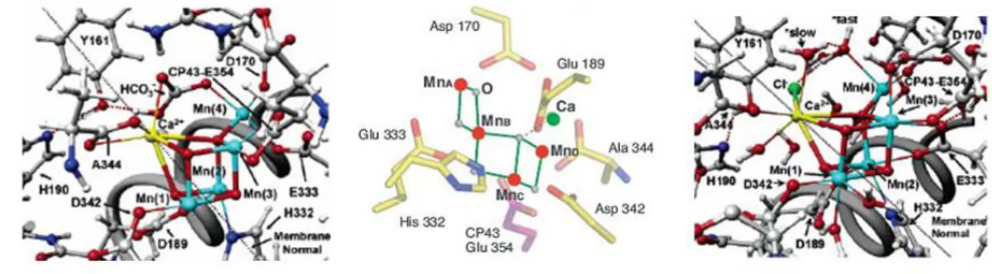
Figure 3: Different models proposed for the structure of the OEC.
Figure 4: Model of a light absorbing triad for oxidation/reduction processes. Figure 5: Schematic of a water splitting photochemical cell.
Figure 6: Pathways for energy transfer from a carotenoid to a phthalocyanine. Figure 7: Structures of carotenoid-phthalocyanin triads and model compounds. Figure 8: Absorption and excitation spectra for dyads 2 (left) and 1 (right). Figure 9: Different energy transfer patways for dyad 2 (left) and 1 (right). Figure 10: Carotenoids involved in zeaxanthin cycle.
Figure 11: Structures of different length caroteno-phthalocyanin dyads and corresponding model
compounds.
Figure 12: (Left) absorption spectra of dyads 6 (red), 7 (blue), 8 (green), and model
phthalocyanin (black). (Right) Emission decays for same compounds.
Figure 13: Physical properties of ground and excited state ruthenium trisbipyridine. Figure 14: Structure of different chromophores.
Figure 15: Structure of different Ruthenium-imidazole-phenol compounds.
Figure 16: Absorption (left) and emission (right) of compounds Ru(bpy)32+ (blue), 16 (green), 17
(black), and 14 (red).
Figure 17: Left: Transient absorption changes at 450 nm for compounds: reference (black), 16
(red), 17 (green), and 14 (blue). Right: Example of transient absorption spectrum monitoring for MV.+ (red line), and 450 nm (blue line). Right bottom: Structure of reference compound (Std).
Figure 18: Proposed reaction mechanism for O2 formation from the reaction of
[(terpy)(H2O)Mn(µ-O)2Mn(H2O)(terpy)] with an external oxidant.
Figure 19: Structure of different Ruthenium-terpyridine compounds.
Figure 20: Ground state absorption spectra in acetonitrile for (a) [Ru(bpy)3]2+, Ru-Terpy and Ru
Terpy-Mn; and (b) [Ru(bpy)3]2+, ester-Ru-Terpy and ester-Ru-Terpy-Mn.
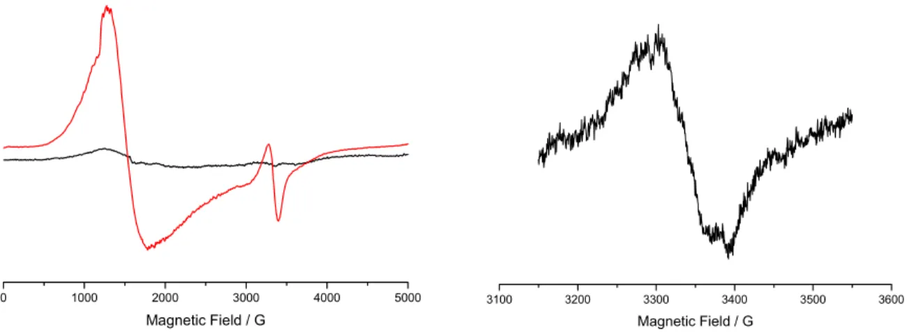
Figure 21: (Left) EPR spectra of a mixture of Ru-Terpy with cobalt pentamine before
illumination (black), and after illumination (red). (Right). Closer look at radical region.
Figure 22: (Left) EPR spectra of a mixture of Ru-Terpy-Mn with cobalt pentamine before
illumination (yellow), and after illumination (blue). (Right). EPR spectra of a mixture of Ester-Ru-Terpy-Mn with cobalt pentamine before illumination (blue), and after illumination (red).
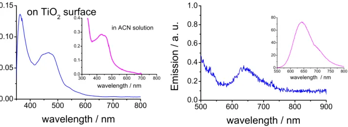
Figure 23: Absorption (left) and emission (right) spectra of compound 19 grafted to a TiO2
surface. Insets indicate results in solution for comparison.
Figure 24: EPR spectra of compound 19a grafted to TiO2 before (red), and after (black)
illumination.
Figure 25: Structures of differnet Mn2 di-μ-oxo dimmers.
Figure 26: Uv/vis spectra of the formation of compound 21.
Figure 27: Uv/vis spectra of the formation of compound 23 (left), and 22 (right). Figure 28: EPR spetra of compounds 23 (left), and 22 (right).
Figure 29: (Top) EPR spectra of a mixture of compound 22 with cobalt pentamine before
illumination and (bottom) after illumination.
Figure 30: Structures of different salen compounds.
Figure 31: Proposed mechanism for salen epoxidation. From Kochi and coworkers
Figure 33: Absorption (left) and emission (right) of compounds Ru Salen (black), Ru Salen Mn
(green), Ester Ru Salen (red), and ester Ru Salen Mn (blue).
Figure 34: EPR parallel mode in methanol of compounds 24a (right), and Salen Mn (left). Figure 35: Uv/vis spectra following the reaction of 24a with cobalt pentamine and light Figure 36: EPR of dark (black), and light (red) reactions of 24a with [CoIII(NH3)5]3+.
Figure 37: Structures of different salophen compounds.
Figure 38: Absorption (left) and emission (right) of compounds Ru Salophen (black), Ru
Salophen Mn (red), Ester Ru Salen (green), Ester Ru Salen Mn (blue), and Ru(bpy)3 (light blue).
Figure 39: EPR spectrum of compound 30a.
Tables
Table 1: Electrochemical data for Ruthenium-Phenol compounds 14, 16-17.In acetonitrile vs.
SCE.
Table 2: Electrochemical data for Ruthenium(II)-Terpyridine-Manganese(II) complexes 14,
16-17. In acetonitrile vs. SCE.
Table 3: Electrochemical data for Ruthenium(II)-Salen-Manganese(III) complexes 24 and 24a as
well as salen Mn(III) for comparison. In acetonitrile vs. SCE.
List of Abbreviations
A Acceptor
Ant Antenna
bpy 2,2’-bipyridine
Car Carotenoid
Catox Catalyst for oxidation
Catred- Catalyst for reduction
Chl Chlorophyll
Co Cobalt
D Donor
db Double bond
e- Electron
EPR Electron paramagnetic resonance
ET Electron transfer
eV Electronvolt
HOMO Highest occupied molecular orbital
ICT Internal charge transfer
LH-I Light-harvesting complex I
LH-II Light-harvesting complex II
LUMO Lowest unoccupied molecular orbital
MLCT Metal to ligand charge transfer
Mn Manganese
µs Microsecond
Nm Nanometer
ns Nanosecond
OEC Oxygen Evolving Complex
PCET Proton coupled electron transfer
Pheo Pheophytin Pc Phthalocyanin ps Picosecond PSII Photosystem II P680 Pigment absorbing at 680 nm Q Quinone Ru Ruthenium Rc Reaction centre S Sensitiser
SCE Standard Calamel Electrode
S0 – S4 Storage states in the Kok cycle
Terpy 2,2’:6’,2’’- terpyridine
THF Tetrahydrofuran
TiO2 Titanium dioxide
TW Terawatt
TyrZ Tyrisine Z
1. Abstract
In natural photosynthesis, light energy is converted into chemical energy by photosynthetic reaction centers. This energy is stored in the form of high energy substances synthesized in the reductive branch of the photosynthetic process. The electrons needed for these processes are furnished by water upon its oxidation by the Oxygen Evolving Complex (OEC) in PSII.
Artificial photosynthesis aims to replicate the reactions that take place in natural organisms in order to i) gain a better understanding of the chemical processes taking place in the natural systems, and ii) strive towards the harnessing of sunlight in order to have access to clean, sustainable fuels. Processes undergone in nature such as light capture, energy transfer, electron transfer, charge separation, activation of catalyst, and reaction at the catalytic site must be accomplished within the framework of artificial systems.
With these concepts in mind we have designed, synthesized and characterized molecules that mimic the reactions performed by antennas and reaction centers present in Photosystem II. These molecules are able to undergo light-induced charge separation, electron transfer, and accumulation of oxidizing/reducing equivalents that mimic the processes occurring in natural systems. The artificial anntenas are composed of carotenoid and phthalocyanin groups. These molecules show large absoption profiles with high extinction coefficients, and are capable of ultra-fast energy transfer processes which lead up to charge separation states. Varying the conjugation length of the carotenoid molecules from 9 double bonds to 11 double bonds, we can show how these molecules may act as energy donors as well as energy dissipators, a process akin to the Non Photochemical Quenching (NPQ) processes which happen during the xanthophyll cycle. The donor side mimics of Photosystem II have also been studied. These supramolecular systems contain a photoactive component covalently linked through a spacer to a cavity where a
metal ion or cluster is located. The photosensitizer used is a [Ru(bpy)3]2+ (bpy = 2,2’-bipyridine)
analogue, a counterpart to P680, which absorbs light in the visible region and triggers an electron
transfer process. The resulting RuIII species has a reversible oxidation potential of 1.30 V vs. SCE, similar to that of P680 (1.25 V vs NHE),17,18 and is, in theory, capable of oxidizing a
Manganese cluster and an electron source. Among the molecules mimicking the donor side of PSII we have synthesized ruthenium-phenol pairs, as well as bimetallic Ruthenium-Manganese systems. Among the latter we have studied those with terpyridine coordination cavities since Mn-di-μ-oxo-Mn dimers of this kind have been reported to catalyze the oxidation of water into molecular oxygen. Other catalytic groups such as salens and salophens have also been studied. These have been reported to perform the two electron oxidation of organic substrates following the same oxygen atom transfer mechanism as the one thought to be responsible for the oxidation of water, and could therefore be useful in achieving our goal. Following the work on synthesis and characterization of molecules capable of harnessing light and using it to drive reduction/oxidation reactions will be presented.
1. Résumé
La photosynthèse est un processus biologique naturel qui convertit l’énergie lumineuse en énergie chimique par l’action de centres réactionnels photosynthétiques. L’énergie convertie est stockée sous forme de produits de haute énergie synthétisés par la branche réductive du processus photosynthétique. Les électrons nécessaires à ces réactions sont fournis par des molécules d’eau lors de leur oxydation par le centre de dégagement de l'oxygène (Oxygen Evolving Complex: OEC) pour le système de photosynthèse II (PSII). La photosynthèse artificielle cherche à
reproduire les réactions qui se produisent dans les organismes naturels afin de i) de mieux comprendre les processus chimiques qui se déroulent dans les systèmes naturels, et ii) de parvenir à exploiter l’énergie solaire pour le développement de carburants propres et renouvelables. Chaque étape qui survient dans le processus de photosynthèse naturelle, telle que la capture de lumière, le transfert d’énergie, le transfert d’électron, la séparation de charge, l’activation du catalyseur et la réaction catalytique doit se produire au sein du système artificiel. La photosynthèse artificielle cherche à reproduire les réactions qui se produisent dans les organismes naturels afin de i) de mieux comprendre les processus chimiques qui se déroulent dans les systèmes naturels, et ii) de parvenir à exploiter l’énergie solaire pour le développement de carburants propres et renouvelables. Chaque étape qui survient dans le processus de photosynthèse naturelle, telle que la capture de lumière, le transfert d’énergie, le transfert d’électron, la séparation de charge, l’activation du catalyseur et la réaction catalytique doit se produire au sein du système artificiel. Avec ces concepts en vue, nous avons conçu, synthétisé et caractérisé des molécules qui imitent les réactions réalisées par les antennes et les centres réactionnels présents dans le photosystème II. Ces molécules sont capables de reproduire la séparation de charges induite par la lumière, le transfert d’électrons et l’accumulation d’équivalents oxydo-réducteurs observés pendant la photosynthèse naturelle. Les antennes artificielles se constituent de caroténoïdes et phthalocyanines. Ces molécules présentent des profiles d’absorption large avec des coefficients d’extinction élevés, et sont capables de supporter des transferts d’énergie ultra rapides qui permettent l’état de séparation de charges. En faisant varier la longueur de la chaine conjuguée des caroténoïdes de neuf à onze liaisons doubles, nous avons pu mettre en évidence comment ces molécules peuvent agir aussi bien comme donneurs que comme agents dissipateurs d’énergie, effet caractéristique qui s’apparente au processus de trempe non-photochimique (Non Photochemical Quenching: NPQ) qui se produit dans le cycle
de la zéaxanthine. Les mimiques des agents donneurs du photosystème II ont aussi été étudiées. Ces systèmes supramoléculaires contiennent une partie photoactive liée de façon covalente par un intermédiaire à une cavité contenant un ion ou un agrégat d’ions métalliques. La photosensibilisateur utilisé est un complexe du ruthénium [Ru(bipy)3]2+ (bpy = 2,2’-bipyridine),
homologue du P680, qui absorbe la lumière dans le spectre visible et déclenche le transfert
d’électron. Les espèces RuIII résultantes ont un potentiel d’oxydation réversible de 1.3 V vs SCE, comparables à celui de P680 (1.25 V vs NHE) et présentent donc la possibilité d’oxyder à la fois
un complexe manganèse ainsi qu’une source d’électron. Concernant les molécules imitant le coté donneur du PSII, nous avons synthétisé des paires ruthénium-phénol, ainsi que des systèmes ruthénium-manganèse bimétalliques. Parmi ces dernières, nous avons étudié celles présentant des cavités de coordination constituées de terpyridines, vu qu’il a déjà été montré que les dimères Mn-di-μ-oxo-Mn de ce type peuvent catalyser l’oxydation de l’eau en oxygène moléculaire. Des salènes et salophènes ont aussi été examinés étant donné que de tels groupes peuvent accomplir l’oxydation à deux électrons de substrats organique. Dans la littérature, ces réactions sont toutes conduites par l’action d’oxydants chimiques externes, tandis que nous avons pour but d’utiliser des espèces oxydantes induites par l’action de la lumière.
2. Introduction
One of the most pressing challenges society is facing today is the transition to a new energy scenario. Increases in world population and the rise of emerging economies have projected an increase in energy consumption from 13 TW today to 30 TW by 2050.1 If we couple the fact that 86 % of this energy comes from fossil fuels to the fact that CO2 levels are the highest they have
been for the past 650 000 years, it is clear that the burn rate that will be needed in 40 year’s time is unacceptable. In order to fill the gap of needed energy we must look for complementary sources that will be reliable enough to fill in the huge gap of demand and sustainable enough so their use will not be deleterious to life on earth. Amongst the possible alternate energy sources, solar power presents itself as the most promising source of renewable energy available. The goal of artificial photosynthesis is to harness this energy to drive the production of high energy chemicals from low energy source compounds.
Nature has given us the guidelines for such a process. During photosynthesis, absorption of light by phototrophic organisms initiates a series of energy and electron transfer processes that conclude with the oxidation of water (eq. 1) to reducing equivalents that can then drive the reduction of CO2 into higher carbohydrates (eq. 2).
2 H2O + 4 hν → O2 + 4 H+ + 4 e- (1)
nCO2 + 2n e- + 2n H+ → (CH2O)n (2)
2 H+ + 2 e- → H2 (3)
Artificial photosynthesis, thus, aims to imitate the above mentioned processes of light absorption, catalysis, and energy conversion. Photo-driven water oxidation, thus, would act as a source of electrons and protons that could be used to drive the production of high energetic fuels such as H2
(eq.3), or reduce carbon dioxide to valuable organic compounds (eq.2). Over the last two decades several advances have been made in this field. Examples of light induced long-lived charge separated states,2-5 electron transfer from chromophores to catalytic sites6,7 and water oxidation catalysis by chemical or electrochemical means8-15 have been reported in the literature. Nevertheless, little success has been achieved when trying to combine all these required processes into one complex. In the following, the work done up to date in our laboratory spanning from artificial antennas to light driven electron transfer systems will be presented.
2.1. Natural photosynthesis
Life on earth can be thought of being, almost entirely solar powered. Oxygenic photosynthetic organisms translate this kind of energy into chemical energy in the form of carbohydrates and oxygen, with the former serving as a source of high energy electrons and the latter as a low energy destination. Photosynthesis begins with the absorption of light. In purple bacteria this process is performed by light-harvesting complexes which are intricate assemblies of low molecular weight proteins and pigments housed within the membrane proteins. These moieties mainly include chlorophylls, quinones and carotenoids.
Most purple bacteria have two types of light-harvesting complexes, designated LH-I and LH-II (Fig 1). LH-I complexes are closely associated with the reaction center and form a so-called 'core' complex, while LH-II complexes are peripheral to this core. When light is absorbed by the peripheral antenna LH-II complexes, energy is transferred to LH-I, which then passes it onto the reaction center, in which a redox reaction causes charge separation across the membrane. All these components interact by three basic photochemical processes:
1) singlet-singlet energy transfer, as is the case in antenna systems consisting of chlorophylls, and carotenoids, which collect light and conduct excitation energy to the reaction center,
Car + hv → 1Car (4)
1Car +Chl → Car + 1Chl (5)
2) triplet-triplet energy transfer, as in the cases of formation of highly reactive singlet oxygen by triplet chlorophylls, and the consequent quenching of these dangerous species by carotenoids,
3Chl + Car → Chl + 3Car (6)
3Chl + 3O
1O
2 + Car → 3O2 + 3Car (8)
3) Photo-initiated electron transfer, involving chlorophylls and quinones which transform excitation energy into chemical potential in the form of long-lived, trans-membrane charge separation.
1Ant + D-A → Ant +1D-A → D+-A- (9)
Figure 1: Antenna and reaction center assembly.
The core complex of PS-II, the reaction centre (RC), contains in itself six chlorophyll a’s and 2 β-carotene pigments2,16. When light is collected by the reaction centre, or the surrounding
antennae systems, the absorbed photon energy is funneled through a series of energy transfer steps to the primary electron donor P680 which is probably made up by the four excitonically
coupled chlorophyll molecules of the PSII core (PD1, PD2, ChlD1, ChlD2 in Fig. 2).
This water oxidizing enzyme is a membrane-spanning, pigment-containing protein made up of several subunits. On one side of the membrane, it performs the two electron reduction of
plastoquinone to plastoquinol (PQ + 2e– + 2H+ → PQH2) while on the other side it catalyses the
four electron oxidation of water, producing molecular oxygen and protons as by products (2H2O
→ 4e- + 4H+ + O
2). The enzyme is thought of as being composed of i) a photochemical part
which upon light absorption, is capable of forming and stabilizing a large charge separated state, thus forming a reductant and a oxidant and ii) a catalytic device made up by a cluster of manganese ions and a redox active tyrosine residue capable of storing oxidizing equivalents, in order to split water.
Figure 2: Cofactors of electron transport chain in PSII.
Upon absorption of harnessed light energy, the sequence of electron transfer reactions can be described as follows:
1. The resulting excited P680 is capable of reducing a nearby pheophytin to produce the
initial charge separation (step 1, figure 2). This yields a cation radical P680+, and a
pheophytin anion radical (Pheo-). P680+ is the most oxidizing species known in biology
2. The pheophytin anion radical donates an electron to a quinone (QA). By this time (200
ps) the electron is 26 Å away from the oxidized P680, thereby stabilizing the charge
separated state (step 2).
3. The highly oxidizing P680+ extracts an electron from a nearby tyrosine residue (TyrZ)
(Step 3). Concomitant with this oxidation, proton loss from the tyrosine’s phenolic hydroxyl group occurs and the neutral tyrosyl radical is formed.
4. The neutral tyrosyl radical oxidizes the Mn ions of the oxygen evolving complex
(designated OEC in Fig. 1).
5. The semiquinone anion formed is further stabilized by a lateral electron transfer step to a second quinone, QB.
The resulting state is stable for seconds and contains a semiquinone (QB–) and a high valence
form of the Mn cluster (S1). In order to complete the cycle, the semiquinone will need a second
reduction process while the manganese cluster will need three additional ones, with each one resulting in an increase in valence (S2 to S0) and the final release of oxygen.
The reaction centre can thus be thought of as a device for generating positive charge equivalents, at the rate of one equivalent per photon. By contrast, as stated above, oxidation of water is a four electron process, and thus this one electron process must be coupled to a multi-electron reaction.
2.2. Water oxidizing complex
In nature, PSII is assembled in the membrane without the presence of the manganese cluster. Subsequent formation of the OEC requires of MnII, Ca2+, Cl- as “reagents”,19,20 light as the energy source, and TyrZ as the oxidant needed to bring the cluster into its high oxidation states. In its
chloride. The structure of the manganese cluster is not clear yet, but it is responsible for charge accumulation and substrate binding. Each absorption of a photon is corresponded by an increase in oxidation state of the cluster. This mechanism must also include the removal of four protons and is known as the Kok cycle, where each step, S0 (most reduced) through S4 (most oxidized),
denotes a storage state of the system. Calcium is required for water oxidation, and its removal from the OEC inhibits the formation of the S3 state. It has been postulated that calcium might be a
substrate for water activation. The function of chloride is less understood, it binds near the water splitting site and its removal slows down the S3 to S4 to S0 water splitting step.
Figure 3: Different models proposed for the structure of the OEC from the groups of Barber21 (left), Yachandra22 (middle), Batista23 (right).
The mechanism for the removal of four electrons and four protons from two substrate water molecules is still subject to speculation.24 While the electronic structure of the different S-states, substrate binding, calcium/chloride binding, and function of amino acid side chains are still a matter of debate, it is widely admitted that i) the two substrate waters of the OEC are bound, one to Ca2+ and the other to one of the Mn ions, ii) the water-bearing Mn ion undergoes oxidation up to the formal +5 oxidation state in the S4 state, iii) the Mn bound water is deprotonated twice
after the S2 state, and iv) that the resulting MnV=O species in the S4 state undergoes a
nucleophilic attack by the calcium bound water producing dioxygen.19,25
3. Artificial photosynthetic devices
Nature thus, has provided us with the inspiration and the basic concepts that should be followed in order to achieve an artificial photosynthetic device. Processes such as light capture, efficient energy transfer coupled to electron transfer, generation of charge separated states, and finally catalytic reaction processes must happen in a concerted fashion. Therefore, in order to achieve these processes when constructing an artificial system one must attend to some basic principles such as architecture of the system, ability to undergo the different and successive processes required for activation (light absorption, electron transfer, charge compensation, charge storage) and use of a suitable catalyst.
The most basic device that could parallel these processes would be composed of a photosensitiser linked to an electron donor on one side and to an electron acceptor on another side. Such a triad would be designed to undergo the following steps:
1. Light absorption by a chromophore (S), or an antenna system followed by energy transfer to the chromophore (S*). D – S – A → D – S* – A
2. Quenching of excited chromophore states by e- transfer D – S* – A → D – S+ – A– or, D – S* – A → D+ – S– – A
3. Electron transfer from donor or sensitizer to form the charge separated state. D – S+ – A- (or D+ – S– – A) → D+ – S – A–.
Studies on triads have been published which show long lifetimes for a charge separated state and multi-electron cascades have been observed.26,27 These examples show that transformation of
light energy into chemical energy in the form of a charge separated state can be achieved. The next step towards water oxidation would require the coupling of pertinent reduction and oxidation catalysts to a triad. The basic requirements for such a particular photo-catalytic system to work are the following: (i) activation by sequential 1e- transfers, including directionality of these processes for each individual charge separation step (ii) resulting redox potentials energetic enough to carry out the final reactions (iii) reaction rates fast enough so as to make photon absorption the rate limiting step.
2 H+ H2 2 H2O O2 + 4H+ x 2 x 4 Catred A S/S* D Catox
Figure 4: Model of a light absorbing triad for oxidation/reduction processes.
In any photo-activated process, electron transfer quenching of the chromophore, whether oxidative or reductive is always in competition with its excited state decay. It is therefore very important to be able to maximize excited lifetimes and tune electronic couplings in order for these processes to happen. Once the electron transfer has happened and the charge separated state has formed –D+ – C – A-–, electron transfer activation to the catalyst must compete with charge recombination between D+ and A- to form the activated species Catox+ – D – C – A – Catred-. For a
second e- transfer, absorption of a photon must be followed by all the above processes with the added difficulty of higher driving forces for electron transfer, and additional recombination steps.
The 4e- process involving water oxidation adds even more competitive pathways which make the process more difficult.
In order to prevent large increases in activation energies due to charge accumulation, alternatives for compensation should be included in the device. Activation of substrate waters is achieved by coupling proton transfer reactions to multi-electron transfer chemistry. Sequential one electron oxidations of the manganese cluster are matched to Mn-aqua → hydroxy → oxo changes in the substrate by the simultaneous release of electrons and protons. In this manner the overall charge of the complex stays fairly constant during the S0 to S4 states and P680+ is capable of inducing the
four oxidation processes.
The last point to mention is the final formation of the O–O bond. In nature this step is believed to be via a nucleophilic attack of a hydroxide bound to a Ca2+ on an electrophilic oxygen from a Mn-oxo bond. The role of Ca2+ is believed to decrease the pKa of the water molecule, producing a
nucleophilic hydroxide that can attack the electrophilic oxo group bound to the manganese ion. Following this example, different models have and could be tried in artificial systems. Different metal-oxo bonds have been studied and are reported to act catalytically in the oxidation of water,60, 68-73 and the epoxidation of olefins.28 Also, activation of the second water molecule could
be accomplished by inserting coordinating basic groups which could lower the pKa of the water
molecule and therefore increase its nucleophilicity.
The overall catalytic system could also be tuned by addition of the pertinent electron withdrawing groups in order to make its high oxidation state neither too reactive so as to undergo self-oxidation, nor too stable so that the reactivity will be hindered.
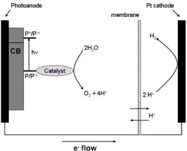
The architecture of the overall system could be changed from solution based to solid state. A system of this kind would result in a more asymmetric system which could be envisaged to help in the overall directionality of the different processes, something akin to the natural enzyme. In such systems, the oxidation catalyst (Catox)would be linked by a photosensitiser to an anode
and the reduction catalyst (Catred) grafted to a cathode29 (see Figure 2). Electrons from the
oxidation half-cell would be transferred to the reduction half-cell, while protons would move in the same direction through a selective membrane. Recent advances in the field of large bandgap semiconductor surfaces such as TiO2, SnO2, or ZnOhave opened the way for new approaches in
light sensitized devices. Amongst the advantages of Graetzel cell type devices are, i) very fast electron injection rates from the excited state of attached chromophores into the conduction band30 of the semiconductor, contributing to the necessary control of directionality of electron transfer, ii) high surface areas up to 1000 times more than those offered by monolayers, iii) efficiency of electron collection in external circuits, iv) ability to tune the electron acceptor states in order to maximize light to electrical energy conversion and, v) high transparency in the visible and infrared regions.
Examples of such devices have been developed with porphyrin-sensitised photoanodes,31,32 which drive oxidation of sugar via a natural enzyme, delivering sufficient reducing potential for H2
Figure 5: Diagram of a water splitting photochemical cell.
The following chapters present the work carried out in relation to the various components used in artificial photosynthesis devices: antennas, photoactive chromophores, and donor groups including manganese complexes.
3.1. Antennas
The role of an antenna is to increase the absorption cross section of the complex and, through a series of energy transfer steps, funnel that energy to a reaction centre where a charge separated state is formed. The solar spectrum has a maximum intensity just below 500 nm35 and extends well into the infrared region where a photon with λ = 1000 nm still has 1.2 eV energy. In nature, the efficient geometrical arrangement of the pigments is related to the tertiary structure of the PS-II antenna subunits (light-harvesting complexes).36 In artificial photosynthesis this spatial arrangement is not easily achievable. The distance between the pigments to be used, their respective angle, and electronic coupling, must be dictated by the covalent bonds that unite them. Two mechanisms are responsible for energy transfer processes. Förster energy transfer occurs in
cases where the distance between the chromophores is larger than their transition dipole strength, the dipoles have sufficient strength, and the lifetime of the emitting chromophore is sufficiently long. Dexter energy transfer takes place when the chromophores are brought into van der Waals contact and there is orbital overlap between them. Systems such as carotenoid-tetrapyrrole triad37 and dyads,38,39 and (Phenylethynyl) anthracene-porphyrin-fullerene heptads40 have been shown to
combine large absorption spectral range (350 nm-700 nm), high extinction coefficients (105 M-1 cm-1), and quantum yields for energy transfer close to 100%.41 Different approaches, such as the use of dendrimers42 or self assembled π-stacked arrays43,44 have also been reported in the
literature.
Work on artificial antennas in the laboratory was carried out by linking carotenoids with phthalocyanins. These pigments are porphyrin-like compounds with extended π-systems and strong absorption in the red to infrared region of the spectrum.
Carotenoids play several roles in the photosynthetic process and are also crucial to the photoprotection of the photosynthetic membranes. As accessory light-harvesting pigments, carotenoids (Car) are found in the chlorophyll-binding antenna proteins of photosynthetic organisms where they absorb light in the blue-green spectral region and efficiently transfer energy to nearby chlorophylls (Chl), as depicted in eqs 4 and 5.
It is well established that carotenoids have two low-lying excited singlet states from which energy transfer to chlorophylls (eq. 5) could occur. Two of the possible singlet-singlet energy transfer pathways are shown schematically in figure 6. Following absorption of light and internal conversion from the S2 (11BBu
+) state, the S
1 (2 A1 g-) state of the carotenoid is populated. Energy
transfer from the S2 state of the carotenoid to the Qx (S2) state of the chlorophyll, in competition
with the ultrafast internal conversion from the S2 to S1 state of the carotenoid, is another one.
Figure 6: Pathways for energy transfer from a carotenoid to a phthalocyanine.
The extremely short lifetime of the S2 state of the carotenoid (~ 200 fs) does not exclude this
second possible pathway. Indeed, spectroscopic studies on the sub-picosecond time scale of some antenna protein complexes of photosynthetic organisms suggest that some energy transfer to the chlorophyll or bacteriochlorophyll energy acceptors occurs from the carotenoid S2 state.45,46
However, the complexity of these natural antenna systems containing multiple chromophores, compounded with the ultrafast time scale, inevitably makes detailed pathway assignments very difficult. Nevertheless, recent results indicate that in some antenna complexes energy transfer proceeds predominantly from the carotenoid S2 state, rather than from the lower energy excited
S1 state as predicted by Kasha’s Rule. Mimicking these unusual photophysics in simple, well
defined model systems offers the opportunity to study the energy transfer pathways and to explore, and perhaps control, the nature of the carotenoid-tetrapyrrole electronic interactions affecting the rates and efficiency of energy transfer. Compounds 1 and 2 are examples of
molecular dyads which were designed to help understand the mechanism(s) of the carotenoid to chlorophyll singlet-singlet energy transfer processes in natural systems.
N N N HN N N NN Si O O O O N N N HN N N NN Si O O O O O O O O N N N HN N N NN Si HO OH 1 2 3 4 5
Figure 7: Structures of carotenoid-phthalocyanin triads and model compounds.
Synthesis of triad 1 was accomplished by coupling of silicon-tetra-tert-butyl-phthalocyanin 5 to the acid chloride derivative of carotenoid 3. This carotenoid was formed by reaction of the
commercially available, 8’-apo-carotenal with phosphonium salt via a Wittig reaction in order to obtain the 10 double bond (db) ester carotenoid, which was further oxidized to its carboxylic acid analogue, and then prepared into its acid chloride synthon.
These molecules show very broad spectral absorptions spanning most of the visible region (300-700 nm). Though their chemical structure is basically the same, and differs only in the extra double bond in each of the carotenoids, their behavior is strikingly different (Figure 9). Triad 2 exhibits near complete light harvesting efficiency (92%) from the carotenoid to the phthalocyanin through three of the available electronic states of the carotenoid, namely S2, S1, and S* as
calculated from the increase in Qy bleaching of Pc.
Triad 1 performs singlet energy transfer at a much lower yield (30%), and it is only the S2 state
which is active in this process, while the S1 shows very little energy transfer, and there is no
evidence for energy transfer from S*. In polar solvents, both triads 1 and 2 exhibit light induced electron transfer from either of the carotenoid molecules to the excited singlet phthalocyanin species with a rise time of about 2 ps. Charge recombination to the singlet ground state occurs in 10 ps for triad 2 and 17 ps for triad 1.
300 400 500 600 700 -0.05 0.00 0.05 0.10 0.15 0.20 0.25 0.30 0.35 Excitation Wavelength, nm A b so rb anc e 300 400 500 600 700 0.00 0.05 0.10 E xcitation W avelength, nm A b so rb anc e 95 % Energy transfer 30 % Energy transfer
Figure 9: Different energy transfer pathways for dyad 2 (left) and 1 (right).
It can thus be concluded that singlet energy transfer from the S2 carotenoid state to the
phthalocyanin molecule can be extremely fast. When the components of the triad are in good spectral overlap and electronic coupling, both upper and lower excited states of the carotenoid can participate in the energy transfer process. Depending on the energetics, electron transfer or back energy transfer from the Pc to the carotenoid can happen, and also important, when in polar solvents carotenoids are efficient electron donors to the tetrapyrrole groups. More results about these molecules are presented in publications I and IV.
3.1.1. Non-photochemical Quenching
Another important function of carotenoids is that of photoprotection. Their mechanism of action here is two-fold. First, they can scavenge singlet oxygen as well as prevent its formation by quenching the triplet state of Chl. Secondly, they can quench the singlet excited state of Chl
S Ener gy, a.u . 0. S2 S1 (2 1 Ag-) S0(11Ag -T S1 (hot -~20% S Q Q T ~10%
{
}
Pc (Si) C(10 db) S Ener gy, a.u . 0. S2 S1 (21Ag -S0(1 1 Ag -T S1 (hot -S Q Q T ~32% ~20% ~8% kT >> (5 ns)-1{
C (9 db) Pc (Si) ~35%under high illumination periods (eg 6-8). This process is known as nonphotochemical quenching and allows the plant to adjust to different light intensity.
The mechanism is triggered enzymatically at high light intensities, when, upon dropping of thylakoid lumen pH, violaxanthin (9 db) converts into antheraxanthin (10 db), which in turn converts into zeaxanthin (11 db). Conversely, under conditions of dim light the cycle is reversed and zeaxanthin is transformed back into violaxanthin.47
HO OH HO HO OH OH O O O Violaxanthin Antheraxanthin Zeaxanthin Low light Excess light
Figure 10: Carotenoids involved in xanthophyll cycle.
The mechanism is still unclear, although there are three proposed hypotheses. The first invokes quenching of the Chl excited state by energy transfer.48 Upon expansion of the polyene chain, the HOMO-LUMO gap would decrease bringing the S1 state of the carotenoid under the Qy of the
Chl and thus allowing back energy transfer from Chl to Car. The second hypothesis involves an electron transfer mechanism.49 As the conjugation of the carotenoid is increased, its first oxidation potential would be lowered and electron transfer from Chl to Car would quench excess energy. The third involves a change in pigment organization.50,51 As violaxanthin is converted
into zeaxanthin, their structural differences could change the way in which the pigment interacts with the protein complex, and this change in pigment organization could trigger the quenching process.
In order to study this process we decided to synthesize three different artificial antennas made up of zinc phthalocyanins covalently linked to three different carotenoids varying in conjugation length from 9 to 11 double bonds.
N N N N N N N N O O O O O O N H Zn R O R O R O 6 7 8 R O R= 9
Figure 11: Structures of different length caroteno-phthalocyanin dyads and corresponding model compounds.
Synthesis of triad 8 was performed by reacting the acid chloride equivalent of carotenoid 8, with the amino phthalocyanin in order to form an amide linkage. The AB3 type zinc phthalocyanin had to be synthesized by reacting a statistical mixture of di-butoxy phthalonitrile and amino phthalonitrile. The 11 double bond (db) carotenoid was obtained by a Wittig reaction of 8’-apo-carotenal (9 db) to yield the corresponding 10 db ester. Reduction to the 10 db alcohol, and oxidation to the 10 db aldehyde allowed for the next Wittig reaction to yield the 11 db ester,
which was further oxidized to the corresponding acid, transformed into its acid chloride derivative and coupled to the amino end of the phthalocyanin.
Figure 12: (Left) absorption spectra of dyads 6 (red), 7 (blue), 8 (green), and model phthalocyanin (black). (Right) Emission decays for same compounds.
UV/Vis results of the individual triads (6-8) are shown on Figure 9 (left). The spectra show that upon complexation of the carotenoid to the phthalocyanin the resulting compound shows broad absorption across the visible region which combines features of both individual components. i.e. bands at 350, 610, and 680 nm due to the phthalocyanin moiety and a broad absorption from 400 to 550 nm due to the carotenoid molecule. It can clearly be seen how upon elongation of the polyene chain by one double bond, the absorption spectrum undergoes a bathochromic shift as conjugation increases (9 db-red, 10 db-blue, 11 db-green).
Figure 12 (right) shows kinetic traces with excitation and detection at 680 nm in THF. The difference in behaviour as one double bond is added from a 9 db polyene chain to a 10 db chain is clearly visible in the shortened recovery time of the Qy state. For model phthalocyanin 9 and 9
db dyad 1, the recovery time is equal at about 3 ns. On the other hand, the lifetime for 10 db dyad 7, decreases by an order of magnitude to 300 ps, while 11 db dyad 8, becomes even a stronger quencher with a Qy lifetime of 56 ps.
Substitution of THF by a more polar solvent makes this effect more pronounced and results in acetone yielded Qy decays of 600 ps for dyad 6, 30 ps (75%) and 120 ps (25%) for dyad 7, and 16 ps (65%) and 90 ps (35%) for dyad 8. This dependence on polarity could suggest an electron transfer process, but no spectral proof for a carotenoid radical cation was found. Instead, the quenching process seems to stem from the S1 state of the carotenoid in combination with an ICT
state which is though to arise from the presence of the carbonyl group in the polyene chain. These results prove that conjugation changes in the polyene backbone of carotenoids are key to thermal dissipation of excess light energy. In this manner it can be observed how a carotenoid of the right length can dissipate Qy energy by shortening its excited state lifetime, and how an increase/decrease of a double bond can turn a carotenoid from a nonquencher into a quencher.
The mechanism seems to be through a carotenoid ICT/S1 state. The Qy state of the
phthalocyanin transfers energy to the ICT state, which in turn transfers energy to the S1 state.
This state is capable of thermal relaxation into the ground state. The effect of the polar solvent seems to be to lower the ICT state making it more available to the Qy state and therefore improving the quenching process. The lowering of the ICT state could be also brought about by conformational changes in the carotenoid, and is a hypothesis that can not be discarded by the experiments presented here. More information about this project is listed in publications III and V.
3.2. Ruthenium based constructs 3.2.1. Photoactive chromophores
A suitable chromophore should replicate the mechanism performed by P680. Energy absorption in
the form of a photon or an energy transfer process, formation of an excited state, and consequent photo-initiated electron transfer.
Several types of molecules have been used as photosensitisers in artificial systems, going from tetrapyrrole4,5,52 systems to transition metal complexes (Ru, Re, Ir, Os)42,53,54.
Another well-known chromophore is the [Ru(bpy)3]2+ complex55-57. This family of molecules has
raised a lot of interest in the last decades in the development of photochemistry, photophysiscs, and photocatalysis.56,58-60
These complexes present an absorption band in the region around 450 nm corresponding to a Metal to Ligand Charge Transfer (MLCT) process with an extinction coefficient of about 13000 M-1.cm-1 61. Upon irradiation in the MLCT band, the input light energy is converted into a 1(dπ6)
→ 1(dπ5π*) excited state, which in turn relaxes to form the lowest triplet state (3MLCT) in less
than a picosecond62-64 with a quantum yield of unity.
[Ru(bpy)3]3+ [Ru(bpy)3]2+ [Ru(bpy)3]+
[Ru(bpy)3]3+ [Ru(bpy)3]2+ [Ru(bpy)3]+ (dπ5π*1)
(dπ5) (dπ6) (dπ6π*1) -0.8 V +0.8 V
+1.3 V -1.3 V 2.1 eV
Figure 13: Physical properties of ground and excited
This state is sufficiently long-lived (1 μs) to encounter other solute molecules and allow energy transfer, as well as reductive or oxidative electron transfer processes to occur before regeneration of the ground state65. As shown in figure 13, *[Ru(bpy)3]2+ has 2.12 eV available for energy
transfer processes and its reduction and oxidation potentials are around 0.80 V vs SCE. In its ground state, the oxidation potential of [Ru(bpy)3]3+ species is around 1.30 V56,63 which is close
to that of the primary donor of PSII, P680+,17,18,66 (1.25 V vs. NHE) therefore making it a suitable
candidate to reproduce the oxidation reactions performed by the natural system.
Synthetic methods are available57,67 for the modification of the bipyridine ligands surrounding the ruthenium atom. In this manner it is possible to obtain mono and di-substituded ligands with either electron donating or withdrawing groups,61,67,68 as well as synthetic handles either for further synthesis53,57,69,70 or for grafting63,70-72 onto surfaces. Oxidation potentials for this group of molecules range from 1.20 to 1.40 V vs SCE, depending on the nature and number of substitutions61,63,67.
These structural changes on the ligand allow the modulation of the photophysical properties of the complex. For instance, direct changes in the relative energies of the dπ and π* orbital levels observed as bathochromic shifts of up to 50 nm54,61,67,73 are achieved by the addition of ester or carboxylate groups to the bipyridine ligands. The emission lifetimes of these compounds are also affected by substitution patterns on the bipyridines61,67,74,75. Electron withdrawing substituents diminish the fluorescence lifetimes and intensities by up to 70% compared to the parent [Ru(bpy)3]2+ 63,67.
The chromophores used in the molecules that will be presented in the following sections are shown in figure 13.
The simplest of these, [Ru(bpy)2Cl2]2+ 13, has been extensively shown in the literature. It is
synthesized by reaction RuCl3 and bipyridine. Coordination into a suitable target molecule is
done by first replacing the two chloride groups by the more labile nitrate groups, and further reaction with a pertinent synthon.
N N N N N N O O Ru O O O O O O O O N N N N N N O O Ru N N N N Cl Cl Ru N N N N N N O O Ru O OH OH O 10 11 12 13
Figure 14: Structure of different chromophores.
Compounds 10-12 are heteroleptic ruthenium compounds that were designed in order to function as precursors in cases where the target molecules would present more than one coordination site for insertion of a metal ion. In these cases, instead of coordinating ruthenium in the latter steps, the chromophore would be added as part of an organic substructure. In these situations the synthetic handle used to build up from was the phendione moiety.
Compound 12 shows the basic bipyridine motif, while compounds 10 and 11 show ligand substitution in the form of ethyl esters and carboxylic acids respectively. As mentioned above,
and shown in later sections, the inclusion of these groups changes both the spectral, and electrochemical properties of the resulting molecules. In the case of compounds 10 and 11, as will be shown in later sections, these modifications will serve as anchoring groups for grafting into TiO2 surfaces as well as potential synthetic handles from which larger molecules could be
constructed.
3.2.2. Donor side
In the “donor” side of PSII, after excitation by light, the oxidized P680+ is reduced by Tyrz, which
in turn oxidizes the OEC, with the ultimate source of electrons being water. This branch, which ultimately leads to one of the most important reactions in nature, is of utmost interest to scientists yet remains poorly understood. Processes such as PCET, and its direct implication in electron transfer and charge compensation, charge accumulation, and the oxidative splitting of water have been studied in the last decade by several groups.76-78 In these cases, the use of simpler artificial systems could allow for the elucidation of pathways which are more complicated to study in natural systems. The following sections, will give an overview of the progress made into ruthenium systems linked to Tyrz synthetic equivalents and manganese complexes.
3.2.2.1. Phenol group
Proton/electron transfer processes in PSII are initiated at TyrZ. It reduces the photoxidised P680,
is involved in PCET processes79 that prevent excessive charge accumulation from making the reaction too endergonic, and has even been thought to intervene in the abstraction of protons from the substrate water molecules.80,81
In PSII, initial charge separation between P680+ and the reduced quinone is preserved by inserting
a tyrosine group between P680 and the manganese cluster. As the OEC undergoes the 4 electron
oxidation, inner reorganization processes, such as changes in bond lengths, might increase the reorganization energy of the cluster and make the electron transfer processes sluggish. A slow electron transfer rate between the OEC and P680+ would favor recombination of the initially
formed P680+-QA- couple, with the consequent loss in efficiency for the overall process. Insertion
of a tyrosine group between the chromophore and the manganese cluster induces fast reduction of P680+ and therefore prevents the recombination of the charge separated pair.
In natural systems, electron transfer processes are often accompanied by proton transfer processes. In the PSII system where a 4e- oxidation is coupled to a 4 proton transfer in order to
produce molecular oxygen this process acquires even more importance. This charge
compensation process allows the OEC to rock within 300 mV between its four oxidation states, therefore avoiding oxidation potentials that could not be reachable by P68066,82,123. The chemical
basis for this proton/electron transfer lies within the chemical properties of the phenol moiety. In aqueous solution, the pKa of a phenol changes from 10 to -2 upon oxidation, with the corollary
that these processes will happen concurrently.
In order to study these processes we have synthesized different ligands sharing a common feature. A ruthenium chromophore attached to a hydrogen-bonded imidazole-phenol couple. By varying the structure of the ligand we intended to change the distance of the couple from the chromophore 15, as well as the angle between the phenol and the imidazole 16 in order to induce some torsional strain between the phenol and imidazole groups, and thus weaken the hydrogen bond between them, and be able to study the differences these structural modifications would cause.
Figure 15: Structure of different Ruthenium-imidazole-phenol compounds.
The UV/Vis absorption of compounds 14, 16, 17, and [Ru(bpy)3]2+ are shown in Figure 16. The
absorption spectrum of these molecules is characteristic of ruthenium compounds and exhibits a strong π-π* transition at 280 nm, an increased absorption at 320 nm due to symmetry breaking around the ruthenium metal, and strong MLCT absorption band at 450 nm which in all cases is red shifted by about 5 nm with respect to [Ru(bpy)3]2+. The emission experiments show bands
with maxima around 610 nm, with lifetimes of 930 ns for compound 16, 1.23 µs for 14, and 1.10 µs for 17. N N N N N N Ru N H N HO 14 N N N N N N Ru N H N N HN OH 15 N N N N N N Ru N H N HO 16 N N N N N N Ru N H N HO 17
300 400 500 600 700 800 900 1000 0,0 0,1 0,2 0,3 0,4 0,5 0,6 0,7 17 14 16 [Ru(bpy)3]2+ Abso rb ance Wavelength (nm)
Figure 16: Absorption spectra of compounds Ru(bpy)32+ (blue), 16 (green), 17 (black), and 14 (red). In acetonitrile.
Cyclic voltammetry studies in acetonitrile show typical ruthenium behavior (Table 1). The ruthenium moiety undergoes oxidation around 1.30 V for all compounds, while on the reduction side, three-one electron processes can be observed for each of the reductions around the ruthenium. The one difference to note is the appearance of a small shoulder in the oxidation of compound 17, which is assigned to the oxidation of the phenol group.
Compound E1/2 RuIII/II E1/2 OH/OH+. E1/2 Ligands
14 1.31 N/O -1.36, -1.58, -1.92
16 1.28 N/O -1.35, -1.58, -1.92
17 1.39 1.12 -1.26, -1.47, -1.82
Table 1: Electrochemical data for 1 mM solutions of Ruthenium-Phenol compounds 14, 16-17 containing .1 M TBAP as supporting electrolyte.In acetonitrile vs. SCE. v = 100 mVs-1. (N/O) Not observed.
N N N N N N Ru N H N HO Std 0 1 2 3 4 5 6 7 8 9 10 -0,012 -0,010 -0,008 -0,006 -0,004 -0,002 0,000 0,002 Δ A45 0 time/ µs Standard 16 17 14 in presence of 10 mM MV2+ (PF6-)2 0 2 4 6 8 10 12 14 16 18 20 -0,008 -0,006 -0,004 -0,002 0,000 0,002 0,004 ΔA time/ µs 450 nm 605 nm 228 + 10 mM MV2+
Figure 17: Left: Transient absorption changes at 450 nm for compounds: Std (black), 16 (red), 17 (green), and 14
(blue). Right: Example of transient absorption spectrum monitoring for MV.+ (red line), and Std (blue line). Right bottom: Structure of reference compound (Std).
Photoinduced electron transfer studies in presence of methyl viologen (fig 17 right) were conducted monitoring the signals at 450 nm arising from Ru(II) and the phenoxyl radical, and 610 nm for reduced methyl viologen. These experiments show that in all cases, upon illumination of the compounds and initial bleaching of the absorption at 450 nm [Ru(II) → Ru(III)], the chromophore is reduced at a faster rate than the decay of the reduced methyl viologen. These results confirm that the Ru(II) is formed by electron transfer from the phenol group and not from the recombination reaction. When comparing between molecules 14, 16, 17, and a previously published molecule bearing two tert-butyl groups ortho and para to the phenol group, it was found, as somewhat expected, that the oxidation of the phenol occurs fastest (black trace) and slowest (blue trace) for the molecules Std and 14 respectively. In the fast case, the more electron rich character of the phenol lowers its oxidation potential and therefore increases the driving force for the reaction. In the case of compound 14 the effect is the opposite and, while formation of the phenoxy radical is observed, the rate is slowest. As usual, it is in the middle of things
where the results are not the most clear. Molecules 16 and 17 were designed to have the same electron density in the phenol group, but vary in the strength of the hydrogen bond between the two groups, and as such it was expected that molecule 17, with a stronger hydrogen bond, would undergo oxidation at a faster rate, a process that was also suggested by cyclic voltammetry which showed that there might be a slightly bigger driving force to do the reaction. Experimentally this was not the case, and it was molecule 16, the twisted molecule, which underwent photo-oxidation faster. Reasons for this behaviour are not clear, and obtaining crystal structures in order to find a value for the degree of twisting as well as NMR experiments in order to try to find a value for the energy of rotation, and therefore the strength of the hydrogen bond, might help elucidate the reasons for these results. Work on the synthesis of molecules containing a phenol group between the chromophore and the catalytic site are currently under progress in the laboratory.
3.2.2.2. Manganese complexes
The ultimate challenge in artificial photosynthesis research is to power a water oxidation catalyst by a photoactive chromophore. Several complexes have been reported to catalyse the oxidation of water chemically. These include manganese cubanes82,83, di μ-oxo manganese terpyridines84,85,
di-manganese polypyridil complexes86, face to face Mn-oxo porphyrins87, and Ruthenium dimers88. All these molecules have the need for large amounts of external oxidizing agents and except for the latter, which have shown truly catalytic behaviour albeit their low rates of turnover, the results for the rest of these molecules have been questioned89 as their mechanisms, as well as
the origin of the oxygen atoms involved in the reaction are unknown. Therefore, it would be very interesting from the point of view of artificial photosynthesis to link any of these putative
catalysts to a chromophore that could provide the oxidative power needed to drive these reactions. There have been several attempts at linking ruthenium choromophores to different manganese containing ligands90-94. To date, none of these experiments have yielded results towards the oxidation of water.
In this section, we will discuss our first attempts at building a molecular system bearing a ruthenium (II) chromophore and a mono- or polinuclear manganese complex as the donor component.
3.2.2.2.1. Terpyridine complexes
Of all the above mentioned manganese catalysts79-84, the compound that has received most attention is that discovered by Brudvig’s group81-82. It utilizes a terpyridine group as the coordinating site for a manganese (II) metal ion, which after further oxidation is transformed into a bis-terpyridine-Mn2-di-µ-oxo complex (cpd 21). Further oxidation of this molecule in a
water/acetonitrile mixture in the presence of oxone was reported to accomplish the catalytic oxidation of water85,95. The mechanism involves the initial formation of a MnIII-MnIV dimer which undergoes oxidation to a MnIV-MnV oxo complex which is thought of being capable of oxidizing water. Upon the 4e-oxidation of water, the metal complex is reduced to the MnII-MnIII state, which replicates the S0 to S4 states in the natural system.
Figure 18: Proposed reaction mechanism for O2 formation from the reaction of [(terpy)(H2O)Mn(µ-O)2Mn(H2O)(terpy)] with an external oxidant. From reference 74.
For this molecule as well as the ones mentioned above there is a caveat. Oxone is a known oxygen transfer reagent, and therefore the origin of the oxygen atoms conforming the resulting molecular oxygen is not clear. It is important to remember that for the water splitting reaction to be catalytic, the O atoms comprising the O2 molecule must both come from water and not any
other reagent. Several studies have been published since trying to prove the origin of oxygen via isotopic labeling and mass spectrometry analyses,84 although the results are not yet clear. It is with this inspiration that we decided to substitute the external oxidant by a ruthenium chromophore, with the goal being to try to instill the required oxidative power by means of a photogenerated oxidant.
Hence, the molecules used for these studies are composed of a ruthenium based chromophore covalently bound to a terpyridine ligand through an imidazole linkage. The photosensitiser acts
as the P680 counterpart, capable of harvesting light and triggering an electron transfer process.
The resulting RuIII species has a reversible oxidation wave occurring at 1.3 eV vs. SCE, and is capable of initiating the processes leading to the ultimate oxidation of a MnII atom which is lodged in the terpyridine cavity.
Photoexcitation: Ru (II) + hv → [Ru (III)(bpy)-(bpy)2]2+*
Reduction of electron acceptor: [Ru (III)(bpy)-(bpy)2]2+ + A → [Ru (III)(bpy)3]3+ + A -Oxidation at catalytic site: Ru (III) + Mn (II) → Ru (II) + Mn (III)
Given the two potential coordinating sites of our target ligand, ie. a phenanthroline, and a terpyridine, the synthetic pathway whereby the ligand is prepared prior to metallation was deemed disadvantageous. In this manner it was chosen to start from a ruthenium (II) heteroleptic core containing either bipyridines, or ester modified bipyridines, attached to a phendione ligand which would serve a synthetic handle. The [Ru(bpy)2(phendione)]2+, and [Ru(ethyl ester
(bpy)2(phendione)]2+ complexes were prepared following modified literature procedures72 and
were further reacted with 4'-formyl terpyridine or 4'-formyl phenyl terpyridine via a Steck-Day reaction. Insertion of the MnIICl2 ion in the terpyridine cavity was performed following modified
N N N N N N Ru O O O O O O O O N N N N N N Ru N N N N N N Ru N H N N H N N H N N N N N N N N N N Mn Cl Cl Mn Cl Cl Mn Cl Cl N N N N N N Ru O O O O O O O O N N N N N N Ru N N N N N N Ru N H N N H N N H N N N N N N N N N N 19 20 20a 19a 18a 18
Figure 19: Structure of different Ruthenium-terpyridine compounds.
Cyclic voltammetry of compounds 18 and 20 show a one electron oxidation of around 1.30 V for the RuII to RuIII step, another around 1.22-.126 attributed to the oxidation of the imidazole group, and three one electron reductions from -1.41 to -1.96 V corresponding to the reduction of the ruthenium ligands (Table 2). Addition of ethyl esters in the bipyridine groups around ruthenium results in positive shifts of about 200 mV for the oxidation process, and between 400 and 200 mV for the reduction processes. These shifts are attributed to the electron withdrawing properties of the four ethyl ester groups in the ruthenium ligands, and the concomitant increase in potential
needed in the oxidation step. Interestingly, in this molecule oxidation of imidazole group was not observed. This effect might be due to the inductive effects of the ester groups on the imidazole moiety. In the bimetallic complexes 18b-20b, an additional reversible peak around 1.10 V is attributed to the MnII to MnIII process. Oxidation of MnII seems to be independent of the substitution pattern of the molecule as the value for its potential only ranges within 50 mV between samples.
Compound E1/2 RuIII/II E1/2 Imid·+/Imid E1/2 MnIII/II E1/2 Ligands ΔG (eV)
Ru-Terpy (18) 1.27 1.22 -1.41, -1.61, -1.96 Ru-Terpy-Mn (18a) 1.30 1.09 -0.21 Ester-Ru-Terpy (19) 1.51 -1.01, -1.24, -1.73 Ester-Ru-Terpy-Mn (19a) 1.54 1.11 -0.43 Ru-Phenyl (20) 1.34 1.28 -1.41, -1.66, -1.96 Ru Phenyl Mn (20a) 1.31 1.06 -0.25
Table 2: Electrochemical data for 1mM solutions of Ruthenium(II)-Terpyridine-Manganese(II) complexes containing .1 M TBAP as supporting electrolyte. In acetonitrile vs. SCE. v = 100 mVs-1.
The ground state absorption spectra of [Ru(bpy)3]2+, and complexes 18, 18a, 19 and 19a in
acetonitrile are reported in Figure 20.*
The absorption spectrum of the ruthenium complexes exhibits the typical transitions. For Ru-Terpy (18), a strong absorption around 280 nm due to a π-π* transition, another band at 320 nm which results from an intraligand charge transfer, and finally a band at 450 nm, the typical MLCT band of ruthenium complexes. For the ester modified complex the ground state absorption spectrum exhibits a broad and red-shifted peak in the UV (308 nm), an enhanced charge transfer band at 350 nm and a broader and red-shifted MLCT band whose maximum occurs at 475 nm.
300 400 500 600 700 0 20000 40000 60000 80000 [Ru(bpy)3]2+ ester Ru-tpy-Mn ester Ru-tpy ε / M -1 cm -1 wavelength/ nm 0 20000 40000 60000 80000
b
[Ru(bpy)3]2+ Ru-tpy Ru-tpy-Mna
Figure 20: Ground state absorption spectra in acetonitrile for (a) [Ru(bpy)3]2+, Ru-Terpy 18 and Ru-Terpy-Mn 18a; and (b) [Ru(bpy)3]2+, ester-Ru-Terpy 19 and ester-Ru-Terpy-Mn 19a.
Insertion of manganese in the second coordination site induces some changes in the absorption spectrum, most notably, a reduction of intensity in the 280 nm band, a new band arising at 350 nm, and a broadening of the MLCT band together with a loss of extinction coefficient.
The strongest changes though are presented in the emission properties of these molecules. Absorption of 450 nm light by the ruthenium complexes induces emission at 610 nm (1.60 µs) for the bipyridine complexes, and 650 nm (1.65 µs) for their ester analogues. The observed red-shift of the optical transitions is a result of a decrease in the HOMO-LUMO gap, due to the electron withdrawing character of ester groups and the greater ease for reduction by the metal. This effect has previously been observed67.
In the case of the dinuclear complexes Ru-Terpy-Mn (18a) and Ester-Ru-Terpy-Mn (19a), the emission is strongly quenched. The lifetimes are biphasic with a main, short living component
(50 and 120 ns respectively) and a small, longer living one (1 µs and 700 ns respectively) in acetonitrile. This quenching process, which has been detected by several groups, has been described as an energy transfer process between ruthenium and manganese. Compounds 20 and
20a were synthesised in order to try to see the effect of increasing the distance between the two
metal centers in this quenching process. Unfortunately, quenching of the emission was just as strong as in the case of molecule 18a. The reason for this might be the increased conjugation that this molecule offers between the two metal centers, which inhibits the distance effect of the extra phenyl group.
Since the goal of conceiving new Ru-Mn complexes consists of being able to photodrive electron transfer processes so that oxidative equivalents can be stored on the Mn ion, the complexes were studied in presence of light and an irreversible external electron acceptor, [CoIII(NH3)5]3+. The
one electron reduction of cobalt pentamine yields ammonia and a CoII ion which is quickly hydrated and renders the reaction irreversible.
For Ru-Terpy (18),these experiments showed that upon illumination of the Ru chromophore, the resulting RuIII species is capable of oxidising the organic ligand around it. Proof for this process is the appearance of an organic radical-like signal around g = 2.01, and the concomitant appearance of a low field band due to the reduced Co (II) species.


![Figure 18: Proposed reaction mechanism for O 2 formation from the reaction of [(terpy)(H 2 O)Mn(µ- O)Mn(µ-O) 2 Mn(H 2 O)(terpy)] with an external oxidant](https://thumb-eu.123doks.com/thumbv2/123doknet/12719497.356600/44.918.178.732.113.536/figure-proposed-reaction-mechanism-formation-reaction-external-oxidant.webp)

![Figure 20: Ground state absorption spectra in acetonitrile for (a) [Ru(bpy) 3 ] 2+ , Ru-Terpy 18 and Ru-Terpy-Mn 18a;](https://thumb-eu.123doks.com/thumbv2/123doknet/12719497.356600/48.918.243.672.140.492/figure-ground-state-absorption-spectra-acetonitrile-terpy-terpy.webp)