HAL Id: tel-02803846
https://hal.inrae.fr/tel-02803846
Submitted on 5 Jun 2020HAL is a multi-disciplinary open access archive for the deposit and dissemination of sci-entific research documents, whether they are pub-lished or not. The documents may come from teaching and research institutions in France or abroad, or from public or private research centers.
L’archive ouverte pluridisciplinaire HAL, est destinée au dépôt et à la diffusion de documents scientifiques de niveau recherche, publiés ou non, émanant des établissements d’enseignement et de recherche français ou étrangers, des laboratoires publics ou privés.
Interactions between the Orange Carotenoid Protein and
the phycobilisomes in cyanobacterial photoprotection
Denis Jallet
To cite this version:
Denis Jallet. Interactions between the Orange Carotenoid Protein and the phycobilisomes in cyanobac-terial photoprotection. Agricultural sciences. 2013. English. �tel-02803846�
HAL Id: tel-00946742
https://tel.archives-ouvertes.fr/tel-00946742
Submitted on 11 Mar 2014HAL is a multi-disciplinary open access archive for the deposit and dissemination of sci-entific research documents, whether they are pub-lished or not. The documents may come from teaching and research institutions in France or abroad, or from public or private research centers.
L’archive ouverte pluridisciplinaire HAL, est destinée au dépôt et à la diffusion de documents scientifiques de niveau recherche, publiés ou non, émanant des établissements d’enseignement et de recherche français ou étrangers, des laboratoires publics ou privés.
Interactions between the Orange Carotenoid Protein and
the phycobilisomes in cyanobacterial photoprotection
Denis Jallet
To cite this version:
Denis Jallet. Interactions between the Orange Carotenoid Protein and the phycobilisomes in cyanobac-terial photoprotection. Agricultural sciences. Université Paris Sud - Paris XI, 2013. English. <NNT : 2013PA112270>. <tel-00946742>
UNIVERSITE PARIS-SUD
ÉCOLE DOCTORALE : SCIENCES DU VÉGÉTAL
Laboratoire des Mécanismes Fondamentaux de la Bioénergétique UMR8221, CNRS/CEA DISCIPLINE : BIOLOGIE
THÈSE DE DOCTORAT
Soutenance prévue le 29/11/2013 parDenis JALLET
Interactions between the Orange Carotenoid
Protein and the phycobilisomes in cyanobacterial
photoprotection
Composition du jury :
Directeur de thèse : Diana KIRILOVSKY DR CNRS, CEA Saclay
Rapporteurs : Cécile BERNARD Professeur, Muséum national d’Histoire Naturelle
Michel HAVAUX DR CEA, CEA Cadarache
Examinateurs : Ondrej PRASIL Professeur, Academy of Sciences of the Czech Republic
Anne-Soisig STEUNOU CR CNRS, CGM Gif-sur-Yvette
Acknowledgements
To my supervisor, Diana, thank you for letting me study such a fascinating topic. When I look back, you were always here to give good advices and to constructively discuss about the experimental results. It was a great pleasure to work with you over the last 3 years. To all my coworkers – past and present – big thanks for everyday’s good mood and for all the help provided. Adjélé, Sandrine, Michal, Rocio, Adrien and Céline (alias Poulpy), I will really miss our 3rd floor.
To Dr. Ryan L. Leverenz, Dr. Marcus Sutter, Dr. Cheryl A. Kerfeld, Dr. Radek Kana and Pr. Ondrej Prasil, thanks for all the fruitful collaborations we have had.
To Dr. Anja Krieger-Liszkay, Dr. Pierre Sétif, Dr. Ghada Ajlani, Dr. Hervé Bottin, Dr. Sun Un in CEA Saclay, thank you for having been always available and ready to share your knowledge with me.
I would also like to acknowledge Dr. Corinne Chauvat and Dr. Catherine de Vitry for participating in my “comité de these” and bringing interesting new ideas into the topic.
To Dr. Jacqui Shykoff, Dr. Marianne Delarue and Martine Fournier from “ED 145 Sciences du vegetal”, thank you for creating a strong connection with Université Paris-Sud 11 and for easing so much the PhD students’ administrative procedures.
To all the administrative staff in iBiTeC-S, particularly Karine, Josiane, Céline, Isabelle, Pascale, Dominique, Sophie, thanks for all your support over the time course of my thesis.
To Amin, Thomas, Sané, Denise, Liz, Margaux, Kathleen (alias Lena?), Mehdi Caherine, Eiri, Stéphanie, Hassina, Tiona, Qian and Eduardo thanks for all the good moments we spent together in Paris (and Brussels!) to relax after hard working.
Finally, thanks a lot to my family and friends from Pyrenees who always were there to encourage me.
This work was funded by the Université Paris-Sud 11, the CNRS, the CEA and the European Training Network Grant HARVEST (FP project n°238017).
TABLE OF CONTENTS
Introduction………1
1. Cyanobacteria ... 3
1.1. Overview of the cyanobacterial phylum ... 3
1.2. A model cyanobacterium: Synechocystis ... 5
2. Photosynthesis in cyanobacteria ... 7
2.1. The linear electron transport chain: PSII, cytb6f and PSI. ... 7
2.2. Phycobilisomes: the cyanobacterial accessory light harvesting complexes ... 11
2.2.1. Composition and structure of the PBs ... 11
2.2.2. PBs function as antennae ... 17
2.2.3. PBs movement and possible implication in State Transitions ... 19
3. Drawbacks of photosystems functioning in an oxygenic medium and defensive strategies ... 22
3.1. Photoinhibition of PSII ... 22
3.2. Photoprotective mechanisms in cyanobacteria: an overview ... 23
3.3. Non-photochemical quenching of the excess light energy harvested. ... 25
4. The Orange Carotenoid Protein (OCP)-related non photochemical quenching of the energy harvested by PBs ... 27
4.1. Discovery... 27
4.2. Distribution and regulation of the OCP ... 29
4.3. Crystal structure of the OCP isolated from Arthrospira ... 31
4.4. Photoactivity of the OCP and Synechocystis mutant studies ... 33
4.5. In vitro reconstitution of the OCP-related photoprotective mechanism ... 37
4.6. FRP allows rebooting the system. ... 39
5. Aims of the thesis ... 41
Chapitre 1: ApcD, ApcF and ApcE are not required for the OCP-related photoprotective mechanism inSynechocystis………...53
Chapitre 2: Specificity of the cyanobacterial Orange Carotenoid Protein: Influences of OCP and phycobilisomes structures………89
Chapitre 3: Structural and functional modularity of the Orange Carotenoid Protein: Distinct roles for the N- and C- terminal domains in cyanobacterial photoprotection………135
ABBREVIATIONS EMPLOYED
ΔAB : Allophycocyanin-deficient Synechocystis PCC 6803 mutant strain αPC/ βPC/αAPC/βAPC : α subunit of phycocyanin/β subunit of phycocyanin/α subunit of
allophycocyanin/β subunit of allophycocyanin A/T/G/C : Adenine/Thymine/Cytosine/Guanine
Anabaena: Anabaena variabilis
APC : Allophycocyanin
ApcD, ApcF, ApcE : Phycobilisome terminal emitters
Arthrospira: Arthrospira platensis PCC 7345 ATP : Adenosine triphosphate Chla : Chlorophyll a
CK: Phycocyanin-deficient Synechocystis PCC 6803 mutant strain
CrtO: β-carotene ketolase (required for echinenone and 3’hydroxy-echinenone synthesis)
CrtR: β-carotene hydroxylase (required for 3’hydroxy-echinenone and zeaxanthin synthesis)
Cytb6f : Cytochrome b6f DNA : Deoxyribonucleic acid
ECN : Echinenone
EM : Electron microscopy
Fd: Ferredoxin
FLIP: Fluorescence Loss In Photobleaching
Flv: Flavodiiron protein
Fm’: Maximal level of fluorescence measured using a PAM fluorometer
FRP: Fluorescence Recovery Protein
FRAP: Fluorescence Recovery After Photobleaching hECN: 3’hydroxy-echinenone
HLIP: High Light Induced Polypeptide LHC: Light Harvesting Complex
M: Molar
Mb: Mega-base
NADP: Nicotinamide Adenine Dinucleotide Phosphate NDH1: NADH:ubiquinone oxidoreductase I
OCPr/OCPo: Red form of the Orange Carotenoid Protein/orange form of the Orange Carotenoid Protein
ORF: Open Reading Frame
P680/P700: Special-pair chlorophyll of photosystem II/special-pair chlorophyll of photosystem I
PAM: Pulse Amplitude Modulation
PB: Phycobilisome
PC: Phycocyanin
PCB: Phycocyanobilin
PCC: Pasteur Core Collection
PDB: Protein Data Bank PEB: Phycoerythrobilin
PQ/PQH2: Plastoquinone/plastoquinol
psbA2: Photosystem II D1 protein encoding gene PSI/PSII: Photosystem I/photosystem II
PTOX: Plastid Terminal Oxidase PXB: Phycobiliviolin
QA/QB: Photosystem II-associated quinines
qE: Energy-dependent non-photochemical quenching qP: Photochemical quenching
RCP: Red Carotenoid Protein RNA: Ribonucleic Acid
ROS: Reactice Oxygen Species
Synechococcus: Synechococcus PCC 7942
Synechocystis: Synechocystis PCC 6803
Thermosynechococcus: Thermosynechococcus elongatus
TyrZ: Tyrozine Z of photosystem II WOC : Water Oxidizing Complex
1
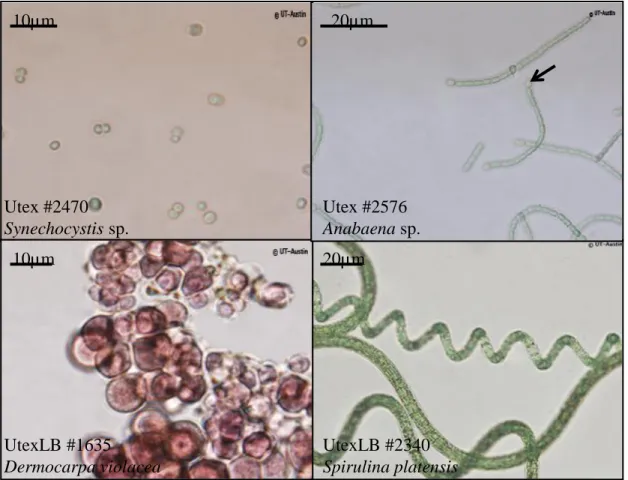
Figure 1. Optical microscopy observation of various cyanobacteria. A. The
unicellular Synechocystis sp. B. The filamentous Anabaena sp., capable of forming heterocysts (indicated with an arrow) for nitrogen fixation C. Dermocarpa violacea, containing red pigments (phycoerythrin) belonging to the phycobiliprotein family D. The filamentous Arthrospira platensis. Copyright: UTEX - The Culture Collection of Algae at The University of Texas at Austin (used with permission)Utex #2470 Synechocystis sp. 10µm 20µm Utex #2576 Anabaena sp. UtexLB #1635 Dermocarpa violacea 10µm UtexLB #2340 Spirulina platensis 20µm 2
3
Introduction
1. Cyanobacteria
1.1. Overview of the cyanobacterial phylum
Cyanobacteria are the only prokaryotic organisms able to perform oxygen evolving photosynthesis. It implies using light as a source of energy and water as a source of reducing power for converting inorganic carbon (CO2) into hydrocarbons (CnH2nOn), oxygen dioxide
(O2) and ATP being also produced. This ability, combined to the discovery of microfossils
testifying their apparition more than 2.5 billion years ago, pointed out cyanobacteria as responsible for the Great Oxydation Event and subsequent life spreading on Earth (Rasmussen et al., 2008). The name blue-green algae was originally proposed because some early-discovered cyanobacterial strains displayed macroscopic, thallus-like structures, containing green (chlorophylls) plus blue (phycobiliproteins) pigments. However, no organelles such as nuclei, mitochondria, endoplasmic reticulum, Golgi apparatus and plastids were found in these organisms. This consequently designated them as prokaryotes unlike green, red and brown algae; the appellation cyanobacterium was suggested, cyano deriving from the greek κύανος (kyanos), blue (Stanier et al., 1978).
Cyanobacteria diverged since their apparition, becoming present in a great diversity of habitats like marine water, fresh water and soils. They have colonized extreme environments such as volcanic lakes (Thermosynechococcus elongatus: Yamaoka et al., 1978) or Antarctic mats (Taton et al., 2003). Some species act as symbionts, for example in lichens or in certain marine invertebrates (Acaryochloris marina: Miyashita et al., 1996); the endosymbiotic theory suggests that chloroplasts derive from cyanobacterial-like cells, originally symbiotically associated to protoeukaryotic hosts but now behaving semi-autonomously (reviewed in: McFadden, 2001). So far, the cyanobacterial phylum includes at least 7500 species distributed in 5 sections and more than 150 genera (NCBI taxonomy database: 11000 entries). The initial classification was built on differences observed among cyanobacteria considering cell diameters (10-7 to 10-5 m), division patterns, branching/non branching filaments formation and potential differentiations for nitrogen fixation (heterocysts) or survival under extreme drought conditions (akinetes) (Rippka et al., 1979) (Fig.1). More recently, 16S RNA sequencing partially confirmed the taxonomic groups previously established (reviewed in: Komárek, 2010). Many cyanobacterial genomes have been released, the amount of data available increasing faster and faster thanks to high-flow techniques
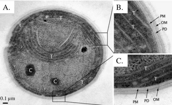
Figure 2. Electron micrograph of a thin section through a Synechocystis
PCC 6803 cell
A. Thin section of a Synechocystis cell revealing the various intracellular structures: ribosomes (r), glycogen granules (g), carboxysomes (C) and thylakoid membranes (T). B. and C.. Enlargments of the boxed areas in A., showing the plasma membrane (PM), the peptidoglycan layer (PD) and the outer membrane (OM). Adapted from Liberton et al., 2006.A.
B.
C.
0.1 µm
5
propagation (deep-sequencing, see (Shih et al., 2013). A rapid survey indicates variable genome sizes (from 2Mb to 13Mb) as well as GC contents (30 to 75%), perfectly reflecting the enormous diversity in morphologies and habitats. Still, some common features exist across the phylum. First of all, even though classical organelles miss, sub-cellular compartments are clearly visible. Particularly, cyanobacterial cells contain membrane stacks that constitute the thylakoids where both photosynthesis and respiration take place (except for Gloaeobacter violaceus: Guglielmi et al., 1981) (Fig.2A). Also, hexagonal structures termed carboxysomes ensure Rubisco’s optimal functioning by creating CO2 enriched/O2 depleted
biochemical environments (reviewed in: Badger and Price, 2003; Price, 2011) (Fig.2A). Hydrocarbons stemming from photosynthesis are converted to glycogen for storage (Fig.2A). Finally a peptidoglycan layer and an outer membrane form the cell wall, which confers some mechanical resistance like in most gram-negative bacteria (see Fig.2B,C).
Certain species chosen to study cyanobacteria physiology and photosynthesis have been used for more than 40 years; among them, the cyanobacterium Synechocystis PCC 6803 (hereafter called Synechocystis) has been employed as a model species in this work.
1.2. A model cyanobacterium: Synechocystis
Synechocystis was isolated from a Californian freshwater lake (Stanier et al., 1971). The round-shaped cells, diameter approximately 2 µm, look greenish when observed using optical microscopy. Their thylakoid membranes form concentric stacks along the plasma membrane as revealed by electron microscopy (recent review: Liberton et al., 2006) (Fig.2A). Synechocystis cells can grow photoautotrophically in mineral media (Stanier et al., 1971). They naturally have the capacity to uptake exogenous DNA and to perform double-homologous recombination, which points them as perfectly suitable for genetic manipulations (Grigorieva and Shestakov, 1982). Moreover, they can grow heterotrophycally in glucose enriched media; under these conditions, mutations impairing the photosynthetic apparatus do not have a lethal effect (Rippka et al., 1979). Synechocystis genome was fully released in 1996, making it the first photosynthetic organism ever sequenced (3.5 Mb, 47.7% GC: Kaneko et al., 1996). Finally, transcriptomic data are also available (Hihara et al., 2001) and recently, RNAseq allowed Transcription Starting Sites mapping (Mitschke et al., 2011). All these points designate Synechocystis as a good model for photosynthesis study.
After giving a general overview about cyanobacteria, the mechanisms underlying cyanobacterial photosynthesis are now going to be addressed.
Figure 3. Components of the oxygenic photosynthesis inside of thylakoid
membranes.
This figure displays the various membrane-spanning complexes implied in oxygenic photosynthesis: Photosystem II (PSII), cytochrome b6f and PSI. They are connected by mobile electron carriers such as plastoquinol (PQH2), plastocyanin (PC) and ferredoxin (Fd). The linear electrons transport chains leads to NADPH accumulation and formation of a proton gradient across thylakoid membranes, used as a motive force for ATP synthesis. Under certain conditions, the cyclic electron transport chain leads to proton gradient formation without any concomitant NADPH production. Adapted from Nevo et al., 2012.7
2. Photosynthesis in cyanobacteria
As said before, the cyanobacterial photosynthesis takes place inside of thylakoid membranes. It implies light energy harvesting and funneling towards special-pair chlorophylls (P680/P700), which upon excitation release low redox potential electrons then sequentially transferred from acceptor to acceptor (reviewed in: Witt, 1996). A proton gradient gets concomitantly created, allowing ATPase functioning; thus, photosynthesis provides NADPH (following NADP+ reduction) and ATP necessary for the Calvin-Benson cycle.
Cyanobacteria, plants and algae actually have similar photosynthetic machinery comprising 2 photosystems (PSI/II), cytochrome b6f (cytb6f) and several mobile components (plastoquinone
(PQ), plastocyanine (PCy), ferredoxin (Fd) working in series (Fig.3).
2.1. The linear electron transport chain: PSII, cytb6f and PSI.
Photosystems are pigment-protein complexes embedded in thylakoid membranes. They contain inner Chlorophyll a (Chla)-binding antennae that harvest light and funnel the associated energy towards reaction centers where photochemistry takes place, i.e. where special-pair chlorophylls (P680/P700) realize charge separations upon excitation. The cofactors receiving electrons from P680/P700 form the acceptor side whereas the ones giving electrons to P680+/P700+ form the donor side; the peptide composition and the cofactors associated to acceptor/donor sides differ in both photosystems, PSII or PSI (Fig.4A,B).
PSII has been extensively studied over the last decades (reviewed in: Nelson and Yocum, 2006; Cardona et al., 2012). 35 Chla molecules appear per monomer, mostly attached to the CP43/CP47 inner antennae. The reaction center comprises 2 proteins, D1/D2, where all cofactors required for photochemistry can be found; it also incorporates a cytochrome b559, absolutely necessary for its correct assembly. Upon excitation, P680 gives an electron to Pheophytin (D1 associated) that is then transferred to QA (D2-associated quinone) and QB
(D1-associated quinone) (Fig.4A). QB can receive 2 electrons before getting protonated; it
subsequently leaves PSII, in the plastoquinol (PQH2) form, by diffusing inside of thylakoid
membranes. On the donor side, Tyrozine Z (Tyr Z, D1 associated) reduces P680+. Remarkably, the Water Oxidizing Complex (WOC) can finally retrieve electrons from water for TyrZ+ reduction, with associated O2 production (reviewed in: Vinyard et al., 2013). 4
successive P680 charge separations can lead to the formation of 1 O2 and 2 PQH2 molecules.4
P680
Pheo
Q
AQ
BWOC
TyrZ
A.
P700
F
XF
AF
BChl A
0Pho A1
B.
Figure
4.
Cofactors
associated to PSII and PSI
for photosynthetic electron
transport.
A. Arrangement of the cofactors in PSII. Upon excitation, the special-pair chlorophyll (P680) gives an electron to the D1-protein associated Pheophytin (Pheo) then transferred sequentially to the D1- protein associated quinone (QA) and the D2-protein associated quinone (QB). QB receives 2 electrons before getting protonated and leaving the binding site as plastoquinol (PQH2). The Water Oxidizing Complex (WOC) retrieves electrons from water and transfers it to P680+ through the Tyrosine Z (TyrZ), with associated O2 emission. PDB file 3ARC (Umena et al., 2011) modified with PyMol. B. Arrangement of the cofactors in PSI. Upon excitation, the special-pair chlorphyll (P700) gives en electron to the Chlorophyll A0 (Chl A0, 2 branches possible: 1 on PsaA, the other on PsaB) then sequentially transferred to Phylloquinone A1 (Pho A1) and to 3 iron-sulphur clusters (FX, FA, FB) before reaching Ferredoxin. On the donor side, pastocyanin reduces P700+. PDB file 1JB0(Jordan et al., 2001) modified with PyMol.
PSII
PSI
9
inside of thylakoids. The first PSII structure ever resolved was obtained from Synechococcu elongatus with 3.8 Å resolution (PDB file 1FE1: Zouni et al., 2001); more recently, Umena et al. got a 1.9 Å structure of Thermosynechococcus vulcanus PSII allowing visualization of its WOC (PDB file 3ARC: Umena et al., 2011).
Cytochrome b6f is another complex spanning thylakoid membranes that provides the
electronic connection between PSII and PSI (3Å resolution crystal structure in Kurisu et al., 2003. PDB file: 1UM3). The Q0 site of cytb6f binds PQH2 and oxidizes it. One electron passes
via a Rieske protein before reaching PCy; the other one follows a different route through 2 b-hemes, eventually reducing semiquinol in the Qi site of cytb6f (reviewed in Osyczka et al.,
2005). For 1 oxidized PQH2, 2 protons accumulate inside of thylakoids and 1 proton is taken
away from cytoplasm (semiquinol reduction/protonation).
Each PSI monomer attaches approximately 90 Chla molecules (versus only 35 in PSII) (reviews about PSI: Brettel and Leibl, 2001; Grotjohann and Fromme, 2005). PsaA/PsaB heterodimers and PsaC constitute the reaction centers. On the donor side, PCy reduces P700+ after charge separation . On the acceptor side, chlorophyll A0, phylloquinone A1 and 3
iron-sulphur clusters (FX, FA, FB) successively accept the electrons which finally are transferred to
the soluble Ferredoxin (Fd) (Fig.4B). An enzyme, the NADP+-oxidoreductase, catalyzes NADPH formation using reduced Fd as substrate. 2 successive P700 charge separations potentially lead to the formation of 1 NADPH molecule, 1 proton being taken away from cytoplasm. Initially, PSI structure was determined in Synechococcus elongatus with 4 Å resolution; the resolution improved to 2.5 Å since that time (PDB file 1JB0: Jordan et al., 2001).
To summarize, electrons can flow from water to PQ via PSII, from PQH2 to PCy via
cytb6f and from PCy to Fd via PSI before reaching NADP+ (Z-scheme of photosynthesis).
During the electron migration a proton gradient is formed with low pH in the lumen and a high pH in the stroma (Fig.3). This constitutes the Linear Electron Transport (LET) chain, producing NADPH and a proton-motive force necessary for ATP synthesis. Interestingly, both the photosynthetic and the respiratory transport chains are situated in the thylakoid membranes when considering cyanobacteria; they share common components, like PQ, cytb6f
Figure 5. Phycobilisomes of the model cyanobacterium Synechocystis PCC
6803. A. Main features of the cyanobacterial phycobiliproteins. Table adapted and upgraded
from Bryant, 1982. B and C. Single particle EM images showing a face view (B) or a top view (C) of Synechocystis phycobilisomes. The bar indicates 10 nm. (from Arteni et al., 2009). D. Schematic representation of the same phycobilisomes and orthogonal projection of their cores. PCB: phycocyanobilin; PEB: phycoerythrobilin; PXB: phycobiliviolin.PC rods:
(αPCβPC) 6LR30LR10 (αPCβPC) 6LR33 (αPCβPC) 6LRC27APC core:
(αβ)3LC8 (αβ)3 (αβ)2ApcEApcF (αβ)2(ApcDβ)LC8B.
D.
C.
Biliprotein Chromophore content of the (αβ) monomer Preferential aggregation state Absorption maximum (nm) Fluorescence maximum (nm) Allophycocyanin (APC) 2 PCB (αβ)3 650 660 C-phycocyanin (PC) 3 PCB (αβ)6 620 645C-phycoerythrin (PE) 5-6 PEB (αβ)6γ 560 577
C-phycoerythrocyanin (PEC) 2 PCB, 1 PXB (αβ)6 570 to 595 625
ApcD 1 PCB isolated protein 671 680
ApcE 1 PCB isolated protein 670 676
A.
11
2.2. Phycobilisomes: the cyanobacterial accessory light harvesting complexes
Going deeper and deeper under water, light quality gets modified by a selective impoverishment in red photons. Since Chla has major absorption bands on the red edge of the spectrum, it becomes less effective for light harvesting in deep water. Photosynthetic organisms living in aquatic media often have developed accessory antennae, containing different kind of pigments that allow using the whole wavelength range available. Most cyanobacteria (and also red algae, not included in this study) employ a special class of complexes called phycobilisomes for that purpose.
Early studies from the 19th century revealed that some cyanobacteria produce soluble pigments, blue or red in color, emitting strong pink fluorescence (review: Marsac, 2003). These pigments were shown to form gigantic granules visible by electron microscopy (EM) on thylakoid membranes and accordingly named phycobilisomes (PBs) (Gantt and Conti, 1966). High concentrations of phosphate (>0.5M) stabilize PBs, permitting to get them intact in a 2 steps process beginning with membrane solubilization using detergents and finishing with sucrose gradients for separation (Gantt et al., 1979).
2.2.1. Composition and structure of the PBs
PBs contain chromophorylated proteins called phycobiliproteins that represent up to 80% of their mass (Marsac and Cohen-bazire, 1977). These phycobiliproteins covalently bind open chain tetrapyrolles of the bilin family through thioether linkages implying one or several cysteines. They exist primarily as (αβ) monomers resulting from the association of 2 subunits, then tending to form (αβ)3 trimers and sometimes (αβ)6 hexamers. The number of cofactors
per (αβ) monomer changes between phycobiliproteins, being for example of 2 in allophycocyanin (APC) and of 3 in phycocyanin (PC). This owes to the fact that αAPC, αPC and βAPC
subunits possess only 1 phycocyanibilin (PCB) binding cysteine whereas βPC has 2 attachment sites for PCB. Phycobiliproteins are classified into 4 families according to the nature of their bilins, to their absorption profile and to their fluorescence emission properties summarized in Fig.5A (adapted from Bryant, 1982). PC and APC are common components of all cyanobacterial PBs, whereas Phycoerythrin and Phycoerythrocyanin production occurs only in certain species (for a review: MacColl, 1998).
A.
B.
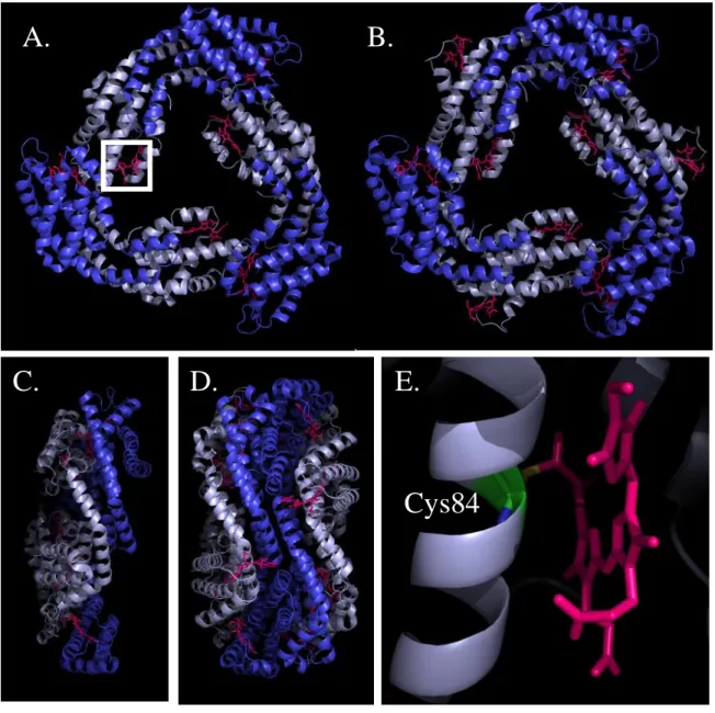
Figure 6. Crystal structures of the phycobiliproteins isolated from
Synechococcus elongatus PCC 7942. A.
Face view and C. side view of an APC trimer;. αAPC subunits are deep blue, β APC subunits are light blue. B. Face view of a PCtrimer and D. side view of a PC hexamer, showing 2 trimers associated back to back. αPC
subunits are deep blue, βPC subunits are light blue. E. Close up of the white frame in A,
showing the thioether bond between Cys84 of βAPC and phycocyanobilin (PCB). PCBs are
shown as purple sticks. PDB files 4F0U and 4H0M (Marx et al., 2013) modified with PyMol.
C.
D.
E.
Cys84
13
In the introduction I will mainly focus on PBs of the cyanobacterium Synechocystis that was used as a model organism in this study. PBs with slightly different architectures and coming from other cyanobacterial strains will be presented in Chapter 2 of the present manuscript. When isolated and observed through EM, Synechocystis PBs appear as hemidiscoidal particles where PC rods (6 in general) radiate from an APC core laying on thylakoid membranes in vivo (Arteni et al., 2009) (Fig. 5B,C). Each rod is made of 2 or 3 PC hexamers. 4 APC trimers give a cylinder and 3 cylinders stack to form the core (Fig.5D). The complex is organized in a way that phycobiliproteins with the most blue-shifted absorption and fluorescence emission properties, namely PC (λAmax=620nm, λFmax=650nm), surround
phycobiliproteins with lower energetic levels (APC, λAmax=650nm, λFmax=660nm). This
allows funneling energy towards APC cores and directional energy transfer to Chla.
Beyond that, no overall tri-dimensional structural picture of PBs is available so far. Isolated phycobiliproteins have been crystallized from diverse organisms in trimeric or hexameric aggregation states and their X-ray diffraction pattern was determined (reviewed in: Adir, 2005). They generally appear as disks of 100Å diameter with a central cavity of 30Å. The crystal structure of trimers or hexamers of Synechocystis PC was not resolved. Only the structure of a monomer of APC was published (PDB file 4F0T; Marx and Adir, 2013). However, crystal structures of PC hexamers and APC trimers of close organisms, for example Synechococcus elongatus PCC 7942 (PDB file 4H0M: Marx and Adir, 2013), are known (Fig.6).
Synechocystis PBs also incorporate colorless linker polypeptides that possibly get inserted into the central cavities of trimers and hexamers of phycobiliproteins (Yu and Glazer, 1982). LRC27 (encoded by the cpcg1 gene) enables the binding of PC rods to the APC core
(Kondo et al., 2005). LR33 (cpcc1 gene) and LR30 (cpcc2 gene) are required for the elongation
of PC rods (Ughy and Ajlani, 2004). LR10 (cpcd gene) has been proposed to cap PC rods and
terminate their elongation, similarly to LC8 (apcc gene) for APC cylinders in the core.
Recently, whole PC rods have been crystallized (including LRC27, LR30, LR33 and LR10) (David
et al., 2011). Authors suggested that even though linkers could not be modeled in the subsequent structure due to symmetry problems, they increased the stability (quantified using the b-factor) of specific PC aminoacids situated on the inner cavity surface. Only one crystal structure exists so far showing linker-phycobiliprotein complexes: the LC8 was shown to be
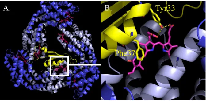
Figure 7. Crystal structure of the L
C8-APC complex isolated from
Mastigocladus laminosus. A.
Face view of an APC trimer with incorporated linker. αAPCsubunits are deep blue, β APC subunits are light blue. The L
C8 linker-polypeptide is represented
in yellow. B. Close up of the white frame in A. showing interactions between LC8 and a βAPC
bond PCB. The contacts have a hydrophobic (bling ring and Phe37 of LC8) or polar (bilin side groups and Tyr33) nature. PCBs are shown as purple sticks. PDB file 1B33 (Reuter et al., 1999) modified with PyMol.
Tyr33
Phe37
A.
B.
15 with 2 out of 3 βAPC
bond PCBs (Reuter et al., 1999) (Fig.7). This agrees well with the idea that linkers are not only necessary for PB assembly but also for tuning phycobiliprotein spectral properties, allowing an optimized polar energy transfer (Lundell et al., 1981a). When purified, PC has its maximum absorbance at 620 nm and fluorescence emission at 645nm (hexameric state). LR33 (res. LRC27) shifts the maximum of absorbance to 623nm (res. 628nm)
and of fluorescence to 648nm (res. 650nm). LC8 containing APC trimers have a red-shifted
emission compared to normal APC trimers (Maxson et al., 1989). Linkers actually change the biochemical environment around PCBs (Fig.7B).
The APC core has a quite intricate organization. It was first described in Synechococcus PCC 6301, thanks to partial dissociation of PBs allowing isolation of “subcore particles” (Yamanaka et al., 1982). The proposed model was then extended to other species after confirmation in Anabaena PCC 7120 (Ducret et al., 1998). Fig.5D shows the tricylindrical APC core of Synechocystis where most trimers contain major APC forms (αβ) that absorb light at 650nm and emit fluorescence at 660nm (see Fig.5A). Differences in spectral properties between PC and APC come from diverging aminoacids surrounding the PCBs (McGregor et al., 2008). αAPC subunits have a more hydrophobic PCB binding pocket than αPC
subunits, responsible for the APC trimers bathochromic shift. The 2 basal cylinders of the core also include trimers with minor APC forms (ApcD, ApcF, ApcE). Purified ApcD (Glazer and Bryant, 1975) and ApcE (Lundell et al., 1981b) have a pronounced red-shift, absorbing light around 670nm then emitting fluorescence at 680nm. They are the lowest energy phycobiliproteins and play the role of PB terminal emitters (Gindt et al., 1994). The differences in the spectral properties are also due to more hydrophobic environments surrounding the bilins (McGregor et al., 2008). ApcE has actually several functions. Its N-terminal part shows some homology with αAPC
subunits, binding 1 PCB thanks to Cys190. Its C-terminal part contains linker-like domains (called REP) separated by spacers (called arms); it acts as a scaffold for APC core assembly and probably fine-tunes APC spectral properties (Houmard et al., 1990). Finally, PB attachment to thylakoid membranes involves polar contacts between ApcE residues and charged lipid head groups, which explains why ApcE is sometimes called LCM (core-membrane linker) (Ajlani and Vernotte, 1998).
We saw that PBs are highly organized complexes. Phycobiliproteins tend to aggregate, forming rows. Colorless linker polypeptides allow the apparition of 2 well defined zones, corresponding to PC rods and to the central APC cores. Spectral properties of
PC
APC
TE
Chla
Absorbance
Emission
620nm
645nm
620nm
660nm
670nm
680nm
0 100 200 300 400 500 620 640 660 680 700 720 740 760 780 F lu o resce n ce (a .u .) Wavelength (nm)PC APC
TE,
PSII
PSII PSI
A.
B.
Figure 8. Energy transfer from phycobilisomes to photosystems in
Synechocystis.
A. Schematic representation of the energy transport pathway in Synechocystis phycobilisomes. B. 77K fluorescence emission spectrum of Synechocystis cells kept in the dark for 10’ then excited using low intensity orange light (590nm) absorbed preferentially by PC.17
phycobiliproteins are fine tuned for efficient energy transfer from PC to APC and finally terminal emitters (ApcD, ApcE, ApcF) (Fig.8A).
2.2.2. PBs function as antennae
PBs funnel light energy to the terminal emitters that in turn equilibrate with Chla from both reaction centers (Fig.8A). When applying an excitation light absorbed preferentially by PC (around 600nm), the fluorescence spectra of cyanobacterial cells at 77K show distinct bands associated with PB components (PC 650 nm, APC 665 nm, TE 683 nm), PSII Chla (CP43 685 nm, CP47 695 nm) and PSI Chla (720 nm) (Campbell et al., 1998) (Fig.8B). In many articles, the ratios between these bands were used as a measure of energy transfer from the PBs to the photosystems (for example Ashby and Mullineaux, 1999). Getting a real quantitative estimate is more complicated. Based on target analysis of spectrally-resolved picosecond fluorescence data, Tian and coworkers (Tian et al., 2011) suggested that 50% of the energy collected by PBs goes to PSI and 50% to PSII in Synechocystis cells grown under their laboratory conditions. Combining fluorescence emission and excitation spectra interpretations, it was proposed that up to 80% of the energy harvested by PBs reaches PSI in Arthrospira platensis cells (Rakhimberdieva et al., 2001). Energy transfer from PBs to PSI could be achieved through a direct interaction between them (Mullineaux, 1992; Mullineaux, 1994) and/or through energy spillover between PSII Chla and PSI Chla (McConnell et al., 2002). Some reports also indicate a possible connection between regular PC rods (Li et al., 2003) or CpcG2-incorporating PC rods (Kondo et al., 2007) and PSI.
There are several terminal emitters per PB (ApcD, ApcF, ApcE) and each of them could create specific routes for energy transfer towards PSI or PSII. ApcD deficient Synechococcus PCC 7002 mutant cells were shown to have impaired energy transfer from PBs to PSI, resulting in a slowed down PSI photoxidation upon orange light illumination (Dong et al., 2009). 77K fluorescence emission spectra of Synechocystis cells lacking ApcD and/or ApcF strongly suggested that in this strain energy flow from PBs to PSI and PSII passes mainly by ApcF (or the associated ApcE) and not by ApcD (Ashby and Mullineaux, 1999). Further investigations are required for determining exactly the role of each of the terminal emitters.
19
2.2.3. PBs movement and possible implication in State Transitions
Strikingly, associations between PBs and PSs are extremely transient (for a review: Mullineaux, 2008). On one hand, FRAP combined to FLIP measurements revealed a very fast diffusion of PBs on thylakoid membranes’ surface (Mullineaux et al., 1997; Sarcina et al., 2001; Yang et al., 2007). On the other hand, PSII complexes are immobile. It is impossible to say whether PSI is mobile or not because it has a low room temperature fluorescence yield making it hardly detectable using confocal microscopy. Anyway, this shows a very dynamic environment where PBs seem to continuously switch from reaction center to reaction center.
It was proposed that light quality is a determining factor that drives variations in the affinity between PBs and PSs through multiple ways. Any light absorbed predominantly by Chla (blue/far red) tends to excite PSI specifically in cyanobacterial cells. Any light absorbed predominantly by phycobiliproteins (green/orange) excites relatively more PSII, at least at low intensities. This can set on an over-oxidation or over-reduction of inter-systems electron transporters and a situation where PSI or PSII becomes rate limiting for photosynthesis (reviewed in: Mullineaux and Emlyn-Jones, 2005). Cyanobacteria, like plants and algae, have developed a mechanism named “state transitions” to cope with the unbalanced PSs’ functioning.
PQ reduction drives transition from State I to State II and a concomitant decrease in PSII fluorescence emission as assessed by 77K spectra and room temperature Pulse Amplitude Modulation (PAM) traces (illustrated in McConnell et al., 2002). PAM fluorometry allows measuring fluorescence yield over time (see Schreiber et al., 1986). A non-actinic modulated detection light (around 650 nm) excites Chl and phycobiliproteins; the emitted fluorescence, after passing through cutoff filters (selecting wavelengths higher than 700 nm), is detected by a synchronous photo-multiplicator. Application of a non-modulated actinic light does not have any impact on signal intensity but can have physiological effects that modify the sample’s fluorescence yield. Several fluorescence levels can be recorded that way: minimum level F0 (no actinic light ON), maximum level Fm (saturating actinic flash ON), steady-state
level Fs’ (continuous, sub-saturating actinic light ON) and maximum level under actinic
illumination Fm’ (see also El Bissati et al., 2000). Transition from State II to State I increases
PSII
PSI
A.
B.
C.
D.
State I
State II
Figure 9. Alternative models for state transitions in Synechocystis. A. In State
I, light preferentially excites PSI and triggers the association of phycobilisomes with PSII. Most of the energy harvested by phycobilisomes then arrives to PSII reaction centers (orange arrow). B. to C. Alternative models for State II. B. Upon State II transition, PSI trimers dissociate into monomers and move for a closer association with PSII. A slight translation of phycobilisomes allows their interaction with PSI, but they remain partly connected to PSII (orange arrows). C. Upon State II transition, phycobilisomes diffuse towards PSI and associate with it (orange arrows). They are no more connected to PSII. D. Similar model to B., adding energy spillover from PSII Chl a to PSI Chl a (drak green arrow).21
It is probable that like in plants and algae the Qo site of cytochrome b6f is involved in
this mechanism (Mao et al., 2002; Huang et al., 2003). According to the mobile antennae model, the changes in PSII fluorescence are related to the movement of PBs from PSII to PSI (or contrary) and the associated diminution (or increase) of the PSII effective antenna size (Joshua and Mullineaux, 2004) (Fig.9) . It is not known how the signal goes from the PQ pool and/or the cyt b6f to the PBs or photosystems. In plants and algae a kinase and a phosphatase
are involved in the signal translation (reviewed in: Ruban and Johnson, 2009). In the past it was suggested that phosphorylation events were also involved in cyanobacterial state transitions (Allen et al., 1985) but these results were not confirmed and the discussion remains open.
The mobile antennae model is largely based on FRAP measurements. In 2004, Joshua and Mullineaux showed that there is a correlation between states transition and PB mobility: in cells treated with high molarity potassium-phosphate buffer or sucrose solution, both processes were inhibited (Joshua and Mullineaux, 2004). Published data about thylakoid membrane organization in Synechocystis WT cells shows colocalization between PSI and PSII; no massive PSI or PSII enriched clumps appear (Folea et al., 2008; Collins et al., 2012). Consequently, if PBs mobility is required for state transitions induction in Synechocystis, local rearrangements or tilting are more probably implied than long range diffusion (Fig.9). In contrast, ultrastructural studies of the thylakoid membranes using EM revealed that there are PSI and PSII enriched domains in Synechococcus PCC 7942 cells (Sherman et al., 1994). Thus, whether or not a long range movement of PBs is required for state transitions could depend on the species considered (see alternative model, Fig.9C).
Suppression of ApcD or ApcF in Synechococcus PCC 7002 (Gindt et al., 1994) and Synechocystis cells (Ashby and Mullineaux, 1999) led to the conclusion that both proteins are required for state transitions. 77K fluorescence emission spectra of ΔApcD (respectively ΔApcF) mutant cells suggest that they are locked in State I (respectively State II). PBs terminal emitters, beyond their functional role, could also act as physical bridges that are required for a correct association between PBs and reaction centers. A reverse genetics approach allowed identifying another protein called Regulation of PBs Association C (RpacC) which suppression impairs state transitions (Emlyn-Jones et al., 1999). RpaC is thought to permit the dynamic association between PBs and PSII (Joshua and Mullineaux, 2005). The absence of similar proteins in organisms other than cyanobacteria and RpaC’s unique predicted folding constitute barriers to understand its exact biochemical role. The mobile
22
antennae model alone does not fully explain state transitions, also observed in mutants lacking assembled PBs (Olive et al., 1997; El Bissati et al., 2000). Movement of photosystems could also be involved in state transitions. PSII forms dimeric rows, particularly visible in State I and tending to disappear in State II (Olive et al., 1997). When present, these rows could prevent PBs attachment to PSI through steric hindrance effects (Bald et al., 1996). PSI exists as a mixture of monomers and trimers (Kruip et al., 1994) arranged more or less randomly on border of the PSII arrays. PSI monomerization increases PBs diffusion rates (Aspinwall et al., 2004) and accelerates state transition kinetics. It has been proposed that spillover from PSII Chla to PSI Chla is partially responsible for PSII fluorescence decrease and concomitant cross-section diminution upon State I-to-II transition (For review: Biggins and Bruce, 1989). This suggests that part of the light energy received by PSII Chl antennae gets transferred to PSI. Very close vicinity between PSII and PSI is required for spillover to occur, probably achieved when PSII dimeric rows disassemble in State II. An extended model has been proposed by McConnell and coworkers (McConnell et al., 2002) which suggests that state transitions are actually due to bot direct changes in PBs energy distribution and spillover from PSII to PSI (fig.9D).
This section aimed at emphasizing that cyanobacteria possess accessory antennae with unique features. The gigantic, soluble, highly organized PBs are designed for harvesting light in the gap form by Chla absorption bands. They are optimized for a very efficient energy transfer towards terminal emitters. They can distribute energy to PSI or PSII, in a very dynamic manner controlled by other cellular processes.
3. Drawbacks of photosystems functioning in an oxygenic medium and defensive strategies
3.1. Photoinhibition of PSII
High light intensities trigger a decrease of PSII activity, irreversible when de novo protein synthesis gets chemically inhibited (lincomycin, chloramphenicol addition), referred to as photoinhibition. The underlying mechanisms are still debated but reactive oxygen species (ROS) generation (particularly singlet oxygen, 1O2) and subsequent oxidative
damages presumably play a central role (reviewed in: Krieger-Liszkay, 2005; Vass, 2012). Under strong illumination or any condition driving PQ over-reduction – like anaerobiosis, CO2 deprivation and low temperatures – PSII acceptor side gets reduced because the QA
23
if there is no oxidized PQ to fill it; as a consequence, forward electron flow gets interrupted and the probability for charge recombination increases. Spontaneous spin conversion of the primary radical pair [P680+•Pheo−•] occurs, going from singlet to triplet state, followed by a possible charge recombination leading to triplet Chl (3P680) formation. 3P680 reacts fast with O2 and the extremely reactive singlet oxygen (1O2) appears, as confirmed by chemical
trapping in Synechocystis cells (Rehman et al., 2013). 1O2 irreversibly damages the D1 protein
then the whole PSII before of eventually impairing the translational and transcriptional machineries required for damaged proteins replacement. Other ROS can participate in photosynthetic apparatus degradation, such as superoxide (O2-.) obtained when QA- reacts with
O2. Donor side effects are observed as well: disruption of the WOC – for example by UV
light – triggers oxidizing radicals (P680+ or TyrZ+) stabilization, which destroy their protein environment. Some groups suggest that visible light absorption by Mn also triggers a dose-dependent degradation of the WOC, directly impairing PSII functioning (reviewed in: Tyystjärvi, 2008).
3.2. Photoprotective mechanisms in cyanobacteria: an overview
Reestablishing a functional photosynthetic apparatus in photoinhibited cyanobacterial cells requires degrading the damaged D1/D2 proteins then assembling new PSII reaction centers, which consumes a lot of ATP. Some cost-effective photoprotective strategies have been developed aimed at limiting ROS production under high light and at preventing their deleterious effects. First of all, cyanobacterial cells possess a ROS scavenging machinery that allows controlling O2-.and H2O2 concentrations in vivo (reviewed in: Latifi et al., 2009); they
also synthesize non-enzymatic antioxidants, such as carotenoids or α-tocopherol, able to quench 1O2 (and lipid peroxides for the second one). Secondly, some alternative transport
routes exist that can remove electrons from PSII acceptor side when the PQ pool is totally reduced: Flv2/Flv4 at the QB site (Zhang et al., 2012), PTOX at the PQH2 level in certain
cyanobacterial strains (reviewed in McDonald et al., 2011). Similarly, Cyclic Electron Transfer around PSI passing by the NDH1 complex then PQ permits ATP production without any associated NADP+ reduction (for a review: Battchikova et al., 2011). Finally, strong irradiances cause changes in the expression levels of many genes (Hihara et al., 2001): this includes an upregulation of the ones encoding for D1 protein paralogues with modified QA
25
redox potential and less prone to perform charge recombination (reviewed in Mulo et al., 2009) or a downregulation of the ones encoding for enzymes implied in photosynthetic pigments synthesis.
In response to high light intensities, cyanobacteria also accumulate High Light Inducible Polypeptides (HLIPs) that show similarities to the Chla/b-binding Light Harvesting Complexes of plants; they attach Chla, possibly carotenoids, and form a single transmembrane α-helix (Dolganov et al., 1995). Synechocystis mutant cells depleted in their HLIPs grow normally under weak illumination but clearly have modified pigment content under strong illumination, accumulating more myxoxantophyll but less Chl than WT Synechocystis cells (Havaux et al., 2003; Xu et al., 2004). HLIPs are thought to stabilize other Chl-containing proteins or to play a role in Chl mobilization under light stress. IsiA constitutes another example of Chla-binding protein induced in response to iron starvation (Havaux et al., 2005) but also to various other abiotic stresses (high light, hyperosmotic medium). 18 IsiA proteins can associate intro a ring that surrounds PSI and acts as an accessory antenna (Melkozernov et al., 2003). Empty IsiA rings also exist, dissipating as heat the light energy they collect and playing a photoprotective role (Ihalainen et al., 2005).
3.3. Non-photochemical quenching of the excess light energy harvested.
An excited photosynthetic pigment can be deactivated by transmitting its exciton to another pigment (until photochemistry occurs, photochemical quenching noted qP), by emitting a photon of fluorescence or by producing heat. The light harvesting antennae associated to photosystems perform very efficient energy transfer to reaction centers, approximately 90% (quantum efficiency) of the collected energy reaching the special-pair chlorophylls under non-saturating illumination. In plants and algae, a photoprotective mechanism increases heat dissipation at the level of Light Harvesting Complexes (LHCs, chla/b-binding antennae) and concomitantly decreases fluorescence emission plus energy transfer to reaction centers under high light conditions (energy-dependent non-photochemical quenching, noted qE. Reviewed in: Müller et al., 2001). qE is induced by the ΔpH formation across thylakoid membranes; it requires the protonation of PsbS (LHC-like protein) and the xantophyll cycle activation (zeaxanthin converted into violaxanthin). Besides that, the exact mechanism underlying heat dissipation remains unsolved. Cyanobacteria do not possess any LHC but harvest light mainly thanks to their PBs. The work presented in this
Figure 10. Blue-light induced OCP-related fluorescence quenching in
Synechocystis cells.
A. and B. Measurements of the fluorescence yield using a PAM fluorometer in dark-adapted wild-type (A) and ΔOCP (B) cells illuminated successively with low-intensity blue-green light (400 to 550 nm, 80 µmol.m-2.s-1 ) and high-intensityblue-green light (740 µmol.m-2.s-1). C. Similar experiments in WT cells showing the
fluorescence recovery without any additions (circle) and in the presence of nigericin (squares) or DCMU (solid line), the Fm’ level only being displayed. Adapted from Wilson et al., 2006.
B.
C.
A.
0 100 200 300 400 500 Time (s) 0 100 200 300 400 500 Time (s) 2627
manuscript focuses on a photoprotective mechanism enabling heat dissipation at the level of PBs under high irradiances.
4. The Orange Carotenoid Protein (OCP)-related non photochemical quenching of the energy harvested by PBs
4.1. Discovery
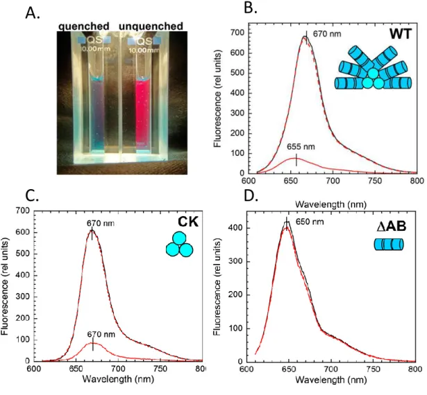
Illuminating Synechocystis cells with strong blue-green light was initially shown to trigger a 30% decrease in the maximal fluorescence level (Fm’) on PAM traces (El Bissati et al., 2000). As soon as illumination stopped, fluorescence started recovering and finished by reaching its initial level (Fig. 10). Strikingly, this phenomenon kept occurring even below the thylakoid membrane phase transition point (25°C) in mutant cells lacking polyunsaturated fatty acids; thus, it was not related to state transitions that were completely blocked in these conditions (El Bissati et al., 2000). The addition of protein synthesis inhibitors had no impact on fluorescence quenching and recovery, which then could not originate from photoinhibition and subsequent photodamage repair (El Bissati et al., 2000). The addition of nigericin, a chemical decoupler, demonstrated there was no correlation between the transthylakoidal ΔpH formation and the fluorescence decrease induction (Wilson et al., 2006) (Fig.10). Taken together, these observations suggested the existence of an unknown cyanobacterial mechanism involving the antennae and diverting energy away from PSII under strong blue-green illumination.
The study of PSII-deficient Synechocystis mutant cells indicated that blue-green light is sensitized by a carotenoid-containing component and actually drives PBs fluorescence quenching (Rakhimberdieva et al., 2004). No fluorescence decrease was observed in PB deficient (PAL) or APC core deficient (ΔAB) Synechocystis mutant cells whereas it was retained in a strain lacking PC rods only (CK) (Wilson et al., 2006). Based on whole cells emission and decay associated spectra, Scott and coworkers arrived to the conclusion that blue-green light has an impact on APC (including red-shifted forms) but not on PC fluorescence emission and decay kinetics (Scott et al., 2006). Thus, the newly described mechanism is associated to APC cores fluorescence quenching.
It requires the presence of the soluble carotenoid-bearing Orange Carotenoid Protein (OCP) as blue-green light does not trigger any fluorescence quenching in OCP deficient Synechocystis mutant cells (Wilson et al., 2006) (see Fig. 10 and next sections for more details).
Figure 11. Distribution of the OCP among cyanobacteria.
A 16S species tree of cyanobacteria with sequenced genomes (including the ones published in Shih et al., 2013). Organism IDs are colored by section (green, Section I; red, Section II; blue, Section III; yellow, Section IV). F = full-length ocp gene, N, C correspond to genes encoding the N- and C-terminal domain, respectively. Adapted from Kirilovsky and Kerfeld, 2012.29
Importantly, oxygen evolution dropped faster and to a more important extent under saturating white light illumination (3000µmol.m-2.s-1) in ΔOCP cells than in WT Synechocystis cells (Wilson et al., 2006). This is due to a decrease of the effective antenna size induced by the OCP related mechanism (Wilson et al., 2006). When this photoprotective mechanism is absent, under high irradiance, more light energy arrives to PSII, which leads to reactive oxygen species formation and to photoinhibition. It was envisioned that OCP sensitizes strong blue-green illuminations then inducing, directly or not, a non-photochemical quenching of the excess light energy harvested by PBs. As a result, energy flow to PSII and fluorescence yield both decrease (Wilson et al., 2006). Measuring P700 oxidation kinetics in PSII deficient cells and fluorescence induction kinetics in PSI deficient cells, Rakhimberdieva and coworkers (Rakhimberdieva et al., 2010) estimated that a 60% decrease in PB fluorescence correlates to a 30-40% drop in quantum efficiency of one or the other photoreaction. Even if the heat dissipation process takes place in APC cores, Chla fluorescence can partly be quenched as uphill energy transfer sometimes occurs (Rakhimberdieva et al., 2007b).
The OCP-related quenching is reversible, fluorescence starting to recover as soon as light source gets turned off (El Bissati et al., 2000). This owes to another actor called the Fluorescence Recovery Protein (Boulay et al., 2010; see also section 4.6.). A better understanding of the OCP related photoprotective mechanism required isolation of the different proteins implied and their subsequent characterization in vitro.
4.2. Distribution and regulation of the OCP
OCP had been detected in Arthrospira maxima, Microcystis aeruginosa and Anabaena flos-aquae long before the discovery of its actual photoprotective role (Holt and Krogmann, 1981). It is encoded by a single gene in Synechocystis (slr1963: Wilson et al., 2006) that has orthologues in most of the PBs-containing cyanobacterial strains sequenced so far (for review: Kirilovsky and Kerfeld, 2012) (Fig.11). The predicted primary structures for OCP homologues show between 62% and 82% identity to that of Synechocystis OCP, certain absolutely conserved aminoacids probably playing key functional roles. Sometimes, short slr1963-like genes also appear, putatively encoding for truncated OCP with a yet to determine function (Fig.11). Cyanobacteria naturally lacking an ocp encoding gene - for example Thermosynechococcus elongatus and Synechococcus elongatus PCC 7942) - are more susceptible to PSII photodamage under high irradiance (Boulay et al., 2008); illumination
Figure 12. Structure of the OCP isolated from Arthrospira maxima.
A. View of an OCP monomer showing the N-terminal domain (residues 1-165) in cyan, the C-terminal domain (residues 186-317) in red and the flexible linker in green. hECN appears as orange sticks, its hydroxyl ring lying in the N-terminal domain and its carbonyl ring in the C-terminal domain. B.Close up showing some conserved OCP aminoacids interacting with hECN and presented with purple sticks. Potential hydrogen bonds are indicated using dashed red lines. PDB file 1M98 modified with PyMol.A.
B.
Tyr44
Trp110
Trp290
Tyr203
H
2O453
3031
with saturating white light (2500 µmolphotons.m-2.s-1) for 3 min triggered a non-reversible fluorescence decrease (15-20%) associated to photoinhibition in both strains, less visible when considering Arthrospira or Synechocystis cells producing OCP. Transcriptomic data indicates that slr1963 expression, which is constitutive, gets upregulated upon high light treatment (Hihara et al., 2001). Iron depletion also leads to OCP over-accumulation in various cyanobacterial strains (Wilson et al., 2007; Boulay et al., 2008). This over-accumulation correlates with an increased amount of blue-green light triggered PB fluorescence quenching; the Fm’ drops by 60% instead of only 30% when considering iron starved Synechocystis cells
(Wilson et al., 2007). OCP acts well as a switch for the non-photochemical fluorescence quenching, in a dose dependent manner.
Non-stressful growth conditions (90µmol.m-2.s-1 white light plus CO2 enriched
atmosphere) lead to the production of 1 OCP for 2-3 PBs in Synechocystis, as revealed by Western Blot analysis (Wilson et al., 2006). OCP is situated on the stromal side of thylakoid membranes as demonstrated by cell fractionation experiments and immunogold labeling (Wilson et al., 2006), thus lying in close vicinity to PBs.
4.3. Crystal structure of the OCP isolated from Arthrospira
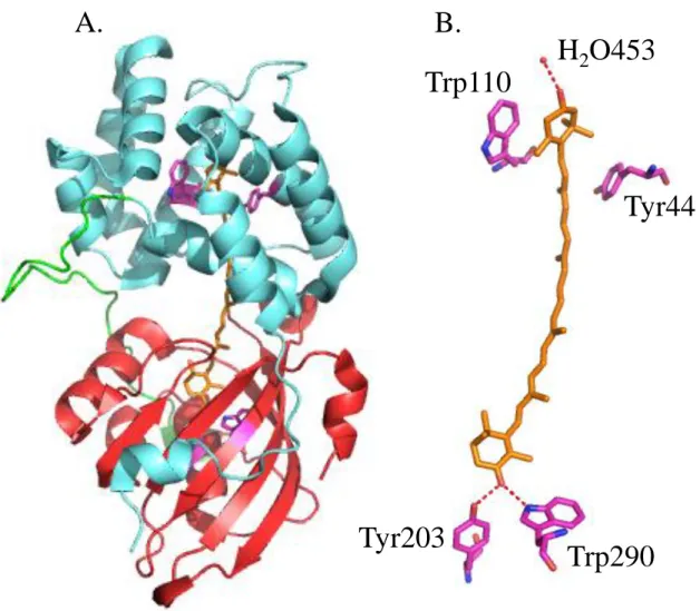
The structure of OCP was resolved before the understanding of its function. Following isolation from Arthrospira maxima cells, OCP appeared as a soluble orange protein with 35 kDa molecular mass (Wu and Krogmann, 1997). Its absorption spectrum exhibited a shoulder at 440 nm plus peaks at 467 nm and 496 nm indicating the presence of a bound carotenoid, identified as 3’-hydroxyechinenone (hECN) (Wu and Krogmann, 1997). The protein environment modifies hECN spectral properties, which normally has a yellowish color when solubilized with organic solvents (Polívka et al., 2005). A 2.1 Å crystal structure was released in 2003 (PDB file: 1M98. Kerfeld et al., 2003) revealing that OCP consists of 2 domains separated by a flexible linker region (Fig.12). The N-terminal domain (residues 1 – 165), made of 2 α-helices bundles (4 helices each, PFAM 09150), is unique to OCP and exists only in cyanobacteria. The C-terminal domain (residues 190 - 317) displays 2 α-helices and a 5 fold β-sheet, reminiscent of the Nuclear Transport Factor 2 family (PFAM 02136). hECN (1 per OCP) spans these domains, its hydroxyl ring getting inserted between the N-terminal α-helices bundles and its keto ring lying in a hydrophobic cleft formed on the C-terminal β-
Figure 13. Isolated Synechocystis OCP responsivity to light.
A. Photograph of isolated OCPo and OCPr. To obtain OCPr the protein was illuminated with blue-greenlight at 740 µmol photons.m-2.s-1 at 12°C for 2 min. B. Absorbance spectra of the dark
orange form (OCPo; black) and the light red form (OCPr; red). C. OCPr accumulation at
11°C and different light intensities: 20 (black), 50 (red), 120 (green),210 (blue), 350 (rose), 740 (orange), and 1,200 (violet) µmolphotons.m-2.s-1 of blue-green light. D.
photoconversion from the OCPo to the OCPr form using a 350 µmol photons.m-2.s-1
blue-green light intensity at different temperatures: 32°C (violet), 28°C (rose), 24°C (blue), 19°C (green), 15°C (red). The protein concentration was OD 0.2 at 495 nm. Adapted from Wilson et al., 2008.
A.
B.
C.
D.
33
sheet. It was found that several absolutely conserved aminoacids among OCP orthologues are situated in close vicinity to hECN and potentially interact with it (Kerfeld et al., 2003) (Fig.12). In the N-terminal domain, Tyr44 and Trp110 possibly establish hydrophobic contacts with the hydroxyl ring. In the C-terminal domain, Tyr203 and Trp290 form hydrogen bonds with the keto-group through their side chains. Also, conserved water molecules can connect hECN hydroxyl-group to the protein backbone through hydrogen bonds. All these interactions are potentially responsible for the fine tuned hECN spectral properties or allow for carotenoid selectivity (Kerfeld et al., 2003).
4.4. Photoactivity of the OCP and Synechocystis mutant studies
Arthrospira maxima cannot be genetically manipulated. Thus, it is impossible to modify Arthrospira OCP to facilitate its isolation via affinity chromatography or to investigate the role of specific aminoacids through targeted mutagenesis. Thus, Synechocystis mutants producing OCP with a C-terminal His-tag for facilitated purification were constructed. Isolated Synechocystis OCP appears orange under dark conditions (OCPo), has a very similar absorption spectrum to that of Arthrospira maxima OCP and binds hECN mainly (Wilson et al., 2008).
Strong blue-green illumination induces OCPo photoconversion to a red from (OCPr) with maximum absorbance at 500 nm (Wilson et al., 2008) (Fig.13A, B). The OCPr accumulation gets fastened and its final amount increases when actinic light intensity rises (Fig.13C). Lowering temperature (16°C to 32°C frame) does not much affect the rate of photoconversion but leads to higher OCPr concentrations at equilibrium. This comes from the fact that OCPr spontaneously reconverts to OCPo, and that low temperatures largely hinder such a recovery (Wilson et al., 2008). The quantum yield of OCP photoactivation was evaluated to approximately 0.03 based on transient absorption spectroscopy, which is quite low and explains why only high intensity blue-green light triggers significant OCPr formation. The metastable OCPr was then proposed to act as a molecular switch turning on the mechanism that leads to PB fluorescence quenching.
Resonance Raman spectroscopy revealed that hECN, which is slightly twisted in OCPo, gets unbent in OCPr resulting in an increased apparent conjugation length (Wilson et al., 2008). The secondary structure of OCP also gets modified upon photoactivation as