Abstract Bony metastases are a fre-quent problem in elderly patients af-fected by cancer, and those with bony metastases involve the spine in ap-prox. 50%. The most frequent spinal metastases (60%) are from breast, lung, or prostate cancer. The chance that an elderly patient (60–79 years old) is affected by bony metastases is four times higher in men and three times higher in women than a mid-dle-aged patient (40–59 years old). Since the medical treatment with all the adjuvant treatment options pro-long the survival of this particular patient group, the spinal metastases may become a mechanical issue, thus requesting surgical treatment. Differ-ent classification systems have been proposed to rationalize surgical indi-cations, some concentrating solely on the local spinal tumor involve-ment and some including the overall clinical situation. Since most of the surgical options are of palliative character, it is more important to base the decision on an overall clinical classification including the different treatment modalities – irradiation, chemotherapy, steroids, bisphospho-nates, and surgery – to make a shared decision. In case surgery is indicated – neural compression, pathological fracture, instability, and progressive deformity, nursing reasons – the most straightforward procedures should be chosen, which may not need an in-tensive care unit stay. In the
thora-columbar spine a posterior decom-pression and posterolateral vertebral body resection through a posterior approach only, with a concomitant reconstruction and stabilization, has shown to work sufficiently well. In the middle and lower cervical spine the anterior approach with anterior decompression and anterior column reconstruction is most effective and has a low morbidity, whereas the oc-cipitocervical junction can generally be treated by posterior resection and stabilization. The outcome should be determined by the survival time in an ambulatory, independent status, where pain is controlled, and the patient is not hospitalized. Surgical manage-ment shows the greatest improve-ment in pain reduction, but also in other domains of quality of life. Since prospective randomized stud-ies comparing different treatment modalities for spinal metastases in-cluding surgery are not available and are ethically difficult to achieve, each case remains an interdiscipli-nary, shared decision making process for what is considered best for a pa-tient or elderly papa-tient. However, whenever surgery is an option, it should be planned before irradiation since surgery after irradiation has a significant higher complication rate.
Keywords Spinal metastases · Vertebral metastases · Elderly · Spinal tumor · Vertebral tumor
Max Aebi
Spinal metastasis in the elderly
Received: 1 August 2003 Accepted: 4 August 2003
Published online: 23 September 2003 © Springer-Verlag 2003
M. Aebi (✉)
Institute for Evaluative Research in Orthopedic Surgery,
University of Berne,
Murtenstrasse 35, P.O. Box 8354, 3001 Berne, Switzerland
Tel.: +41-31-6328713, Fax: +41-31-6320928, e-mail: maebi@orl.mcgill.ca
Introduction
Bony metastases are a frequent event in breast, prostate, lung, kidney urinary bladder, and thyroid cancer as well as in multiple myeloma and other hematological malignan-cies which may, however, be considered as primary tu-mors. About 10% of the cancer patients are attained by metastases located in the spine [23, 36] (incidence 1999, SEER and NPCR Registries, United States Cancer Statis-tics; SEER Cancer Statistics Review 1975–2000, National Cancer Institute). Among adults 60% of spinal metastases are either from breast, lung, or prostate cancer. Renal and gastrointestinal malignancies each account for about 5% of spinal metastases, and thyroid carcinomas and melanomas occurring with a lesser frequency [2, 24] (incidence 1999, SEER and NPCR Registries, United States Cancer Statis-tics; SEER Cancer Statistics Review 1975–2000, National Cancer Institute). Since these tumors are increasingly ac-cessible to treatment by surgery, radiation therapy, and chemotherapy, thus prolonging the survival of the affected patients, there is also an increased probability of them be-ing affected by metastases, i.e., with the improved sur-vival, previously silent spinal metastases are becoming clinically apparent and significantly impairing quality of life. Metastatic disease involving the spine most often af-fects the vertebral bodies of the thoracic, lumbar, cervical, and sacral spine. Siegal et al. [46] estimated that approx. 5% of patients with cancer metastases develop cord com-pression. In patients with spinal metastases approx. 20% have a cord compression.
Many of the above primary tumors affect persons of ad-vanced age (60% of cancer patients are older than 65 years; incidence 1999, SEER and NPCR Registries, United States Cancer Statistics; SEER Cancer Statistics Review 1975–2000, National Cancer Institute; World Health Or-ganization report: “Pain in the elderly with cancer,” www. whocancerpain.wisc.edu), and therefore the metastases become a major issue in the elderly. The average age of patients affected by secondary spinal tumors is 55 – 60 years [23] when considering all metastases; however, it is sig-nificantly higher when considering tumors that are more
prevalent in the elderly such as prostate cancer and multi-ple myeloma (Table 1). Prostate cancer, for exammulti-ple, is at least six times more frequent in men aged 60 – 79 years than in those 40 – 59 years old. Breast cancer is almost double and lung cancer five times higher in the elderly (60 – 79 years) than in the middle-aged (40 – 59 years). Al-though cancer is one of the major causes of morbidity and mortality, elderly persons are often excluded not only from clinical cancer studies but also from standard treat-ment, and generally also from cancer screening because comorbidity and frailty alter the risk benefit of screening (World Health Organization report: “Pain in the elderly with cancer,” www.whocancerpain.wisc.edu). There is clearly an underrepresentation of older persons in drug studies, as documented by the United States Food and Drug Administration (http://cbsnewyork.com, 19 July 2003). Spinal metastases can become a major burden for el-derly because it usually affects the quality of life by re-ducing the endurance, the capacity to ambulate, and the ability for physical activity. Due to their age these patients often have other diseases which already limit their quality of life or have metastases in other skeletal areas, therefore limiting even more the therapeutic options which may still be considered in younger patients.
Pathological anatomy and classification
Malignant metastatic cells most frequently spread to the spine hematogenously with tumor emboli following the paravertebral plexus (plexus of Batson) [3, 11, 45, 53] that is characterized by a lack of valves. It is postulated that the venous blood return is shifted into the paravertebral plexus via the intervertebral and basivertebral veins due to increased intra-abdominal and intrathoracic pressure. As a result metastases which follow this pathway result in the characteristic pattern of bony spread because tumor cells are seeded by this mechanism into the capillary network of the vertebral bodies. Due to its avascular nature the disc is usually spared from tumor involvement: however, the most frequently and severely affected part of the ver-tebra is the verver-tebral body (in about 80%) followed by the pedicles and the posterior elements. This constellation ex-plains why most of the spinal metastasis are located in front of the spinal cord or dural sac ending up with an an-terior epidural compression. More than 90% of spinal metastases are extradural and only 5% intradural and less than 1% intramedullar [45]. Less frequently cancer cells spread into the spine through aortic segmental arteries, for example, in lung cancer [45, 49]. Finally there is also the option of direct spread through direct tumor infiltration into the spine, e.g., the Pancoast’s tumor of the lung.
There have been several attempts to classify and stage spinal tumors [7, 8, 9, 13, 16, 17, 27, 28, 50, 51]. DeWald et al. [13] suggested a classification system for spinal metastases that is oriented mainly towards surgical treat-Table 1 Probability of developing invasive cancer (percentages)
at selected ages with spinal metastasis (from [23])
40–59 years old 60–79 years old Breast cancer 4.06 (1 in 25) 6.88 (1 in 15) Prostate cancer 1.90 (1 in 53) 13.69 (1 in 7) Lung cancer Male 1.29 (1 in 78) 6.35 (1 in 16) Female 0.94 (1 in 106) 3.98 (1 in 25) All sites Male 8.17 (1 in 12) 33.65 (1 in 3) Female 9.23 (1 in 11) 22.27 (1 in 4)
ment. They proposed the following five classes with sub-groups covering most of the possibilities of spinal metas-tases appearance:
– Class I: destruction without collapse but with pain. – Class II: the addition of moderate deformity and
col-lapse with immune competence. This class is consid-ered a good risk for surgery.
– Class III: patients are immunocompromised with mod-erate deformity and collapse. This class carries greater risk for surgery.
– Class IV: includes patients with paralysis, collapse, and deformity with immune competence. This class is con-sidered a relative surgical emergency.
– Class V:adds immune incompetence to paralysis, col-lapse, and deformity. This class is not considered a good operative risk.
This classification allows consideration of the tumor, po-tential instability, and patient physiology, which is a sen-sible approach to a difficult problem. Enneking et al. [17] developed a staging scheme for malignant tumors of the spine in particular in adaptation to the staging of muscu-loskeletal tumors in general. The WBB Surgical Staging System was been introduced in 1997 primarily for pri-mary bone tumors of the spine [9]. This can be applied for metastatic spine tumors; however, there are presently few reports on the system’s correlation with, for example, out-come when applied for surgical indications. Tokuhashi et al. [50] introduced a scoring system for the preoperative evaluation of metastatic spine tumor prognosis that, in-stead, allows a correlation of the tumor extent with the
prognosis [51]. The system differentiates between intra-compartmental, extraintra-compartmental, and multiple tumor involvement. The first two categories include types 1 – 3 and types 4 – 6, respectively, whereas multiple tumor in-volvement is categorized as type 7 (Fig. 1). This scoring system found increasing application in recent years as a baseline in publications to make the results comparable among different scientific publications. K. Tomita et al. [51] applied this system to propose their surgical strategy in spinal metastatic disease.
Clinical presentation and Imaging
The clinical presentation of metastatic spine disease is pre-dominantly pain, neurological deficit, progressive defor-mity, and general weakness. Pain may be localized to a certain structure and region of the spine and may be of radicular or medullary origin. The pain is either caused by increased intraosseous pressure in the vertebral bodies due to cellular invasion of the cancellous bone, by com-pression of neural structures such as roots or nervous fibers, by a secondary instability due to the osteoligamen-tous destruction of parts of the axial skeleton, or by the in-filtration of the dura or other neuroanatomical structures. Pain is usually indicated as more or less constant, dull, however with a predominance of night pain and often not to be influenced by the regulation of the physical activi-ties. Generally speaking, slowly progressive, dull neck or back pain which occurs in a patient with a known cancer disease or which may become apparent in an elderly pa-Fig. 1 Tokuhashi et al. [50]
scoring system to establish pre-operative prognosis of metasta-tic spine tumor
tient without a history of a tumor, should be considered as caused by a spinal metastases until proven otherwise [20]. The neurological deficit appears clearly with a delay of weeks to months after the initial presentation of pain. The period between initial pain and neurological deficit is for the cervical and thoracic spine weeks to months but in the lumbar spine days to weeks [1, 31]. The patients may have motor or sensory deficit or both, whereas there is the option of pure radicular and/or a medullary compression. Since most tumors start in the vertebral body, an anterior cord compression can be expected which is represented by a deficit of the corticospinal pathways with the clinical presentation of a spastic paraparesis which may finally re-sult in an inability to ambulate [20, 46]. Spastic parapare-sis appears usually before sensory disturbances. It can progress slowly but always have the potential to deterio-rate within days.
Many patients who present to the spine surgeon with a paraparesis reveal a long history of preliminaries for weak-ness when specifically asked [2]. The loss of the ambula-tory capacity may arrive quickly. Sensory disturbances may start with tingling sensation and other dysesthesias that may, again, fairly quickly convert into a loss of most the sensory modalities, even within hours. Further com-pression may lead to a paresis of the bladder and sphinc-ter and sensory deficits as well as sensory dysfunction in general may become apparent and finally incapacitate the patient. Bladder and sphincter dysfunction are usually ir-reversible if they last more than 48 h or even shorter [12, 13, 18, 25]. Sphincter disturbances also present rather late, and in elderly persons less attention may be given to this issue, since men may have preexisting micturation difficulty with a prostate problem and women with the bladder/uterus relationship as well as a weak pelvic floor. Obviously there may be an urine retention present or dif-ficulty to initiate the micturation as well as a bladder with an overflow or a weakness, presenting as incontinence. These clinical presentations are often irreversible and are nonfavorable prognostic factors.
The cerebrospinal fluid acts as a puffer for a compres-sive process, and even in case the cord is already com-pressed it is first a deterioration in the capillary circulation in the spinal cord which only secondary causes relevant cord damage [26]. Segmental or even multisegmental in-stability may be a major pain generator as well as genera-tor for neurological functional deficit through temporary or dynamic mechanical compression of neurostructures. This instability occurs with the destruction of the domi-nant stabilizing elements of the spine, i.e., the posterior el-ements such as the facet joints, pedicles, laminae, and spinous processes including the soft tissue including liga-ments and joint capsules which all contribute to the stabil-ity. Since most of the vertebral metastases affect primarily the vertebral bodies which are the major structure of the anterior column, metastases do not necessarily coinci-dence with instability, as long as the vertebral body con-tours are intact. Only when the bony structure of the
ver-tebral body is weakened by the replacement of bone by tu-mor tissue (osteolytic metastases) with the result of a pathological fracture, may the anterior column be weak-ened sufficiently to make it collapse. Usually the posterior elements are also involved to some extent at this point and render the segment definitely unstable. Osteoblastic tumor metastases are prone to pathological fractures with frag-ment displacefrag-ments only if there is a certain mix with os-teolytic components. Osteoblastic metastases can reach a considerable hardness which makes a fracture rather im-probable; however, they can initiate radicular or medullar compression due to the solidity of the tumor tissue.
In elderly patients who complain of slowly increasing pain which occurs also during sleeping in the low back re-gion, gluteal rere-gion, groin, knee, or generally in the lower extremity, may have a hip or knee problem, however, re-main suspicious for a metastatic bone cancer, specifically if they have a tumor history or clinical signs of a consum-ing disorder. Also newly appearconsum-ing neck pain in an elderly person should be taken seriously by the first consulted physician and not just automatically considered as an ex-pression of a degenerative cervical spine disease.
The advent of magnetic resonance imaging (MRI) has certainly added a new dimension to the tumor diagnostic of the spine, although computed tomography (CT), specif-ically combined with myelography may still have a rele-vant role to play, since CT may show more precisely the bony involvement. However, as a search methodology and for appreciation of the spinal tumor involvement MRI is the diagnostic tool of choice. It is noninvasive, in con-trast to myelography, which may even be promoting a neurological deterioration combined with CT. It cannot be overlooked, however, that MRI may be overinterpreted by the examiner, and sometimes in cases in which a precise preoperative diagnostic work-up is necessary for the sur-gical planning CT may be more appropriate. The MRI of-fers a good visualization of the soft tumor involvement. In T1-weighted images metastatic tumors appear usually in a hypodense form, whereas in T2-weighted images tumors of the spine are rather hyperdense as an expression of an increased water content or replacement of the fatty mar-row of the bone by tumor cells [26]. Metastases show gadolinium enhancement. In the tumor work up a bone scintigraphy may play its role as search tool for skeletal metastases. A radioisotopic study has a sensitivity of 65– 70%; however, it is preferred to the other studies because the whole body can be searched. For a more specific search in an anatomical region, for example, the cervical, thoracic, or lumbosacral spine the MRI has a higher sen-sitivity than the bone isotope study [20].
Treatment modalities
Although there is no class I evidence (double-blind ran-domized placebo-controlled trial) for any of the treatment modalities indicated in the treatment of spinal metastases,
there are several treatment options recommended. In the case of neurological deficit dexamethasone is the only treatment, which has proven evidence of therapeutic effi-cacy [29, 35, 40, 52]. The therapeutic decision in elderly
frail patients is particularly difficult when they also have significant comorbity. Nevertheless there are today essen-tially four modalities of treatment available after the ad-ministration of steroid: (a) irradiation, (b) surgery, (c) bis-phosphonates, and (d) rarely chemotherapy and hormonal therapy as an adjuvant therapy in well defined tumor types [47]. A fifth possibility is a combination of all the above. The efficacy of these diverse treatment modalities and the survival rate of patients depend on the histological tumor type, tumor stage, therapeutic control of the primary tu-mor, and tumor spread. Overall survival in this patient cat-egory is around 12 months [12, 15, 33, 48, 51, 54, 56].
The indications for treatment are given not merely by the neurocompression but also alsol by the major determi-nants of quality of life: (a) pain, be it radicular, medullar, or of dural origin caused by direct or chronic compression through instability and/or progressive deformity of the vertebral column, or be merely by intravertebral pressure elevation due to tumor invasion, (b) loss of mobility, and (c) nursing reasons. This decision-making process is diffi-Fig. 2 Long fixation in progressing deformity and instability a A
62-year-old woman with multiple-level involvement of the cervi-cal, thoracic, and lumbar spine metastases of a breast cancer with neurological deficit and pain due to progressing deformity and in-stability. b Long fixation (sublaminar wiring-metal-cement com-pound) and partial correction from C1 to the lower thoracic spine in combination with irradiation was most efficient in reducing pain and neurological deficit for more than 3 years. c A 58-year-old man with a hypernephroid carcinoma and cervical involvement had previous anterior surgery and a cement block posteriorly (as-terisk) with consecutive progression of the tumor, loosening of the fixation and a nonunion at the cement-bone interface (arrow). d Posterior removal of the cement block and stabilization were fol-lowed by e anterior revision and restabilization after a previous embolization of the tumor and occlusion of one of the vertebral ar-teries. The patient died 2 years after this surgery from metastatic complications other than the cervical spine
cult since a surgical option is often declined because of the possible comorbities, which, however, have never been evaluated in an appropriate controlled study.
Nevertheless it is clinical experience that patients who had surgery and were not delayed in the postsurgical re-covery phase due to relevant medical problems and com-plications belong to the most grateful patients in spinal surgery although the surgery is purely palliative. This ob-viously raises the question of whether the surgery can be simplified and minimized in elderly patients to prevent as much as possible the adverse effects of surgery [37, 38]. Furthermore there is a still ongoing debate as to whether patients should be treated with radiation therapy alone or in combination with decompression, both modalities en-hanced by the administration of high-dose steroids [14, 18, 58]. The general opinion has long been influenced – and still is – by a study in the 1980s which showed no sig-nificant difference between patients who had irradiation alone or decompression through laminectomy alone [58] with respect to pain relief, motor performance, and sphincter function. The combination of radiotherapy and laminec-tomy did not change the outcome significantly compared to radiation therapy alone. A major argument today, how-ever, is that decompression alone in form of a laminec-tomy without a concomitant stabilization is in most cases insufficient to affect the pain relevantly; in fact decom-pression alone may even increase the instability and fur-ther contribute to pain syndrome and neurological deficit. Furthermore a laminectomy compared to a vertebrectomy or at least an anterior decompression cannot achieve the same degree of decompression since 80% of the tumor compressions arise anteriorly where it cannot be reached by laminectomy. The role of the decompression through laminectomy in spinal metastases has become increas-ingly debatable with the enhanced experimental biome-chanical knowledge as well as in vivo studies in monkeys, where the spinal cord hemodynamics could never be re-stored after laminectomy alone demonstrating the insuffi-cient effect of a laminectomy alone [14]. The clinical
ex-perience with the introduction of instrumentation shows that the realignment of a multiply involved collapsing spine has significantly improved the neurological deficit of patients with spinal metastases (Fig. 2) [5, 6, 10, 13, 32, 41, 48, 57].
Today the debated question is whether irradiation alone is sufficient for most of the patients or whether it must be combined with decompression and stabilization, and, if so, whether the surgery comes first followed by the irradi-ation or in the opposite sequence. From the surgical stand point of view surgery should definitely be before irradi-ation if there is any probability that irradiirradi-ation alone may not be sufficient to treat the patient (Fig. 3). Surgery into irradiated tissue has a significantly higher infection rate (30%) and is more difficult to perform than done before the irradiation [12, 15, 21, 34].
Surgical options
Indications for surgery are:
– Pain due to mechanical compression of the different pain-producing structures or clear instability
– Symptomatic mechanical compression of neurostruc-tures (neurological deficit)
– Rapidly progressing neurological deficit due to me-chanical compression
– Unknown primary tumor with clearly defined metasta-tic involvement of the spine
– Radioresistant tumor
– Neurological deterioration or increasing pain during or after radiotherapy (should be avoided by a careful eval-uation of the tumor potential before irradiation is de-cided) [21]
Surgery generally is said to be indicated when the patient is still in a general condition which safely allows surgery, and if life expectancy is at least 6 months. The latter in-creasingly depends on the kind of surgical procedures and approaches which need to be chosen. This 6-month rule may be overruled by the possibilities of less invasive sur-gical procedures which allow a faster recuperation and cause less surgical trauma.
Many of the criteria are used to make a surgical indi-cation cannot be handled rigidly and must be weighted in an interdisciplinary decision-making process. For exam-ple, there is substantial debate over what is exactly an un-stable spine, and consequently there may be patients who are definitely overtreated with all the technical options available today on the base of an obscure understanding of instability. For example, applying the Denis classifica-tion for traumatic thoracolumbar fractures may not be ap-propriate as indication basis for surgical indications. There are more appropriate concepts developed in oncological surgery which should be applied to the metastatic spine [13, 16, 32, 50, 51].
Fig. 3 Surgery ideally should be carried out before irradiation [1]. Irradiation which preceding surgery [2] has a significantly higher complication rate [21]
Fig. 4 Anterior surgery for metastatic spine disease. a Woman with a kidney cancer, metastasing in the middle-thoracic spine. b Anterior resection and stabilization by a metal-cement compound and subse-quent irradiation. c A 62-year-old woman with a breast cancer me-tastases into the C7 vertebra. d Resection, reconstruction with a tri-calcium bone substitute block, and plating with consecutive irradia-tion
In most instances the need to operate as radically as possible is usually also an overkill since radicality in most instances is not really possible, and most studies show that the local surgery of the spine does not fundamentally change the survival rate of these tumor patients, and very rarely the operated local spinal tumor is the cause of the mortal-ity [16, 24, 25, 33, 36, 54, 55, 56]. This, again, needs to be kept in mind when deciding for surgery. The severity and extent of surgery can be influenced by adjuvant measures that may moderate the surgical intervention to an accept-able degree. One such measure is the preoperative em-bolization in vascularized spinal metastases or primary tu-mors. This can reduce blood loss and consequently mor-bidity and mortality drastically and facilitate the surgeon’s work significantly. Kidney tumors, multiple myeloma, and thyroid tumors should definitely be considered for preop-erative embolization to reduce the blood loss.
Technically a spinal tumor located predominantly in the vertebral body can be approached by anterior surgery
alone (Fig. 4) or in combination with a posterior proce-dure (Fig. 2c–e), or it can be performed entirely through a posterior approach leaving the patient with less morbidity (Fig. 5). However, is must be recognized that endoscopic anterior surgery for vertebral tumors, specifically in the thoracic spine, where the surgeon can profit from the nat-ural thoracic cavity in contrast to the lumbar spine, may considerably diminish the morbidity of extensive anterior surgery in the elderly. The goal is in any case to operate on the patient in such a way that stay in the intensive care unit can be avoided. Again, with modern retractor systems and less invasive technology it is possible to perfect the posterolateral approach to the anterior spine elements of the thoracolumbar spine through a midline incision which allows a laminectomy, a vertebral body resection, the an-terior column reconstruction and posan-terior stabilization in a single approach (Fig. 5) [41, 42]. In the middle and lower cervical spine the anterior approach is most straightfor-ward and yields little morbidity (Fig. 4c–d). In rare cases
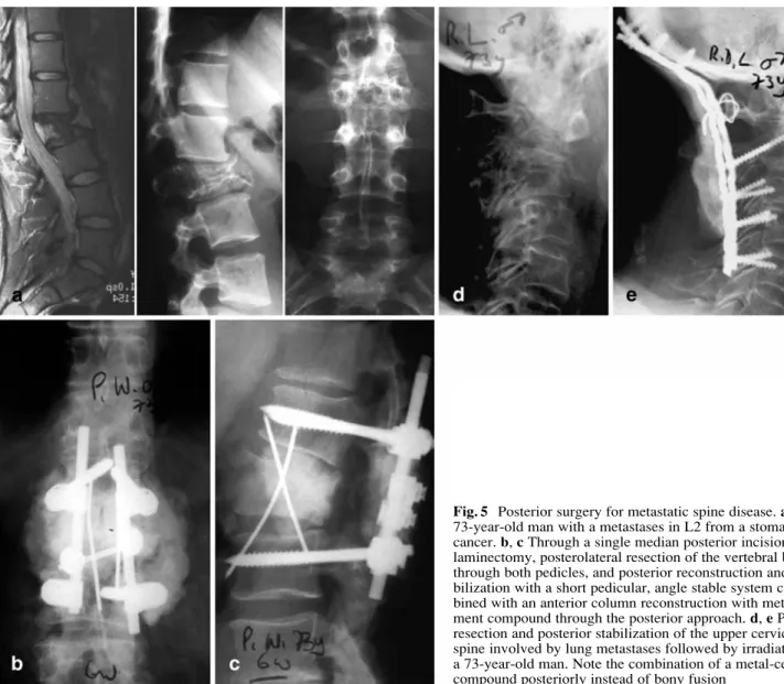
Fig. 5 Posterior surgery for metastatic spine disease. a A 73-year-old man with a metastases in L2 from a stomach cancer. b, c Through a single median posterior incision laminectomy, posterolateral resection of the vertebral body through both pedicles, and posterior reconstruction and sta-bilization with a short pedicular, angle stable system com-bined with an anterior column reconstruction with metal-ce-ment compound through the posterior approach. d, e Partial resection and posterior stabilization of the upper cervical spine involved by lung metastases followed by irradiation in a 73-year-old man. Note the combination of a metal-cement compound posteriorly instead of bony fusion
a combined procedure may be indicated to control the pain mostly due to the instability (Fig. 5). At the occipito-cervical junction a posterior resection and stabilization combined with irradiation is generally sufficient as pallia-tive measure. Some authors have recently enthusiastically advocated minimally invasive technology to approach certain lesions in particular in the vertebral body involve-ment: Vertebroplasty or kyphoplasty as palliative technique may increasingly gain significance in patients with high morbidity index or elevated risk for open surgery [37, 38]. Reconstruction of the anterior column for stability rea-sons as well as realignment of the spine is rarely carried out with autologous bone because the average life ex-pectancy does not justify it, and a possible postoperative irradiation would damage the healing potential of an auto-graft. Today this reconstruction is performed either with a metal-cement compound as in building construction or with the use of metal or ceramic spacers in combination with cement, which may or may not be filled with bone substitutes. Major allograft may be an alternative; how-ever, the biological conditions for its integration are not satisfactory, specifically in the case of adjuvant irradiation and possible chemotherapy.
The stability of a diseased segment after tumor resection can certainly be enhanced by a strong posterior instru-mentation in combination with the anterior reconstruction of the anterior column and is biomechanically superior to a purely anterior reconstruction, even with anterior instru-mentation [32]. The surgeon needs to keep in mind that the major goal of the surgery is to put the patient in a con-dition to be as soon as possible independently mobile without any brace, which is an additional burden in those severely ill and often rather cachectic patients with the po-tential of pressure sores and unease with external fixation devices.
Option of irradiation
The general principles that govern the outcome of treat-ment of patients with malignant tumors of the spine are the same as those for tumors at any other site. First, for pa-tients to be considered cured all tumor cells at the primary, regional, and distant sites must be inactivated or removed. Second, the determinants of probability of success are the anatomical site and size of the tumor and the histopatho-logical type and grade of the tumor. Malignant lesions of the spine are often not respected with secure margins be-cause of the constraints imposed by the proximity of the spinal cord and nerve roots, major vessels (especially along the thoracic column), and organs (e.g., esophagus). An in-tact spine is critical to an individual’s anatomical integrity. Also, the role of radiation therapy for malignant tumors of the spine is often severely limited by the necessity to in-clude the spinal cord in the high-dose region because tu-mor abuts on the dura and/or cord.
The patients in whom symptomatic spinal cord com-pression develops often represent a debilitated and elderly population with considerable surgical risks. Not all pa-tients can safely undergo surgery either anteriorly or pos-terolaterally or even in combination – although mostly not necessary – with appropriate stabilization procedures. Nev-ertheless, a considerable number of these are sufficiently treated by irradiation, either because there are only mini-mal neurological symptoms, or because an aggressive sur-gical approach is deemed inappropriate at initial presenta-tion [12]. The widespread use of MRI of the spine to de-tect metastatic disease in patients with cancer, results in the early diagnosis of epidural metastatic disease, which often is irradiated since not really symptomatic. For many reasons therefore more previously irradiated patients pre-sent to the hospital with symptomatic spinal cord com-pression. The number of major wound complications is high in this population. Recent studies showed that spinal irra-diation before surgical decompression for spinal cord com-pression is associated with a significantly higher major wound complication rate. In addition, preoperative spinal irradiation might adversely affect the surgical outcome [4], (Fig. 3).
Irradiation is an appropriate palliative pain treatment in many patients; however, the indications need to be ratio-nalized if we do not want to deal increasingly with cases after irradiation who need surgery because irradiation did not stop the tumor. Therefore the indications for irradiation in most of the frequent bony and spinal metastases (breast, prostate, lung, colon cancer, and multiple myeloma) are [40]:
– Radiosensitive tumor (malignant lymphoma, myeloma, small-cell lung cancer, seminoma, neuroblastoma, and Ewing’s sarcoma).
– A lesion to the spine which does not compromise the stability or the neurological function of the spinal cord or its roots, but where the leading symptom is pain which is difficult to control by medication alone.
– Mild compression of neurostructures without relevant clinical neurological signs where it can be anticipated that the irradiation will stop the further progression of the tumor, or the patient’s life expectancy is less than 3–6 months.
– Paraplegia more than 24 h.
– Multiple level involvement of the spine where surgery may be useless to control the metastatic disease. In this case the irradiation is a desperate attempt to palliatively influence the bony pain and to delay neurological com-plication depending from the biological/histological char-acteristics of the tumor.
– Disseminated disease with life expectancy less than 3–6 months.
– Tumor involvement for which recalcification of the ir-radiated vertebra can be anticipated from the biological behavior of the tumor more rapidly than a pathological fracture in a weakened vertebra.
– A general condition of the patient with a reduced resis-tance rendering a surgical intervention impossible. Patients who have a relevant symptomatic neurocompres-sion or instability or a failed pain management after irra-diation should no longer undergo irrairra-diation, but a surgi-cal option needs to be evaluated. This shared decision-making process, once again should, be handled in a multi-disciplinary team. Irradiation generally should not be per-formed without a histological diagnosis, with very few exceptions. In all those cases in which the primary tumor is unknown or not sure, a biopsy is recommended of the suspected vertebra either by a posterolateral percutaneous approach or by the pedicle of the patient with a Yamshidi needle of sufficient diameter (≥3 mm), usually in local anesthesia and by image guidance to obtain a proper tis-sue sample allowing a histological diagnosis. This can be a simple hand-guided biopsy under image intensifier or a computer-assisted one.
There is no radiotherapeutic regimen showing consis-tent superiority in the treatment of spinal metastases, al-though multiple treatment protocols have been carried out. Usually 30y in 10 fractions (over 2 weeks) are applied. Other commonly used regimens vary between 8 Gy in a single fraction and 40 Gy in 20 fractions over 4 weeks [12].
Pharmacological options
Here we may consider chemotherapy, bisphosphonates, and in some specific tumors hormonal therapy (breast, prostate, thyroid cancer) and as a general medication steroids such as dexamethasone. This is the most frequently used cortico-steroid despite the fact that in the literature there is no valid comparison of dexamethasone and methylprednisolone [35, 40]. Two dosing regimen are used: the high-dose dexameth-asone regimen comprises an initial bolus of 100 mg with subsequent dose of 96 mg/day. This regimen seems to have only a historical value since significant side effects have been associated with its use. It should be administered only to patients with rapidly progressing neurological deficit. The moderate-dose dexamethasone regimen starts with 10 mg intravenous bolus and continues with 16 mg/day four times daily [40, 52]. This dosage is well tolerated, and it is the regimen of choice in symptomatic patients. No steroids are proposed in nonparetic ambulatory patients.
Recently a new dimension in the treatment of bony metastases has been advocated. Since it is well established that bony metastases in general and of the spine in partic-ular increase treatment costs and may significantly pro-long hospital stay, new means of simple treatment of bony metastases are being evaluated [24]. Bisphosphonates have stood the test of time in the treatment of bony com-plications because they stop the vicious circle of tumor progression and pathological bone turnover. Under the ef-fect of the tumor cells the balance between bone resorp-tion and new bone formaresorp-tion is disturbed; tumor cells seed
in the bone under the attraction of growth factors [43]. There they deliberate mediators which stimulate both the osteoclasts and osteoblasts, which start to turnover the bone in an unphysiological way. Again, growth factors are released which stimulate tumor cells for proliferation. The vicious circle of pathological bone remodeling and tumor progression starts. Subsequently bone quality and bone density diminish. The stability of the bone strongly de-creases. Bisphosphonates show a high affinity to bone and are augmented mainly in locations with high bone turn-over. They are therefore ideal medications to stop the vi-cious circle of bone metastasing and damaging [42]. The most successful medication is pamidronate (second-gen-eration bisphosphonate) which is successful mostly in bony metastases of breast cancer and in osteolysis in multiple myeloma [4]. Zoledronic acid is one of the most recently developed agents and is characterized by an imidazol ring. In animal experiments the effect was 100–850 times better than that with the older pamidronate [30, 39, 44].
The objective clinical success of the bisphosphonate depends significantly on the reduction and delay of skele-tal complications (SREs=pathological fracture, spinal cord compression, need for irradiation or surgery for stabiliza-tion) [19, 22]. It can be anticipated today that the bisphos-phonates have an immediate antitumoral effect. Bisphos-phonate treatment has the goal of diminishing the inci-dence of bony complications, vertebral body fractures, pain, and osteoporosis. The outcome should be determined by the survival time – once a spinal metastasis is detected – in an ambulatory, independent status, where pain is con-trolled, and the patient is not hospitalized. The mean sur-vival time is 14– 18 months depending obviously on the patient’s condition before entering treatment for the spinal problem. Wise et al. [56] report a mean survival time of 15.9 months after surgery for spinal metastasis, whereas Weigel [55] reports a 13.1 months mean survival time with 11.1 months mean time at home after surgery. In our own material of 67 fully documented cases between 1996 and 2001 the mean survival after surgery was 14.2 months (un-published data). Tomita et al. [51] (un-published recently sur-vival times that were longer in cases in which wide or marginal excision was made (38.2 months), with only 7% local tumor recurrence, and the survival time in patients treated with intralesional excision was 21.5 months and 31% local tumor recurrence whereas only in patients with pallia-tive surgery and stabilization the survival was 10.1 months and the local tumor recurrence 28%. They based their sur-gical decision making on a new prognostic scoring sys-tem. Sundaresan et al. [49] reported a mean survival time of 30 months in patients with surgery for solitary metas-tases of the spine and with a survival of 5 years and more in 18% of their cases. Mazel et al. [42] achieved a mean survival rate of 16.7 months in 21 of 35 patients who died and 38.2 months in 14 of 35 patients who were alive at follow-up with a so-called radical excision of tumors of thoracic and cervicothoracic metastases.
These results also suggest a concept of differentiated surgery with more radical options than just palliative sur-gery. The neurological outcome is crucial and depends on the initial neurological deficit before surgery. About one-half of the paraparetic patients at the time of diagnosis re-gain the ability to walk, but only fewer than 5% of pa-tients, who are paraplegic regain ambulation [2]. Postop-erative complications are frequent and are found in 15 – 30% of cases [55, 56].
Wai et al. [54] assessed prospectively the overall qual-ity of life after surgical management of metastatic spine
disease, using a validated global health status quality-of-life instrument (Edmonton Symptoms Assessment Scale). They found the greatest improvement in the domain of pain reduction, but there was also improvement in other domains of quality of life. The clinical results of nonsur-gical treatment for spinal metastases has been presented in a prospective analysis of 101 patients who were treated with radiation therapy and/or chemotherapy. Of these, 66% remained neurologically stable or improved after treatment; 67% had pain relief, and 64% improved func-tionally, which was more related to the general debility than local tumor recurrence [33]. Unfortunately no prospective study has compared nonsurgical and surgical treatment of spinal metastases with clearly defined condi-tions and parameters to allow a differentiated decision about the best solution for the patient. It has also been considered that such a study may be extremely difficult to execute also for ethical reasons.
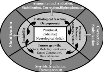
This leaves us with the necessity to assess every pa-tient individually and to weigh the different elements in shared decision making of an interdisciplinary team to-gether with the patient. It is a complex algorithm tailored to the patient’s individual problem and therapeutic options available (Fig. 6). It cannot be emphasized enough that a decision for a conservative treatment, specifically with ir-radiation, should not be taken unless there is a clear un-derstanding that a later surgical option is very improbable. There is no doubt that preoperative irradiation has a sig-nificantly negative effect on surgical outcome [21]. Fig. 6 Decision algorithm of the treatment tailored to the
individ-ual patient’s need and therapeutic option
1. Abdu WA, Provencher M (1998) Pri-mary and metastatic tumors of the cer-vical spine. Spine 23:2767–2776 2. Bartanusz V, Porchet F (2003) Current
strategies in the management of spinal metastatic disease. Swiss Surg 9:55–62 3. Batson OV (1940) The function of the
vertebral veins and their role in the spread of metastases. Am Surg 112: 138–145
4. Berenson JR, Hillner BE, Kyle RA, Anderson K, Lipton A, Yee GC, Bier-mann JS, American Society of Clinical Oncology Bisphosphonates Expert Panel (2002) American Society of Clinical Oncology practice guidelines: the role of bisphosphonates in multiple myeloma. J Clin Oncol 20:19–36 5. Bilsky MH, Boland P, Lis E, Raizer JJ,
Healey JH (2000) Single-stage postero-lateral transpedicle approach for spondylectomy, epidural decompres-sion, and circumferential fusion of spi-nal metastases. Spine 25:2240–2250
6. Bilsky MH, Shannon FJ, Sheppard S, Prabhu V, Boland PJ (2002) Diagnosis and management of a metastatic tumor in the atlantoaxial spine. Spine 27: 1062–1069
7. Boriani S, Chevalley F, Weinstein JN, Biagini R, Campanacci L, De Iure F, Piccill P (1996) Chordoma of the spine above the sacrum. Treatment and out-come in 21 cases. Spine 21:1569–1577 8. Boriani S, Biagini R, De Iure F,
Bertoni F, Malaguti MC, Di Fiore M, Zanoni A (1996) En bloc resections of bone tumors of the thoracolumbar spine. A preliminary report on 29 pa-tients. Spine 21:1927–1931
9. Boriani S, Weinstein JN, Biagini R (1997) Primary bone tumors of the spine. Terminology and surgical stag-ing. Spine 22:1036–1044
10. Bridwell KH, Jenny AB, Saul T, Rich KM, Grubb RL (1988) Posterior seg-mental spinal instrumentation (PSSI) with posterolateral decompression and debulking for metastatic thoracic and lumbar spine disease. Limitations of the technique. Spine 13:1383–1394
11. Coman DR, DeLong RP (1951) The role of the vertebral venous system in the metastasis of cancer into the spinal cord: experiments with tumor cell sus-pensions in rats and rabbits. Cancer 4: 610–618
12. DeLaney TF, Suit HD (2000) Treat-ment of spine tumors: radiation ther-apy. Curr Opin Orthop 11:502–507 13. DeWald RL, Bridwell KH, Prodromas
C, Rodts MF (1985) Reconstructive spinal surgery as palliation for metasta-tic malignancies of the spine. Spine 10: 21–26
14. Doppman JL, Girton RT (1976) Angio-graphic study of the effect of laminec-tomy in the presence of acute anterior epidural masses. J Neurosurg 45:195– 202
15. Dürr HR, Wegener B, Krödel A, Müller PE, Jansson V, Refior HJ (2002) Mul-tiple myeloma: surgery of the spine. Spine 27:320–326
16. Enkaoua EA, Doursounian L, Chatel-lier G, Mabesoone F, Aimard T, Sail-lant G (1997) Vertebral metastases. A critical appreciation of the preopera-tive prognostic Tokuhashi Score in a series of 71 cases. Spine 22:2293–2298 17. Enneking WF, Spanier SS, Goodman
MA (1980) A system for surgical stag-ing of musculoskeletal sarcoma. Clin Orthop 153:106–120
18. Findlay GFG (1984) Adverse effects of the management of malignant spinal cord compression. J Neurol Neurosurg Psychiatry 47:761–768
19. Fleisch H (2000) Bisphosphonates in bone disease. From the laboratory to the patient. Academic, San Diego 20. Gerszten CP, Welch W (2000) Current
surgical management of metastatic spi-nal disease. Oncology 14:1013–1024 21. Ghogawala Z, Mansfield FL, Borges LF (2001) Spinal radiation before sur-gical decompression adversely affects outcomes of surgery for symptomatic metastatic spinal cord compression. Spine 26:818–824
22. Green JR, Müller K, Jaeggi KA (1994) Preclinical pharmacology of CGP 42’446, a new, potent, heterocyclic bis-phosphonate compound. J Bone Miner Res 9:745–751
23. Greenlee RT, Murray T, Bolden S, Wingo PA (2000) Cancer statistics, 2000. CA Cancer J Clin 50:7–33 24. Groot MT et al (2003) Costs of
prostate cancer metastasis to the bone in The Netherlands. Eur Urol 43:226– 232
25. Grosman R. Rouchal M, Chalupka R (2000) The long-term results of surgi-cal management of spine metastatic tu-mours from breast cancer. Scripta Med (Brno) 73:169–172
26. Grossman RI, Yousem DM (1994) Neuroradiology. The requisites. Mosby Year Book, St. Louis, pp 491–495 27. Gunterberg B (1997) Point of view: a
system for surgical staging and man-agement of spine tumors. A clinical outcome study of giant cell tumors of the spine. Spine 22:1783
28. Hart RA, Boriani S, Biagini R, Currier B, Weinstein JN (1997) A system for surgical staging and management of spine tumors. A clinical outcome study of giant cell tumors of the spine. Spine 22:1773–1782
29. Heiss JD, Papavassiliou E, Merrill M et al (1996) Mechanism of dexametha-sone suppression of brain tumor-asso-ciated vascular permeability in rats. J Clin Invest 98:1400–1408
30. Hortobagyi GN, Theriault RL, Lipton A et al (1998) Long-term prevention of skeletal complications of metastatic breast cancer with pamidronate. J Clin Oncol 16:2038–2044
31. Jenis LG, Dunn EJ, An HS (1999) Metastatic disease of the cervical spine. Clin Orthop 359:89–103 32. Kanayama M, Cunningham BW,
Abumi K, Kaneda K, McAfee PC (1999) Biomechanical analysis of ante-rior versus circumferential spinal re-construction for various anatomic stages of tumor lesions. Spine 24: 445–450
33. Katagiri H, Takahashi M, Inagaki J, Kobayashi H, Sugiura H, Yamamura S, Iwata H (1998) Clinical results of non-surgical treatment for spinal metas-tases. Int J Radiat Oncol Biol Phys 42: 1127–1132
34. Khan DC, Malhotra S, Stevens RE, Steinfeld AD (1999) Radiotherapy for the treatment of giant cell tumor of the spine: a report of six cases and review of the literature. Cancer Invest 17:110– 113
35. Koehler PJ (1995) Use of corticoste-roids in neuro-oncology. Anticancer Drugs 6:19–33
36. Landis SH et al (1999) Global cancer statistics. CA Cancer J Clin 49:33–64 37. Lemke DM, Hacein-Bey L (2003)
Metastatic compression fractures – ver-tebroplasty for pain control. J Neurosci Nurs 35:50–55
38. Lieberman IH, Dudeney S, Reinhardt MK et al (2001) The initial outcome and efficacy of ‘kyphoplasty’ in the treatment of painful osteoporotic verte-bral compression fractures. Spine 26: 1631–1636
39. Lipton A, Theriault RL, Hortobagyi GN et al (2000) Pamidronate prevents skeletal complications and is an effec-tive palliaeffec-tive treatment in women with breast carcinoma and osteolytic bone metastases. Long-term follow-up of two randomized, placebo-controlled trials. Cancer 88:1082–1090 40. Loblaw DA, Laperriere NJ (1998)
Emergency treatment of malignant ex-tradural spinal cord compression: an evidence-based guideline. J Clin Oncol 16:1613–1624
41. Marchesi D, Arlet V, Aebi M (2003) Treatment of spinal metastases by pos-terolateral decompression and pedicle screw fixation. Spine (in press) 42. Mazel C, Grunenwald D, Laudrin P,
Marmorat JL (2003) Radical excision in the management of thoracic and cer-vicothoracic tumors involving the spine: results in a series of 36 cases. Spine 28:782–792
43. Mundy GR (1999) Bisphosphonates as cancer drugs. Hosp Pract (Off Ed) 34: 81–94
44. Rosen LS, Harland SJ, Oosterlinck W (2002) Broad clinical activity of zole-dronic acid in osteolytic to osteoblastic bone lesions in patients with a broad range of solid tumors. Am J Clin On-col 25:19–24
45. Schick U, Marquardt G, Lorenz R (2001) Intradural and extradural spinal metastases. Neurosurg Rev 24:1–5 46. Siegal T, Siegal T (1989) Current
con-siderations in the management of neo-plastic spinal cord compression. Spine 14:223–228
47. Smith MR et al (2003) Zometa in-creases bone mineral density in men receiving androgen deprivation therapy for nonmetastatic prostate cancer. J Urol 169:2008–2012
48. Sundaresan N, Galicich JH, Lane JM, Bains MS, McCormack P (1985) Treatment of neoplastic epidural cord compression by vertebral body resec-tion and stabilizaresec-tion. J Neurosurg 63: 676–684
49. Sundaresan N, Rothmann A, Manhart K, Kelliher K (2002) Surgery for soli-tary metastases of the spine. Rationale and results of treatment. Spine 27: 1802–1806
50. Tokuhashi Y, Matsuzaki H, Toriyama S. Kawano H, Ohsaka S (1990) Scor-ing system for the preoperative evalua-tion of metastatic spine tumor progno-sis. Spine 15:1110–1113
51. Tomita K, Kawahara N, Kobayashi T, Yoshida A, Murakami H, Akamaru T (2001) Surgical strategy for spinal metastases. Spine 26:298–306
52. Vecht CJ, Haaxma-Reiche H, van Putten WLJ, de Visser M, Vries EP, Twijnstra A (1989) Initial bolus of conventional versus high-dose dexamethasone in metastatic spinal cord compression. Neurology 39:1255–1257
53. Vinholes J et al (1996) Effects of bone metastases on bone metabolism: impli-cations for diagnosis, imaging and as-sessment of response to cancer treat-ment. Cancer Treat Rev 22:289–331 54. Wai EK, Finkelstein JA, Tangente RP,
Holden L, Chow E, Ford M, Yee A (2003) Quality of life in surgical treat-ment of metastatic spine disease. Spine 28:508–512
55. Weigel B, Maghsudi M, Neumann C, Kretschmer R, Müller FJ, Nerlich M (1999) Surgical management of symp-tomatic spinal metastases. Postopera-tive outcome and quality of life. Spine 24:2240–2246
56. Wise JJ, Fischgrund JSA, Herkowitz HN, Mongomery D, Kurz LT (1999) Complications, survival rates, and risk factors of surgery for metastatic dis-ease of the spine. Spine 24:1943–1951 57. York JE, Berk RH, Fuller GN, Rao JS,
Abi-Said D, Wildrick DM, Gokaslan ZL (1999) Chondrosarcoma of the spine: 1954 to 1997. J Neurosurg 90 [Suppl 1]:73–78
58. Young RF, Post EM, King GA (1980) Treatment of spinal epidural metas-tases: randomized prospective compar-ison of laminectomy and radiotherapy. J Neurosurg 53:741–748
![Fig. 3 Surgery ideally should be carried out before irradiation [1].](https://thumb-eu.123doks.com/thumbv2/123doknet/14834489.621495/6.892.150.378.802.961/fig-surgery-ideally-carried-irradiation.webp)