Psychophysiology 2019 Mar 2:e13354 (advanced online publication) doi: 10.1111/psyp.13354.
Structural brain differences predict early traumatic memory processing
Geraldine Gvozdanovic,1,2*, Philipp Stämpfli1,5,Erich Seifritz1,4 & Björn Rasch3,4*
1 Department of Psychiatry, Psychotherapy and Psychosomatics, Psychiatric Hospital,
University of Zürich, Zürich Switzerland
2 Institute of Psychology, University of Zürich, Zürich, Switzerland
3 Department of Psychology, University of Fribourg, Fribourg, Switzerland
4 Competence Center of Sleep & Health Zurich, University of Zürich, Switzerland
5 MR-Center of the Department of Psychiatry, Psychotherapy and Psychosomatics and
the Department of Child and Adolescent Psychiatry, Psychiatric Hospital of the University of Zürich, Zürich, Switzerland.
Key Words: Structural MRI, voxel-based morphometry (VBM), trauma film paradigm, early
intrusive memories, PTSD
Running head: Brain structures predict early traumatic memories
*Corresponding authors: Geraldine Gvozdanovic, University of Zurich; [email protected], and Björn Rasch, University of Fribourg; [email protected]
Abstract
Intrusive memories are a key symptom of posttraumatic stress disorder (PTSD). They emerge early after trauma exposure and are predictive for PTSD development. There is a high
relevance in evaluating the neurobiological mechanisms of early stages of intrusive symptom development to provide a further understanding of PTSD.
In the present study we explore structural differences in healthy young females preceding experimental trauma exposure and their relationship to early intrusive memory development using a traumatic film paradigm. With voxel-based morphometry we demonstrate that smaller insular volume was associated with an increased number of early intrusive film memories. Moreover, larger lingual gyrus/cerebellar and inferior frontal gyrus/precentral gyrus volumes were also related to an increased number of early intrusive film memories. Our results identify unique brain areas associated with early experimental trauma memory processing and highlight the necessity of evaluating early symptom stages relevant for personalized PTSD prevention and treatment.
1. Introduction
Posttraumatic stress disorder (PTSD) is a psychiatric condition that develops after a traumatic event and is associated with intrusive and avoidant symptoms, increases in arousal, and negative alterations in cognition and mood (APA, 2013). Especially intrusive memories are of central importance in PTSD. They are perceived as highly disturbing and emerge already in early stages after trauma exposure. Most importantly, they are predictive for the
development of PTSD (APA, 2013; Brewin, 2015; Elbert & Schauer, 2002; Michael, Ehlers, Halligan, & Clark, 2005).
From a neurobiological perspective, PTSD is associated with alterations in brain function and structure. Functionally, altered activity has been mostly found in the amygdala, ventromedial prefrontal cortex, anterior cingulate cortex (ACC) and insula during trauma related stimuli (Pitman et al., 2012). Structurally, consistent evidence suggests that hippocampal volume is reduced in PTSD compared to healthy controls and traumatized subjects without PTSD (Karl et al., 2006; Kitayama, Vaccarino, Kutner, Weiss, & Bremner, 2005). In addition, reduced hippocampal volumes can be found bilaterally (Smith, 2005) and have been proposed to predict PTSD symptom severity (Pitman et al., 2012). Furthermore, volume reductions in the amygdala are associated with PTSD, specifically on the left side (Karl et al., 2006). Insula reductions of the surface area have been found after childhood maltreatment ((Kelly et al., 2013). Most pertinent to our present study, reductions in the surface area of the insula predicted the later development of PTSD (Hu et al., 2018). Further structural alterations in PTSD patients’ samples are reported in the anterior cingulate cortex (ACC) (Kasai et al., 2008; Kitayama, Quinn, & Bremner, 2006), the ventromedial prefrontal cortex (vmPFC) (Carrion, Weems, Richert, Hoffman, & Reiss, 2010), and the orbitofrontal cortex (Pitman et al., 2012; Sekiguchi et al., 2013).
Structural brain differences have been discussed both as a consequence of extreme stress damaging the brain (state), and also as a vulnerability factor (trait). Toxic effects can emerge on hippocampal cells due to glucocorticoids (Sapolsky, Uno, Rebert, & Finch, 1990) and stress in general (Bremner, 1999). Stress has the capacity of altering and reorganizing function and structure of several biological systems (Elbert & Schauer, 2014), the genome (Radtke et al., 2011), and the functional (Lanius et al., 2001; Lanius et al., 2004) and
structural (Karl et al., 2006) integrity of the brain. Thus, structural brain alterations in PTSD patients could be viewed as a consequence of exposure to an extreme stressor followed by increases in persistent stress.
At the same time, brain atrophies might also serve as a vulnerability factor for individual differences in PTSD development after exposure to extreme stress. For example, a smaller hippocampal volume in monozygotic twins was a predisposing factor for the development of combat associated PTSD (Gilbertson et al., 2002). Therefore, individual differences in brain structures in healthy subjects might be an important predisposition factor for later PTSD development.
To further examine the relationship between brain structure and PTSD symptoms, here we investigate the association between intrusive memories and volume of specific brain areas in healthy subjects. We employed a highly controlled and well established setting of an
experimental medicine model (Holmes & Bourne, 2008). Healthy subjects watched a trauma film after structural scan measurements, and symptoms of intrusions had to be reported in a diary across the following week. We conducted voxel-based morphometry (VBM) of whole brain magnetic resonance imaging (MRI) data as an unbiased approach on whole brain level to identify structural differences related to early intrusive symptoms.
We used this study design specifically to focus on a very early time window after
highly intrusive symptoms emerge (Bremner, 2005). Evaluating this early phase is relevant to identify predictive variables to enable preventive and therapeutic measures in PTSD,
especially for core symptoms. We hypothesize that intrusive symptoms during the earliest stages after experimental trauma exposure are associated with reduced volumes in the hippocampus, the amygdala and the insula as these regions are tightly linked to memory processing, fear conditioning, and intrusive memories (Dolan & Vuilleumier, 2003; Liberzon & Abelson, 2016; Osuch et al., 2001; Phelps et al., 2001).
2. Method 2.1. Subjects
Seventy-four healthy female subjects (mean age 23.4 years, range 18 - 36 years) went through a general screening on past medical and psychiatric disorders and were excluded depending on their medical record. The experience of a “real-life” traumatic event in the past was taken as an exclusion criterion. Additionally, subjects were screened for standard MRI compatibility. Subjects signed their written informed consent after the procedure of the study had been explained in detail. The study protocol was in line with the Declaration of Helsinki and approved by a local ethical review board. Subjects were reimbursed for their
participation (CHF 25/h).
2.2. Experimental procedure
Subjects arrived at the imaging center, they were informed about the procedure and signed their informed consent. After entering the scanner, all subjects went through a structural T1 scan and subsequently watched a trauma film. The high resolution structural T1 scan lasted 3 min and 40 sec while the trauma film had a duration of 14 min. After the experiment, subjects received an intrusion diary with a detailed explanation for filling in the intrusion diary for the following seven days (Figure 1).
Research on functional data using the trauma film paradigm has been published in previous work (Gvozdanovic, Stampfli, Seifritz, & Rasch, 2017).
2.3. Trauma film paradigm:
Subjects watched a trauma film in accordance with the trauma film paradigm (Holmes & Bourne, 2008). The trauma film of this study displayed rape and further physical assault (film called “Irreversible”, (Noé, 2002)) and was associated with strong emotional and
physiological reactions in previous research (Weidmann, Conradi, Groger, Fehm, & Fydrich, 2009)
After being originally developed in the 60s (Horowitz, 1969; Lazarus, 1964), the trauma film paradigm has been validated in over 70 studies only in recent years (James et al., 2016). The trauma film paradigm is per definition an experimental medicine model. Experimental medicine models try to model deviant mechanisms in non-patient samples in order to
evaluate processes involved in the development of clinically relevant symptoms (James et al., 2016). In this case, the experimental medicine model evaluates behavior in non-patient groups to examine factors resulting in PTSD (James et al., 2016). After a trauma film, i.e., an experimental trauma, reactions similar to trauma in real life have been systematically
observed, including hallmark symptoms of PTSD. Changes in reactions can be found in physiological arousal, intrusive memories, negative cognitions and mood (APA, 2013; Butler, 1995; Holmes, Brewin, & Hennessy, 2004; James et al., 2016; Weidmann et al., 2009). Symptoms emerging after an experimental trauma last solely for hours or days and diminish within one week (Holmes et al., 2004; James et al., 2016).
In our study the traumatic film depicted a rape scene, displaying characteristics of witnessing a traumatic event. Diagnosis criteria for PTSD include both an immediate exposure to a traumatic event and also witnessing trauma (APA, 2013). To examine early PTSD-like symptoms, the trauma film paradigm is an ideal model to bridge the gap between non-clinical and clinical samples based on its strongly controllable setting (Clark & Mackay, 2015).
2.4. Intrusion diary and questionnaires
The intrusion diary had to be completed at home and consisted of a seven-day diary. Subjects were instructed on how to complete the intrusion diary in detail after the scanning session. Intrusive memories that emerged had to be filled in the intrusion diary. Describing the content in detail, making notes on perceived vividness and distress, and writing down further intrusive characteristics for every single memory were part of the subjects’ daily task for seven days. In order to analyze the intrusion diaries, the sum of reported intrusions per week was used. Scoring criteria for the diagnosis of intrusions were ratings on distress, vividness and the sudden occurrence of the reported memories.
Participants also filled in the following questionnaires: state trait anxiety inventory (STAI) for state and trait, Becks depression inventory (BDI), a questionnaire on the perception of the film including the following items: film perception in general, stress, vividness, anger, sadness, anxiety, disgust, helplessness, wakefulness, and concentration, resulting in a total score of the film perception questionnaire.
2.5. MRI data acquisition
Measurements were performed on a Philips Achieva 3.0 Tesla TX whole-body magnetic resonance scanner (Philips Healthcare, Best, The Netherlands), equipped with a thirty-two channel receive-only head coil array. High resolution structural 3D T1-weighted anatomical data were acquired via a 3D magnetization-prepared rapid gradient-echo sequence (MP-RAGE) with the following parameters: voxel size = 1.0x1.0x1.0 mm3, time between two inversion pulses = 2170 ms, inversion time = 1000 ms, inter-echo delay = 9.3 ms, flip angle = 8°, field of view = 240x240 mm2, 160 sagittal slices. The trauma film was displayed on
MR-compatible video goggles (Resonance Technology, Northridge, USA). Moreover, headphones especially designed for MR measurements (MR confon GmbH, Magdeburg, Germany) were used by subjects during the film presentation.
2.6. MRI data analyses for voxel-based morphometry (VBM)
Standard image data preparation, preprocessing, and statistical analysis were performed using statistical parametric mapping SPM12 (www.fil.ion.ucl.ac.uk/spm) in Matlab 2017b.
Structural data preprocessing included segmentation of the structural data into grey matter, white matter and CSF. An additional preprocessing step was performed using an MR bias correction. With the use of the Dartel algorithm (Ashburner, 2007) in SPM12 images were spatially normalized to standard Montreal Neurological Institute (MNI) space and voxels were resliced to a voxel resolution of 1,5 mm x 1,5 mm x 1,5 mm. Spatially normalized and segmented images were modulated with the Jacobian determinants from the transformation with the aim of adjusting for the resulting volume changes (Ridgway et al., 2008). Smoothing was done using a full width at half maximum Gaussian kernel (FWHM) of 4 mm.
For statistical analyses on group level, multiple regression analyses were performed. As a regressor of interest the sum of individual intrusions reported in the intrusion diary during the subsequent week after the experiment was entered into the design matrix. In order to deal with individual differences in brain sizes, global normalization with respect to whole brain volume was taken into account and an overall grand mean scaling was computed. Statistical significance was set at pFWE.cluster < 0.05, with a cluster defining threshold (CDT) of p = 0.001 for whole brain analyses (Eklund, Nichols, & Knutsson, 2016).
Further region of interest (ROI) based VBM analyses were conducted in order to evaluate ROIs that were previously reported in PTSD (i.e. hippocampus, amygdala, vmPFC and orbitofrontal cortex) and were preprocessed using the above-mentioned preprocessing steps.
Masks were taken from predefined automatic anatomical labelling atlas (AAL, (Rolls, Joliot, & Tzourio-Mazoyer, 2015; Tzourio-Mazoyer et al., 2002) and were resliced to match the preprocessed Dartel images (1.5 mm x 1.5 mm x 1.5 mm). In order to also look at subject specific parcellations, we additionally preprocessed our images with VBM and used the CAT 12 toolbox (Computational Anatomy Toolbox 12), implemented in SPM12. Images were segmented into gray matter (GM), white matter (WM), and cerebrospinal fluid (CSF) by using the default settings. Images were spatially normalized with DARTEL and smoothed with a FWHM of 4 mm.
By multiplying the preprocessed structural images of each subject with the ROI masks, a calculation of the total ROI volume was made in milliliters. The extracted data was then correlated with intrusive symptoms and questionnaires on the perception of the film in SPSS24 using Spearman correlations.
3. Results
3.1. Behavioral Results: 3.1.1. Anxiety and Intrusions
The reported intrusion data contained a minimum intrusion of minintrusion = 0 and a maximum
sum of maxintrusions = 13 during the week after exposure to the experimental-trauma film.
Mean number of intrusions was μ = 3.23 and a standard deviation of SD = 2.74.
The sample displayed no depressed symptoms and revealed trait anxiety under the cut-off point of clinically relevant anxiety symptoms during the experiment (Julian, 2011). As expected, anxiety level significantly increased due to the trauma film. Paired t tests between pre and post film STAI state measures revealed that there was a significant increase in anxiety after the experimental-trauma film compared to before the film (mean anxiety pre film μ = 36.71; mean post film anxiety μ = 46.81; t = -6.5; p < 0.001) (Table 1).
At the end of the experiment, anxiety measures were still elevated and significantly increased compared to baseline anxiety (mean anxiety pre experiment μ = 36.71; mean anxiety post experiment μ = 40.48; t = -2.7; p < 0.01), see Table 1. Therefore, after the trauma film, participants reached a clinically relevant score (Julian, 2011).
Moreover, Spearman correlations revealed that the higher the anxiety measured after the film, the higher the perceived subjective distress during the intrusions in the subsequent week (r = .261; p < 0.05).
-- insert table 1 about here--
3.2. Structural MRI Results:
In general, there was no correlation between intrusions and extracted total grey matter volume (p > 0.1).
3.2.2. Whole-brain VBM: Negative intrusion contrast
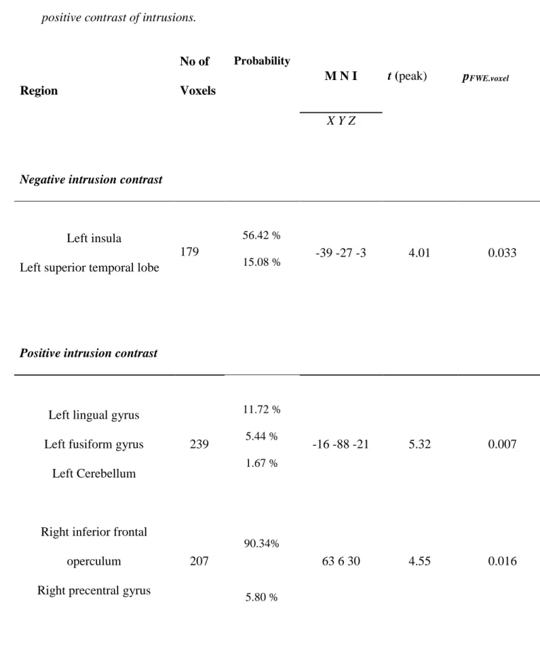
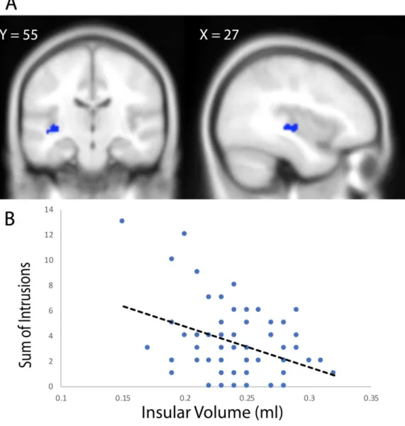
Multiple regression analyses on a whole brain level using VBM revealed that the development of intrusive memories within one week after the experimental trauma was associated with smaller volumetry in the left insula and the left superior temporal lobe (Fpeak = 4.01), both pFWE.cluster < 0.05, CDT of p = 0.001, see Figure 2 and Table 2. Thus, more reported intrusions were associated with less insular volume. For the insula, we had strong a priori and directional hypothesis on smaller insula being associated with an increased number of intrusions.
-- insert figure 2 about here --
3.2.3. Whole-brain VBM: Positive intrusion contrast
Intrusive memories analyzed from the diaries were positively associated with larger structural volumetry in left cerebellum, left lingual – and left fusiform gyrus (Fpeak = 5.32), with
pFWE.cluster < 0.05, CDT of p = 0.001, see Figure 3 and Table 2. Moreover, increases in volumetry were found in the right precentral gyrus and the right inferior frontal operculum (Fpeak = 4.55), with pFWE.cluster < 0.05, CDT of p = 0.001. As these results were of an
explanatory, bidirectional nature, we have adjusted our pFWE values for two sided tests, with a new p value threshold of pFWE < 0.025. Results reveal that larger volumetry in the lingual
gyrus/cerebellum and inferior frontal gyrus/precentral gyrus is associated with more intrusions.
--insert table 2 about here—
-- insert figure 3 about here --
3.2.4. Brain behavior correlates
An additional questionnaire on the perception of the film (items: film perception in general, stress, vividness, anger, sadness, anxiety, disgust, helplessness, wakefulness and
concentration) revealed that the total score on the perception of the film negatively correlated with our reported structural differences in the insula (r = -0.266, p < 0.05). This correlation was of the same directionality as the association between the insula and the intrusions. Moreover, the film perception also positively correlated with intrusions (r = 0.219; p < 0.05. Additional detailed analyses on the intrusions revealed that the insula (r = - 0.280), inferior frontal gyrus/precentral gyrus (r = 0.232), and lingual gyrus/cerebellum (r = 0.212)
significantly correlated with subjective distress of the intrusions (p < 0.05). For vividness, only the insula (r = - 0.268) and inferior frontal gyrus/precentral gyrus (r = 0.239) were significantly correlated (p < 0.05).
In order to control for age and anxiety, we entered these variables as a covariate to the data. Partial correlation analyses revealed that age and anxiety trait measured by the STAI did not have a significant effect on the correlation between intrusion and the insula, inferior frontal gyrus/precentral gyrus, and lingual gyrus/cerebellum (all, p < 0.05).
In order to additionally evaluate structural regions of interest that were reported in PTSD patients, we separately calculated the volumes per individual for the hippocampus, amygdala, anterior cingulate cortex, and ventromedial prefrontal cortex and orbitofrontal cortex. We found no significant correlation between the structural ROIs reported in PTSD and early intrusive symptoms, all p > 0.1.
Results on subject specific parcellations (CAT12) revealed no significant correlation between the hippocampus, amygdala, orbitofrontal cortex, medial prefrontal cortex, and the anterior cingulate cortex.
4. Discussion
The present study evaluates structural differences associated with early intrusive memories. Structural T1 scans were acquired before experimental trauma exposure and healthy subjects had to fill in an intrusion diary during the subsequent week. Our results reveal that a higher number of early intrusive memories was associated with a smaller volume in the left insula. Moreover, larger volumes in the left lingual gyrus/cerebellum and right inferior frontal gyrus/ precentral gyrus were positively associated with the number of intrusions. We found no correlation between previously reported ROIs in PTSD and early traumatic memory
processing. Overall, our results display specific structural brain volume changes that predict at an early stage the number of later intrusive memories.
The smaller volume of the insula and its association with intrusive memories is consistent with previous data in PTSD patients, where inverse relationships between insular volume and PTSD symptoms were found (Chen et al., 2006; Hu et al., 2018; Kelly et al., 2013; Meng, 2014; Perez et al., 2017). Also on a functional level, differences in insular activity have been associated with PTSD in general (Kolassa & Elbert, 2007; Pitman et al., 2012), and with the development of PTSD (Liberzon & Martis, 2006), and fear processing (Phelps et al., 2001). Importantly, functional differences in the insula have been associated with intrusive
memories (Osuch et al., 2001). Meta analyses in healthy subjects revealed that the insula is functionally associated with emotional recall and emotional tasks with cognitive demands (Phan, Wager, Taylor, & Liberzon, 2002). Increased activity in the insula was also found during aversive conditioning in healthy subjects (Buchel, Morris, Dolan, & Friston, 1998; Marschner, Kalisch, Vervliet, Vansteenwegen, & Buchel, 2008).
As we additionally found a significant correlation between structural differences in the insula and the perception of the film, we could conclude that the volumetric changes in the insula
are related to specific differences in traumatic memory and not only to memory processes in general. Functional changes in the insula have not been exclusively associated with PTSD patients, but were also found in other anxiety disorders (Pitman et al., 2012). The finding from our study might therefore indicate that the insula is associated with a general
susceptibility for negative intrusive memories rather than a disorder-specific change in brain volume.
Our further findings on a larger volume of the lingual gyrus/cerebellum/fusiform gyrus and its relation to intrusions highlights the importance of these regions in PTSD like symptoms. Altered grey matter volume in the lingual gyrus has been associated with chronic PTSD (Tan et al., 2013). In healthy subjects, the fusiform and the lingual gyrus have been associated with processing of uncertain cues (Zhang et al., 2016) and emotions (Fairhall & Ishai, 2007). Altered surface areas in the lingual and fusiform gyri have been associated with anxiety and depression symptomology (Couvy-Duchesne et al., 2018). Interestingly, while the cerebellum was previously mainly associated with planning and executing motor functions, this brain structure has recently been increasingly connected to neuropsychiatric symptoms (Buckner, 2013). A recent study by (Rabellino, Densmore, Theberge, McKinnon, & Lanius, 2018) on resting state functional connectivity in PTSD and its dissociative subtype found that different subregions of the cerebellum are of relevance in the psychopathology of PTSD. As the cerebellum is also involved in emotion regulation (Schutter & van Honk, 2009), a dysregulation of emotional memory processing due to an enlarged cerebellum might be fundamental for intrusive symptom development. In addition, on a structural level, the cerebellum has been repeatedly connected to PTSD. Specifically, enlargements of the
cerebellum have been associated with combat related PTSD (Sussman, Pang, Jetly, Dunkley, & Taylor, 2016) and rape victims with PTSD (Sui et al., 2010). Childhood-trauma related pediatric PTSD has also been related to structural changes in the cerebellum (De Bellis &
Kuchibhatla, 2006). Thus, our findings support the notion that the cerebellum seems to be an underestimated region of relevance in traumatic memory processing, with effects occurring already at early symptom stages.
The precentral gyrus, as also found in our results, is associated with motor functions (Semmes & Chow, 1955). Interestingly, the amydala is known for its projections to motor cortices including the precentral gyrus (Grezes, Valabregue, Gholipour, & Chevallier, 2014), a connection which has been associated with emotional modulations (Rizzo et al., 2018). Changes in functional connectivity between the precental gyrus and PTSD relevant regions have been reported in a trauma film group compared to a control film group during intrusive film picture presentation (Gvozdanovic et al., 2017). Thus, potentially due to the limbic-motor interface properties of the precentral gyrus and its associated wide range brain connections (Rizzo et al., 2018), changes in the precentral gyrus seem to be of relevance in intrusive symptom development. Our findings in the inferior frontal gyrus give indications for a potential malfunctioning of inhibition and altered salience processing, two potentially important factors in intrusion development. Activity in the inferior frontal gyrus has been associated with proactive inhibition in functional MRI research in PTSD patients (van Rooij et al., 2014). In healthy participants it has been associated with detecting salient stimuli and response inhibition (Hampshire, Chamberlain, Monti, Duncan, & Owen, 2010).
In order to evaluate further specific structural ROIs reported in previous PTSD research, we also conducted volumetric ROI analyses. We found no significant difference between
previously reported ROIs and early traumatic memory processing. This demonstrates that our reported whole brain VBM findings are unique to an early stage and that it is of high
relevance to examine different stages after experimental trauma exposure to provide a detailed understanding of structural brain processes involved in PTSD.
Interestingly, we found no significant differences in hippocampal volume. A possible explanation might be that structural changes in the hippocampus are not specifically associated with early traumatic memory processing or alternatively that atrophies in hippocampal volumes are a consequence of extreme stress exposure rather than a
predisposition factor. As the hippocampus is important for contextualization (Liberzon & Abelson, 2016), it might also be that the hippocampal volume is rather related to other PTSD symptom clusters that are primarily involved in putting traumatic memories in to context. Concerning the debate on whether structural differences in general emerge as a consequence of trauma or serve as a vulnerability factor, our results rather suggest that volumetric changes serve as a vulnerability factor for development of early intrusive symptoms and potentially of PTSD. However, this might only account for our reported structural regions in the insula, lingual gyrus/cerebellum and inferior frontal gyrus/precentral gyrus. Nevertheless, we have to emphasize that our study only entails correlational analyses of structural differences
comparing the state before experimental trauma exposure with early intrusive memories development.
It is also important to highlight that PTSD is a more complex disorder which cannot be entirely depicted by an experimental medicine model. The trauma film paradigm primarily focuses on early intrusive memory processing. Therefore, early differences in hippocampal volume might nevertheless be found in patient samples. A disadvantage of measuring only patient samples, however, is that a high comorbidity with other psychiatric disorders might be responsible for inconsistent findings (Eckart et al., 2012).
Our reported findings could not have been related to previous traumatization which may be part of a building block effect (Neuner et al., 2004), as past traumatization was an exclusion criterion in our study. Exposure to childhood trauma is also essential for PTSD development
(Teicher, 2010). We did not formally examine childhood trauma in detail through additional assessment of further questionnaires.
A limitation to this study is that we have only measured intrusions during the subsequent week after experimental-trauma film exposure as according to the trauma film paradigm. Future research should also focus on intrusions at times beyond the first week. Moreover, further information concerning type and other characteristics of the intrusive memories should be additionally assessed in the diaries.
As our sample of healthy subjects only included females, this further limits our findings. In PTSD patients, up to 50% of females develop a PTSD after sexual assault (Chivers-Wilson, 2006). Moreover, female and male brain structures differ in both the insula, inferior frontal gyrus/precentral gyrus and lingual gyrus/cerebellum (Ruigrok et al., 2014). In this sense, the generalization of our findings might be further limited.
As an additional limitation, only an experimental medicine model was deployed in healthy subjects, and the applied experimental trauma mainly includes characteristics of witnessing a trauma. We expect PTSD patient samples to display further and stronger structural
differences associated with intrusive symptoms developed after “real” trauma exposure. Thus, our study has to be transferred to a sample with real life traumatized subjects to both replicate our current findings and to evaluate whether structural differences associated with early traumatic memory processing are of predictive value for PTSD development.
In sum, within the complexity of previous findings our study provides evidence that the insula, the lingual gyrus/cerebellum and the inferior frontal gyrus/precentral gyrus are correlated with early intrusion development. These findings provide new insights in the relevance of structural differences in early intrusive symptoms and open opportunities for precise diagnosis, and for personalized treatment and prevention.
5. Author note
The project was supported by the European Research Council (ERC) under the European Union's Horizon 2020 research and innovation program (grant agreement n° 677875), the Forschungskredit of the University of Zuerich (grant no FK-16-074) and the Clinical Research Priority Program ‘Sleep and Health’. There are no conflicts of interests.
6. References
APA, A. P. A. (2013). Diagnostic and statistical manual of mental disorders (5th ed.).
Washington, DC. https://doi.org/10.1176/appi.books.9780890425596
Ashburner, J. (2007). A fast diffeomorphic image registration algorithm. Neuroimage, 38(1), 95-113. https://doi.org/10.1016/j.neuroimage.2007.07.007
Bremner, J. D. (1999). Does stress damage the brain? Biol Psychiatry, 45(7), 797-805. https://doi.org/10.1016/s0006-3223(99)00009-8
Bremner, J. D. (2005). Effects of traumatic stress on brain structure and function: relevance to early responses to trauma. J Trauma Dissociation, 6(2), 51-68.
https://doi.org/10.1300/J229v06n02_06
Brewin, C. R. (2015). Re-experiencing traumatic events in PTSD: new avenues in research on intrusive memories and flashbacks. Eur J Psychotraumatol, 6, 27180.
https://doi.org/10.3402/ejpt.v6.27180
Buchel, C., Morris, J., Dolan, R. J., & Friston, K. J. (1998). Brain systems mediating aversive conditioning: an event-related fMRI study. Neuron, 20(5), 947-957.
https://doi.org/10.1016/s0896-6273(00)80476-6
Buckner, R. L. (2013). The cerebellum and cognitive function: 25 years of insight from anatomy and neuroimaging. Neuron, 80(3), 807-815.
https://doi.org/10.1016/j.neuron.2013.10.044
Butler, G., Wells, A., & Dewick, H. . (1995). Differential Effects of Worry and Imagery After Exposure to a Stressful Stimulus: A Pilot Study. Behavioural and Cognitive
Psychotherapy, 23, 45–56. https://doi.org/10.1017/s1352465800017628
Carrion, V. G., Weems, C. F., Richert, K., Hoffman, B. C., & Reiss, A. L. (2010). Decreased prefrontal cortical volume associated with increased bedtime cortisol in traumatized youth. Biol Psychiatry, 68(5), 491-493.
https://doi.org/10.1016/j.biopsych.2010.05.010
Chen, S., Xia, W., Li, L., Liu, J., He, Z., Zhang, Z., . . . Hu, D. (2006). Gray matter density reduction in the insula in fire survivors with posttraumatic stress disorder: a voxel-based morphometric study. Psychiatry Res, 146(1), 65-72.
https://doi.org/10.1016/j.pscychresns.2005.09.006
Chivers-Wilson, K. A. (2006). Sexual assault and posttraumatic stress disorder: a review of the biological, psychological and sociological factors and treatments. Mcgill J Med,
9(2), 111-118. https://doi.org/10.1002/9781118269718.ch9
Clark, I. A., & Mackay, C. E. (2015). Mental Imagery and Post-Traumatic Stress Disorder: A Neuroimaging and Experimental Psychopathology Approach to Intrusive Memories of Trauma. Front Psychiatry, 6, 104. https://doi.org/10.3389/fpsyt.2015.00104 Couvy-Duchesne, B., Strike, L. T., de Zubicaray, G. I., McMahon, K. L., Thompson, P. M.,
Hickie, I. B., . . . Wright, M. J. (2018). Lingual Gyrus Surface Area Is Associated with Anxiety-Depression Severity in Young Adults: A Genetic Clustering Approach. eNeuro,
De Bellis, M. D., & Kuchibhatla, M. (2006). Cerebellar volumes in pediatric maltreatment-related posttraumatic stress disorder. Biol Psychiatry, 60(7), 697-703.
https://doi.org/10.1016/j.biopsych.2006.04.035
Dolan, R. J., & Vuilleumier, P. (2003). Amygdala automaticity in emotional processing. Ann N
Y Acad Sci, 985, 348-355. https://doi.org/10.1111/j.1749-6632.2003.tb07093.x
Eckart, C., Kaufmann, J., Kanowski, M., Tempelmann, C., Hinrichs, H., Elbert, T., . . . Kolassa, I. T. (2012). Magnetic resonance volumetry and spectroscopy of hippocampus and insula in relation to severe exposure of traumatic stress. Psychophysiology, 49(2), 261-270. https://doi.org/10.1111/j.1469-8986.2011.01303.x
Eklund, A., Nichols, T. E., & Knutsson, H. (2016). Cluster failure: Why fMRI inferences for spatial extent have inflated false-positive rates. Proc Natl Acad Sci U S A, 113(28), 7900-7905. https://doi.org/10.1073/pnas.1602413113
Elbert, T., & Schauer, M. (2002). Burnt into memory. Nature, 419(6910), 883. https://doi.org/10.1038/419883a
Elbert, T., & Schauer, M. (2014). Wenn Gegenwart zur Illusion wird. Spuren belastender Lebenserfahrungen in Genom,
Gehirn und Geist. Nova Acta Leopoldina, NF 120(405), 3–19.
Fairhall, S. L., & Ishai, A. (2007). Effective connectivity within the distributed cortical network for face perception. Cereb Cortex, 17(10), 2400-2406.
https://doi.org/10.1093/cercor/bhl148
Gilbertson, M. W., Shenton, M. E., Ciszewski, A., Kasai, K., Lasko, N. B., Orr, S. P., & Pitman, R. K. (2002). Smaller hippocampal volume predicts pathologic vulnerability to psychological trauma. Nat Neurosci, 5(11), 1242-1247.
https://doi.org/10.1038/nn958
Grezes, J., Valabregue, R., Gholipour, B., & Chevallier, C. (2014). A direct amygdala-motor pathway for emotional displays to influence action: A diffusion tensor imaging study.
Hum Brain Mapp, 35(12), 5974-5983. https://doi.org/10.1002/hbm.22598
Gvozdanovic, G. A., Stampfli, P., Seifritz, E., & Rasch, B. (2017). Neural correlates of experimental trauma memory retrieval. Hum Brain Mapp.
https://doi.org/10.1002/hbm.23613
Hampshire, A., Chamberlain, S. R., Monti, M. M., Duncan, J., & Owen, A. M. (2010). The role of the right inferior frontal gyrus: inhibition and attentional control. Neuroimage,
50(3), 1313-1319. https://doi.org/10.1016/j.neuroimage.2009.12.109
Holmes, E. A., & Bourne, C. (2008). Inducing and modulating intrusive emotional memories: a review of the trauma film paradigm. Acta Psychol (Amst), 127(3), 553-566.
https://doi.org/10.1016/j.actpsy.2007.11.002
Holmes, E. A., Brewin, C. R., & Hennessy, R. G. (2004). Trauma films, information processing, and intrusive memory development. J Exp Psychol Gen, 133(1), 3-22.
https://doi.org/10.1037/0096-3445.133.1.3
Horowitz, M. J. (1969). Psychic trauma. Return of images after a stress film. Arch Gen
Psychiatry, 20(5), 552-559. https://doi.org/10.1001/archpsyc.1969.01740170056008
Hu, H., Sun, Y., Su, S., Wang, Y., Qiu, Y., Yang, X., . . . Wang, Z. (2018). rtical surface area reduction in identification of subjects at high risk for post-traumatic stress disorder: A pilot study. Aust N Z J Psychiatry, 1. https://doi.org/10.1177/0004867417750757 James, E. L., Lau-Zhu, A., Clark, I. A., Visser, R. M., Hagenaars, M. A., & Holmes, E. A. (2016).
psychological trauma: intrusive memories and beyond. Clin Psychol Rev, 47, 106-142. https://doi.org/10.1016/j.cpr.2016.04.010
Julian, L. J. (2011). Measures of anxiety: State-Trait Anxiety Inventory (STAI), Beck Anxiety Inventory (BAI), and Hospital Anxiety and Depression Scale-Anxiety (HADS-A).
Arthritis Care Res (Hoboken), 63 Suppl 11, S467-472.
https://doi.org/10.1002/acr.20561
Karl, A., Schaefer, M., Malta, L. S., Dorfel, D., Rohleder, N., & Werner, A. (2006). A meta-analysis of structural brain abnormalities in PTSD. Neurosci Biobehav Rev, 30(7), 1004-1031. https://doi.org/10.1016/j.neubiorev.2006.03.004
Kasai, K., Yamasue, H., Gilbertson, M. W., Shenton, M. E., Rauch, S. L., & Pitman, R. K. (2008). Evidence for acquired pregenual anterior cingulate gray matter loss from a twin study of combat-related posttraumatic stress disorder. Biol Psychiatry, 63(6), 550-556. https://doi.org/10.1016/j.biopsych.2007.06.022
Kelly, P. A., Viding, E., Wallace, G. L., Schaer, M., De Brito, S. A., Robustelli, B., & McCrory, E. J. (2013). Cortical thickness, surface area, and gyrification abnormalities in children exposed to maltreatment: neural markers of vulnerability? Biol Psychiatry, 74(11), 845-852. https://doi.org/10.1016/j.biopsych.2013.06.020
Kitayama, N., Quinn, S., & Bremner, J. D. (2006). Smaller volume of anterior cingulate cortex in abuse-related posttraumatic stress disorder. J Affect Disord, 90(2-3), 171-174. https://doi.org/10.1016/j.jad.2005.11.006
Kitayama, N., Vaccarino, V., Kutner, M., Weiss, P., & Bremner, J. D. (2005). Magnetic resonance imaging (MRI) measurement of hippocampal volume in posttraumatic stress disorder: a meta-analysis. J Affect Disord, 88(1), 79-86.
https://doi.org/10.1016/j.jad.2005.05.014
Kolassa, I. T., & Elbert, T. (2007). Structural and Functional Neuroplasticity in Relation to Traumatic Stress. Current Directions in Psychological Science, 16(6), 321-325. https://doi.org/10.1111/j.1467-8721.2007.00529.x
Lanius, R. A., Williamson, P. C., Densmore, M., Boksman, K., Gupta, M. A., Neufeld, R. W., . . . Menon, R. S. (2001). Neural correlates of traumatic memories in posttraumatic stress disorder: a functional MRI investigation. Am J Psychiatry, 158(11), 1920-1922.
https://doi.org/10.1176/appi.ajp.158.11.1920
Lanius, R. A., Williamson, P. C., Densmore, M., Boksman, K., Neufeld, R. W., Gati, J. S., & Menon, R. S. (2004). The nature of traumatic memories: a 4-T FMRI functional connectivity analysis. Am J Psychiatry, 161(1), 36-44.
https://doi.org/10.1176/appi.ajp.161.1.36
Lazarus, R. S. (1964). A laboratory approach to the dynamics of psychological stress.
American Psychologist,, 19(6), 400–411. https://doi.org/10.1037/h0041245
Liberzon, I., & Abelson, J. L. (2016). Context Processing and the Neurobiology of Post-Traumatic Stress Disorder. Neuron, 92(1), 14-30.
https://doi.org/10.1016/j.neuron.2016.09.039
Liberzon, I., & Martis, B. (2006). Neuroimaging studies of emotional responses in PTSD. Ann
N Y Acad Sci, 1071, 87-109. https://doi.org/10.1196/annals.1364.009
Marschner, A., Kalisch, R., Vervliet, B., Vansteenwegen, D., & Buchel, C. (2008). Dissociable roles for the hippocampus and the amygdala in human cued versus context fear conditioning. J Neurosci, 28(36), 9030-9036.
Meng, Y. Q., C.; Zhu, H.; Lama, S.; Lui, S., Gong, Q.; Zhang, W. (2014). Anatomical deficits in adult posttraumatic stress disorder: A meta-analysis of voxel-based morphometry studies. Behavioural Brain Research, 270, 307–315.
https://doi.org/10.1016/j.bbr.2014.05.021
Michael, T., Ehlers, A., Halligan, S. L., & Clark, D. M. (2005). Unwanted memories of assault: what intrusion characteristics are associated with PTSD? Behav Res Ther, 43(5), 613-628. https://doi.org/10.1016/j.brat.2004.04.006
Neuner, F., Schauer, M., Karunakara, U., Klaschik, C., Robert, C., & Elbert, T. (2004). Psychological trauma and evidence for enhanced vulnerability for posttraumatic stress disorder through previous trauma among West Nile refugees. BMC Psychiatry,
4, 34. https://doi.org/10.1186/1471-244X-4-34
Noé, G. (Writer). (2002). Irreversible [Motion Picture]. In C. Rossignon (Producer). France: Mars Films.
Osuch, E. A., Benson, B., Geraci, M., Podell, D., Herscovitch, P., McCann, U. D., & Post, R. M. (2001). Regional cerebral blood flow correlated with flashback intensity in patients with posttraumatic stress disorder. Biol Psychiatry, 50(4), 246-253.
https://doi.org/10.1016/s0006-3223(01)01107-6
Perez, D. L., Matin, N., Barsky, A., Costumero-Ramos, V., Makaretz, S. J., Young, S. S., . . . Dickerson, B. C. (2017). Cingulo-insular structural alterations associated with psychogenic symptoms, childhood abuse and PTSD in functional neurological disorders. J Neurol Neurosurg Psychiatry, 88(6), 491-497.
https://doi.org/10.1136/jnnp-2016-314998
Phan, K. L., Wager, T., Taylor, S. F., & Liberzon, I. (2002). Functional neuroanatomy of emotion: a meta-analysis of emotion activation studies in PET and fMRI.
Neuroimage, 16(2), 331-348. https://doi.org/10.1006/nimg.2002.1087
Phelps, E. A., O'Connor, K. J., Gatenby, J. C., Gore, J. C., Grillon, C., & Davis, M. (2001). Activation of the left amygdala to a cognitive representation of fear. Nat Neurosci,
4(4), 437-441. https://doi.org/10.1038/86110
Pitman, R. K., Rasmusson, A. M., Koenen, K. C., Shin, L. M., Orr, S. P., Gilbertson, M. W., . . . Liberzon, I. (2012). Biological studies of post-traumatic stress disorder. Nat Rev
Neurosci, 13(11), 769-787. https://doi.org/10.1038/nrn3339
Rabellino, D., Densmore, M., Theberge, J., McKinnon, M. C., & Lanius, R. A. (2018). The cerebellum after trauma: Resting-state functional connectivity of the cerebellum in posttraumatic stress disorder and its dissociative subtype. Hum Brain Mapp. https://doi.org/10.1002/hbm.24081
Radtke, K. M., Ruf, M., Gunter, H. M., Dohrmann, K., Schauer, M., Meyer, A., & Elbert, T. (2011). Transgenerational impact of intimate partner violence on methylation in the promoter of the glucocorticoid receptor. Transl Psychiatry, 1, e21.
https://doi.org/10.1038/tp.2011.21
Ridgway, G. R., Henley, S. M., Rohrer, J. D., Scahill, R. I., Warren, J. D., & Fox, N. C. (2008). Ten simple rules for reporting voxel-based morphometry studies. Neuroimage, 40(4), 1429-1435. https://doi.org/10.1016/j.neuroimage.2008.01.003
Rizzo, G., Milardi, D., Bertino, S., Basile, G. A., Di Mauro, D., Calamuneri, A., . . . Cacciola, A. (2018). The Limbic and Sensorimotor Pathways of the Human Amygdala: A Structural Connectivity Study. Neuroscience, 385, 166-180.
Rolls, E. T., Joliot, M., & Tzourio-Mazoyer, N. (2015). Implementation of a new parcellation of the orbitofrontal cortex in the automated anatomical labeling atlas. Neuroimage,
122, 1-5. https://doi.org/10.1016/j.neuroimage.2015.07.075
Ruigrok, A. N., Salimi-Khorshidi, G., Lai, M. C., Baron-Cohen, S., Lombardo, M. V., Tait, R. J., & Suckling, J. (2014). A meta-analysis of sex differences in human brain structure.
Neurosci Biobehav Rev, 39, 34-50. https://doi.org/10.1016/j.neubiorev.2013.12.004
Sapolsky, R. M., Uno, H., Rebert, C. S., & Finch, C. E. (1990). Hippocampal damage associated with prolonged glucocorticoid exposure in primates. J Neurosci, 10(9), 2897-2902. https://doi.org/10.1523/jneurosci.10-09-02897.1990
Schutter, D. J., & van Honk, J. (2009). The cerebellum in emotion regulation: a repetitive transcranial magnetic stimulation study. Cerebellum, 8(1), 28-34.
https://doi.org/10.1007/s12311-008-0056-6
Sekiguchi, A., Sugiura, M., Taki, Y., Kotozaki, Y., Nouchi, R., Takeuchi, H., . . . Kawashima, R. (2013). Brain structural changes as vulnerability factors and acquired signs of post-earthquake stress. Molecular Psychiatry, 18, 618 - 623.
https://doi.org/10.1038/mp.2012.51
Semmes, J., & Chow, K. (1955). Motor effects of lesions of precentral gyrus and of lesions sparing this area in monkey. Archives of Neurology & Psychiatry, 10(1001), 546 - 556. https://doi.org/10.1001/archneurpsyc.1955.02330110062009
Smith, M. E. (2005). Bilateral hippocampal volume reduction in adults with post-traumatic stress disorder: a meta-analysis of structural MRI studies. Hippocampus, 15(6), 798-807. https://doi.org/10.1002/hipo.20102
Sui, S. G., Zhang, Y., Wu, M. X., Xu, J. M., King, M. E., Duan, L., . . . Li, L. J. (2010). Abnormal cerebellum density in victims of rape with post-traumatic stress disorder: Voxel-based analysis of magnetic resonance imaging investigation. Asia-Pacific Psychiatry,
2, 129-135. https://doi.org/10.1111/j.1758-5872.2010.00076.x
Sussman, D., Pang, E. W., Jetly, R., Dunkley, B. T., & Taylor, M. J. (2016). Neuroanatomical features in soldiers with post-traumatic stress disorder. BMC Neurosci, 17, 13. https://doi.org/10.1186/s12868-016-0247-x
Tan, L., Zhang, L., Qi, R., Lu, G., Li, L., Liu, J., & Li, W. (2013). Brain structure in post-traumatic stress disorder: A voxel-based morphometry analysis. Neural Regen Res, 8(26), 2405-2414. https://doi.org/10.3969/j.issn.1673-5374.2013.26.001
Teicher, M. H. (2010). Commentary: Childhood abuse: new insights into its association with posttraumatic stress, suicidal ideation, and aggression. J Pediatr Psychol, 35(5), 578-580. https://doi.org/10.1093/jpepsy/jsq018
Tzourio-Mazoyer, N., Landeau, B., Papathanassiou, D., Crivello, F., Etard, O., Delcroix, N., . . . Joliot, M. (2002). Automated anatomical labeling of activations in SPM using a macroscopic anatomical parcellation of the MNI MRI single-subject brain.
Neuroimage, 15(1), 273-289. https://doi.org/10.1006/nimg.2001.0978
van Rooij, S. J., Rademaker, A. R., Kennis, M., Vink, M., Kahn, R. S., & Geuze, E. (2014). Impaired right inferior frontal gyrus response to contextual cues in male veterans with PTSD during response inhibition. J Psychiatry Neurosci, 39(5), 330-338. https://doi.org/10.1503/jpn.130223
Weidmann, A., Conradi, A., Groger, K., Fehm, L., & Fydrich, T. (2009). Using stressful films to analyze risk factors for PTSD in analogue experimental studies--which film works best? Anxiety Stress Coping, 22(5), 549-569.
Zhang, M., Ma, C., Luo, Y., Li, J., Li, Q., Liu, Y., . . . Qiu, J. (2016). Neural basis of uncertain cue processing in trait anxiety. Sci Rep, 6, 21298. https://doi.org/10.1038/srep21298
Table 1. Mean sample scores on clinically relevant measures BDI score: mean (SD) STAI Trait: mean (SD) STAI State baseline / pre film: mean (SD)
STAI State post film: mean (SD) STAI State post experiment: mean (SD) Sample 5.7 (4.9) 37 (8.3) 36.9 (10.6) 46.9 (11.3) 40.5 (9.6)
Table 2. Clusters found during whole brain VBM analyses when looking at the negative and positive contrast of intrusions.
Region No of Voxels Probability M N I t (peak) pFWE.voxel X Y Z
Negative intrusion contrast
Left insula
Left superior temporal lobe
179
56.42 %
15.08 % -39 -27 -3 4.01 0.033
Positive intrusion contrast
Left lingual gyrus Left fusiform gyrus
Left Cerebellum
Right inferior frontal operculum Right precentral gyrus
239 207 11.72 % 5.44 % 1.67 % 90.34% 5.80 % -16 -88 -21 63 6 30 5.32 4.55 0.007 0.016
Figure1. Study design: Subjects went through structural scanning before watching a trauma film. During the subsequent week subjects had to fill in an intrusion diary while reporting on experienced intrusive memories.
Figure 2. Insula volume and intrusive symptoms: (A) Smaller volume of the insula / superior temporal gyrus predicts more intrusive symptoms in healthy subjects after trauma film exposure. This association is significant in the left insula / superior temporal gyrus (179 voxels, peak at [-39 -27 -3]; pFWE.cluster < 0.05, CDT of p = 0.001), as revealed by a multiple regression analyses on whole brain level during VBM. Coronal and sagittal slices are shown at a threshold of pFWE.cluster = 0.05, superimposed on a canonical normalized image of
SPM12 (mean T1 image, avg152T1.nii). (B) Scatterplot of the negative association between the volume of the insula / superior temporal gyrus and intrusive symptoms reported during the subsequent week.
Figure 3. Volume of inferior frontal gyrus/precentral gyrus and the lingual gyrus/cerebellum and intrusive symptoms. (A) Volume in the inferior frontal operculum/ right precentral gyrus (207 voxels) and (B) left lingual gyrus/fusiform gyrus/ cerebellum (239 voxels) positively predicted the sum of memory intrusions measured over seven days in healthy subjects after trauma film exposure (both pFWE.cluster < 0.05, CDT = 0.001). Coronal and sagittal slices are shown at a threshold of pFWE.cluster = 0.05, superimposed on a canonical normalized image of
SPM12 (mean T1 image, avg152T1.nii). The scatterplots show the positive associations between memory intrusions measured during seven days after film exposure and (C) volume in the right precentral gyrus / inferior frontal operculum and (D) left lingual