HAL Id: hal-01139992
https://hal-univ-rennes1.archives-ouvertes.fr/hal-01139992
Submitted on 7 Apr 2015HAL is a multi-disciplinary open access archive for the deposit and dissemination of sci-entific research documents, whether they are pub-lished or not. The documents may come from teaching and research institutions in France or abroad, or from public or private research centers.
L’archive ouverte pluridisciplinaire HAL, est destinée au dépôt et à la diffusion de documents scientifiques de niveau recherche, publiés ou non, émanant des établissements d’enseignement et de recherche français ou étrangers, des laboratoires publics ou privés.
Computational models of epileptiform activity.
Fabrice Wendling, Pascal Benquet, Fabrice Bartolomei, Viktor Jirsa
To cite this version:
Fabrice Wendling, Pascal Benquet, Fabrice Bartolomei, Viktor Jirsa. Computational models of epileptiform activity.. Journal of Neuroscience Methods, Elsevier, 2016, 260, pp.233-251. �10.1016/j.jneumeth.2015.03.027�. �hal-01139992�
!" "
Computational models of epileptiform activity
Fabrice Wendling
1,2, Pascal Benquet
1,2, Fabrice Bartolomei
3,4, Viktor Jirsa
3,41 INSERM, U1099, Rennes, F-35000, France 2 Université de Rennes 1, LTSI, F-35000, France 3 INSERM, U1106, Marseille, F-13000, France
4 Aix Marseille Université, Faculté de Médecine, Marseille, F-13000, France
Corresponding author: [email protected]
Submitted to the Journal of Neuroscience Methods Special issue on “Methods and Models in Epilepsy Research” Guest Editors: Massimo Avoli, Vincenzo Crunelli, John Jefferys Revised version, submitted March 23, 2015 – Accepted March 24, 2015
#" "
!"#$%&'$(
We reviewed computer models that have been developed to reproduce and explain epileptiform activity. Unlike other already-published reviews on computer models of epilepsy, the proposed overview starts from the various types of epileptiform activity encountered during both interictal and ictal periods. Computational models proposed so far in the context of partial and generalized epilepsies are classified according to the following taxonomy: neural mass, neural field, detailed network and formal mathematical models. Insights gained about interictal epileptic spikes and high-frequency oscillations, about fast oscillations at seizure onset, about seizure initiation and propagation, about spike-wave discharges and about status epilepticus are described. This review shows the richness and complementarity of the various modeling approaches as well as the fruitful contribution of the computational neuroscience community in the field of epilepsy research. It shows that models have progressively gained acceptance and are now considered as an efficient way of integrating structural, functional and pathophysiological data about neural systems into “coherent and interpretable views”. The advantages, limitations and future of modeling approaches are discussed. Perspectives in epilepsy research and clinical epileptology indicate that very promising directions are foreseen, like model-guided experiments or model-guided therapeutic strategy, among others.
Keywords: epilepsy, epileptiform activity, computational model, neural mass, neural field, detailed network, formal mathematical model, interictal epileptic spike, high-frequency oscillations, fast onset, seizure initiation, seizure propagation, spike-wave discharge, status epilepticus
)*(+,$%-./'$0-,(
Epileptiform activity refers to brain activity recorded during epileptic phenomena that cover not only the seizure episodes but also the plethora of abnormal transient events occurring outside seizures like, for instance, interictal spikes or high-frequency oscillations in partial epilepsies.
To date, significant progress has been achieved regarding the numerous techniques aimed at recording brain activity. Epilepsy research has always benefited from these advances. Epileptiform activity can be recorded either globally using EEG/MEG, fMRI or SPECT instruments as well as very locally using intracerebral micro- and macro-electrodes (intracellular, MUAs, LFPs, iEEG) or microscopic imaging. The multiplicity of recording techniques, each characterized by its own time and space resolution, has also led to a need i) for advanced information processing techniques aimed at extracting/describing the multimodal information conveyed by the observations, on the one side and ii) for computational models aimed at decoding/explaining the mechanisms underlying their generation, on the other side.
$" "
This demand explains why the field of computational modeling in epilepsy has grown rapidly over the past decades. Numerous models were proposed to i) investigate the complex pathophysiological factors leading to ictogenesis and/or epileptogenesis and often resulting from multiple causes and ii) help the interpretation of epileptiform activity, whatever the recording technique. These models have progressively been more widely accepted and are now considered as an efficient way of integrating structural, functional and dynamical data about neural systems (coming from neurobiology, neurophysiology and neurology research) into “coherent and interpretable views”. Models have the unique ability to identify key - possibly hidden - variables and relate these variables across multiple levels of descriptions. For instance, computational models of fast ripples observed in electrophysiological signals recorded from epileptogenic zones allowed for connecting pyramidal cell abnormal firing patterns to pathological oscillations in small-scale neural networks. Another recognized virtue of models is the capacity to generate hypotheses that can be tested experimentally. For example, some models have successfully predicted the alteration of GABAb receptor-mediated responses on thalamocortical cells in the genesis of spike-wave patterns in absence seizures.
In this article we review computer models that have been developed to reproduce and explain epileptiform activity. Conversely to reviews already published (Lytton, 2008)" (Wendling, 2008) or available online (http://www.scholarpedia.org/article/Models_of_epilepsy) which are based on the type of modeling approach (typically, from microscopic to mesoscopic or macroscopic level) or on the mechanisms (typically excitability and synchronization), this review follows a less traditional outline in the sense that its starts from the type of epileptiform activity under study. For both interictal and ictal activity, in the context of partial and generalized epilepsies, we reviewed most of the models proposed so far and classified them according to the following taxonomy: neural mass models (NMMs), neural field models (NFMs), detailed networks and formal mathematical models. This review shows the richness and complementarity of these various modeling approaches as well as the extreme productivity of the computational neuroscience community in the field of epilepsy research. This review ends with a discussion about the advantages, limitations and future of modeling approaches along with some perspectives in epilepsy research and clinical epileptology.
%" "
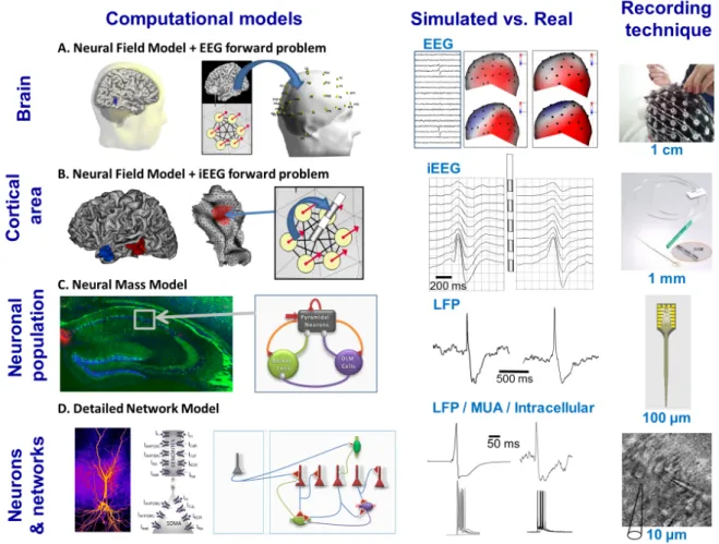
1*(23,3%&4(5%0,'0543#(-6(.3$&043.(,3$7-%89(,3/%&4(:&##9(,3/%&4(6034.(&,.(
6-%:&4(:&$;3:&$0'&4(:-.34#(
"
The challenge faced by any modeling approach is to capture the essential features of the system under study in a simple - but not too simple - description of its behavior (defined by the model variables) under controlled conditions (defined by the model parameters). In the brain, the level of modeling, i.e. the level at which this description is elaborated, is a crucial choice and strongly depends on i) the type of behavior to be reproduced in the model and ii) the nature of the data it relates to, from single-unit recordings, through local field potentials (LFPs) to electroencephalographic (EEG) and magnetoencephalographic (MEG) signals and functional magnetic resonance imaging (fMRI). In the field of epilepsy, models at nearly all levels of description were proposed since epileptiform activity may involve small clusters of neurons (e.g. in epileptic high-frequency oscillations) or, conversely, extended systems like the thalamocortical loop (e.g. in spike-wave discharges). This diversity ranging from models lying at sub-cellular level to large-scale systems involving cortical (and possibly subcortical) regions is illustrated in Figure 1. At the microscopic level, detailed network models include explicit representations of principal neurons and interneurons interconnected by means of synapses and/or gap junctions (Figure 1D). In these networks, single neuron models use compartments to represent the structural and functional properties of actual neurons. Parameters related to morphology (dendrites, soma, axon), passive (membrane capacitance, axial resistivity, leak conductance, membrane time constant, leak reversal potential) and active electrical properties (voltage-dependent ion channels) are taken into account in these single cell models, most of them still being based on the Hodgkin-Huxley formalism (Hodgkin and Huxley, 1952). Nowadays, a considerable number of neuron models is available (Gerstner and Kistler, 2002) that typically vary in the range of the dynamic repertoire of their behaviors, or in the mathematical model (differential or integral equations, conductance based or normal form, etc.) producing the behaviors. Models of networks of interconnected neurons offer the advantage of investigating epileptiform activity mechanisms, both at cellular (e.g. channelopathies) and network level (e.g. altered synchronization or connectivity patterns, effects of synaptic and or electronic couplings).
&" "
The mesoscopic level can be viewed as intermediate between the micro- and macro-scopic levels. Neural mass models (NMMs, Figure 1C), also referred to as “neuronal population models”, consider the average activity of interconnected subpopulations of principal neurons and interneurons without explicit representation of mechanisms lying at cellular level, conversely to ensembles of explicit neuron models. The theoretical bases of these models were established in the early 1950’s (Beurle, 1952) and were further developed by Ventriglia (Ventriglia, 1974)"(Ventriglia, 1978) and by Wilson and Cowan (Wilson and Cowan, 1972). These approaches were applied in mesoscopic models of the olfactory system (Freeman, 1963, 1973, 1968) and of the alpha rhythm of the EEG (Lopes da Silva et al., 1974; Lopes da Silva et al., 1976). Although they correspond to lumped descriptions of neuronal assemblies, these models are inspired from neurophysiology. The two relevant variables are the firing rate and the average post-synaptic potentials, either excitatory (EPSPs) or inhibitory (IPSPs), at each sub-population. Interactions among subpopulations can also be adjusted by coupling constants representing the average number of synaptic contacts between the considered cell types. It worth mentioning that the recent past years have witnessed a considerable increase of interest for this approach, as relatively extended networks need to be accounted in many forms epilepsy (Soltesz and Staley, 2008). A single NMM can only represent for the temporal dynamics of a neuronal assembly. Neural field models (NFMs) account for this limitation and describe the spatiotemporal evolution of coarse grained variables such as synaptic or firing rate activity in coupled populations of neurons. Formally, they are governed by integro-differential equations and can be viewed as an extension of NMMs based on local differential equations. For some conditions they can be expressed as Partial Differential Equations (PDEs) (Jirsa and Haken, 1996) and allow for the study of travelling waves and spatiotemporal patterns of brain activity (Coombes, 2005) either at the level of one or several circumscribed region(s) of the cortex (Figure 1B) or for larger scale systems like the entire neocortex (Figure 1A). In the field of epilepsy, NFMs were developed in conjunction with the solution to the depth-EEG/EEG/MEG forward problem in order to help the interpretation of epileptiform activity as reflected by magneto-electro-physiological signals collected in patients. Besides the aforementioned models inspired from neurophysiology, an alternative that recently developed is to make use of so-called “generic”, “canonical” or “formal mathematical models”. These phenomenological models are
'" "
intended to capture some dynamical properties of neural systems into relatively simple coupled differential equations that do not attempt to “copy” the underlying neurophysiological mechanisms. They offer a number of advantages for the formal study (typically a thorough bifurcation analysis), the modeling and the simulation of large-scale brain networks, as those involved during the propagation of seizures.
[Insert Figure 1 about here]
<*(=-.34#(-6(0,$3%0'$&4(&'$0>0$?(
<*)(+,$3%0'$&4(350435$0'(#5083#(&,.("/%#$#(
Interictal epileptic spikes (IESs) are very often observed in human partial epilepsies as well as in most experimental models of focal epilepsy (Schwartzkroin and Wheal, 1984). During epileptogenesis, a number of experimental studies also reported the appearance of isolated epileptic spikes during the latent period (Avoli et al., 2006; Staley and Dudek, 2006). It is commonly admitted that IESs are polymorphic events (Alarcon et al., 1994). Nevertheless, to a large extent, IESs correspond to transient signals (a few hundred ms) presenting with a more or less sharp initial component (often referred to as the “spike”) sometimes followed by a slower, more or less pronounced, component of opposite polarity, referred to as the “wave” (Figure 2A). Chauvière et al. (Chauviere et al., 2012) distinguished two types of interictal spikes: type 1, with a spike followed by a long-lasting wave; and type 2 with a spike without wave. Reconstruction of the flow in state space demonstrated that type 1 and 2 spikes could be understood as the dynamic class of excitable systems. Excitable systems comprise two state variables, which have one stable equilibrium point and a threshold (separatrix). When the threshold is crossed, then a fast large-amplitude discharge (spike) occurs and the state variables slowly return to equilibrium (wave). This establishes the type 1 spikes; discharges that do not cross the threshold return to equilibrium fast (no wave) and establish type 2. Although IESs have long been recognized as electrophysiological markers of epileptic processes ((Matsumoto and Ajmone-Marsan, 1964), review in (de Curtis and Avanzini, 2001)), their relationship to seizures is still elusive (Avoli et al., 2006) and their potential value as a biomarker of epileptogenic regions is not demonstrated yet. In this context, as
(" "
reviewed below, computational models may provide insights into the mechanisms underlying the generation of IESs and help the interpretation of their features, as reflected in LFPs or EEG data.
Neural mass models. Almost 40 years ago, a first attempt to study paroxysmal spikes in a neural mass
model (NMM) was reported in (Zetterberg et al., 1978). At that time, an electronic model was built to accurately analyze the waveforms of output signals under both stable and unstable conditions. This model was intended to represent a local neuronal population containing three subsets of neurons (two excitatory and one inhibitory) interconnected with positive and negative feedback loops. As in the computer implementations of NMMs, it contained both linear dynamic and nonlinear static elements. The analysis of its behavior revealed that the input noise level may lead to network instability (appearance of limit cycles). Very interestingly, from this model, authors could bring up the hypothesis that “epileptic spikes are generated in a population of neurons that operate close to instability” and that “spikes may be viewed as borderline cases between normal background activity and seizure activity”.
NMMs were also used in experimental studies of the epileptogenesis process defined as the structural and functional changes leading a normal brain to produce recurring seizures. Indeed, epileptic spikes appear relatively early during the latent period defined as the time from initial insult to spontaneous seizures. In the pilocarpine model of TLE (Curia et al., 2008), changes in the glutamatergic and GABAergic drives during epileptogenesis were investigated in a NMM of the CA1 hippocampal region (El-Hassar et al., 2007) based on recordings performed at early (3 days post injection), late (10 days post injection) and at chronic stage (characterized by recurrent spontaneous seizures). Conditions to reproduce the observed interictal-like activity (in term of spike morphology and occurrence frequency) were derived from extensive simulations where model parameters (EPSP and IPSP amplitude, rise and decay time constants) were varied at the soma and dendrites of the pyramidal cell subpopulation. The model (described in (Wendling et al., 2002)) predicted that the increase of the glutamatergic/GABAergic drive ratio is a sufficient condition for the occurrence of epileptic spikes (Figure 2C) and that this ratio impacts their occurrence frequency. This prediction was verified and explained by several neurobiological findings obtained in vitro, including changes in the rise and
)" "
decay time constants of actual post-synaptic currents. In the kainate model, recent reports also demonstrated that the occurrence frequency of epileptic spikes (ESs) increases during epileptogenesis (White et al., 2010). NMMs provided the opportunity to explain this change in frequency as well as changes in the morphological features (Huneau et al., 2013). In this study, authors developed signal processing methods to automatically detect and characterize ESs over long duration periods (30 days). The shape changes of ESs as a function of time were first characterized and then reproduced in a NMM model. Some key parameters that impact the ESs morphology could be identified. Results showed that hyperexcitability stems from the progressive diminution of GABAergic inhibition. This model-based counter-intuitive hypothesis was experimentally verified, both in vivo and in an in vitro. Based on these results, a novel electrophysiological marker computer from LFPs was proposed which provides information about the progress of the disease after initial insult.
Neural field models. NFMs have the unique advantage to account for the spatial features (geometry,
extent, connectivity) of neuronal sources, in addition to their temporal features like the synchronization level among neurons. In order to progress in the interpretation of IESs recorded by depth-EEG electrodes, a NFM was developed and reported in (Cosandier-Rimele et al., 2007). This model consists in an extended source (neocortical patch) combining two levels of representation: i) coupled NMMs to describe the temporal dynamics of neuronal sources within the patch and ii) distributed current dipoles to describe the electrical contribution of neuronal sources to electrode contacts depending on their spatial location over the patch."From this NFM, LFPs were simulated by solving the depth-EEG forward problem which consists in computing the electrical potentials at electrodes contacts. This computation makes use of the dipole theory. It takes into account a number of physical factors like the distance between the current dipole sources and the electrode contacts, their orientation and moment, as well as the volume conductor properties like the conductivity of the different brain tissues. Transient epileptiform activities (IESs) were simulated for various conditions regarding the synchronization of neuronal populations and the spatial extent of the source, providing insights into the relationship between the spatiotemporal properties of cortical neuronal sources and IESs as recorded by depth electrodes (see Figure 1B).
*" "
Detailed network models. At single neuron level, epileptiform activity related to sustained
depolarizations (SDs) and paroxysmal depolarizing shifts (PDSs) often associated with interictal epileptic spikes simultaneously observed in LFPs was studied in (Heilman and Quattrochi, 2004). Six-compartment models of increasing detail level were analyzed according to their ability to generate SDs and PDSs. Reported findings suggested the dominance of neurophysiological elements (calcium-related transmembrane currents, in particular) over morphological elements (accurate representation of dendritic trees) in the generation and sculpting of epileptiform activity.
A two-compartment model of the CA1 pyramidal neuron designed using Hodgkin–Huxley formalism was reported in (Demont-Guignard et al., 2009). This model accounts for a variety of somatic and dendritic currents of potential interest in epilepsy. For instance, in addition to the classical voltage gated sodium, potassium (BK, SK) and calcium (T-type, R-type, L-type) channels, the model also includes hyperpolarization-activated cationic current (Ih) and a rapidly inactivating potassium current (IKA), present only in the dendritic compartment. This latest channel allowed for mimicking the effect of 4-aminopyridine (IKA blocker) on pyramidal neuron discharges and for comparing the change in firing patterns with real experimental data obtained on hippocampal slices. Based on this neuron model, a CA1 subfield network was created by synaptically connecting 20% of pyramidal cells to 20% of dendritic and somatic-projecting GABAergic interneurons (Demont-Guignard et al., 2009). Simulated intracellular activity of several hundreds of GABAergic and glutamatergic cells were produced, simultaneously with an epileptic spike reflected in the LFP (Figure 2B). Results showed that epileptic spikes are i) generated by the synchronous discharges of a large number of pyramidal cells and ii) followed by a slower negative wave explained by the sustained activity of interneurons generating feedback inhibition.
Another detailed network model of interictal epileptic spikes in CA1 has been recently published (Ratnadurai-Giridharan et al., 2014). Also based on the Hodgkin-Huxley formalism, glutamatergic pyramidal cells were modeled according to (Golomb et al., 2006), basket cells were modeled from (Wang and Buzsáki, 1996) and oriens-alveus (OA) cells were taken from (Wang, 2002). As in Demont-Guignard et al. (2009), the CA1 network was constructed with a 80%-20%
excitatory-!+" "
inhibitory neuron ratio using 225 pyramidal cells, ~22 basket cells, and ~22 OA cells. AMPA and GABAa conductance (but not NMDA) were used to simulate synaptic interactions. Conditions on neuronal synchronization to evoke paroxysmal depolarization shift (PDS) events in CA1 pyramidal cells were determined. Parameters were adjusted to fit experimental measurements of PDS burst width and PDS after-hyperpolarization duration. Axonal sprouting was modeled by increasing the average number of pyramidal-to-pyramidal synapses. This model nicely produced spontaneous interictal spikes as the sprouting-induced recurrent connectivity of the CA1 was sufficently increased, even if the Schaffer collateral input is not highly synchronized.
[Insert Figure 2 about here]
<*1(+,$3%0'$&4(@0A;(B%3C/3,'?(D#'044&$0-,#E(F05543#(G)1HI1JH@KL(&,.(B&#$(
F05543#(G1JHIMHH@KL(
During the past decade considerable effort has been devoted to the study of high-frequency oscillations (HFOs, 80-600 Hz), often classified as high-gamma activity (80-120 Hz), ripples (Rs, 120-250 Hz) and fast-ripples (FRs, 120-250-600 Hz). Ripples and FRs are brief (a few tens of ms), observed in signals recorded with intracerebral electrodes located in brain structures. HFOs were first reported in experimental in vivo models of epilepsy (rat, kainate models) (Bragin et al., 1999c) and then in the human epileptic brain (intracranial recording from hippocampus and entorhinal cortex) (Bragin et al., 1999a). At that time, they were hypothesized to be a marker of brain areas involved in spontaneous seizures (Bragin et al., 1999b). This clinical value was confirmed later in a number of papers (Jacobs et al., 2009; Urrestarazu et al., 2007) showing that i) the occurrence rate of HFOs is higher within the seizure onset zone and ii) the resection of brain sites generating HFOs favorably correlates with surgical outcome (Jacobs et al., 2010). Regarding the cellular and network mechanisms involved in the generation of ripples and FRs which dominant frequency exceeds the maximal neuronal firing rates, several hypotheses were raised. These are mainly related to i) the timing of APs generated in neighboring pyramidal cells, ii) the unreliable firing and weak synchronization of neurons in disorganized networks in conjunction with enhanced glutamatergic synaptic activity (Dzhala and Staley, 2004; Foffani et al., 2007; Staley, 2007) and iii) the electronic coupling between pyramidal
!!" "
neurons (Draguhn et al., 1998; Traub et al., 2002). These hypotheses raised from experimental studies explain why modeling approaches were essentially conducted at the level of detailed network models where neuron features, cellular firing patterns and synchronization among cells can be explicitly assessed.
Ripples (120-250 Hz). Traub et al. proposed a detailed model for studying high frequency oscillations.
(Traub et al., 1999). The neuronal network contained a large number pyramidal cells (n=3072), but no interneurons. Each pyramidal neuron was constructed from 64 compartments. At this time the model did not comprise synaptic connections but already implemented axo-axonal gap junctions between pyramidal cells. Action potentials could be reproduced in cell somata and the network was able to generate spikelets (defined as small spike-like membrane potential deflections) at about 140-172 Hz. These results suggested that a purely excitatory network can produce ripples if gap junctions are implemented, even without synaptic connections.
The next step consisted in introducing inhibitory GABAergic interneurons within the glutamatergic network (Traub and Bibbig, 2000). The model also comprised 1) excitatory afferent input on pyramidal neurons and interneurons, 2) interneuron inhibition and 3) pyramidal cell self-excitation. Despite the presence of chemical synaptic interactions, this study also confirmed a key role of gap junctions between pyramidal cells in ripple oscillations generation. But in this case, synaptic connections modulated the oscillations (around 182 Hz). Interneurons fire at high rate at the top of slow depolarization whereas pyramidal cells fire at lower rates. For strong GABAergic input onto pyramidal cells, the frequency of resulting HFOs depends of the time course of GABAa IPSCs.
Taxidis and colleagues (Taxidis et al., 2012) built a model of CA3 hippocampal subfield connecting the CA1 subfield in a feed-forward manner. A total of 1000 pyramidal cells and 100 interneurons were represented. Synaptic connections used fast AMPA and GABAa conductances. Population bursts starting from the CA3 subfield could evoke sharp-wave-like slow depolarization in the CA1 dendritic compartment associated with ripple-like oscillations (200 Hz) in the CA1 somatic layer. Interestingly, GABAergic interneurons and fast recurrent inhibition in CA1 strongly modulated the frequency and
!#" "
synchrony of these oscillations. Overall the model was able to generate sharp wave ripples solely through synaptic interaction, without gap junctions.
A detailed model of the dendate gyrus in three dimensions was recently developed (Kobayashi et al., 2014), including 10,000 granular cells with corresponding 300 mossy cells, 100 basket cells and 120 hilar interneurons. This biology-inspired model contained multiple ionic currents, including sodium, rectifier potassium channels, A-type potassium, hyperpolarization-activated nonselective cation channels (Ih) and several types of voltage-gated calcium channels. Regarding the cell types, the number of compartments in a model cell was between 9 and 17, and the number of dendrites per cell was around 4. When no sclerosis is simulated, the loss of inhibition evoked multiple bursts of ripple-like activity around 150 Hz. HFOs with peak frequencies at around 250 Hz could be produced in the model with 100% sclerosis, dense connectivity, and no inhibition (Kobayashi et al., 2014). This study shows that each positive peak of HFOs largely reflects action potentials overriding a large and slow depolarization (reminiscent of PDS).
Fast ripples (250-600 Hz). Increasing evidence show that fast ripple (FR) oscillation represents a
valuable biomarker of the epileptogenic zone. To date, at least four different computational models were able developed to understand the pathophysiological mechanisms underlying FR generation. The first one (Roopun et al., 2010) is a cortical column model built from 4750 multi-compartment cortical neurons. Each of these model neurons included a soma, an axon initial segment, and several dendrites containing Na+, K+, Ca2+, and rectifier conductances. Main excitatory cells were interconnected with several types of interneurons: basket, axo-axonic, low threshold spiking and neurogliaform cells. Both glutamatergic excitatory synapses (AMPA receptors) and inhibitory synapses (GABAA receptors) were implemented along with interneuronal dendritic gap junctions and axo-axonic gap junctions between homologous types of glutamatergic neurons. Using both experimental data from human temporal neocortical slices and in silico experiments, this study showed that the reduction of the GABAa receptor function failed to reduce HFO occurrence. Conversely, this occurrence was reduced by the non-specific blocker carbanoxolone (CBX). These results suggest that HFOs could be produced even without major contribution of interneurons, mainly in a non-synaptic
!$" "
manner through a key role of gap junctions between excitatory cells. However, one should notice that i) the intriguing question of the existence of gap junctions between pyramidal cells remains to be clarified (for review, see (Mercer, 2012)) and ii) the direct involvement of gap junction is difficult to demonstrate as pharmacological blockers of gap-junctions are largely non-specific. Indeed CBX broadly affects several neuronal membrane conductances independently of gap junctions, including AMPA/NMDA mediated EPSCs and GABAa-mediated IPSCs (Tovar et al., 2009).
The second computational model of fast ripples (FRs) is the one proposed by Ibarz et al. (Ibarz et al., 2010). The authors started from experimental data obtained in epileptic rat hippocampus in which the emergence of HFOs (> 250 Hz) was experimentally investigated (Foffani et al., 2007). This model consisted of 120 neurons, each composed of 19 compartments representing the soma, the basal and the apical dendrites of pyramidal cells (based on (Traub et al., 1991)). As the main objective was to look at the effect of out-of-phase firing, the authors simulated two clusters of 60 neurons that burst at different delays. Using faster Na+ dynamics, this model was able to produce FRs without gap junctions nor depolarizing GABA (reversal potential for GABAa-like synaptic currents was -75 mV), with two different firing regimes. Within the first type of cluster, if abnormally synchronized single-cells burst together in phase, the model produces oscillations in the FR frequency band (>250Hz). But if clustered bursting neurons are slightly desynchronized then LFP oscillations occur at much higher frequency. In this case, the “out-of-phase” firing mode of small clusters of neurons produces fast ripples (>400 Hz).
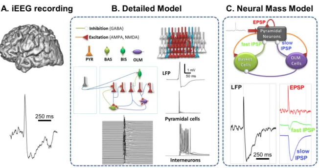
The third detailed model (figure 3) was designed to understand the mechanisms underlying both fast ripples (FRs) and epileptic spikes in the hippocampal CA1 area (Demont-Guignard et al., 2012). The model started from a previously-published study (Demont-Guignard et al., 2009). The network included 1182 pyramidal neurons, 52 oriens-lacunosum moleculare (OLM) interneurons, 52 basket cells (BAS) and 52 bistratified cells (BIS), connected via inhibitory GABAergic and excitatory glutamatergic (AMPA/NMDA) synapses. Depolarizing GABA was also implemented in the model as the reversal potential for a proportion of GABAa IPSCs was shifted to more depolarized value at some GABA synapses. Interestingly, the model could switch from FRs to epileptic spikes depending on excitability conditions. This study revealed that these two epileptic biomarkers share some common
!%" "
mechanisms (depolarized GABAa reversal potential in some neurons, presence of intracellular paroxysmal depolarization shift - PDS - and altered synaptic transmission). But epileptic spikes were produced by a larger group of highly excitable hypersynchronized pyramidal cell, having a large spatial distribution whereas FRs were produced by small clusters of weakly synchronized, less excitable neurons firing together, in line with the “out of phase” discharge mode of Ibarz’s model. Importantly the model predicted a functional link between epileptic spikes and FRs that was verified on organotypic slices. Depending on the degree of alteration of GABAergic and glutamatergic transmission, the field response evoked by single pulse in control condition changes into FRs (350 Hz) when AMPA/NMDA conductance were increased and GABAa conductance was moderately decreased. Further increase of the network excitability transforms FRs into epileptic spikes.
Very recently another detailed model was used to study the transition from ripples to fast ripples (Simon et al., 2014). The authors started from the pyramidal neuron model described in Traub et al. (2012) comprising soma, branching dendrites, and a 24-compartment axon with gap junctions localized in the distal axonal branch. Network activity was generated by spreading waves and/or cycles of axonal firing. In this model, ripples (< 250 Hz) were amplified when action potential could be conducted from an axon to another through electrical coupling and FRs (>200Hz) were elicited by ectopic spikes that randomly occur. However it is worth noting that this interesting model of transition between ripple and fast ripple did not include interneurons whereas other studies showed that interneuron firing and consecutive depolarizing GABA might play a key role in the generation of high frequency oscillations (Alvarado-Rojas et al., 2014; Demont-Guignard et al., 2012; Pallud et al., 2014; Wendling et al., 2012).
[Insert Figure 3 about here]
N*(=-.34#(-6(0'$&4(&'$0>0$?
N*)(O&%$0&4(350435#03#(
!&" "
In partial epilepsies, seizures are often characterized by the appearance of fast oscillations in LFPs or EEG signals, sometimes referred to as “chirps” (Schiff et al., 2000). Typically, in human neocortical epilepsies, rapid discharges observed at seizure onset range from 70 to 120 Hz. They constitute a characteristic electrophysiological pattern in focal seizures characterized by a noticeable increase of intracerebral EEG signal frequency (Lee et al., 2000)"(Wendling et al., 2003). In mesial temporal lobe epilepsies (mTLE), fast oscillations at seizure onset were also observed in the hippocampus, amygdala and entorhinal cortex (Bartolomei et al., 2001). These HFOs are usually associated with a lower frequency range (beta and low gamma, 20-40 Hz) as compared with those recorded from the neocortex (Figure 4). Interestingly, as fast oscillations at seizure onset seem to be a hallmark of focal seizures, they were recently utilized to quantify the degree of epileptogenicity of brain structures recorded by depth-EEG electrodes during the pre-surgical evaluation of patients candidate to surgery ((Bartolomei et al., 2008)" (David et al., 2011)" (Gnatkovsky et al., 2011), see also (Andrzejak et al., 2014) for comparative study of quantitative methods). The mechanisms underlying the generation of HFOs at seizure onset have been a topic of increasing interest over the ten past years. In TLE, a number of experimental studies suggest that fast-onset seizures may result from the synchronous activity of GABA releasing cells (Gnatkovsky et al., 2008)"(Shiri et al., 2014). In temporal lobe “plus” epilepsies (TLE+), very fast oscillations (VFOs, > 80 Hz) prior to seizures would involve neocortical ripples that actively participate in their initiation (Grenier et al., 2003). Another hypothesis supported by in vivo and in vitro recordings is that VFOs observed in electrocorticography are generated by networks of pyramidal neurons coupled by gap junctions (Traub et al., 2010). This brief overview on fast oscillations at seizure onset shows that this type of epileptiform activity is of major importance for two reasons, at least. First, it may provide some hints about key mechanisms involved in the transition from interictal to ictal activity. Second, it may be used to elaborate quantitative indexes aimed at revealing the epileptogenicity of the underlying brain tissue. In this context, a number of computational modeling approaches were proposed to specifically analyze HFOs ranging from 70 to 120 Hz (i.e. beyond the gamma frequency band) typically observed at the onset of partial seizures. These approaches made use of lumped-parameter models (Molaee-Ardekani et al., 2010), detailed network models (Traub et al., 2010; Traub et al., 2008; Traub et al., 2001) and formal models
!'" "
(Vladimirov et al., 2011). Interestingly, these modeling approaches are based on two fundamentally different assumptions that are still a matter of debate. In the former, HFOs are produced by synchronous firing of fast-spiking interneurons. The LFP basically reflects IPSPs generated onto pyramidal neurons, as reported experimentally. In the latter, HFOs are caused by gap junctions among pyramidal axons as experiments show that they persist under the blockade of synapses and that they disappear upon action of gap junction blockers.
Neural Mass models. In order to specifically explain the mechanisms underlying the generation of
HFOs (>80 Hz) observed in depth-EEG signals at the onset of neocortical seizures, a neural mass model was proposed (Molaee-Ardekani et al., 2010). Following the classical “neuronal population model” approach, the proposed model included two sub-populations of cells: i) one sub-population of pyramidal neurons and ii) one sub-population of interneurons targeting the perisomatic region of pyramidal cells where fast GABAergic currents are mediated. From this relatively simple model, the authors could accurately reproduce “chirp-like” rapid discharges (70-110 Hz) while respecting physiological values for the kinetics (rise and decay time) of two key variables in NMMs, namely the average glutamatergic EPSPs and GABAergic IPSPs. Interestingly, the model indicated that mutual inhibition was a key factor for the generation of seizure-onset HFOs, in line with previous experimental studies suggesting that a possible substrate for fast activity (gamma range) reflected in LFPs is the presence of mutual GABAa-mediated synaptic inhibition within the interneuronal network ((Wang and Buzsaki, 1996), review in (Bartos et al., 2007)). Finally, in this model, the simulation of a fast onset activity that reproduces the actual onset pattern (in term of frequency, energy and bandwidth) could only be obtained by a gradual decrease of both the EPSP and IPSP amplitudes (that dramatically increased with respect to “normal” values).
Neural field models. The recording of HFOs from scalp electrodes is still on open issue. In the field of
epilepsy, it is admitted that fast oscillations at seizure onset are not clearly discernible in scalp EEG due to low signal-to-noise ratio. In order to investigate the observability of these so-called “rapid discharges”, a NFM-based approach was reported in (Cosandier-Rimele et al., 2012). Using coupled NMNs distributed over cortical “epileptic” patches, authors examined the impact of several factors (electrode-source distance, source extension and synchrony, background activity) on the observability
!(" "
of fast oscillations in simultaneously simulated depth and scalp EEG signals. They could identify critical factors that prevent rapid discharges to be detected from scalp EEG.
Detailed network models. The generation of very fast subdural EEG oscillations (>70 Hz) occurring at
the onset of focal seizures was accurately studied by Traub and collaborators (Traub et al., 2001). The authors designed a detailed network model that included 3072 pyramidal cells and 384 interneurons. In this network, both synaptic and gap junctional couplings were represented. Based on extensive computer simulations combined with intra- and extracellular recordings performed in rat hippocampal slices, authors could show that gap junctions play a crucial role in the generation of EEG oscillations similar to those observed in patients with drug-resistant epilepsy. More particularly, simulations could replicate actual data when synapses were blocked while axonal gap junctions between principal neurons did exist. It is noteworthy that very high frequency oscillations could also be simulated in interneuron networks when axonal gap junctions were included between interneurons. A few years later, the same group (Traub et al., 2010) proposed a novel detailed network model to explain electrocorticographic (ECoG) very fast oscillations (> 80 Hz) prior to human neocortical seizures. This model included 15,000 neurons (one type only: layer 5, tufted, intrinsically bursting pyramidal cell) interconnected by axonal gap junctions according to a globally random topology. Each neuron was represented by a 61-compartment (1 for soma, 6 for axon and 54 for dendrites) model and multiple voltage-dependent membrane conductances were included. Field and intracellular patterns similar to those observed experimentally could be simulated when pyramidal cells were coupled by axonal gap junctions and when synaptic transmission was absent. Interestingly, as in actual recordings, simulations showed that spikelets on hyperpolarized somatic membrane potentials were more frequent than full action potentials. To end with, it worth mentioning that a number of theoretical studies based upon the above detailed network models have specifically analyzed the role of electrical couplings among neurons in the generation of HFOs (Lewis and Rinzel, 2000; Munro and Borgers, 2010).
Formal mathematical models
Cellular-automata approach. A cellular automaton is a discrete model consisting of a grid of cells. A finite number of states is associated with each cell. In various disciplines of science (mathematics,
!)" "
physics, computational biology, …), cellular automata (CellAut) have been extensively used to model dynamical systems provided that these systems i) are discrete in time and space, ii) operate on a uniform, regular lattice and iii) are characterized by local interactions. It is therefore not surprising that this modeling approach was used to analyze the spatio-temporal features of self-organized oscillations in neural networks. In the context of epileptiform activity, this approach was first followed by Traub et al. (Traub et al., 1999) to" assess the contribution of gap junctions to the generation and shaping of high-frequency (> 100 Hz) neuronal population oscillations in the hippocampus and then used again in the specific context of HFOs (> 80 Hz) observed at the onset on seizures (Traub et al., 2010). In this latter study, authors designed a 2D network of excitable cells with spatially constrained connections (electrically coupled pyramidal neurons with 3 states - resting, firing, refractory -, no synaptic transmission) to predict wave propagation. The assumption supporting this simple modeling approach is that HFOs (> 80 Hz) would be generated by networks of axons electrically-coupled by gap junctions. Strikingly, and although the biological realism of CellAut models is questionable in the context on neural network dynamics, the model was found to reproduce spatio-temporal patterns of activity (referred to as ‘‘blobs’’) which also appear during actual HFOs at seizure onset. Finally, using a similar modeling approach, a number of theoretical results could be established regarding the influence of the network topology and the connection length, among other factors (Vladimirov et al., 2011).
Dynamic system approach. Acknowledging the time scale separation present in seizure evolution, Jirsa and colleagues (Jirsa et al., 2014) used the theory of fast-slow systems in nonlinear dynamics to develop a taxonomy of seizures characterized by bifurcations for seizure onset and offset. They identified from experimental seizure data of various species a predominant class of bifurcation pairs, integrated into a phenomenological dynamic model called Epileptor (Jirsa et al., 2014, comment in (Friston, 2014)). The Epileptor comprises two ensembles and a slow permittivity variable (see Figure 5). The first ensemble is linked to the generation of fast oscillations, the second ensemble to the interictal spikes and the often-present spike-wave complex close to seizure offset. The slow permittivity variable accounts for, presumably predominantly, extracellular effects related to energy
!*" "
consumption and oxygen and captures details of the autonomous slow evolution of interictal and ictal phases, as well as various details of seizure evolution during each phase. Under the hypothesis that the pre-ictal state can be understood as an equilibrium point attractor, there is a finite small number of ways to enter into an oscillatory state: the super- and sub-critical Hopf bifurcations, the saddle-node bifurcation and the saddle-node-on-invariant-circle (SNIC) bifurcation. Each bifurcation has clearly identifiable characteristics with regard to their amplitude and frequency scaling as the oscillation evolves after seizure onset. Canonical mathematical models for seizure onset oscillations can unambiguously capture these characteristic behaviors. The same line of reasoning can be performed for the seizure offset bifurcation that also comprises four bifurcations from the ictal to the non-ictal state, which are the SNIC, the supercritical Hopf, the fold limit cycle and the homoclinic bifurcation. Across three species (Zebra fish, mouse, and human) the saddle-node bifurcation at seizure onset and homoclinic bifurcation at seizure offset appears to be predominant.
[Insert Figure 5 about here]
"L(=-.34#(-6(6-'&4(#30K/%3#9(#30K/%3(5%-5&A&$0-,(&,.($%&,#0$0-,(6%-:(0,$3%0'$&4($-(0'$&4( &'$0>0$?(
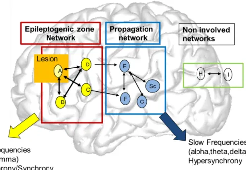
Focal (partial) seizures are limited to a relatively-well circumscribed area of the brain (referred to as the epileptogenic zone - EZ -), conversely to generalized seizures in which both hemispheres are involved (see section 3.2). Other the past two decades, and following the pioneer work of Bancaud and Talairach (Talairach et al., 1962) who introduced the functional stereotaxic exploration of epilepsies, the notion of “epileptic focus” has evolved toward the now well-accepted concept of epileptogenic network (Nair et al., 2004). Indeed, based on intracerebral EEG recording, many studies reported that the EZ very often involves several distinct and distant brain areas, not only at seizure onset but also during seizure propagation (Bartolomei et al., 2001). In this respect a number of signal processing methods were developed to identify epileptogenic networks and characterize their dynamics from depth-EEG recordings. Some of these methods were based on functional connectivity, typically quantified by association/synchronization measures among signals (Pijn et al., 1990)"(Schindler et al., 2007)" (Lehnertz et al., 2009; Wendling et al., 2010)) while some other methods were based on the
#+" "
computation of “epileptogenicity” indexes (Andrzejak et al., 2014). From the modeling viewpoint, the analysis and the description of focal seizure dynamics (initiation, propagation and termination) has long been - and is still - a topic of large interest. Over the past years, models were proposed at any level of description, from the single neural mass to formal mathematical models.
Neural mass models. More than twenty-five years ago, Freeman and colleagues showed that the
NMM they initially proposed for the olfactory system could produce realistic EEG signals, as compared with segments of EEGs recorded from rats during seizures initiated by a few seconds of high intensity repetitive electrical stimulation (Freeman, 1987). The relevance of NMMs, also referred to as nonlinear lumped-parameter models, in the analysis of depth-EEG epileptic signals was then emphasized by Wendling and colleagues (Wendling et al., 2000) who published a series of papers showing that NMMs can produce epileptiform signals strikingly similar to those recorded in mTLE. In the hippocampus, these authors proposed a NMM more specifically adapted to the hippocampal cytoarchitecture (Wendling et al., 2002). In particular, this model includes two subpopulations of GABAergic interneurons (mediating GABAa slow and fast currents onto pyramidal cells), conversely to the “generic” NMM that classically includes one interneuron type. Importantly, this model, reviewed in (Lytton, 2008; Wendling et al., 2012), explains the transition from interictal to ictal activity by a progressive decrease of slow dendritic inhibition. Highly realistic epileptiform signals and transitions of activity could be simulated as compared to actual recordings performed in patients with temporal lobe epilepsy (TLE) and in experimental models of TLE (figure 4). Interestingly, in this model, the fast onset activity (around 25-30 Hz) is explained by IPSPs produced by the fast feedback inhibitory loop. Experimental evidence was reported a few years later (Gnatkovsky et al., 2008) (Shiri et al., 2014). Following the same approach, NMMs were then elaborated to describe the transition from interictal to ictal activity in the entorhinal cortex (EC) from recording performed in the isolated brain preparation (Labyt et al., 2007; Labyt et al., 2006). Methods were also developed to automatically identify model parameters from real signals (Wendling et al., 2005). As NMMs formally correspond to nonlinear dynamical systems, they have also been used in theoretical studies aimed at analyzing their behavior with respect to endogenous or exogenous parameter changes. A number of stability and bifurcations studies (Blenkinsop et al., 2012; Touboul et al., 2011) were conducted both
#!" "
in the generic NMM and in the Wendling’s model. Similar models were used to investigate seizure generation scenarios (Kalitzin et al., 2011). The novelty consisted in the simulation of several interconnected neuronal populations, each one representing a set of excitatory pyramidal cells and the other representing inhibitory interneurons. Assuming that the connectivity between neuronal populations controls the dynamic spectrum of the system, the main objective was to determine whether model parameters are correlated with the observable features of the simulated EEG signals (covariance functions). To proceed, authors introduced a signal measure called “ictality” that quantifies the length and amplitude of the ictal phase. A number of scenarios were identified that explain how a neuronal system exhibiting multistability can switch from a “normal ongoing activity” state to a “paroxysmal EEG activity state” and then to an “epileptic seizure” state.
[Insert Figure 4 about here]
Neural field models. Although NMMs proved efficient to produce realistic epileptiform signal
dynamics and transitions, they cannot account for the spatial features of epileptogenic networks that often involve distant and extended brain areas. This consideration motivated the development of neural field models (NFM) in which the multiplicity of interconnected brain sources, their complex geometry (folded brain surface) as well as their synchronization level are taken into account. In addition to the temporal dynamics at each source, these factors that intervene in the EEG forward problem have also an impact on signals reconstructed at the level of electrodes (intracerebral or scalp). To address this issue, a NFM was proposed in (Cosandier-Rimele et al., 2010) and used to study relationship between the spatiotemporal features of neuronal generators (convoluted cortical dipole layers) and the EEG signals recorded at electrodes. This model was based on coupled neurophysiologically-relevant NMMs and allowed for investigation of several factors regarding the location, area, geometry, and synchronization of neocortical sources as represented by neuronal populations. This type of approach contributes to the joint analysis and interpretation of simultaneously-recorded depth and scalp EEG signals which remains a difficult task.
Single neuron and detailed network models. Early modeling studies by Traub and collaborators at
##" "
critical for the production of epileptiform activity. Typically, network models combined with in vitro data showed the impact of three important parameters on the generation of synchronized afterdischarges: the synaptic strength, the synaptic density and the refractoriness of neurons following a period of excitation (Traub et al., 1984). The mechanisms underlying propagation of epileptiform field potentials from CA2 to CA3 were investigated in computer simulations (up to 1000 cell arrays of model neurons). Results showed that i) the propagation of epileptiform activity was plausible even if one considers the slow conduction along axons interconnecting CA2-CA3 neurons, provided that there are sufficiently many interconnections and ii) when the number of synchronously activated cells initiating a population burst increases then the number of interconnections required to propagate seizures decreases (Traub et al., 1987). Following this pioneer work on hippocampal area CA3 that has been one of the most intensively studied brain region for computational models of epileptiform activity (review in (Lytton et al., 2005)), a number of detailed models were then developed to investigate the network mechanisms underlying focal seizure initiation and propagation in other brain structures. In order to evaluate the relationship between the strength of network connectivity and seizure-like activity patterns generated by neocortical networks, a detailed model was developed by van Drongelen et al. (van Drongelen et al., 2005). It included 656 excitatory and inhibitory neurons of 4 types (regular-firing pyramidal, bursting pyramidal, basket cell, chandelier cell, Hodgkin-Huxley formalism). The impact of synaptic strengths on the simulated extracellular activity was investigated revealing the essential parameter changes to generate seizure activity. Indeed, the network model could generate a large variety of behaviors that critically depended on the strength of synaptic connections. Interestingly, and in contrast to the common belief that strong excitatory coupling is needed to synchronize bursting, results obtained from this computational model indicated that neural networks can generate and sustain seizure-like activity if the excitatory coupling strength falls below a critical value.
Formal mathematical models. Recently, Kalitzin and colleagues introduced a simplified, analytical
model which can mimic the behavior of more realistic computational models of neural dynamics like NMMs (Kalitzin et al., 2014). An essential feature of this model, referred to as Z6, is that it exhibits multistability. It is defined by a single ordinary differential equation where 2 real constant coefficients
#$" "
and complex constant coefficient control the behavior of the system. A major advantage of the Z6 model is that it is simple and analytical allowing for i) formal mathematical analysis and ii) creation of large-scale networks. To some extent, this is also a limitation since model parameters cannot be directly interpreted in physiological terms. Nevertheless, this study contributes to the understanding of oscillatory states in models of collective neuronal behavior and emphasizes the existence of multiple stable or quasi-stable states as a necessity to account for the variety of epileptiform activity observed in the epileptic brain.
Drawing upon concepts from the cellular automaton framework, Goodfellow and colleagues (Goodfellow et al., 2012b) investigated the effects of spatial heterogeneity on the large-scale spatio-temporal dynamics of epileptiform rhythms. The overall model was characterized according to excitability and a classification of dynamical behaviors was established as a function of model parameters. Distinct regimes could be identified like “sustained oscillatory activity” with complex spatiotemporal dynamics or “stable rest state” in which a perturbation can induce travelling waves, among others. Interestingly, this model also raised conditions on tissue heterogeneities that favor normal and abnormal spreading of activity, like the existence of area-specific connectivities, which is an essential notion in epilepsy. Along the same idea, the impact of the pattern of connections in brain networks on the focal or generalized aspect of EEG discharges was investigated in (Terry et al., 2012) using a phenomenological model initially reported in (Benjamin et al., 2012). The model is expressed as a single complex equation that is a special case of a more general form introduced by Kalitzin et al. (Kalitzin et al., 2010)."Computer simulations showed that the spatial features of EEG discharges may depend of subtle changes in network structure, without the requirement for any localized pathologic brain region. Results also showed that the introduction of “pathological” regions can give rise to primary or secondary generalized seizures depending on the network structure. Interestingly, they also showed that a decrease of connectivity may lead to increased frequency of seizure-like activity.
Several mechanisms of seizure recruitment have been tested in computational models with varying levels of mathematical abstraction, mostly with a particular focus on either propagation through continuous sheets of neural populations (Kramer et al., 2005, 2007; Ursino and La Cara, 2006; Kim et al., 2009; Hall and Kuhlmann, 2013), or seizure recruitment of single cell activity within a population
#%" "
(Miles et al., 1988; Golomb and Amitai, 1997; Compte et al., 2003; Bazhenov et al., 2008; Fröhlich et al., 2008). For either approach, it is not evident how much we can infer about seizure recruitment of downstream brain regions that are distant, and often the biophysics of the slow processes is not well enough known to allow for explicit modeling or simply too complex (see Marder and Taylor, 2011). As an alternative, Proix et al. (2014) investigated seizure recruitment using the Epileptor and generalized forms of coupling, in particular focused on the Epileptor’s slow permittivity variable. Arguments based on time scale separation in the theory of nonlinear dynamic systems suggest that fast couplings through synapses or gap junctions will not qualitatively change the behavior on the slow time scales. For this reason multi-scale seizure recruitment necessitates the inclusion of slower homeostatic mechanisms affecting the permittivity of cell tissue. Coupling through permittivity variables then has to model the extracellular/intracellular effects of local and distant discharges. Though of phenomenological and abstract nature, the network model with the permittivity based coupling allowed highlighting the repertoire of clinically well-known recruitment scenarios (Proix et al., 2014). Then again, counterintuitive phenomena were also encountered when multiple brain regions are mutually coupled through permittivity. For instance, if some of the coupled brain regions are epileptogenic, then the overall effect may render the network behavior non-epileptogenic. Counter-intuitive behaviors as such offer novel perspectives and explanatory approaches to so far not understood phenomena such as unexpected relapse after surgery (Hennessy et al 2000). Often these relapses cannot be understood solely on the basis of local arguments (such as missed lesions identified in postoperative MRI) and necessitate other explanatory approaches as the one proposed here on the basis of network effects.
Networks of permittively coupled Epileptors are capable of demonstrating multiscale effects as observed clinically in seizure propagation. Recruitment of brain regions far from the epileptogenic zone may occur many seconds after seizure onset and is thus significantly slower than the fast discharges on the time scale of 10 to 50 msec. To illustrate the spatiotemporal spread of activity through a realistic brain network, we coupled 190 Epileptors with permittive coupling (Proix et al., 2014) using a large-scale connectivity matrix, the connectome, derived from tractographic data and solve it computationally using methods of the neuroinformatics platform The Virtual Brain
(Sanz-#&" "
Leon et al., 2015; Sanz Leon et al., 2013). In particular, we add linear and multiplicative Gaussian white noise to the model equations to enhance the degree of variability and achieve better realism. To this end we also compute the forward solution for stereotactic EEG (sEEG; see Figure 6). All parameters were set identical across the network. Only the epileptogenicity parameter was enhanced in the epileptogenic zone comprising the amygdala and the hippocampal region.
[Insert Figure 6 about here]
<*1(23,3%&40K3.(350435#03#(
a) Spike-wave discharges and absence seizures
Spike-wave (or “spike-and-wave”) discharges denotes a regular, symmetrical and generalized pattern observed in the EEG, typically during absence seizures (Seneviratne et al., 2012). Experimental and clinical findings have given insight into some basic mechanisms of spike-wave (SW) discharges (seven decades of research are reviewed in (Avoli, 2012)). However the mechanisms that are responsible for the spontaneous transition between normal ongoing activity and paroxysmal SW activity are not completely resolved. Typically, issues like thalamocortical vs. cortical mechanisms of SW generation or role of GABAa vs. GABAb are still debated. Over the past decade, these issues, among others, were dealt with using a number of modeling approaches, at different levels of description.
Neural mass models. SW discharges and absence seizures were extensively studied in Lopes da
Silva’s group, both in humans and in rats (WAG/Rij model). This group also significantly contributed to the development and investigation of thalamocortical computational models of paroxysmal SW activity at neural mass level. In (Suffczynski et al., 2004), a thalamocortical network model based on an extension of a lumped model of alpha rhythm generation (Lopes da Silva et al., 1974) is described. This model consists of two mutually interconnected circuits (cortical and thalamic). The thalamic circuit includes a population of thalamocortical (TC) cells that projects to a population of reticular thalamic (RE) cells that, in turn, inhibit TC cells via GABAa and GABAb synapses. The cortical circuit includes both pyramidal cells and inhibitory interneurons and is structurally and functionally
#'" "
very close to the generic NMM reported in (Jansen et al., 1993). The model was found to produce SW-like discharges (9 Hz, large amplitude oscillations) occurring spontaneously during the ongoing activity, i.e. without any parameter change. Bifurcation analysis confirmed that the model exhibits bistable dynamics, i.e. attractors corresponding to normal and paroxysmal states coexist. The noise present in the network is responsible for the switch between both stable dynamics. From extensive computer simulations, a prediction could be made regarding the statistical distribution of the duration of ictal and seizure-free epochs. It was then experimentally verified in the WAG/Rij model. As theoretically conjectured, simulations also confirmed the possibility of stopping seizures using a single pulse perturbation applied at the appropriate time during the transition to ictal activity. At the same time, Robinson et al. (Robinson et al., 2003) proposed a structurally-similar corticothalamic model in which bifurcation and parameter sensitivity analyses were performed. The model was shown to exhibit a number of “normal” behaviors with associated waves (slow waves, theta, alpha, spindles) as well as “pathological” ones evocative of Petit Mal seizures (now more often referred to as absence seizures). The latter were found to be associated with a 3 Hz spike-wave cycle and mathematical analysis showed that this cycle consisted of an alternation between two states. The same corticothalamic model is used in (Roberts and Robinson, 2008) with the objective to elucidate the temporal relationships between the features of waveforms recorded in the cortex and thalamus, during absence seizures. This study revealed how seizure waveforms depend on parameters governing thalamocortical, corticothalamic, intracortical, and synaptodendritic delays and provided insights into the interpretation of delays between peaks occurring in 3 Hz SW discharges generated in the cortex and thalamus."It is worth mentioning that, besides modeling studies, a multi-objective genetic algorithm was developed to estimate the free parameters of this model from clinical data recordings (Nevado-Holgado et al., 2012). Finally, a thorough bifurcation analysis was conducted in the same corticothalamic model (Breakspear et al., 2006). It led, for the first time, to a parsimonious and unifying explanation of the defining features of the 2 major types of human generalized seizures, namely the absence (3Hz) seizures and the tonic–clonic (10 Hz) seizures. In the former, a Hopf bifurcation explains the transition from ongoing resting EEG to 3Hz SW discharges, as confirmed in (Rodrigues et al., 2009). Here simulations shared close similarity with scalp EEG data recorded from patients with absence seizures.
#(" "
In contrast, the bifurcation diagram for tonic–clonic (10 Hz) seizures showed an abrupt transition from fixed-point dynamics to large-amplitude oscillations. This bifurcation was found to be subcritical, i.e. associated with bistable behavior. This seminal modeling study about the potential mechanisms underlying generalized seizures was pushed forward in (Yan and Li, 2011) where an integrative view of mechanisms involved in SW discharges is proposed, along with potential implications for optimal therapy. Starting from a classification of the model behavior into five regimes (resting, spindle, delta, SW, and extreme prolonged firing), authors analyzed a number of factors leading to model bifurcations, like the interplay of GABA-mediated inhibitions or AMPA-mediated excitations within and between the cortical and thalamic modules. Interestingly, some factors are related to the action of anti-epileptic drugs showing that optimal treatments are highly variable and that personalized therapy is needed."
Detailed network models. SW discharges observed in absence epilepsy were largely studied using
both single neuron and detailed network models. Authors may refer to (Destexhe, 2014) for a recent and comprehensive review of network models of absence seizures. Early models date back to the 90’s. Starting from in vitro experiments suggesting that 3 Hz paroxysmal oscillations in the thalamus are caused by a reciprocal interaction between TC and RE cells, with GABAb IPSPs (RE/TC) and AMPA EPSPs (TC/RE), a simple computational model consisting of a single TC cell reciprocally connected to a single RE cell was proposed in (Destexhe et al., 1993)." This model could simulate essential features of slow thalamic oscillations, with however, physiologically implausible slowing down of GABA currents. This discrepancy pointed toward that the necessity i) to correctly capture this mechanism to explain the emergence of oscillations observed in absence seizures and ii) to include nonlinear “cooperative” GABAb-mediated responses in computational models (Destexhe and Sejnowski, 1995). Interestingly, further models that included the cooperative GABAb-responses could explain the genesis of SW patterns in LFPs (Destexhe, 1998; Wallenstein, 1994). In addition, the role of GABAa conductances was also investigated in detailed models (Destexhe, 1999). Results confirmed that the frequency of oscillations in absence seizures critically depend on GABAergic conductances. While this frequency ranged from 2-4 Hz in thalamic relay cells for strong GABAb conductances, it increased to 5-10 Hz when GABAa conductances became dominant. Therefore, the
#)" "
model suggested that two types of SW oscillations can be generated in thalamocortical circuits. Their frequency is determined by the type of GABA receptors in thalamic relay cells. Following with descriptions at single cell level, a computer model of an individual thalamocortical neuron was presented in (Lytton and Sejnowski, 1992) and used to better understand the effect of ethosuximide, a common anti-epileptic drug used to control absence seizures. This realistic neuron model included nine voltage-dependent ionic channels and accounted for dendritic morphology. In addition to reproducing some major responses observed in slices, the model could simulate the change in low-threshold calcium current with ethosuximide. It showed a selective alteration of the dynamics of slow bursting in thalamic cells that intervene in SWs observed extracellularly. Another mechanism that was analyzed using detailed network models is the quiescence in TC neurons (Lytton et al., 1997). Indeed, during spontaneous SW seizures recorded in cats, thalamic reticular neurons were found to discharge with long spike bursts riding on a depolarization, whereas thalamocortical neurons could be either involved into the seizures or quiescent. Large multicolumnar simulations led the authors to hypothesize the existence of a spatial “center-surround pattern” in generalized SW seizures. In the center, TC neurons would not fire. In the surround, low-threshold spikes would occur as TC neurons are less hyperpolarized. This surround, named “epileptic penumbra,” would be the forefront of expanding epileptic waves at the onset of generalized seizures.
Formal mathematical models. In order to explore the interplay between node dynamics and network
structure in the emergence of hypersynchrony, a phenomenological was developed (Schmidt et al., 2014), consisting of coupled Kuramoto models (mathematical oscillator), each representing the activity of a mass of neurons. Based on this model, authors show that the emergence of synchrony may be caused by two distinct mechanisms. The first mechanism termed “network-driven synchrony” is characterized by cycles occurring within the macroscopic network. The second mechanism, referred to as “node-driven”, which is characterized by the ability of individual nodes to induce synchrony in whole network. Depending on the level of local and global synchrony controlled in the model, a phase transitions could be simulated. To some extent, some of them resulted in emergent large amplitude oscillations, analogous to the transition between ongoing and SW activity observed in generalized seizures.