HAL Id: inserm-01899804
https://www.hal.inserm.fr/inserm-01899804
Submitted on 19 Oct 2018
HAL is a multi-disciplinary open access archive for the deposit and dissemination of sci-entific research documents, whether they are pub-lished or not. The documents may come from teaching and research institutions in France or abroad, or from public or private research centers.
L’archive ouverte pluridisciplinaire HAL, est destinée au dépôt et à la diffusion de documents scientifiques de niveau recherche, publiés ou non, émanant des établissements d’enseignement et de recherche français ou étrangers, des laboratoires publics ou privés.
A filter affinity transfer method for the analysis of
rheumatoid factors.
Pierre Aucouturier, Jean-Paul Maillochon, Guy Joseph-Théodore, Françoise
Duarte, Jean-Louis Preud’Homme
To cite this version:
Pierre Aucouturier, Jean-Paul Maillochon, Guy Joseph-Théodore, Françoise Duarte, Jean-Louis Preud’Homme. A filter affinity transfer method for the analysis of rheumatoid factors.. Journal of Clinical Laboratory Analysis, Wiley-Blackwell, 1991, 5 (6), pp.378-81. �inserm-01899804�
Journal of Clinical Laboratory Analysis 5:378-381 (1991)
A Filter Affinity Transfer Method
for the Analysis of Rheumatoid Factors
Pierre Aucouturier, Jean-Paul Maillochon, Guy Joseph-Théodore,
Françoise Duarte, and Jean-Louis Preud'homme
Laboratory of lmmunology and /mmunopathology (CNRS URA 1172), Poitiers University Hospital, Poitiers, France
Transfer of serum proteins separated by thin layer agarose electrophoresis onto nitro cellulose sheets precoated with purified hu man polyclonal lgG followed by revelation with enzyme-coupled anti-µ or anti-a antisera resulted in the specific detection of rheuma toid factors (RF) belonging to the lgM or lgA classes. Mono- or polyclonality of such RF can be evaluated from the patterns of the
blots (sharp bands). ln addition, their light chain type can be determined using affinity filters coated with a 'Y heavy chain disease protein or with lgG Fe fragments. This sim ple and rapid procedure allows an easy char acterization of monoclonal RF, even if they are present in minute amount amongst poly clonal RF as in certain sera from rheumatoid arthritis patients.
Key words: anti-lgG antibodies, monoclonal immunoglobulins, Western blotting, affinity blot ting, rheumatoid arthritis
IN T RODUCTION
High level rheumatoid factors (RF ) are present in the serum of patients with various autoimmune diseases, especiall y rheu matoid arthritis (RA), the Sjôgren's syndrome and idiopathie mixed cryoglobulinemia. On the other hand, a strikingly large proportion of serum monoclonal immunoglobulins (lg) dis plays a RF activity. Such monoclonal RF may belong to any of the three main lg classes, predominantly IgM. They may result in clinical manifestations such as vasculitis, hyper gammaglobulinemic purpura or symptoms due to type II cryoglobulinemia (1,2). lt is therefore of clinical interest to characterize serum monoclonal RF. Severa! procedures have been developed to analyse RF charge heterogeneity and light chain restriction. These methods are based upon the isola tion of RF by affinity chromatography, followed by conven tional immunochemical analysis or on the revelation by conjugated aggregated IgG of blots of zone electrophoresis or isoelectric focusing of serum proteins (2-5). Such meth ods are technically complex and time consuming and they can hardly be used in large-scale studies. F ilter affinity trans fer (FAT), which consists in a specific transfer of electropho retically separated proteins onto ligands (antigens, antibodies, lectins) bound to a solid phase (6) offers an elegant alterna tive. It was previously used to detect small amounts of serum monoclonal Ig using anti-Ig antibody-coated filters (7) or to characterize, with antigen-coated filters, monoclonal Ig reac tive with viral, proteic or gangliosidic antigens (8-10).
We developed a rapid and easy method for the analysis of lgM and IgA RF by FAT after thin layer agarose
electropho-resis. This inexpensive method is applicable to large-scale studies of the clonality of RF and to screening of monoclonal Ig for RF specificity.
MATERIALS AND METHODS
The principle of the method is to separate serum proteins by high resolution electrophoresis and to transfer them on an uncoated nitrocellulose sheet to characterize serum lg using conjugated anti-Ig antibodies, as previously described (11, 12), and on IgG-coated nitrocellulose to specifically detect RF. Preliminary experiments allowed to determine technical con ditions under which the same electrophoresis can be efficiently successively blotted onto uncoated and lgG-coated blots.
Sera
RF containing sera included 17 sera from RA patients and 4 sera which contained monoclonal lgM or IgA of known RF activity (3). As controls, we used a normal serum pool and 37 monoclonal IgM containing sera and a monoclonal IgA containing serum from patients with Waldenstrôm's macroglobulinemia (WM), lymphoma or chronic lymphocytic leukemia (CLL) with no known or an irrevelant (cold agglu tinin, anti-myelin associated glycoprotein, MAG) antibody activity of the monoclonal Ig.
Received June 28, 1991; accepted July 5, 1991.
Address reprint requests to Pierre Aucouturier, CNRS URA 1172, CHUR La Milétrie, BP 577, F86021 Poitiers, France.
Filter Affinity Transfer for RF 379
for 10 minutes on uncoated nitrocellulose, and revelation by conjugates specific for the various Ig heavy and light chains.
Preparation
of
Affinity FiltersHuman polyclonal IgG were prepared from Cohn fraction
I1 (Sigma, St. Louis, MO) by DEAE-trisacryl (IBF, Villeneuve La Garenne, France) chromatography in 1 0 mM Tris-HC1 pH 7.5 buffer. The y heavy chain disease (HCD) protein Riv, which is a short y chain made up of the hinge region and of
the two last constant domains of the y l chain (13) and IgG Fc fragments prepared by papain digestion of a monoclonal IgGlX, were kind gifts of Dr. E. Mihaesco (Paris). Nitrocel- lulose sheets (HAHY 304, Millipore, Bedford, MA) were incubated for 1 hr at room temperature with solutions of polyclonal IgG at 10 mg/ml in 0.01 M phosphate, 0.15 M NaCl, pH 7.4, buffer (PBS) or of the y HCD protein or Fc fragments at 5 mg/ml in PBS and rinsed once with PBS. IgG filters were saturated by incubation for 1 hr in 5% skimmed milk in PBS and rinsed in PBS. Saturation of
y HCD or Fc fragments filters required an additional step of incubation with a 10 mg/ml solution of bovine serum albumin in PBS (PBS-BSA). Such affinity filters can be stored at - 20°C for at least three months with no noticeable loss of antigenic activity.
Filter Affinity Transfer
Thin layer (0.4 mm) agarose electrophoresis of 2 p l of serum diluted 1/20 in saline was performed using the Paragon'" kit (Beckman, Gagny, France). A first short blotting onto uncoated
nitrocellulose was performed under pressure (15 g per cm2 for 10 seconds). Then the IgG affinity filter was applied to the same agarose gel under the same pressure for 7 minutes. The uncoated nitrocellulose was saturated with 5% skimmed milk in PBS. Then, both types of membranes were incubated for one hour with alkaline phosphatase-conjugated anti-p or anti-a antibodies (Biosys, Compiegne, France) diluted 1/500 and 1/5,000 in PBS-BSA, respectively, washed six times with PBS containing 0.05% Tween 20 (Merck, Darmstadt, Germany) (PBS-Tween). Bound antibodies were revealed with 0.4 mM tetrazolium nitroblue (Sigma), 0.4 mM 5-bromo-4-
chloro-3-indolylphosphate (p-toluidine salt, Sigma) in 0.1 M Tris, 0.1 M NaCl, 50 mM MgC12, pH 9.5, buffer. The reac- tion was stopped in distilled water. Light chains were simi- larly detected on uncoated nitrocellulose or on yHCD or Fc fragments FAT filters using conjugated anti-x and anti-A antibodies (Sigma) or the monoclonal antibodies HP 6023 (anti-x) or HP 6024 (anti-A) (both kind gifts of Dr. C. Reimer, Atlanta, GA) followed by revelation with alkaline phosphatase- coupled anti-mouse IgG antibodies extensively absorbed on human IgG (12), as above.
Standard lrnmunoblots
The sera were also analysed under our standard conditions of Western blotting (1 1,12): electrophoresis of sera diluted 1/50 to 111 ,OOO according to serum Ig levels, pressure blotting
RESULTS
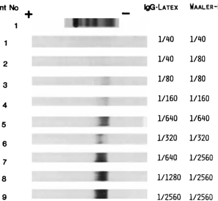
RF were undetectable by FAT in normal serum pools (not shown). In contrast, FAT allowed an easy detection of RF in RA sera with RF titers as low as 1/40 by the IgG-latex agglu- tination test and the Waaler-Rose reaction (Fig. 1) and in the sera containing monoclonal IgM or IgA with known RF activ- ity (Fig. 2). It is worth noting that several RA sera (at least Nos. 3 to 6 on Fig. 1) clearly contained small amounts of homogeneous RF (narrow bands on the IgG-coated nitrocel- lulose). These narrow bands were undetectable by electro- phoresis nor on standard immunoblots. As for monoclonal RF containing sera, comparison of the blots of the same elec- trophoresis onto uncoated and IgG-coated nitrocellulose allowed an easy identification of monoclonal RF. Monoclonal Ig lacking RF activity were consistently negative. In some of the latter sera, polyclonal RF or discrete narrow bands of monoclonal R F which were clearly distinct from the major monoclonal Ig were detectable, generally together with a weak RF activity by agglutination tests. The internal controls in the experiment shown on Fig. 1 are sera containing monoclonal IgM (lanes 1) or IgA (lanes 3) without RF activity. As other examples of specificity controls of the FAT, Figure 3 shows a CLL serum with no detectable RF by the latex agglutination test and Waaler-Rose reaction (this serum contained small amounts' of a monoclonal R F which was otherwise undetect- able) and Figure 4 the serum from a patient with WM and a peripheral neuropathy and an anti-MAG activity of the monoclonal IgM (latex agglutination test and Waaler-Rose reac- tion titers were 1/360 and 1/40, respectively).
In addition to their charge homogeneity, monoclonal RF may also be identified on the basis of light chain restriction using the yHCD protein or IgG Fc fragments as antigen in the FAT. However, a correct saturation of the blots (and hence specificity) was more difficult to obtain than with IgG coated nitrocellulose and the specific staining of bound RF was not as strong.
DISCUSSION
We describe a new method for the characterization of RF which appears to be especially suitable for an evaluation of their monoclonality on the basis of charge homogeneity and light chain restriction. This method is simple and rapid and it does not need any sophisticated material or reagent, when applied to the detection of R F belonging to Ig classes other than IgG. It can be used on a large scale to screen monoclonal IgM or IgA for RF activity or to evaluate the mono- or oligoclonality of RF in inflammatory diseases. Conceivably, FAT could also be used to detect IgG RF, using Fc fragments as antigen in the FAT and antibodies specific for IgG Fab frag- ments as revelating antibodies, for instance. RF display a
A
+
PatientNo
1 I IQG-LATEXWAALER-ROSE
1/40 1/40 12
3
4 1/40 1/80 1/80 1/80 1/1601/160
1/640 1/640 1/320 1/320 1/640 1/2560 5 6 7 8 1/1280 1/2560 91/2560
1/2560Fig. 1. FAT study of nine individual RA sera (revelation with anti-p con-
jugate). The electrophoresis of the first RA serum is shown for comparison and the RF titers of the RA sera are indicated on the right part of the figure.
EP N IgG EP N IgG
Fig. 2. Comparative study of monoclonal IgM and IgA with and without
known anti-IgG antibody activity. The FAT blots are revealed with anti-p
(A) or anti-a (B) conjugates. Lanes 1 contain a WM serum without known antibody activity of the monoclonal IgM, lanes 2 a WM serum with a
monoclonal IgMh displaying a RF activity (as shown by the study of RF isolated by affinity chromatography and by a latex agglutination test posi- tive at lo-&), lanes 3 a control serum with a non-RF monoclonal IgA and lanes 4 the serum from a patient with hypergammaglobulinemic purpura
and a monoclonal IgAK RF (3). EP, agarose electrophoresis; N, FAT experi-
ment, normal uncoated nitrocellulose; IgG, FAT experiment, polyclonal IgG- coated nitrocellulose.
stronger affinity for polymeric than for monomeric IgG. IgG bound to a solid phase are therefore well suited for their detec- tion. Even IgG-RF complexes preexisting in vivo appear to be displaced enough for RF binding to the affinity filter to occur. Monoclonal IgM with known R F specificity yielded narrow bands by FAT whereas the monoclonal IgA from the patient with hypergammaglobulinemic purpura did not (Fig. 2B). This IgA was electrophoretically homogeneous when isolated but was diffuse in the whole serum due to high level of binding to polyclonal IgG (3). Residual complexes bound to the affinity filter likely explain the charge heteroge- neity of this monoclonal IgA by FAT. This example illustrates a limitation of the method for evidencing the monoclonality of certain RF. The difference between this IgA RF and IgM RF is possibly due to a stronger affinity of the IgA for mono- meric IgG which might reflect differences in the variable regions orland polymerization of the monoclonal IgA (14). In such cases, other methods, such as affinity chromatogra- phy isolation of RF after immune complex dissociation (3),
appear to be required.
Under the present technical conditions, FAT is specific, as shown by the negative internal controls with irrevelant WM sera. This specificity is dependent on the efficacy of the sat- uration of the coated nitrocellulose. Filters were easily and regularly properly saturated after coating with polyclonal IgG.
A
B
EP N W
Fig. 3. Electrophoresis (EP), standard Western blots ( A ) and FAT (B, N,
normal uncoated nitrocelllose; IgG, polyclonal IgG-coated nitrocellulose) of a CLL serum containing oligoclonal Ig (IgMX, IgMh, IgGK, 1gGX) with- out RF activity by FAT and a narrow band of IgM anti-IgG that is undetect- able by electrophoresis and on standard blots.
-
EP
Fig. 4. Study of a WM serum with a monoclonal IgM anti-MAG. The latter
monoclonal Ig is negative by FAT, as expected, but FAT discloses the presence of three narrow bands of IgM anti-IgG that are otherwise undetectable. After coating with the y HCD protein or with Fc fragments, an additional saturation step was required. There is, hence, clearly a need to control the binding specificity of every newly prepared affinity filter. In fact, we include an internal control using a WM serum with no RF activity of the monoclonal IgM in every experiment.
FAT detects polyclonal RF in RA sera with RF titers as low as 1/40 by agglutination tests. Being a solid phase assay, it is likely to detect monomeric IgM RF which might be pres- ent in certain RA sera and do not yield positive agglutination reactions (15). It is not sensitive enough to detect RF in nor- mal sera, which is rather an advantage in clinical terms. FAT is highly resolutive, and narrow bands of IgM with RF activ-
Filter Affinity Transfer for RF 381
ity can be detected in the serum of patients with immuno- proliferative disorders or even with autoimmune disease. Such oligoclonal RF are likely present in minute amounts since they were undetected by standard immunoblotting which allows the identification of monoclonal Ig in concen- trations as low as 25 pgiml (11). The finding of homoge- neous RF amongst polyclonal RF in autoimmune processes was not unexpected but its significance remains to be estab- lished (1,2,4,5). In the present study, we merely examined technical parameters but in the near future we intend to apply FAT to the screening of a large number of sera from autoim- mune patients to evaluate the precise incidence of oligoclonal RF under such conditions.
REFERENCES 1. 2. 3. 4. 5. 6. 7. 8. 9. 10. 11. 12. 13. 14 15
Carson DA, Chen PP, Fox RI, Kipps TJ, Jirik F, Golfien RD, Silverman
G, Radoux V, Fong S: Rheumatoid factors and immune networks. Annu Rev Immunol5:109-126, 1987.
Shakib F (ed): Autoantibodies to immunoglobulins. Monogr Allergy 26:l-81, 1989.
Preud’homme JL, Duarte F, Aucouturier P Isolation of monoclonal rheu- matoid factors in hypergammaglobulinemic purpura. Diagn Immunol Chu JL, Gharavi AE, Elkon KB: Spectrotypic analysis of IgM and IgA rheumatoid factors. Clin Exp Immunol63:601-607, 1986.
Bouvet JP, Pillot J: Restricted heterogeneity of polyclonal rheumatoid factors. Arthritis Rheum 30:998-1005, 1987.
Erlich HA, Levinson JR, Cohen SN, McDevitt HO: Filter affinity trans-
fer. A new technique for the in situ identification of proteins in gels. J
Biol Chem 254:12240-12247, 1979.
MacLachlan R: Monoclonal immunoglobulins: affinity blotting for low concentrations in serum. Clin Chem 35:478-480, 1989.
Domes R, Ter Menlen V: Detection and identification of virus-specific oligoclonal IgG in unconcentrated cerebrospinal fluid by immunoblot technique. J Neuroimmunol7:77-89, 1984.
CNZ M, Olsson T, Ernerudh J, Hojeberg B, Link H: Immunoblot detec- tion of oligoclonal anti-myelin basic protein IgG antibodies in cerebro- spinal fluids in multiple sclerosis. Neurol 37: 1515- 15 19, 1987. Jauberteau MO, Cook JM, Drouet M, Preud’homme JL: Affinity immunoblotting detection of serum monoclonal immunoglobulins reac- tive with glycosphingolipids. J Immunol Methods 134:107-112, 1990. Aucouturier P, Capella M, Briault S , Danon F, Intrator L, Preud’homrne JL: Caractkrisation des immunoglobulines monoclonales dans les liquides biologiques par immunoempreinte sur nitrocellulose. Rev Inst Pasteur Lyon 20:147-153, 1987.
Briault S , Courtois-Capella M, Duarte F, Aucouturier P, Preud’homme
J L Isotypy of serum monoclonal immunoglobulins in human immuno- deficiency virus-infected adults. Clin Exp Immunol74: 182-184, 1988. Mihaesco E , Guglielmi P, Brouet JC, Mihaesco C: Biochemical and biosynthetic studies of a crystallizable human 1 heavy chain disease protein. Scand JImmunol 18:145-151, 1983.
Schrohenloher RE, Koopman WJ, Moldoveanu Z, Solomon A: Activ-
ity of rheumatoid factors of different molecular sizes: comparison of autologous monomeric and polymeric monoclonal IgA rheumatoid fac- tors. J Immunol 134:1469-1474, 1985.
Bonagura VR, Mendez L, Agostino N, Pernis B: Monomeric ( 7 s ) IgM found in the serum of rheumatoid arthritis patients share idiotypes with pentameric (19s) monoclonal rheumatoid factors. J Clin Invest 79:813-818, 1987.