HAL Id: cea-01119512
https://hal-cea.archives-ouvertes.fr/cea-01119512
Submitted on 24 Feb 2015
HAL is a multi-disciplinary open access
archive for the deposit and dissemination of
sci-entific research documents, whether they are
pub-lished or not. The documents may come from
teaching and research institutions in France or
abroad, or from public or private research centers.
L’archive ouverte pluridisciplinaire HAL, est
destinée au dépôt et à la diffusion de documents
scientifiques de niveau recherche, publiés ou non,
émanant des établissements d’enseignement et de
recherche français ou étrangers, des laboratoires
publics ou privés.
Distributed under a Creative Commons Attribution - ShareAlike| 4.0 International
License
Optimization of sample preparation for MRI of
formaldehyde-fixed brains
Yann Leprince, Benoît Schmitt, Christophe Destrieux, Laurent Barantin,
Alexandre Vignaud, Denis Rivière, Cyril Poupon
To cite this version:
Yann Leprince, Benoît Schmitt, Christophe Destrieux, Laurent Barantin, Alexandre Vignaud, et al..
Optimization of sample preparation for MRI of formaldehyde-fixed brains. 23rd Annual Meeting of
ISMRM, International Society for Magnetic Resonance in Medicine, May 2015, Toronto, Canada.
�cea-01119512�
3616
Optimization of sample preparation for MRI of formaldehyde-fixed brains
Yann Leprince1,2, Benoît Schmitt1, Élodie Chaillou3, Christophe Destrieux4, Laurent Barantin4, Alexandre Vignaud1, Denis Rivière1, and Cyril Poupon1
1NeuroSpin, CEA, Saclay, France, 2Université Paris-Sud, Orsay, France, 3INRA, Tours, France, 4Université François-Rabelais, Tours, France
Target audience: researchers who perform MR acquisitions on formaldehyde-fixed post-mortem brains.
Purpose: Magnetic resonance imaging of post-mortem brains allows long acquisition times up to several days and can be used to obtain high-resolution images at high field (7 T) which can be readily correlated with histological examination of the tissue. However, death and formaldehyde fixation are known to modify severely the relaxivity and diffusion properties of brain tissue.1,2 In particular, formaldehyde is known to shorten T
2, which drastically
reduces SNR.
In order to counteract this effect and recover better SNR, free fixative can be washed out by soaking the sample in isotonic saline solution. This has been demonstrated in small biopsy-sized tissue samples,3 but little data is available
concerning whole brain specimens.2
This study was designed to describe the kinetics of the change of relaxivity and diffusion properties of whole brain specimen at 7 T, during fixation, and during soaking in saline solution, in order to determine optimal soaking times. Methods: Two healthy ewes (adult female sheep ovis aries, 2 years old) were used for this study, in accordance with local animal regulation (Authorization A37801 at the French Ministry of Agriculture). The animals were euthanized through massive injection of barbiturate, after having received heparin to prevent coagulation. The head was separated from the body, and perfused through the carotid arteries with 4 L of PFA at 4°C (4% formaldehyde in phosphate-buffered saline, prepared from paraformaldehyde powder). The perfusion
happened within a few minutes of death, hence no degradation should happen during the post-mortem interval (PMI) between death and fixation. The brain was then extracted, immersed into PFA for post-fixation, and kept at 4°C before the first scan.
Samples were scanned repeatedly (23 and 19 times respectively during post-fixation of both samples in PFA, 19 times during saline soaking for one sample) on a clinical 7 T MRI system, mapping the following quantitative parameters over the whole brain. T1 was
mapped using variable flip angle–actual flip angle imaging4 (VAFI) at 1 mm isotropic
resolution. T2 was mapped at 1.7 mm isotropic resolution using spin echo EPI repeated
several times for a large window of TE. T2* was mapped using a multi-echo gradient echo
sequence with 1 mm isotropic resolution. Both T2 and T2* were fitted with least-squares
regression of a single exponential decay. Diffusion-weighted images were acquired using single shot spin echo EPI with 2 mm isotropic resolution, 256 diffusion directions, b = 4500 s/mm2. Mean diffusivity and fractional anisotropy were calculated with a
first-order tensor model. Regions of interest were drawn manually in 3D in sub-cortical white matter and caudate nuclei (see Figure 1), and were transformed into the referential of each quantitative map using rigid registration. Voxels affected by partial volume or artefacts due to air bubbles were excluded.
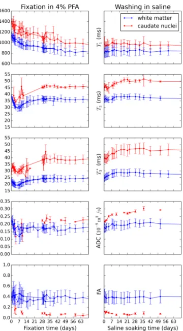
Results: The time course of the measured parameters is represented on Figure 2, over 10 weeks during fixation and saline soaking. The data for caudate nuclei is noisy with a few outliers, because they contain very few voxels for lower resolution measurements. T1
decreases during fixation for approximately 8 weeks, and is restored only marginally by saline. T2 and T2* decrease for the first week of fixation, then grow back to a plateau in
approximately 6 weeks. They grow during the first two weeks during saline soaking, then seem to decrease very slowly. The mean diffusivity decreases for the first week of fixation, and increases slightly in saline. Fractional anisotropy does not change significantly during the whole experiment.
Discussion: Good reproducibility is observed between subjects where the curves overlap. The slow decrease of T1 is consistent with previously published data,1 as well as the
decrease of diffusivity and stability of anisotropy.2 On the other hand, the rebound of T 2
and T2* after one week was not expected. A similar phenomenon was previously
attributed5 to early decomposition of unfixed deep tissue. However, the present study uses
perfusion fixation, which should stop such degradation by exposing all the tissue to fixative. Instead, we hypothesize that this rebound is due to the degradation of the PFA solution itself through precipitation of monomeric formaldehyde into paraformaldehyde, effectively lowering the formaldehyde concentration as the solution degrades. This could be avoided by regularly changing the fixative or by using formalin, which includes methanol to prevent polymerization of formaldehyde. Washing the tissue in saline had the intended effect of restoring higher T2, T2*, and diffusivity, although to a rather modest
degree, which can be explained if the fixative concentration decreased as hypothesized. Conclusion: The usefulness of the saline soaking procedure is assessed in terms of restoring T2, T2*, and diffusivity, which is particularly beneficial to post-mortem diffusion
imaging. The fixation was found to stabilize after approximately 8 weeks, and the optimal duration of saline soaking is found to be around 3 weeks. These durations can be expected to be longer for larger specimen, such as human brains, which require longer penetration times.
References: 1. Tovi M. and Ericsson A. Acta Radiol. 33, 400–404, 1992. 2. D’Arceuil H. E. et al. NeuroImage 35, 553–565, 2007. 3. Shepherd T. M. et al. MRM 62, 26–34, 2009. 4. Hurley S. A. et al. MRM 68, 54–64, 2012. 5. Dawe R. et al. MRM 61, 810–818 (2009).
Figure 1: 2D snapshot of a high-resolution Turbo Spin Echo scan of one subject and associated 3D ROIs.
Figure 2: time course of measurements during fixation and saline soaking. Error bars represent mean ± standard deviation in each
![[PDF] Cours Analyse et Conception Merise Pdf](data:image/gif;base64,R0lGODlhAQABAIAAAP///wAAACH5BAEAAAAALAAAAAABAAEAAAICRAEAOw==)