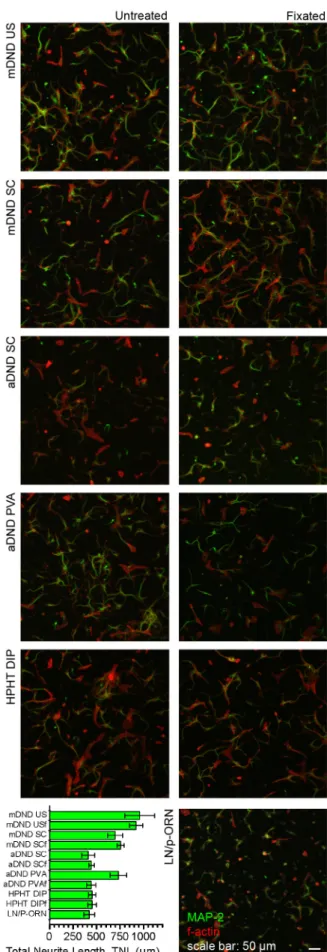
Patterned neuronal networks using nanodiamonds and the effect of varying nanodiamond properties on neuronal adhesion and outgrowth
Texte intégral
Figure


Documents relatifs
14 plots the evolution of the normalised real contact area as a function of the normal load for respectively (i) the thermal models, (ii) the elastic GW model and, (iii) the FE
We presented a complete framework for continuous prediction of human emotions based on features characterizing head movements, face appearance and voice in a
After regressing the cross-sectional Vasicek adjusted betas over the excess returns of the stocks for each one of the 44 years for which data were available,
In each session, multi-unit activity (MUA) and local field potential (LFP) were recorded bilaterally, while monkey performed these three tasks. In the following
Here we provide evidence that Piwi is expressed, but normally at hardly detectable levels, in the Drosophila brain and VNC, while it is strongly induced in those tissues
Nous serons ainsi en situation de réelle simulation et pourrons constater les effets que ces quantités d’eau soutirées de la réserve d’eau chaude peuvent avoir sur le chauffage de
In contrast, the excess charge found for GlyT2a (z T ≅ +2) is not compatible with this stoichiometry. The additional charge does not result from an absence of net Cl -
The reduction in Q max (see table 2), which is proportional to the number of functional transporters present in the plasma membrane (see methods), indicates that the loss
