Original article
11
C-acetate PET in the early evaluation of prostate cancer
recurrence
Susanne Albrecht1, Franz Buchegger1, 2, Dmitri Soloviev1, Habib Zaidi1, Hansjoerg Vees3, Haleem G. Khan4, Alain Keller1,
Angelika Bischof Delaloye2, Osman Ratib1, Raymond Miralbell3, 5
1Service of Nuclear Medicine, University Hospital of Geneva, Rue Micheli-du-Crest 24, 1211 Geneva 14, Switzerland 2Service of Nuclear Medicine, University Hospital of Lausanne, Lausanne, Switzerland
3Service of Radiation Oncology, University Hospital of Geneva, Geneva, Switzerland 4Institute of Radiology Jean Violette, Geneva, Switzerland
5Instituto Oncológico Teknon, Barcelona, Spain
Received: 14 February 2006 / Revised: 11 April 2006 / Accepted: 30 April 2006 / Published online: 11 July 2006 © Springer-Verlag 2006
Abstract. Purpose: The first aim of the study was to investigate the diagnostic potential of 11C-acetate PET in the early detection of prostate cancer recurrence. A second aim was the evaluation of early and late PET in this context.
Methods: The study population comprised 32 prostate cancer patients with early evidence of relapse after initial radiotherapy (group A) or radical surgery (group B). The median PSA of group A (n=17) patients was 6 ng/ml (range 2.6–30.2) while that of group B (n=15) was 0.4 ng/ ml (range 0.08–4.8). Pelvic-abdominal-thoracic PET was started 2 min after injection of 11C-acetate and evaluated after fusion with CT.
Results: Group A: Taking a SUVmax≥2 as the cut-off, PET showed local recurrences in 14/17 patients and two equivocal results. Distant disease was observed in six patients and an equivocal result was obtained in one. Endorectal MRI was positive in 12/12 patients. Biopsy confirmed local recurrence in six of six (100%) patients. PET was positive in five of the six patients with biopsy-proven recurrences, the result in the remaining patient being equivocal. Group B: Among the 15 patients, visual interpretation was positive for local recurrences in five patients and equivocal in four. One obturator lymph node was positive. Endorectal MRI was positive in 11/15 patients and equivocal in two. Positional correlation of positive/equivocal results on PET and endorectal MRI was observed in seven of nine patients. PSA decreased significantly after salvage radiotherapy in 8/14 patients, providing strong evidence for local recurrence. PET of the eight patients responding to RT was positive in three and equivocal in two.
Conclusion:11C-acetate PET was found to be valuable in the early evaluation of prostate cancer relapse. Optimising scanning time and use of modern PET-CT equipment might allow further improvement.
Keywords:11C-acetate – PET – Prostate cancer relapse – PSA
Eur J Nucl Med Mol Imaging (2007) 34:185–196 DOI 10.1007/s00259-006-0163-x
Introduction
Conventional radiology of recurrent prostate cancer has a low sensitivity and specificity because of difficulties in differentiating between recurrent tumour and repair tissue after surgery and/or radiation therapy (RT).18 F-fluoro-deoxyglucose positron emission tomography (FDG-PET) also has a low sensitivity owing to the modest glucose consumption of these tumours and the proximity of the urinary tract, with high background activity [1,2].
Promising results in the detection of recurrent prostate cancer have been obtained with the newer PET tracers11 C-acetate, 11C-choline and 18F-fluorocholine [3–7]. Radio-labelled acetate and choline are partially integrated as phosphatidylcholines into the cellular membranes of proliferating cells, another part being catabolised [8]. PET with these tracers could thus show biological functionality of radiologically suspicious lesions.
Recently, nanoparticle-enhanced magnetic resonance imaging (MRI) has emerged, yielding encouraging results [9,10]. Endorectal MRI could provide the most interesting information concerning local disease [11,12], notably in distinguishing T2 and T3 stages [13]. However, the interpretation following radiotherapy remains challenging owing to the difficulty in differentiating between post-RT
Susanne Albrecht ()) Service of Nuclear Medicine, University Hospital of Geneva, Rue Micheli-du-Crest 24, 1211 Geneva 14, Switzerland
e-mail: [email protected] Tel.: +41-21-3723311, Fax: +41-21-3727169
signal modifications and tumour relapse [14]. In the latter situation, a higher specificity has been described for MR spectroscopy [14] than for endorectal MRI.
Independent of the radiological evaluations, tumour relapse is, as would be expected, more frequent in high-risk prostate cancer patients. A consensus statement of the American Joint Committee on Cancer (AJCC) defined advanced T staging (T≥pT2c), high tumour grades (sum of Gleason score ≥8) and high pre-surgical serum prostate-specific antigen (PSA) values (>20 ng/ml) as the three major risk parameters of prostate cancer [15]. Positive surgical margins, seminal vesicle invasion and capsular involvement represent further risk parameters [16–18].
Several of the initial11C-acetate or11C- or18F-choline PET studies [3–7] were performed in patients with rather high PSA values, suggestive of advanced disease. A more significant contribution for patient management might be expected from PET performed at earlier stages of tumour recurrence. However, a first evaluation of11C-acetate PET showed a low sensitivity of only 7% in post-surgical patients with PSA values below 3.0 ng/ml [4].
Definition of biochemical recurrence is clearly different after initial radical surgery or radiotherapy. Radical surgery eliminates all prostate tissue and PSA is expected to fall below the detection limit. Thus, recurrence in these patients is already suspected at the earliest rise in PSA [19–21]. It has been shown that even very low PSA values of 0.1– 0.2 ng/ml following radical surgery represent a probability for further rise in PSA and tumour recurrence of 90% [22]. The risk would be 67% at PSA values≤0.1 ng/ml according to this study [22].
Regarding patients with first-line RT, definition of recurrence by reference to rising PSA remains a matter of debate. An ASTRO consensus statement of 1997 required the measurement of three consecutive increases in PSA [23]. More recently, a retrospective analysis of 4,800 patients [24] showed that other definitions of recurrence would be slightly more sensitive and specific than the ASTRO criterion. After RT, measurement of two con-secutive PSA increases of at least 0.5 ng/ml each, or a PSA value ≥2 ng/ml above nadir, would provide sensitivities and specificities for recurrence of between 64% and 78% [24]. Considering these rather modest results for sensitivity and specificity, it is obvious that the definition of treatment failure following RT remains difficult. A strong suspicion of recurrence will generally be established at PSA values significantly higher than 2 ng/ml.
Salvage RT of post-surgical patients with recurrent cancer has been studied by different groups. It has been shown that a PSA value >2 ng/ml is a bad prognostic factor [25]. Accordingly, salvage RT should be performed as early as possible. As a challenge for PET, the most interesting patient population in the post-surgical situation would therefore present very low PSA values. Tumour size in these patients will be particularly small and PET partial volume effects will be frequently observed. As a consequence, the predictive value of the standardised uptake value (SUV) in these patients might be compro-mised. As mentioned, a first evaluation of11C-acetate PET
in post-surgical patients with PSA values below 3 ng/ml showed a disappointingly low sensitivity [4]. A commen-tary questioned the methodology of this study, such as the absence of SUV measurement and the low number of histological confirmations [26].
The first aim of the present study was to investigate the diagnostic potential of 11C-acetate PET in the early detection of prostate cancer recurrence. Patients were studied at biochemical relapse after initial RT (group A) or radical surgery (group B). Based on the above-mentioned considerations, SUVmax, though calculated for all areas of focal uptake, was not used in the PET interpretation of patients from group B.
A second aim was the evaluation of early and late PET. In fact, while several studies have suggested that tumour uptake of 11C-acetate is very rapid [7, 27], the retention time in tumours of individual patients has not been described. Late PET was performed with the intention of ascertaining whether individual tumours remained positive.
Materials and methods Patient population
All patients gave their written informed consent to the study protocol, which had been approved by the ethical commission of the hospital and the Swiss authorities (Swissmedic and Federal Office of Public Health, Section of Radioprotection). The consecutive PET studies presented here were performed from December 2004 to July 2005.
Two groups were evaluated. Patients of group A (n=17) had been treated initially with RT. Table1summarises their clinical status and their biochemical status initially and at PET. Median tumour radiation dose was 74 Gy (range 64–78.4 Gy). Nine (53%) of the 17 patients presented one or more of the three high-risk factors as previously described [15]. The rise in PSA after nadir post RT was significant and confirmed at least once.
Patients of group B (n=15, numbered 18–32) were treated initially with radical surgery. Table2summarises their clinical status and their biochemical status initially and at PET. All 15 patients presented one or more of the three high-risk factors [15]. Many patients of this group presented further risk factors [16–18], such as histologically positive surgical margins (n=11), invasion of seminal vesicles (n=4), capsular invasion (n=9) or perineural invasion (n=13).
11
C-acetate 11
C-acetate was prepared at the cyclotron unit of Geneva University Hospital from 11C-carbon dioxide produced on an IBA 18/9 cyclotron, according to a modified [28], previously published procedure [29]. In brief, 11C-acetate was prepared based on a combination of the captive solvent radiolabelling“Grignard” reac-tion [30] conducted in the sterile catheter extension tube, followed by the convenient solid-phase extraction purification [29] on a series of ion-exchange cartridges. The requested radiochemical purity of the patient formulations, as determined by high-performance liquid
chromatography, was ≥95%. Mean radiochemical purity of 11 C-acetate at injection was 98.2±1.6%.
PET, CT and fusion
The PET scanner used was the ECAT ART (Siemens/CTI, Knoxville, TN, USA). After bladder voiding, patients were placed in the scanning position and six-run transmission scanning (5 min/bed position) using 137Cs single-photon point sources was recorded starting at the prostate bed. The laser-defined starting position was marked in ink on the patient’s leg. Patients were then injected, applying standard precautions, with an activity of 520 MBq 11 C-acetate. This activity was selected in order to cover the long scanning time of a trunk scan as performed here. The emission scan was started 2 min after acetate injection, comparable with previous studies [7,
27]. The initial run of 10 min was centred on the prostate bed, followed by five runs of seven minutes each covering the rest of the pelvis, abdomen and thorax. A final run of 10 min, started at 47 min post injection, was again again on the prostate bed.
In a typical PET scan and for a patient injected with 520 MBq
11
C-acetate, the number of trues per bed position was about 12– 18×106counts on the first six runs and about 8×10⁶counts on the
final prostate bed-centred run. The initial modest number of trues per time unit reflects the low sensitivity of the partial-ring tomograph and its low count rate performance for the relatively high activity injected since the peak noise equivalent count rate for a 20-cm-diameter cylinder is 27 kcps at a concentration of 15 kBq/ml [31]. It should be emphasised that the performance of the camera can be improved by operating the scanner with a decreased block integration time and reduced coincidence time window [32].
CT imaging was performed between 2 and 48 h after the PET scan on a Siemens Sensation 16 scanner. The starting position of the dedicated fusion CT, marked with two metallic needles, was identical to that of the PET scan (ink marks), as verified by the same physician. PET-CT image fusion in the first four patients used the standard contrast-enhanced thoracic-abdominal-pelvic CT. It showed the major difficulties in the alignment of PET and CT data acquired in different scanning positions and with different breathing. Subse-quently, patients had a non-contrast-enhanced CT for image fusion acquired in the PET position (arms held along the body) and with flat breathing. The non-contrast-enhanced CT was finally modified to a low-dose CT [33] in the PET position and flat breathing using 120 kV, 60 mAs and a pitch of 1.5 and a 1-s rotation. Under the latter condition, the mean effective radiation dose for an adult patient was 3.5 mSv, as calculated using the IMPACT CT patient dosimetry calculator (http://www.impactscan.org/ctdosimetry.htm).
PET images were reconstructed using iterative attenuation-weighted ordered subset expectation maximisation (OSEM). An attenuation correction matrix was calculated by segmenting the attenuation map, followed by forward projection at appropriate angles of the transmission image [34]. The generated attenuation correction map was used to reconstruct the emission data. The images were scatter corrected and reconstructed by using normalised attenuation-weighted, OSEM iterative reconstruction implemented with the ECAT 7.2 software. The default parameters used were OSEM iterative reconstruction with two iterations and eight subsets followed by a post-processing Gaussian filter (kernel full-width half-maximal height, 6 mm). The voxel size was set to 3.4×3.4×3.4 mm3. Attenuation-corrected views were obtained in transaxial, coronal and sagittal planes.
One of the surgically treated patients (patient 26) had a hip prosthesis that led to a CT artefact projecting on the prostate bed. His PET/CT fusion scan was therefore analysed without and with attenuation correction. All other patients had their attenuation-corrected PET/CT fusion scan evaluated.
PET to CT image co-registration was performed using the commercial Hermes multi-modality fusion software (Nuclear Diag-nostics AB, Stockholm, Sweden).
Standardised uptake values (SUV) were calculated [35] as follows: SUV=lesion activity per gram/(injected activity/patient body mass (g)). Mean and maximal SUV were calculated for ROIs of focal uptake and compared with adequate control regions to derive tumour to non-tumour (T/N) ratios.
A standard, contrast-enhanced CT was available for all patients. It was consulted for PET interpretation if suspicious foci of uptake in the abdomino-pelvic region suggested the presence of adenopathies or if other structures generated focal uptake, such as degenerative osseous disease, and the low-dose CT did not present the necessary resolution to allow their clear definition.
Table 1. Characteristics of 17 group A patients treated initially by RT Age (yrs) Mean 72.5 Range 58–87 Initial stage cT1 n=4 cT2a–b n=4 cT3a n=7 n.a. n=2 Gleason score 4–6 n=7 7 n=5 >7a n=2 n.a. n=3 Initial PSA (ng/ml) <10 n=2 10–20 n=9 >20a n=4 n.a. n=2
Initial RT dose (Gy)
Mean 73.9 Range 64–78.4 PSA at PET (ng/ml) Mean 10.4 Range 2.6–30.2 Median 6.0
PSA doubling time (months)
Mean 8.2
Range 1–24
Time from RT (months)
Mean 61.7
Range 27.5–100.7
PET interpretation was performed by two experienced nuclear medicine physicians who gave their final consensus statement in the clinical report as tumour positive, equivocal or tumour negative.
Salvage RT and follow-up
Salvage RT in post-surgical patients was performed with 18 MV-X-rays using a Clinac 2100-C linear accelerator and an irradiation technique of four fields (anterior, posterior and two lateral). RT was delivered in five weekly fractions of 2 Gy/day to a total of 32 fractions (64 Gy) directed on the prostate bed. A boost dose of five fractions (10 Gy) was delivered to tumour recurrences, whenever such a recurrence was shown by endorectal MRI and/or11C-acetate PET.
Provided there was an adequate interval from initial RT and absence of distant disease in patients of group A, in these patients a second, salvage RT was proposed after hormonal therapy. Secondary salvage RT used 18-MV X-rays generated by the Clinac 2100-C linear accelerator and administered using the standard protocol of 5× weekly at 1.8 Gy/day up to 45 Gy (25 fractions). External beam RT was then completed with 18 Gy brachytherapy.
All included patients accepted the complementary evaluation with endorectal MRI and a diagnostic contrast-enhanced thoraco-abdominal-pelvic CT. Bone scintigraphy was performed in eight patients of group A and in seven patients of group B.
Patients of group A had various further evaluations. Biopsies as the gold standard for local disease were indicated prior to salvage RT in the absence of evidence of systemic disease. Endorectal MRI was performed in 12 patients and completed with MR spectroscopy in nine [14]. Patients with disseminated disease as shown by radiology received hormonal therapy alone.
Patients who had undergone initial radical surgery were proposed salvage RT based on their situation of high-risk prostate cancer in the absence of evidence, with the exception of a single positive obturator lymph node in one patient, of dissemination both at initial surgery and at relapse. One patient of this group with a particularly aggressive tumour (Gleason 5+5) was initially given anti-hormonal treatment, followed by salvage RT.
Response to salvage RT was taken as a safe indication for local relapse. Thus, a reduction in the PSA value by≥50% 6 weeks after completion of RT, compared with the initial value, served as surrogate proof for the presence of tumour. Biochemical response after salvage RT, whereby PSA values had decreased to less than 0.04 ng/ml (detection limit), indicated an optimal local response, providing the strongest evidence for the presence of localised disease alone.
Statistical analysis
For evaluation of the statistical correlation between early and late prostate PET, the inter-category variation kappa test was performed
Table 2. Characteristics of 15 group B patients treated initially by radical surgery Patient Age (yrs) Stage Gleason score PSA initial/after surgery(ng/ml) Surgical margins
Infiltrations PSA doubling time (months)
Time from surgery (months)
PSA at PET (ng/ml) 18 48 pT3ba 3+4 79.5a/0.5 Neg. Perineural, seminal
vesicle
7 18.3 2.4
19 70 pT3aa 4+3 8.5/0.11 Pos. Perineural, capsular,
vascular
1 10.0 3.6
20 68 pT3aa 3+4 10.5/0.05 Pos. Perineural, capsular 15 47.7 0.76
21 62 pT3aa 3+4 7.8/<0.1 Neg Perineural, capsular 6 34.0 0.33
22 64 pT3aa 4+3 n.a./0.2 Pos. Perineural 30 71.8 4.8
23 63 pT2ca 3+3 13.2/0.06 Pos. Perineural 12 24.1 0.26
24 73 pT3aa 3+4 9.1/0.05 Neg. Perineural, capsular 24 52.4 0.3
25 73 pT3ba 3+3 10/0.24 Pos. Perineural, capsular,
seminal vesicle
Postop. 3.0 0.24
26 55 pT2b 9a 6.2/0.9 NA NA 12 52.0 3.10
27 65 pT3aa 4+5a 33.2a/0.36 Pos. Perineural Postop. 5.0 0.40
28 71 pT3ba 4+3 7.1/<0.04 Pos. Perineural, capsular, seminal vesicle
12 27.3 0.08
29 63 pT3ba 5+5a 6.3/0.51 Pos. Perineural, capsular, seminal vesicle
Postop. 3.1 0.51
30 67 pT3aa 4+3 4.9/0.05 Pos. Perineural, capsular 2 7.0 0.19
31 70 pT3aa 3+4 5.5/<0.04 Pos. Perineural, capsular 8 14.0 0.20
32 66 pT2b 3+3 23.0a/0.19 Pos. Neg. 36 44.4 0.39
Mean 65.2 7.3 16.1/0.22 13.8 27.6 1.17
SD 6.7 1.2 19.9/0.25 10.9 21.8 1.52
Median 66.0 7.0 8.8/0.11 12.0 24.1 0.39
Neg. negative, Pos. positive, NA not available, Postop. postoperative aIndicates high-risk patient characteristics
using the UNISTAT 5.5 statistical package for Windows (2002 edition, Unistat Ltd, London, England, run on Windows XP) comparing positive, suspicious and negative results. Ap value <0.05 was considered significant. Un-weighted kappa values of 0.01–0.19, 0.2–0.39, 0.4–0.59, 0.6–0.79 and 0.8–1.0 were rated as slight, fair, moderate, substantial and almost perfect agreement, respectively [36].
Results
All 11C-acetate injections (518±18 MBq, radiochemical purity 98.2±1.6%) were well tolerated.
The principal characteristics of the 17 patients in group A, treated initially with RT, are summarised in Table 1. Nine patients (53%) presented one or more high risk factors. The rise in PSA in these patients was significant, reaching a median value of 6 ng/ml at PET. The median delay between initial radiotherapy and PET was 61.7 months.
The characteristics of the 15 patients in group B are presented in Table2. All were at high risk, presenting one or more of the three major risk determinants according to the consensus statement (Table 2). Eleven patients had histologically positive surgical margins, highly suggestive of residual local disease. Twelve patients had a confirmed increase in PSA observed 7–52 months after surgery. The three other patients underwent the 11C-acetate PET between 3 and 5 months post radical surgery based on a PSA that did not return to baseline but remained elevated at between 0.24 and 0.51 ng/ml. These elevated values were confirmed before salvage RT. The median delay between surgery and PET was 24.1 months, and a median PSA of 0.4 ng/ml was measured within 1 month of PET. PSA was below 0.8 ng/ml in 11 of 15 patients.
PET in patients treated initially with RT
In group A, focal uptake with an SUVmax≥2 were considered abnormal. Focal uptake <2 was considered equivocal. According to these criteria, 14 patients (82%) were positive (Figs.1,2) and two (12%) equivocal for local recurrence. Mean SUVmaxof positive and equivocal results was 2.9 and the mean T/N ratio was 2.4 (Table3).
With regard to lymph node disease, five patients showed focal11C-acetate uptake. In detail, three patients had lymph nodes with a SUVmaxhigher than 2.0, another patient had one node that was clearly positive and one that was equivocal, and one patient had one equivocal result. In comparison with CT, three patients (patients 2, 3 and 9) had positive or equivocal11C-acetate PET for adenopathies as defined by CT with lymph node diameters ≥1 cm. Two patients had a positive or equivocal PET scan (patients 16 and 13, respectively) for lymph nodes smaller than 1 cm in diameter that were not considered suspicious on the basis of radiological criteria.
With regard to M staging, a biopsy-proven metastasis of the corpus cavernosum was highly positive on PET (SUVmax 6.3). Contrast-enhanced CT, performed in all
patients, and bone scintigraphy, performed in eight patients, were positive or suspicious for bone metastases in four patients. However, only two of the multiple vertebral metastases were rated positive by PET (in patient 15) while three suspicious lesions of the ribs and two lumbar vertebrae were negative on PET. One suspicious lesion of the skull was outside the PET scanning field.
At late pelvic PET, following the observation of decreasing SUVmaxof initially positive lesions, a positive cut-off level of 1.5 was used. Late PET of the prostate bed
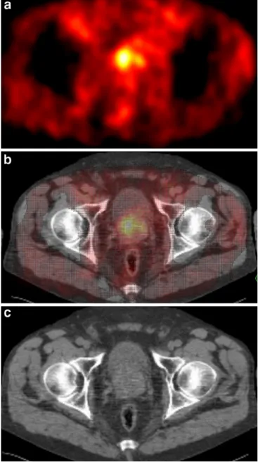
Fig. 1.11C-acetate PET of patient 8 performed 30 months post RT. PSA before RT combined with hormonal therapy was 21.8 ng/ml and it fell to 0.3 ng/ml after treatment. PSA later rose rapidly to 8.0 ng/ml at PET, with a short doubling time of 2 months. PET scan at seminal vesicle level (a) is shown, together with the correspond-ing fused imagcorrespond-ing (b) and CT (c). Note the focal uptake on the right, with a SUVmaxof 2.0, and a smaller focus on the left. Endorectal
MRI (not shown) completed with MR spectroscopy showed an extended lesion infiltrating bilaterally into the seminal vesicles
was positive for local recurrence in 13 patients (77%) and equivocal in three (18%). The mean SUVmax of positive/ equivocal results was 2.0±0.7 and the mean T/N ratio, 2.3±0.6. When analysing early and late PET for correlation of positive, equivocal and negative results of local
recurrence, concordance (p<0.05) was observed but kappa was moderate, with a value of 0.49.
Endorectal MRI was performed in 12 patients and completed with MR spectroscopy in nine. It indicated the presence of local recurrence(s) in all evaluated patients.
Definitive confirmation by histology (the gold standard for the presence of local disease) was indicated in patients who presented evidence of exclusive local recurrence. Biopsies were obtained from six patients and were all positive (100%). In comparison to initial grading, the Gleason score remained identical in three patients and increased by 1 point in the other three.
With regard to biopsy-proven recurrences (n=6), PET was positive in five of these patients and equivocal in one.
PET in surgically treated patients
The prostate bed of patients of group B was assumed to be empty of any prostate tissue after initial radical surgery. Furthermore, the interval between surgery and PET was long, at 3 months or more. Any post-surgical inflammatory response was therefore expected to have disappeared. Furthermore, the very low PSA values in many of these patients indicated the presence of very small tumour recurrences/residues. This hypothesis was supported by endorectal MRI, which showed the prevalence of fre-quently single, small tumour lesions. For these reasons, the SUVmax values, while determined, were not used in the interpretation of the PET scan, since partial volume effects would artificially diminish these values.
Reading of the attenuation-corrected PET/CT fusion scans revealed five positive (Figs.3,4) and four equivocal results for local recurrence (Table4). A single lymph node of the obturator region was interpreted as positive in patient 32, its SUVmax being 2.6. No further metastasis was detected in this group. The mean SUVmax of locally positive and equivocal results was 2.1 and 1.5, respectively, while the corresponding T/N ratios were 1.9 and 1.6.
Eleven patients of group B had a PSA value <0.8 ng/ml at PET. Four were positive and two equivocal for a local recurrence.
The late PET centred on the prostate bed showed five positive and three equivocal results. In comparison to the early PET, the late scan showed one additional equivocal result while one early equivocal result became positive on the late scan. When analysing the correlation of early and late PET for local recurrence according to positive, equivocal and negative results, a significant concordance (p<0.001) was observed; however, the kappa coefficient was moderate, with a value of 0.59. Mean SUVmaxof the late positive and equivocal results was 2.0 and 1.8, respectively, while the corresponding T/N ratios were 2.1 and 1.6 (Table4).
Endorectal MRI was performed in all 15 post-surgical patients (Figs.3,4, Table4). It was interpreted as locally positive in 11 and suspicious in two cases. In the two patients with negative endorectal MRI, 11C-acetate PET was also negative in one patient and equivocal in the late
Fig. 2.11C-acetate PET of patient 7 performed 62 months post RT. PSA before RT was 6.3 ng/ml and it later rose again to 4.5 ng/ml at PET, with a doubling time of 8 months. PET scan at the prostate level (a) is shown, together with the corresponding fused imaging (b) and CT (c). Note the focal uptake on the right, with a SUVmaxof
2.7, and a smaller focus on the left, correlated with positive endorectal MRI. Rectal activity was high in this patient
scan in the other. Positive/equivocal results of endorectal MRI in these patients concerned mostly a single focal lesion. An analysis of the spatial correlation between PET and MRI was performed according to the location of the observed recurrence. For the nine patients with positive/ equivocal results on both examinations, a spatial correla-tion was observed in seven (78%).
With regard to the single positive lymph node observed by PET in patient 32, this node was negative by other radiological criteria: ferromagnetic nanoparticle-enhanced abdominal MRI was interpreted as negative owing to the small size of the node (0.7 cm, measured on CT), a frank hypo-signal and uptake of the contrast medium. Endorectal MRI showed only a non-specific obturator lymph node signal.
Overall, no further suspicion of metastases in group B patients was noted except the obturator lymph node in patient 32. Contrast-enhanced whole-body CT was nega-tive in all patients. Complementary bone scintigraphy performed in seven patients confirmed the suspicion of Paget’s disease in one patient but was negative for bone metastases. All patients were therefore amenable to salvage RT that also encompassed the suspicious obturator lymph node.
Fourteen patients were evaluated for PSA evolution after salvage RT. A significant reduction in PSA by≥50% after salvage RT was observed in eight patients (57%). Reduction in PSA after radiotherapy directed to the prostate bed is, in our opinion, close to representing
proof of local tumour recurrence or residue. Of the eight patients with positive response to RT, three had a positive PET while in two other patients PET was equivocal. PSA fell below the detection limit in three patients, suggesting eradication of recurrent local disease. Interestingly, the three patients with optimal PSA response after salvage RT presented a PSA at RT initiation of <0.4 ng/ml (range 0.19– 0.33), underlining the need to evaluate these patients at the earliest indication of recurrent disease. Only one patient (no. 19) had a frankly increasing PSA value after 1 month of RT, which was stopped and replaced by hormonal therapy. Five patients had no significant evolution of PSA at 6 weeks post salvage RT. One of these patients had a PSA of 0.08 ng/ml, the clinical relevance of which remains unclear currently.
Discussion
This study was performed in two groups of patients. Results of 11C-acetate PET in post-RT patients showed a high number of local recurrences. In all six patients in whom biopsies were obtained, histology confirmed the local recurrences. The PET results in this group of patients presenting moderate PSA values are thus in agreement with previous reports.
The second group of patients, who had initial radical surgery, was studied at very low PSA values with a median of only 0.4 ng/ml. Here,11C-acetate PET was positive or
Table 3. Results of 17 group A patients treated initially with RT. Results are indicated as positive (+), equivocal (Equiv) or negative (Neg)
Pat. no. 11C-acetate PET Endorectal MRI
(spectroscopy)
Biopsy Salvage RT post hormonal therapy T PET early T PET late N M T SUVmax early T/N ratio early N SUVmax M SUVmax
1 Equiv + Neg Neg 1.9 1.9 + + Yes
2 + + + Neg 2.4 4.0 4.4/3.0 + +
3 Neg Neg + Neg 5.8 ND
4 + Equiv Neg Neg 2.5 2.7 + + Yes
5 Equiv Equiv Neg Neg 1.6 1.3 +
6 + + Neg Neg 3.8 3.8 + + Yes
7 + + Neg Neg 2.7 2.1 +
8 + Equiv Neg Neg 2.0 2.2 +
9 + + + + 4.1 2.7 3.4/1.8 6.3 ND +
10 + + Neg Neg 2.1 1.9 ND
11 + + Neg Neg 3.9 3.3 + + Yes
12 + + Neg Neg 3.3 2.0 + 13 + + Equiv Neg 2.3 1.1 1.2 + 14 + + Neg Neg 3.0 3.0 + 15 + + Neg + 3.5 2.3 2.8 + 16 + + + Neg 3.2 1.2 2.8 ND 17 + + Neg Neg 4.1 3.4 ND Meana 2.9 2.4 SDa 0.8 0.9 Mediana 2.9 2.3
aMean±1 standard deviation and median SUV
maxand T/N ratios are given for positive and equivocal results combined
equivocal for local recurrence in 9 of 15 patients. Even in the subgroup of 11 patients with PSA values <0.8 ng/ml, evidence for local recurrence was observed in a similar percentage. This result contrasts with the finding of a previous study, where a sensitivity of only 7% was reported for 11C-acetate PET in patients presenting PSA values below 3.0 ng/ml [4].
Tumour-positive biopsies in group A and response to RT in group B (as reflected by reduction in the PSA value) represent the strongest evidence for local disease in these patients. Of the patients meeting these criteria, five out of six in group A and three out of eight in group B were positive by PET. One further patient of group A and two patients in group B showed equivocal PET scans. The suboptimal percentage of PET-positive patients in group B might be related to the limited acquisition potential of the available PET scanner, as discussed below. The equivocal PET scans also might be a direct consequence of this fact. Several factors might have contributed to the better results observed here in patients with low PSA values compared with the aforementioned report [4]. Firstly, the patient population studied here presented mainly stage 3 disease and positive surgical margins, highly suggestive of local recurrent or residual disease. In comparison, patients in the previous study presented mostly stage 2 prostate cancer, where rising PSA was possibly more frequently
related to distant recurrence [26]. Secondly, we used image fusion of PET with CT. We feel that it is very difficult to evaluate small-volume areas of focal uptake in the prostate bed region given that rectal accumulation of 11C-acetate can be quite high. Thirdly, PET interpretation was specifically tuned in order to deal with the expected small-volume recurrences and consequently partial volume effects in the post-surgical patients. This appeared com-patible with the expected absence of residual prostate tissue and absence of any post-surgical inflammatory response.
In the follow-up of the post-surgical patients, histolog-ical confirmation of local recurrence was not performed, mainly for two reasons: first, the indication for salvage RT was supported sufficiently by the available data; second, the risk of a negative biopsy result was high in the presence of very limited disease. However, in these patients PSA decrease after salvage RT was taken as a safe indication of local tumour response. This interpretation is supported by the fact that after radical surgery, PSA values as low as 0.1 ng/ml most reliably reflect prostate cancer recurrence and/or residual disease [22]. In this respect, the single patient evaluated and re-treated at a PSA below 0.1 ng/ml (patient 28) should be interpreted with caution, since the 3-year survival rate without further increase in PSA in such patients may be as high as 33% [22].
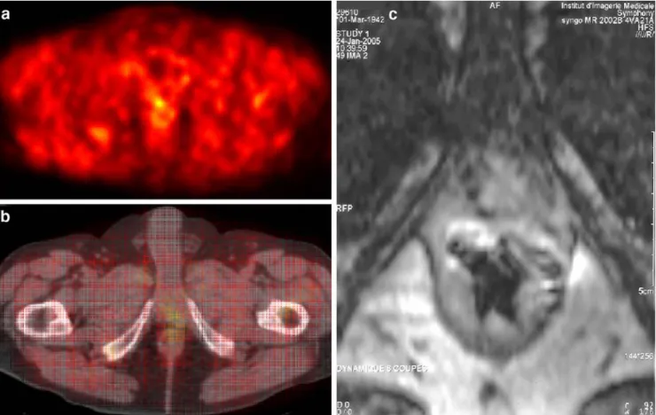
Fig. 3. 11C-acetate PET of patient 23, performed 24 months post surgery. The pre-surgical PSA value of 13.2 ng/ml fell to 0.06 ng/ml before rising again to 0.26 ng/ml at PET. Prostate bed-centred PET scan (a) is shown, together with corresponding fused PET-CT (b) and endorectal MRI (c). Note the small area of focal uptake
with a SUVmaxof 2.6. The focal PET uptake correlated with an
endorectal MRI finding suspicious for a recurrence close to the urethral anastomosis. Salvage RT was rapidly initiated within 1 month of11C-acetate PET and resulted in a decrease in PSA to 0.06 ng/ml 6 weeks after RT termination
The significance of stable (four patients) or rising PSA (one patient) after salvage RT in the remaining post-surgical patients remains unclear currently. Presence of minor distant disease cannot be excluded. However, since PSA was measured only 6 weeks after RT, the PSA response could not be evaluated with certainty.
Given the high number of group A patients who were positive for local tumour recurrence on PET (many of these results being confirmed by histology), one might argue that in such patients11C-acetate PET could be performed earlier in the course of suspicious PSA measurements. On the other hand, it needs to be borne in mind that, particularly in younger patients, normal prostate tissue takes up 11 C-acetate in significant amounts [37]. However, it is not known to what degree normal prostate tissue of older patients in the post-RT situation accumulates 11C-acetate, in particular after a long delay post RT. Our PET results clearly showed the presence of focal uptake while the background activity of normal prostate tissue was below SUVmax2, as shown by the T/N ratios.
PET was positive or equivocal for metastases in six patients of group A. These observations concerned mostly lymph node metastases, several of which were confirmed by CT. Another obturator lymph node was positive on PET in group B. Overall, three lymph nodes less than 1 cm in
diameter were positive or equivocal on PET despite being unsuspicious by radiological criteria. However, it has not yet been possible to confirm these results, the follow-up being too short to date.
Despite the positive observations concerning lymph node disease, the sensitivity of PET for detection of distant disease appeared limited, notably for osseous metastases. This limited ability to detect distant metastases might have been partially related to the significant tracer uptake in the pancreas, intestinal tract and bone marrow in some patients. Focal uptake was also observed in degenerative bone and joint disease. Furthermore, the limited resolution of the available PET and the need to fuse PET with CT from a different machine might have contributed to a sub-optimal detection rate of metastases.
With regard to optimisation of the PET scanning time, tumour recurrences remained positive or equivocal in the large majority of patients at the late scanning time, two physical half-lives after the injection of 11C-acetate. However, this study was not designed to identify the optimal timing for scanning. Dynamic scanning has been proposed previously [26]. Recently, in another study,18 F-fluorocholine PET was investigated in the staging of primary prostate cancer. Results showed that late PET, performed 1 h after injection, allowed better differentiation
Fig. 4. 11C-acetate PET of patient 31 performed 14 months post surgery. The pre-surgical PSA value of 5.5 ng/ml fell to <0.04 ng/ml after surgery before rising again to 0.20 ng/ml at PET. Early PET scan at the upper bulb level (a) is shown, together with the corresponding CT (b) and fused imaging (c) as well as endorectal
MRI (d). Note the small area of focal uptake with a SUVmaxof 2.7.
The focal PET uptake correlated with the positive endorectal MRI for a small recurrence at the upper side of the penile bulb. Salvage RT was initiated within 40 days of11C-acetate PET and resulted in a decrease in PSA to 0.10 ng/ml 6 weeks after RT termination
between benign and malignant prostate tumours when compared with early imaging [38].
Two other recent studies addressed the value of integrated 18F-fluorocholine PET/CT in patients with biochemical relapse when PSA values are less than 5 ng/ ml [39, 40]. Both studies showed the feasibility of fluorocholine PET at these PSA values, with a high number of positive findings. In comparison, in the group of post-surgical patients evaluated here, PSA was <0.8 ng/ml in 11 patients. Despite the very low PSA values,11C-acetate was positive or equivocal in the majority of these patients. Further studies are therefore warranted to establish the value of 11C-acetate based on integrated PET/CT instru-mentation and to compare these results with those obtained using18F-fluorocholine.
Conclusion
The results of this study show that 11C-acetate PET may have significant potential for the detection of recurrent prostate cancer, most notably in the very early work-up of post-surgical patients. While in group A the number of locally positive PET studies was very high, suggesting that the examination was performed too late, in group B the
number of positive patients was slightly lower, suggesting a limited sensitivity that might be improved in future evaluations. Some limitations were observed in this study that may have been related in part to the available PET scanner. While performance of the machine was tuned to a maximum, its acquisition potential remained limited by saturation, and in addition the resolution might have been lower than on modern PET/CT equipment. Further improvement might be obtained by PET/CT co-registration and by optimisation of scanning time, as indicated by the results shown here in the late pelvic scan. This study therefore shows, in contrast to a previous one, that 11 C-acetate PET may be of interest particularly in patients presenting very low PSA values following initial radical surgery, and possibly also earlier when there is a suspicious increase in PSA post RT. These situations might merit further evaluation of11C-acetate PET using more advanced PET/CT instrumentation.
Acknowledgements. We acknowledge with thanks the generous donation from the foundation Cellex International that made this study possible. We also express our gratitude to the staff of the services of nuclear medicine and radiology who performed the imaging and to Mrs. Frances Godson for reviewing the manuscript.
Table 4. Results of 15 group B patients treated initially with radical surgery. Results are given as positive (+), equivocal (Equiv) or negative (Neg)
Pat. no. 11C-acetate PET Endorectal MRI Salvage RT (Gy) PSA post RTa
T PET early T PET late N/M T SUVmaxearly T/N ratio early
18 Neg Neg Neg/Neg – + 67 ↔
19 Equiv + Neg/Neg 1.0 1.4 + Stoppedb ↑
20 Neg Neg Neg/Neg – + 74 ↔
21 Neg Neg Neg/Neg – + 74 ↓↓
22 Equiv Equiv Neg/Neg 2.3 1.8 + 74 ↓
23 + + Neg/Neg 2.6 2.2 Equiv 74 ↓
24 Neg Neg Neg/Neg + 74 ↓↓
25 Equiv Neg Neg/Neg 1.2 1.7 + 74 ↓
26 + + Neg/Neg 1.8 1.6 + 74 ↓
27 Neg Equiv Neg/Neg Neg 64 ↔
28 + + Neg/Neg 2.0 2.0 + 74 ↔
29 Equiv Equiv Neg/Neg 1.4 1.3 + Ho+RTc
30 Neg Neg Neg/Neg Neg 64 ↓↓
31 + Neg Neg/Neg 2.7 1.9 + 74 ↓
32 + + +/Neg 1.6 1.7 Equiv 74 ↔
Meand 2.1/1.5 1.9/1.6
SDd 0.5/0.6 0.2/0.2
Mediand 2.0/1.3 1.9/1.6
aPSA evolution after RT is shown: a decrease or increase by≥50% in PSA 6 weeks post therapy compared with the initial value is indicated by↓ and ↑, respectively. A PSA decrease to below the detection limit (0.04 ng/ml) after salvage RT is indicated by ↓↓. PSA variations of <50 % are denoted by↔
bSalvage RT was interrupted because of a significant increase in PSA after 1 month therapy
cPatient 29 presented a highly aggressive tumour (Gleason 5+5). He was re-treated with anti-hormonal therapy followed by RT and was therefore not evaluated for PSA evolution
References
1. Schoder H, Larson SM. Positron emission tomography for prostate, bladder, and renal cancer. Semin Nucl Med 2004;34:274–92
2. Sanz G, Rioja J, Zudaire JJ, Berian JM, Richter JA. PET and prostate cancer. World J Urol 2004;22:351–2
3. Price DT, Coleman RE, Liao RP, Robertson CN, Polascik TJ, DeGrado TR. Comparison of [18F]fluorocholine and [18F] fluorodeoxyglucose for positron emission tomography of androgen dependent and androgen independent prostate cancer. J Urol 2002;168:273–80
4. Oyama N, Miller TR, Dehdashti F, Siegel BA, Fischer KC, Michalski JM, et al.11C-acetate PET imaging of prostate cancer: detection of recurrent disease at PSA relapse. J Nucl Med 2003;44:549–55
5. Kotzerke J, Volkmer BG, Glatting G, van den Hoff J, Gschwend JE, Messer P, et al. Intraindividual comparison of [11C]acetate and [11C]choline PET for detection of metastases of prostate cancer. Nuklearmedizin 2003;42:25–30
6. Seltzer MA, Jahan SA, Dahlbom M, Sathyamurthy N, Barrio JR, Phelps ME, et al. Combined metabolic imaging using C-11 acetate and FDG PET for the evaluation of patients with suspected recurrent prostate cancer. J Nucl Med 2003;44:132P 7. Fricke E, Machtens S, Hofmann M, van den Hoff J, Bergh S, Brunkhorst T, et al. Positron emission tomography with11 C-acetate and18F-FDG in prostate cancer patients. Eur J Nucl Med Mol Imaging 2003;30:607–11
8. Yoshimoto M, Waki A, Obata A, Furukawa T, Yonekura Y, Fujibayashi Y. Radiolabeled choline as a proliferation marker: comparison with radiolabeled acetate. Nucl Med Biol 2004;31:859–65
9. Harisinghani MG, Barentsz J, Hahn PF, Deserno WM, Tabatabaei S, van de Kaa CH, et al. Noninvasive detection of clinically occult lymph-node metastases in prostate cancer. N Engl J Med 2003;348:2491–9
10. Winter PM, Caruthers SD, Kassner A, Harris TD, Chinen LK, Allen JS, et al. Molecular imaging of angiogenesis in nascent Vx-2 rabbit tumors using a novel alpha(nu)beta3-targeted nanoparticle and 1.5 tesla magnetic resonance imaging. Cancer Res 2003;63:5838–43
11. Siegel C. Organ-confined prostate cancer: effect of prior transrectal biopsy on endorectal MRI and MR spectroscopic imaging. J Urol 2005;174:569
12. Brassell SA, Rosner IL, McLeod DG. Update on magnetic resonance imaging, ProstaScint, and novel imaging in prostate cancer. Curr Opin Urol 2005;15:163–6
13. Cheng GC, Chen MH, Whittington R, Malkowicz SB, Schnall MD, Tomaszewski JE, et al. Clinical utility of endorectal MRI in determining PSA outcome for patients with biopsy Gleason score 7, PSA <or=10, and clinically localized prostate cancer. Int J Radiat Oncol Biol Phys 2003;55:64–70
14. Coakley FV, Teh HS, Qayyum A, Swanson MG, Lu Y, Roach M, et al. Endorectal MR imaging and MR spectroscopic imaging for locally recurrent prostate cancer after external beam radiation therapy: preliminary experience. Radiology 2004;233:441–8
15. Cox JD, Gallagher MJ, Hammond EH, Kaplan RS, Schellhammer PF. Consensus statements on radiation therapy of prostate cancer: guidelines for prostate re-biopsy after radiation and for radiation therapy with rising prostate-specific antigen levels after radical prostatectomy. American Society for Therapeutic Radiology and Oncology Consensus Panel. J Clin Oncol 1999;17:1155 16. Ward JF, Slezak JM, Blute ML, Bergstralh EJ, Zincke H.
Radical prostatectomy for clinically advanced (cT3) prostate cancer since the advent of prostate-specific antigen testing: 15-year outcome. B J U Int 2005;95:751–6
17. Winkler MH, Khan FA, Shabir M, Okeke A, Sugiono M, McInerney P, et al. Contemporary update of cancer control after radical prostatectomy in the UK. Br J Cancer 2004;91:1853–7 18. Ward JF, Zincke H, Bergstralh EJ, Slezak JM, Myers RP, Blute ML. The impact of surgical approach (nerve bundle preserva-tion versus wide local excision) on surgical margins and biochemical recurrence following radical prostatectomy. J Urol 2004;172:1328–32
19. Ward JF, Moul JW. Biochemical recurrence after definitive prostate cancer therapy. Part I: defining and localizing biochemical recurrence of prostate cancer. Curr Opin Urol 2005;15:181–6
20. Ward JF, Moul JW. Biochemical recurrence after definitive prostate cancer therapy. Part II: treatment strategies for biochemical recurrence of prostate cancer. Curr Opin Urol 2005;15:187–95
21. D’Amico AV, Whittington R, Malkowicz SB, Schultz D, Blank K, Broderick GA, et al. Biochemical outcome after radical prostatectomy, external beam radiation therapy, or interstitial radiation therapy for clinically localized prostate cancer. JAMA 1998;280:969–74
22. Freedland SJ, Sutter ME, Dorey F, Aronson WJ. Defining the ideal cutpoint for determining PSA recurrence after radical prostatectomy. Prostate-specific antigen. Urology 2003;61:365–9 23. Consensus statement: guidelines for PSA following radiation therapy. American Society for Therapeutic Radiology and Oncology Consensus Panel. Int J Radiat Oncol Biol Phys 1997;37:1035–41
24. Horwitz EM, Thames HD, Kuban DA, Levy LB, Kupelian PA, Martinez AA, et al. Definitions of biochemical failure that best predict clinical failure in patients with prostate cancer treated with external beam radiation alone: a multi-institutional pooled analysis. J Urol 2005;173:797–802
25. Stephenson AJ, Shariat SF, Zelefsky MJ, Kattan MW, Butler EB, Teh BS, et al. Salvage radiotherapy for recurrent prostate cancer after radical prostatectomy. JAMA 2004;291:1325–32 26. Dimitrakopoulou-Strauss A, Strauss LG. PET imaging of
prostate cancer with11C-acetate. J Nucl Med 2003;44:556–8 27. Kotzerke J, Volkmer BG, Neumaier B, Gschwend JE,
Hautmann RE, Reske SN. Carbon-11 acetate positron emission tomography can detect local recurrence of prostate cancer. Eur J Nucl Med Mol Imaging 2002;29:1380–4
28. Soloviev D, Tamburella C. Technical report: captive solvent [11C]acetate synthesis in GMP conditions. Appl Radiat Isotopes 2006; in press
29. Roeda D, Dolle F, Crouzel C. An improvement of11C acetate
synthesis-non-radioactive contaminants by irradiation-induced species emanating from the11C carbon dioxide production target. Appl Radiat Isotopes 2002;57:857–60
30. Davenport RJ, Dowsett K, Pike VW. A simple technique for the automated production of no-carrier-added [1–11
C]acetate. Appl Radiat Isotopes 1997;48:1117–20
31. Bailey DL, Young H, Bloomfield PM, Meikle SR, Glass D, Myers MG, et al. ECAT ART—a continuously rotating PET camera: performance characteristics, initial clinical studies, and installation considerations in a nuclear medicine department. Eur J Nucl Med 1997;24:6–15
32. Townsend DW, Beyer T, Jerin J, Watson CC, Young J, Nutt R. The ECAT ART scanner for positron emission tomography. 1. Improvements in performance characteristics. Clin Positron Imaging 1999;2:5–15
33. Brix G, Lechel U, Glatting G, Ziegler SI, Munzing W, Muller SP, et al. Radiation exposure of patients undergoing whole-body dual-modality18F-FDG PET/CT examinations. J Nucl Med 2005;46:608–13
34. Zaidi H, Gomez M, Boudraa A, Slosman DO. Fuzzy clustering-based segmented attenuation correction in whole-body PET imaging. Phys Med Biol 2002;47:1143–60
35. Allal AS, Dulguerov P, Allaoua M, Haenggeli CA, El-Ghazi EA, Lehmann W, et al. Standardized uptake value of 2-[18F] fluoro-2-deoxy-D-glucose in predicting outcome in head and
neck carcinomas treated by radiotherapy with or without chemotherapy. J Clin Oncol 2002;20:1398–404
36. Malpica A, Matisic JP, Niekirk DV, Crum CP, Staerkel GA, Yamal JM, et al. Kappa statistics to measure interrater and intrarater agreement for 1790 cervical biopsy specimens among twelve pathologists: qualitative histopathologic analysis and methodologic issues. Gynecol Oncol 2005;99:S38–52
37. Kato T, Tsukamoto E, Kuge Y, Takei T, Shiga T, Shinohara N, et al. Accumulation of [11C] acetate in normal prostate and benign prostatic hyperplasia: comparison with prostate cancer. Eur J Nucl Med Mol Imaging 2002;29:1492–5
38. Kwee SA, Wei H, Sesterhenn I, Yun D, Coel MN. Localization of primary prostate cancer with dual-phase18F-fluorocholine PET. J Nucl Med 2006;47:262–9
39. Heinisch M, Dirisamer A, Loidl W, Stoiber F, Gruy B, Haim S, et al. Positron emission tomography/computed tomography with F-18-fluorocholine for restaging of prostate cancer patients: meaningful at PSA <5 ng/ml? Mol Imaging Biol 2006;8:43–8
40. Schmid DT, John H, Zweifel R, Cservenyak T, Westera G, Goerres GW, et al. Fluorocholine PET/CT in patients with prostate cancer: initial experience. Radiology 2005;235:623–8
![Do we have to withdraw antiandrogenic therapy in prostate cancer patients before PET/CT with [11C]choline?](data:image/gif;base64,R0lGODlhAQABAIAAAP///wAAACH5BAEAAAAALAAAAAABAAEAAAICRAEAOw==)