Treating Displaced Distal Forearm Fractures
in Children
Joerg Schneider
1, Georg Staubli
2, Stephan Kubat
3, Stefan Altermatt
1Abstract
Purpose: Distal forearm fractures are among the most common fractures in children. In the past few years the option of percutaneous pinning has gained more attention in the treatment of unstable fractures. However, it remains unclear in which cases a fracture or its reduction should be considered unstable. Study Design: In order to evaluate which type of frac-tures profit most from additional pinning after closed reduction, we performed a retrospective analysis of 225 consecutive cases using the recently published AO pediatric classification of long bone fractures.
Results: After closed reduction, position in the cast was lost in 23% of the cases. The proportion of unstable reductions was much higher in completely displaced fractures. The amount of dislocation was more impor-tant than the type of fracture according to the AO classification proposal.
Conclusions: Fully displaced fractures should always be reduced in a setting with pins immediately available. If anatomical reduction cannot be achieved, pinning is advocated. The AO proposal for pediatric long bone fracture classification could be a useful tool to render the diverse studies more comparable. However, the important feature of complete versus subtotal dis-placement is lacking.
Key Words
Children
Æ
FractureÆ
Distal forearmÆ
AO pediatric classificationEur J Trauma Emerg Surg 2007;33:619–25 DOI 10.1007/s00068-007-6204-8
Introduction
Distal forearm fractures are the most common frac-tures at any age. While treatment of displaced fracfrac-tures in adults has changed more towards operative man-agement, closed reduction and splinting is still the most used treatment option in children. If and in which cases there is an indication for operative management is still under discussion.
There is no agreement in the literature on how much displacement of forearm fractures in children can be accepted and left to normal remodelling. Von Laer reports that dorsal displacements up to 40° will re-model in children under the age of 10 years [1]. Even so some authors suggest reposition at displacements as low as 15° under the age of 10 years and 10° for older children [2, 3]. Others accept up to 20° angulation at any age [4, 5] (Table 1). The fact that the long-term result is determined by the position at fracture con-solidation and that loss of position in the cast may be more important than the initial displacement [6], makes it difficult to establish rules. Additionally, comparing different studies is problematic because so far there is no clear definition of the metaphysis in the child. The recently published AO proposal of pediatric long bone fracture classification may help to overcome this problem [7].
However, there is an agreement that repetitive treatment should be avoided because the need for repetitive manipulation is known to be associated with an augmented number of growth disturbances [5, 8]. Nevertheless, remanipulation rates of 20–39% are reported [4, 9–13], with one study showing a redis-placement rate of 90% in isolated radius fractures [2].
1Department of Surgery, University Children’s Hospital, Zurich,
Switzerland,
2Department of Emergency, University Children’s Hospital, Zurich,
Switzerland,
3Department of Radiology, University Children’s Hospital, Zurich,
Switzerland.
Received: November 22, 2006; revision accepted: February 19, 2007; Published Online: June 27, 2007
The factors influencing redisplacement are not well known. Complete displacement and failure to achieve perfect anatomical reduction are important [4, 9–11]. Other reasons mentioned are isolated fracture of the radius, not well fitting casts, and experience of the surgeon [2, 9, 14].
While it is quite clear that percutaneous pinning can effectively reduce the risk for secondary dis-placement [12, 15, 16], it is not known which frac-tures will profit from this method and in which cases this may be an overtreatment. McLauchlan advocates pinning in all fully displaced fractures [12]. Choi limits the indication to displacements of more than half of the diameter [15]. Gibbons suggests pinning in isolated radius fractures [2]. Proctor recommends pinning of all fractures where anatomical reduction cannot be achieved [4]. Mani indicates ‘‘severe frac-tures’’ with ‘‘inadequate reduction’’ as candidates for K-wires. Most studies are, however, performed with too few patients to perform subgroup analysis of fracture type.
To investigate which distal fractures are prone to secondary dislocation, we retrospectively analyzed all manipulated distal forearm fractures at our clinic over a period of 2 years.
Patients and Methods Patients
All cases of children with distal forearm fractures presented at our emergency department between Jan-uary 1st 2004 and December 31st 2005 were analyzed. All patients needing reduction or operation were in-cluded in the study and their X-rays reviewed. Patients with pathological fractures and patients initially trea-ted at an outside facility were excluded. Patients who had only X-rays at the outside facility but manipulation at our institution were included.
Fractures
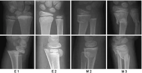
Patients with epiphyseal and metaphyseal fractures where included in the study. A metaphyseal fracture was defined as a fracture within a square over the epiphyseal plate of both bones on the AP-X-ray according to the AO proposal [7] (Figure 1).
Treatment
The initial treatment option was chosen by the emer-gency physician/surgeon on duty according to our internal guidelines:
Distal forearm fractures with more than 20° angulation in a child younger than 10 years and any
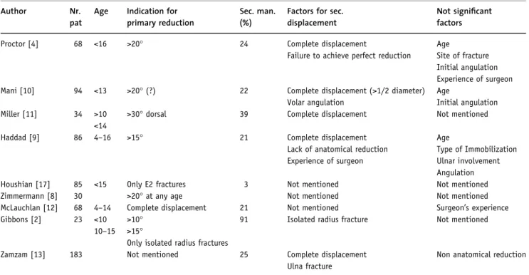
Table 1. Summary of relevant studies.
Author Nr.
pat
Age Indication for primary reduction
Sec. man. (%)
Factors for sec. displacement
Not significant factors
Proctor [4] 68 <16 >20° 24 Complete displacement
Failure to achieve perfect reduction
Age
Site of fracture Initial angulation Experience of surgeon
Mani [10] 94 <13 >20° (?) 22 Complete displacement (>1/2 diameter)
Volar angulation
Age
Initial angulation Miller [11] 34 >10
<14
>30° dorsal 39 Complete displacement Not mentioned
Haddad [9] 86 4–16 >15° 21 Complete displacement
Lack of anatomical reduction Experience of surgeon
Age
Type of Immobilization Ulnar involvement Angulation
Houshian [17] 85 <15 Only E2 fractures 3 Not mentioned Not mentioned
Zimmermann [8] 30 >20° at any age Not mentioned Not mentioned
McLauchlan [12] 68 4–14 Complete displacement 21 Not mentioned Surgeon’s experience
Gibbons [2] 23 <10 10–15
>10° >15°
Only isolated radius fractures
91 Isolated radius fracture Not mentioned
Zamzam [13] 183 Not mentioned 25 Complete displacement
Ulna fracture
angulation in older children were considered an indi-cation for reduction.
Depending on the age of the child and his or her preference, general anesthesia or regional (IV) anes-thesia was chosen for the reduction.
If the fracture was not stable under fluoroscopic control after reduction, two diverging K-wires were percutaneously inserted radially. Follow-up X-rays were obtained 5 and 10 days later in non-operated patients and at 3 weeks and 3 month in all patients. If a redisplacement was noted on day 5 or day 10 X-ray controls, the decision for re-reduction and radial K-wire fixation was judged by the same criteria as in the post accident X-ray.
After reduction, fractures were immobilized in an above-elbow cast, either with a plaster of Paris or a ScotchcastÒ (3M, Minnesota). The plaster of Paris was arranged as a sandwich of a volar and a dorsal splint, unified by a paper and an elastic bandage. The ScotchcastÒ was applied as a mixed soft-scotch cast with a dorsal and a volar splint of ScotchcastÒunified by a circular layer of SoftcastÒ. The type of plaster and the position of the wrist in the plaster (flexion, neutral or extension) were chosen by the physician on duty.
Analyzed Parameters
Radius and ulna fractures were classified separately according to the AO proposal [7] with additional denomination of the radius fractures as fully or non-fully displaced (Figure 2). The angulations in the sag-ital and frontal plane as well as the lateral translation
were measured on the initial and post reduction X-rays. Anatomical reposition was defined as no angulation and no translation in any direction.
Re-displacement was defined as the need for a secondary reposition and consecutive operation.
Statistics
Data were analyzed with Vassar Stats website for sta-tistical computation using two-sided Chi-Square and Fisher’s Exact Test where appropriate. A p < 0.05 was regarded as significant difference.
Results
A total of 241 patients with 244 distal fractures were treated over the 2 years period at our institution. Three patients suffered simultaneous fractures of both arms. Due to their small numbers, for the statistical analysis, they were regarded as independent fractures. Nineteen fractures were excluded from the statistical analysis; 11 of them were initially treated at an outside facility, two had pathologic fractures, and the X-ray dossiers of six patients were not complete. Of the remaining 222 pa-tients with 225 fractures 184 had metaphyseal fractures and 41 had physeal fractures Salter-Harris type I or II. There were no Salter-Harris type III or IV fractures of the radius in our collective. Ipsilateral humerus frac-tures were observed in four cases.
Patients’ Demographics
Seventy-five patients were girls, 150 boys. The left arm was involved in 128 cases, the right 97 times. Mean age
Figure 1. Definition of the metaphysis.
Figure 2. AO Classification: E1: physeal injury with complete separation of epiphysis (Salter Harris type I); E2: physeal injury with metaphyseal fragment (Salter Harris type II); M2: incomlete metaphyseal fracture (One broken corticalis); M3: Complete metaphyseal fracture (two broken corticales).
was 10 years (3–16). In 180 cases the fracture was displaced dorsally, in 45 cases palmarly.
Secondary Dislocation Rate
Of the included 225 fractures, 20 were operated immediately and 205 were initially reduced. Of the initially reduced patients 47 (23%) had a secondary displacement which needed a secondary operation. In three cases an external fixator was used and in one case internal fixation with a plate was performed. All remaining patients underwent percutaneous pinning. None of the 67 primary or secondary pinned patients needed reintervention because of redislocation.
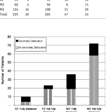
Subgroup Analysis See Table 2 and Figure 3.
E1. There were no full displacements among the eight E1 type fractures. All of them could be anatomi-cally reduced and secondary displacement was never observed.
E2. All 33 patients suffering an E2 type fracture were primary reduced and none initially operated. The position was lost in 10 (30%) of these patients.
If an anatomical reduction was achieved (14 cases) the position in the cast was always maintained. In the 19 fractures where anatomical reduction could not be achieved, the position in the cast was lost ten times (53%) (p < 0.05).
Full displacement was noted in 10 out of the 33 cases. Redisplacement occurred six times (60%), all after non-anatomical reduction. Of the 23 not fully displaced fractures only four (19%) needed a second-ary reduction, although in thirteen cases anatomical reduction had not been achieved (p < 0.05).
M2. An M2 type fracture was diagnosed 60 times. Of them, four were initially pinned and 56 initially re-duced. Anatomical reduction was possible nine times and redislocation never occurred. Though an anatom-ical reduction was not achieved in 47 cases, a secondary intervention was necessary only six times (13%) (p = 0.57).
M3. In 124 patients a M3 type fracture was observed. A primary operation was performed in 16 cases and a primary reposition in 108 cases. A total of 31 (29%) patients showed secondary displacement.
Of the 108 primary reduced fractures, 36 were initially fully displaced. Of them 17 (47%) redislocated opposed to 14 out of 72 (19%) initially not fully dis-placed fractures (p < 0.05).
Anatomical reduction was possible in 16 cases with one secondary displacement (6%). In the 92 cases without anatomical reduction, redisplacement was ob-served in 30 cases (33%) (p < 0.05).
Risk Factors for Secondary Displacement After Primary Reduction
Degree of Dislocation (Table 3)
Of the 46 fully displaced and reduced fractures, 23 (50%) had to undergo a secondary operation as com-pared to 24 (15%) of the 159 not fully displaced frac-tures (p < 0.001).
Angle and Direction of Dislocation
Reintervention was required after 39 (24%) of 162 dorsally displaced fractures, versus eight (19%) of the 43 palmarly displaced fractures. This difference was, however, not significant (p = 0.54).
Neither the angle of dislocation in the frontal or sagital, nor the lateral dislocation showed any signifi-cance.
Table 2. Overview of the results split by AO classification. AO class. Total Primary operation Primary reduction Secondary operation Redislocation rate (%) E1 8 0 8 0 0 E2 33 0 33 10 30 M2 60 4 56 6 11 M3 124 16 108 31 29 Total 225 20 205 47 23
Figure 3. Fully versus not fully displaced fractures; split by AO classification.
Involvement of the Ulna
Overall, 118 patients had an additional ulna fracture, 15 of them underwent primary operation. After pri-mary reduction, 29 (28%) of 103 showed a secondary dislocation.
Instable ulna fractures (ulna M3) resulted in a dis-location in 15 out of 39 cases (38%), compared to 12 out of 60 (20%) stable ulna fractures (ulna M2) (p < 0.05). An isolated radius fracture existed in 107 patients of which five were initially operated. A secondary dislocation occurred in 18 (18%) of 102 patients after primary reposition (p = 0.07).
E2. Involvement of the ulna was present in eleven patients with E2 type fractures; a secondary interven-tion was needed three times (27%). An isolated radius fracture existed in 22 cases; six (27%) of them had to undergo a second procedure (p = 0.46).
M2. A redislocation was noted in three (13%) out of 23 patients with an additional ulna fracture. Sec-ondary dislocations occurred in two (6%) out of 32 cases where no ulna fracture existed (32 cases) (p = 0.65).
M3. Patients with an M3 type fracture suffered an additional ulna fracture in 68 cases. In 22 (32%) of them, a secondary dislocation was noted compared to nine (23%) of the 40 patients with isolated radius fractures (p = 0.32).
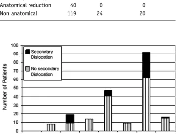
Anatomical Reduction
Anatomical reduction could be achieved in 47 patients; one (2%) of them needed a secondary intervention. In the 158 patients where anatomical reduction could not be achieved, reintervention was needed in 46 patients (29%) (p < 0.001) (Figure 4).
Cast and Position in the Cast
In the 79 cases where a plaster of Paris was applied, redisplacement occurred 20 times (25%). If a Scotch-castÒwas used (126 cases), redisplacement occurred 27 times (21%) (p = 0.51). In 104 cases the wrist was casted in a flected position. A redisplacement was diagnosed in 16 (15%) of them. A neutral position was chosen 66 times and redisplacement noted in 17 cases (26%). After applying a cast with the wrist in extended position two (11%) out of 18 redislocated (p = 0.10).
Age
A secondary dislocation occurred in 23 (27%) out of 84 children under the age of 10 compared to 24 (20%) out of 121 children over 10 years of age (p = 0.20).
Discussion
A few prospective studies about the pinning of distal forearm fractures in children [11, 12] have recently been published. However, all of them had a very lim-ited number of patients. Even though our study was conducted retrospectively, the large number of patients allows for the extraction of results from subgroup analysis. It is also the first time that the new AO classification proposal was used for fracture classifica-tion. The fact that the AO classification is a proposal implies that it still has some limitations. In particular, the important distinction between fully and non-fully displaced fractures is missing in the classification.
In this study, the overall secondary dislocation rate was not high compared to other studies (Table 1). One reason may be, that only dislocations leading to an operation were considered for our study. Re-disloca-tions within our limits (same limits as for the initial decision for reduction) were not considered. The pa-tient is not interested in angles; what matters for him are a good outcome, i.e. range of motion and how many times he has to undergo an intervention.
Table 3. Summary of major risk factors in primary reduced fractures.
Total Secondary
operation
Re-dislocation rate (%) Complete displaced fracture
Anatomical reduction 7 1 14
Non anatomical 39 22 56
Not complete displaced fracture
Anatomical reduction 40 0 0
Non anatomical 119 24 20
Figure 4. Influence of anatomical reduction (ana: Anatomical reduction; non: Non anatomical reduction).
The limits of dislocation and angulation are arbi-trary. Anything between 15° and 30° is found in the literature (Table 1). As proposed by other authors [4, 8, 10], we chose a limit of 20° to avoid a prolonged time of remodeling and visible angulation at the time of cast removal.
In accordance with other studies, complete placement was a major risk factor for secondary dis-placement [4, 9, 10, 13]. If these fractures are anatomically reduced, the risk of secondary displace-ment can be minimized. However, this may be very difficult to accomplish in M3 type fractures where anatomical reduction could only be achieved in 3 out of 36 cases (8%). In completely displaced E2 type frac-tures, this goal could be achieved in 6 out of 10 patients (60%) (p < 0.001).
In contrast to the results presented by Gibbons [2], isolated radius fractures were less prone to secondary displacement than fractures of both bones. Especially instable fractures of the ulna (ulna M3) are significantly more prone to secondary dislocation. Exceptions to this finding were E2 fractures where isolated radius fractures were more prone to secondary displacement.
Dorsally displaced fractures had a higher tendency of secondary displacement than palmarly displaced ones. This may, however, be attributed to the higher energy normally involved in these fractures and therefore the higher number of fully displaced frac-tures. If fully and not fully displaced fractures are analyzed separately there is no difference.
In contrast to other studies, epiphyseal fractures were included in our analysis. Despite their small prevalence, E1 fractures seem to be unproblematic. In contrast, anatomical reduction appears to be crucial in E2 fractures. We cannot confirm the small number of secondary manipulations presented by Houshian [17] and the question remains if our indication for re-reduction is too rigorous in these cases.
The type of cast may be less important in the management of distal forearm fractures. Good results can be achieved either with ScotchcastÒ or Plaster of Paris. Tilting the wrist in the opposite direction of the fracture dislocation may offer an advantage. However, this finding was not significant in our study and ran-domized prospective studies are needed to clarify this question.
A retrospective study points out the fact that a below-the-elbow plaster may be advantageous for the treatment of these fractures [18]. At the beginning of our study, the evidence for this procedure was weak. Therefore, an above-the-elbow cast was chosen. It was thought that additional prevention of pro-/suppination
could potentially lead to a better stability. Recently however, the efficacy of below-the elbow plaster has been supported by two prospective studies. Therefore, the application of short arm plasters should be con-sidered in future studies.
Conclusions
Completely displaced distal forearm fractures should always be reduced in the disposition of pins. If ana-tomical reduction cannot be achieved, pinning is advocated.
If some additional features are included, the AO proposal of pediatric long bone fracture classification may become a useful tool to render different studies more comparable in the future.
Acknowledgment
We would like to thank Marianne Sauter and Monika Fa¨ssler from the Department of Radiology for searching all the X-ray dossiers and Kristopher Kubow of the ETH Zu¨rich for his help with editing.
References
1. Von Laer L, Hasler C. Spontaneous corrections, growth disorders and post-traumatic deformities after fractures in the area of the forearm of the growing skeleton. Handchir Mikrochir Plast Chir 2000;32:231–41.
2. Gibbons CL, Woods DA, Pailthorpe C, Carr AJ, Worlock P. The management of isolated distal radius fractures in children. J Pediatr Orthop 1994;14:207–10.
3. Jones K, Weiner DS. The management of forearm fractures in children: a plea for conservatism. J Pediatr Orthop 1999;19:811–5. 4. Proctor MT, Moore DJ, Paterson JM. Redisplacement after
manipulation of distal radial fractures in children. J Bone Joint Surg Br 1993;75:453–4.
5. Zimmermann R, Gschwentner M, Pechlaner S, Gabl M. Remodeling capacity and functional outcome of palmarly ver-sus dorsally displaced pediatric radius fractures in the distal one-third. Arch Orthop Trauma Surg 2004;124:42–8. 6. Younger AS, Tredwell SJ, Mackenzie WG. Factors affecting
fracture position at cast removal after pediatric forearm frac-ture. J Pediatr Orthop 1997;17:332–6.
7. Slongo T, Audige L, Schlickewei W, Clavert JM, Hunter J. Development and validation of the AO pediatric comprehensive classification of long bone fractures by the Pediatric Expert Group of the AO Foundation in collaboration with AO Clinical Investigation and Documentation and the International Asso-ciation for Pediatric Traumatology. J Pediatr Orthop
2006;26:43–9.
8. Zimmermann R, Gabl M, Angermann P, Lutz M, Reinhart C, Kralinger F, Pechlaner S. Late sequelae of fractures of the distal third of the forearm during the growth period. Handchir Mi-krochir Plast Chir 2000;32:242–9.
9. Haddad FS, Williams RL. Forearm fractures in children: avoiding redisplacement. Injury 1995;26:691–2.
10. Mani GV, Hui PW, Cheng JC. Translation of the radius as a predictor of outcome in distal radial fractures of children. J Bone Joint Surg Br 1993;75:808–11.
11. Miller BS, Taylor B, Widmann RF, Bae DS, Snyder BD, Waters PM. Cast immobilization versus percutaneous pin fixation of dis-placed distal radius fractures in children: a prospective, ran-domized study. J Pediatr Orthop 2005;25:490–4.
12. McLauchlan GJ, Cowan B, Annan IH, Robb JE. Management of completely displaced metaphyseal fractures of the distal radius in children. A prospective, randomised controlled trial. J Bone Joint Surg Br 2002;84:413–7.
13. Zamzam MM, Khoshhal KI. Displaced fracture of the distal ra-dius in children: factors responsible for redisplacement after closed reduction. J Bone Joint Surg Br 2005;87:841–3. 14. Voto SJ, Weiner DS, Leighley B. Redisplacement after closed
reduction of forearm fractures in children. J Pediatr Orthop 1990;10:79–84.
15. Choi KY, Chan WS, Lam TP, Cheng JC. Percutaneous Kirschner-wire pinning for severely displaced distal radial fractures in children. A report of 157 cases. J Bone Joint Surg Br 1995;77:797–801.
16. Muratli HH, Yagmurlu MF, Yuksel HY, Aktekin CN, Bicimoglu A, Tabak AY. Treatment of childhood unstable radius distal methaphysis fractures with closed reduction and percutaneous Kirschner wires. Acta Orthop Traumatol Turc 2002;36:52–7. 17. Houshian S, Holst AK, Larsen MS, Torfing T. Remodeling of
Salter-Harris type II epiphyseal plate injury of the distal radius. J Pediatr Orthop 2004;24:472–6.
18. Chess DG, Hyndman JC, Leahey JL, Brown DC, Sinclair AM. Short arm plaster cast for distal pediatric forearm fractures. J Pediatr Orthop 1994;14:211–3.
Address for Correspondence Stefan Altermatt, MD Department of Surgery University Children’s Hospital Steinwiesstrasse 75
8032 Zurich Switzerland
Phone (+41/44) 266-7111, Fax -7171 e-mail: [email protected]