Walter Knirsch Oliver Kretschmar Maren Tomaske Kathrina Stutz Nicole Nagdyman Christian Balmer Achim Schmitz Dominique Béttex Felix Berger Urs Bauersfeld Markus Weiss
Cardiac output measurement in children:
comparison of the Ultrasound Cardiac Output
Monitor with thermodilution cardiac output
measurement
Received: 11 September 2007 Accepted: 14 January 2008 Published online: 23 February 2008 © Springer-Verlag 2008
All authors are members of the Working Group on Non-invasive Haemodynamic Monitoring in Paediatrics.
W. Knirsch (u) · O. Kretschmar · M. Tomaske · K. Stutz · C. Balmer · U. Bauersfeld
University Children’s Hospital, Division of Pediatric Cardiology, Steinwiesstrasse 75, 8032 Zurich, Switzerland e-mail: walter.knirsch@kispi.uzh.ch Tel.: +41-44-2667617 Fax: +41-44-2667981
K. Stutz · A. Schmitz · D. Béttex · M. Weiss University Children’s Hospital, Department of Anesthesia,
Steinwiesstrasse 75, 8032 Zurich, Switzerland
N. Nagdyman · F. Berger German Heart Center, Department of Congenital Heart Disease,
Augustenburger Platz 1, 13353 Berlin, Germany
Abstract Objective: To compare the assessment of cardiac output (CO) in children using the nonin-vasive Ultrasound Cardiac Output Monitor (USCOM) with the in-vasive pulmonary artery catheter (PAC) thermodilution cardiac output measurement. Design and setting: Prospective observational study in a tertiary center for pediatric cardiol-ogy of a university children’s hospital.
Patients: Twenty-four pediatric
pa-tients with congenital heart disease without shunt undergoing cardiac catheterization under general anes-thesia. Measurements and results: CO was measured by USCOM using a suprasternal CO Doppler probe in children undergoing cardiac catheter-ization. USCOM data were compared to CO simultaneously measured by PAC thermodilution technique. Mea-surements were repeated three times within 5 min in each patient. A mean percentage error not exceeding 30% was defined as indicating clinical useful reliability of the USCOM.
CO values measured by PAC ranged from 1.3 to 5.3 l/min (me-dian 3.6 l/min). Bias and precision were –0.13 and 1.34 l/min, respec-tively. The mean percentage error of CO measurement by the USCOM compared to PAC thermodilution technique was 36.4% for USCOM.
Conclusions: Our preliminary data
demonstrate that cardiac output measurement in children using the USCOM does not reliably represent absolute CO values as compared to PAC thermodilution. Further studies must evaluate the impact of incorporating effective aortic valve diameters on CO measurement using the USCOM.
Keywords Cardiac output · Pul-monary artery catheter · Monitoring · Hemodynamic · Children · Noninva-sive
Introduction
In critically ill pediatric patients the measurement of car-diac output (CO) is of considerable interest for the cardio-vascular assessment and hemodynamic management [1]. In contrast to adult patients, in whom the thermodilution method by pulmonary artery catheter (PAC) is widely ac-cepted and used for CO measurement, this technique is not routinely used in pediatric critical care due to its increased
risk of complications in children (infections, thrombosis, embolism) and the size of the catheter, which is not con-venient for continuous monitoring process in small chil-dren [2].
The Ultrasound Cardiac Output Monitor (USCOM, Sydney, Australia) is a noninvasive CO monitor based on continuous-wave Doppler ultrasound (Fig. 1) for adults and children. USCOM has been evaluated for dogs [3] and for adult patients comparing it to CO thermodilution
technique [4–7] and has been used in various clinical settings [8–10]. No data are currently available with regard to reliability of the USCOM for CO measurement in children. The aim of this study was to compare CO measurement by the noninvasive USCOM with the ther-modilution technique during cardiac catheterization for CO assessment in pediatric patients.
Materials and methods
TechniqueAfter starting the USCOM device (Fig. 1) patient data, in-cluding height, weight, and gender, are typed in as well as the chosen vessel (aorta) is indicated. In children Doppler flow curves by the USCOM are obtained by a 3.3-MHz transducer placed in the suprasternal notch to obtain an optimal aortic flow signal at the aortic valve. With contin-uous slight movements of the USCOM probe the operator aims to assess a clear systolic beginning and end, full sys-tolic timing, and the highest velocities of the Doppler flow curve. On the monitor display the Doppler curve is pre-sented as a time velocity curve. A representative Doppler curve is saved for further calculations. The CO value is calculated using the flow integral, and the aortic valve di-ameter as given by the USCOM internal algorithm based on height and gender.
Patients
Hospital ethics committee approval and written parental consent were obtained for all patients. Pediatric patients with congenital heart disease undergoing diagnostic and/or interventional cardiac catheterization under general anesthesia were prospectively enrolled. Patients with residual intracardiac or extracardiac shunt, tricuspid or pulmonary valve regurgitation, body weight below 3 kg, and age older than 18 years were excluded. The analysis included 24 patients aged 0.1–16.7 years (median 7.6) and weighing 3.4–51.0 kg (23.0 kg). Indications for cardiac catheterization were device closure of an atrial septal defect in nine and ventricular septal defect in six, balloon dilatations of the pulmonary valve in two and of branches of the pulmonary arteries in three, stenting of a coarctation of the aortic arch in one, and for other diagnostic reasons in three. Premedication and induction of anesthesia (inhalational or intravenous) depended upon the patient’s medical condition and anesthesiologist’s preferences. After induction of general anesthesia and after establishing neuromuscular blockade the patient was intubated and ventilated by the anesthesia respirator. Prior to the CO measurements we excluded a residual shunt following device closure or a residual pulmonary valve
Fig. 1 Ultrasonic cardiac output monitor
regurgitation by transesophageal echocardiography and/or angiography.
Cardiac output measurements
CO measurements were performed at the end of the cardiac catheterization procedure under hemodynamic steady-state conditions with the pulmonary artery modilution catheter and USCOM device in place. For ther-modilution CO assessment a 5-F balloon-tipped pulmon-ary artery catheter (Edwards Lifescience, Irvine, Calif., USA) was inserted via a 6-F sheath in the femoral vein and directed to the pulmonary artery under fluoroscopic control. CO was measured by PAC with 5-ml bolus injec-tion of NaCl 0.9% at a temperature of 4°C, as described elsewhere [2]. After a test injection three consecutive measurements were performed. CO values for the US-COM measurements were recorded immediately before injection of each of the three repeated thermodilution fluid boli. The USCOM measurement was performed by a single experienced consultant pediatric cardiologist after a learning curve prior the start of the study. The cardiologist performing the USCOM measurement was blinded to the CO measurements by the thermodilution technique. A total of 72 paired CO measurements by thermodilution and USCOM were recorded.
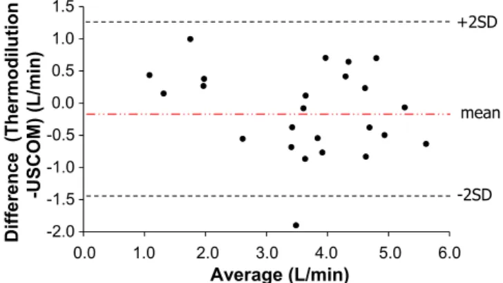
Fig. 2 Bland–Altman analysis to compare cardiac output values ob-tained by the USCOM and the thermodilution technique in 24 car-diac pediatric patients
Statistics
Mean values of the three measurements for both methods were calculated per patient and used for further statistics. Intraobserver variability was calculated for the two meth-ods. Bland–Altman analysis was performed to compare CO values obtained by the USCOM with those obtained by PAC thermodilution technique [11]. The mean propor-tional error (2 SD/mean CO× 100) was calculated. Data are presented as median (range). A mean percentage error not exceeding 30% was defined as indicating clinical use-ful reliability of the USCOM [12, 13].
Results
Mean CO values measured by PAC thermodilution tech-nique ranged from 1.30 to 5.30 l/min (median 3.57) and mean CO values measured by USCOM ranged from 0.86 to 5.93 l/min (4.04). Bias (mean difference) between the two methods was –0.13 l/min and precision (± 2 SD of differences) was 1.34 l/min (Fig. 2). The signal quality of the USCOM Doppler was comparable and good in all pa-tients independently of age. The intraobserver variability was 3.1% by the PAC thermodilution technique and 5.7% by USCOM measurement. The mean percentage error of CO measurement was 36.4% for USCOM vs. to PAC thermodilution technique.
Discussion
This study compared CO measurements between the new noninvasive continuous-wave Doppler based monitoring system USCOM and the thermodilution technique with pulmonary artery catheter in pediatric patients with congenital heart disease without shunts from infancy to adolescence. The USCOM is a noninvasive cardiac
output monitor based on the transthoracic measurement of Doppler flow velocity of the large intrathoracic vessels. It is easy to operate, and CO is easily displayed by the USCOM device. The technique is reported to be easily learned even by noncardiologists and nursing staff [14]. The usefulness of fast and easy assessment of CO mon-itoring by USCOM was supported by a previous study by Knobloch et al. [8] in adult patients for hemodynamic assessment in preclinical emergency medicine.
The main findings were that measurement of CO with USCOM does not reliably represent absolute values as compared to pulmonary artery catheter thermodilution technique with a mean percentage error higher than 30%. We believe that user dependency and technical factors may contribute to this disagreement. User-dependent factors must be taken into account such as the insonation angle during measurement. Further technical factors of the Doppler method are well known limitations, such as the circumstance that the Doppler beam is one-dimensional in a typical three-dimensional structure, and its flow is considered laminar and described as plug flow, although it is well known to be skewed.
Other patient-dependent factors such as implication of the type of congenital heart defect in regard to the growth of the aortic valve must be considered. The technique itself may entail certain factors influencing the measurement. One of these is the estimated aortic valve diameter. The algorithm for the calculation of the aortic and pulmonary valve diameter incorporated in the USCOM software is based on the data of Nidorf et al. [15] and may involve a systemic error during growth of the heart. These normal values of cardiac dimensions published by Nidorf et al. were evaluated for healthy children while in our study children with congenital heart disease were examined, which may contribute as a source of error. Further studies could rule out this error by measuring the true aortic valve diameter by transthoracic echocardiography and to “calibrate” and adjust the USCOM measurement. The impact of the direct measurement of the aortic valve diameter to the measurement of the cardiac output by conventional transthoracic Doppler echocardiography has been shown [16].
In addition, the limitations of transthoracic pulsed Doppler cardiac output measurement in comparison to cardiac output measurement by the thermodilution technique has been recognized [17]. On the other hand, transesophageal continuous-wave Doppler cardiac output measurement in comparison to the thermodilution tech-nique seems to be suitable [18]. Therefore the Doppler flow quality remains an important influencing factor. Doppler flow signal quality may have been affected by intrathoracic air, patient position, and operator’s learning curve. In our study, however, the modalities of the USCOM were optimal standardized (anesthesia, pediatric patients with excellent access, and good sonographic window) so that the impact of these factors may be rather low.
Table 1 Summary of literature evaluating reliability of CO measurement by USCOM (d.n.a., data not available; LOA, limits of agreement; calc., calculated)
Critchley et al. [3] Tan et al. [4] Chand et al. [5] Knobloch et al. [6] Chan et al. [7] Setting Animal lab. model, Postcardiac surgery, Postcardiac surgery, Postcardiac surgery, Postcardiac surgery,
anesthetized dogs adult patients adult patients adult patients adult patients Reference method Ultrasonic flow probe Thermodilution Thermodilution Thermodilution Thermodilution
Patients n = 6 n = 22, n = 50 n = 36 n = 26
mean age: 63.5 years mean age: 59.2 years mean age: 67.2 years mean age: 60.6 years Simple regression d.n.a d.n.a d.n.a. R = 0.870, p< 0.01 R = 0.46, p< 0.01
Cardiac output: −0.01 l/min 0.18 l/min −0.14 l/min −0.23 l/min 0.22 l/min bias (LOA) (−0.34 to 0.31) (−1.43 to 1.78) (−1.43 to 1.58) (−2.52 to 1.79) (−1.17 to 1.62)
Mean error 13% d.n.a. 34% (calc.) 42% (calc.) 54% (calc.)
To our knowledge, this is the first study in pediatric patients validating CO measurement by USCOM with CO measurement by PAC thermodilution considered as gold standard (Table 1). USCOM has demonstrated reliable results of CO measurements in laboratory animal studies by using aortic flow probes in anesthetized dogs. The investigators found 95% limits of agreement of –0.34 and 0.31 l/min over a wide range of cardiac output values [3].
Tan et al. [4] recently evaluated the USCOM vs. ther-modilution cardiac output measurements in adult patients after cardiac surgery and found, comparable to our results, 95% limits of agreement for the two techniques of –1.43 and 1.78 l/min. Chand et al. [5] and Chan et al. [7] con-firmed this level of agreement of the two techniques in a similar clinical setting with adult patients after cardiac surgery. For both studies we calculated the mean percent-age error, which exceeds 30% (Table 1). Although their agreements were less than those obtained in our study, the authors judged the USCOM to be a reliable technique for CO assessment.
The fact that our study included no patients with nor-mal cardiac anatomy included may be considered as limi-tation of this study. However, cardiac catheterization with the opportunity of PAC thermodilution CO assessment in patients without heart disease is very rare, and limits this type of investigation to pediatric patients with congenital
heart disease or critically ill pediatric patients on the inten-sive care unit. In this study a single operator performed all USCOM measurements. Further studies must investigate interrater and intrarater reliability of the USCOM method. Since the aortic valve diameter may be a crucial and un-known factor in the calculation of absolute CO value, the USCOM may become a trend monitor rather than a tool for the assessment of absolute CO values. To support this ap-plication of USCOM in pediatric patients further research on long-term and repeated CO measurements is needed.
In the future, USCOM may rather serve as a feasible, fast usable, valuable diagnostic tool to measure cardiac output and to demonstrate therapeutic hemodynamic ef-fects, for example, of fluid challenge and intropic supports in critical ill children, by comparing pre- and postthera-peutic CO values with USCOM. In this clinical settings the absolute value of CO may be rather unimportant as the fact the CO has increased from a “low” CO to a “normal” or even “high” CO.
In conclusion, based on our findings USCOM cannot be recommended for the assessment of absolute CO values in pediatric cardiac patients. Further studies must determine whether the incorporation of measured aortic valve diameter can improve accuracy of the USCOM, and whether the device can be reliably used as a trend monitor for cardiac output assessment.
References
1. Egan JR, Festa M, Cole AD, Nunn GR, Gillis J, Winlaw DS (2005) Clinical assessment of cardiac performance in infants and children following cardiac surgery. Intensive Care Med 31:568–573
2. Webster CS, Merry AF, Emmens DJ, Van Cotthem IC, Holland RL (2003) A prospective clinical audit of central venous catheter use and complications in 1000 consecutive patients. Anaesth Intensive Care 31:80–86
3. Critchley LA, Peng ZY, Fok BS, Lee A, Phillips RA (2005) Testing the reliabil-ity of a new ultrasonic cardiac output monitor, by using aortic flowprobes in anesthetized dogs. Anesth Analg 100:748–753
4. Tan HL, Pinder M, Parsons R, Roberts B, van Heerden PV (2005) Clinical evaluation of USCOM ultra-sonic cardiac output monitor in cardiac surgical patients in intensive care unit. Br J Anaesth 94:287–291
5. Chand R, Mehta Y, Trehan N (2006) Cardiac output estimation with a new Doppler device after off-pump coronary artery bypass surgery. J Cardiothorac Vasc Anesth 20:315–319
6. Knobloch K, Lichtenberg A, Winter-halter M, Rossner D, Pichlmaier M, Phillips R (2005) Non-invasive cardiac output determination by two-dimensional independent Doppler during and after cardiac surgery. Ann Thorac Surg 80:1479–1484
7. Chan JS, Segara D, Nair P (2006) Measurement of cardiac output with a noninvasive continuous wave Doppler device versus the pulmonary artery catheter: a comparative study. Crit Care Resusc 8:309–314
8. Knobloch K, Hubrich V, Rohmann P, Lupkemann M, Gerich T, Krettek C, Phillips R (2006) Feasibility of preclini-cal cardiac output and systemic vascular resistance in HEMS in thoracic pain-the ultrasonic cardiac output monitor. Air Med J 25:270–275
9. Knobloch K, Hoeltke V, Jakob E, Vogt PM, Phillips R (2007) Non-invasive ultrasonic cardiac output monitoring in exercise testing. Int J Cardiol 2007 (epub ahead of print: 26 April)
10. Siu CW, Tse HF, Lee K, Chan HW, Yung C, Lee S, Lau CP (2007) Cardiac resynchronization therapy optimization by ultrasonic cardiac output moni-toring (USCOM) device. Pacing Clin Electrophysiol 30:50–55
11. Bland JM, Altman DG (1986) Statis-tical methods for assessing agreement between two methods of clinical measurements. Lancet I:307–310 12. Tibbals J, Hochmann M, Osborne A,
Carter B (1992) Accuracy of the BoMED NCCOM3 bioimpedance cardiac output monitor during in-duced hypotension: an experimental study in dogs. Anaesth Intensive Care 20:326–331
13. Critchley LAH, Critchley JAJH (1999) A meta-analysis of studies using bias and precision statistics to compare cardiac output measurement techniques. J Clin Monit 15:85–91
14. Dey I, Sprivulis P (2004) Emergency physicians can reliably assess emer-gency department patient cardiac output using the USCOM continuous wave Doppler cardiac output monitor. Emerg Med Australas 17:193–199
15. Nidorf SM, Picard MH, Triulzi MO, Thomas JD, Newell J, King ME, Weyman AE (1992) New perspectives in the assessment of cardiac cham-ber dimensions during development and adulthood. J Am Coll Cardiol 19:983–988
16. Gardin JM, Tobis JM, Dabestani A, Smith C, Elkayam U, Castleman E, White D, Allfie A, Henry WL (1985) Superiority of two-dimensional mea-surement of aortic vessel diameter in Doppler echocardiographic estimates of left ventricular stroke volume. J Am Coll Cardiol 6:66–74
17. Nottermann DA, Castello FV, Stein-berg C, Greenwald BM, O’Loughlin JE, Gold JP (1989) A comparison of ther-modilution and pulsed Doppler cardiac output measurement in critically ill children. J Pediatr 115:554–560 18. Murdoch IA, Marsh MJ, Tibby SM,
McLuckie A (1995) Continuous haemo-dynamic monitoring in children: use of transoesophageal Doppler. Acta Paediatr 84:761–764