ORIGINAL ARTICLE
Diagnosis of tetrahydrobiopterin deficiency using filter paper
blood spots: further development of the method
and 5 years experience
Thomas Opladen&Bettina Abu Seda&Anahita Rassi&
Beat Thöny&Georg F. Hoffmann&Nenad Blau
Received: 3 December 2010 / Revised: 9 February 2011 / Accepted: 10 February 2011 / Published online: 17 March 2011 # SSIEM and Springer 2011
Abstract In every newborn with even mild hyperphenyla-laninemia (HPA) tetrahydrobiopterin (BH4) deficiencies
need to be excluded as soon as possible. Differential diagnosis is most commonly performed by analysis of urinary neopterin and biopterin. In 2005 a new method for the measurement of neopterin, biopterin and other pterins in dried blood spot (DBS) on filter paper was introduced. In order to evaluate the usefulness of this method as a standard tool for differential diagnosis of HPAs we analyzed neopterin, biopterin, pterin and dihydropteridine reduc-tase activity in DBS from 362 patients with HPA over the period of five years. Age-dependent reference values were established for the HPA population. Sixty-four patients with BH4 deficiency (27 patients with
6-pyruvoyl-tetrahydropterin synthase deficiency, seven with GTP cyclohydrolase I deficiency, and 30 with
dihydrop-teridine reductase) were identified. Reference values for neopterin and biopterin in DBS were calculated for each of the variants. 6-pyruvoyl-tetrahydropterin synthase and GTP cyclohydrolase I deficiency can be diagnosed by neopterin and biopterin analysis alone, while for diagnosis of dihydropteridine reductase deficiency additional deter-mination of enzyme activity from the same DBS is essential. Regarding test sensitivity, the interpretation of neopterin and biopterin concentration per hemoglobin is more valid than the interpretation of neopterin and biopterin per liter. Percentage of biopterin, of the sum of neopterin and biopterin should always be calculated. In addition, determination of hemoglobin concentration is essential as a measure for efficient extraction of neopterin and biopterin. Although the measurement of neopterin and biopterin in urine is more sensitive due to the higher concentrations present, our data prove the usefulness of their measurement from DBS for the routine diagnosis of BH4deficiencies.
Abbreviations
BH4 Tetrahydrobiopterin
DBS Dried blood spot
DHPR Dihydropteridine reductase GTPCH GTP cyclohydrolase I HPA Hyperphenylalaninemia PCD Pterin-4a-carbinolamine dehydratase PKU Phenylketonuria PTPS 6-pyruvoyl-tetrahydropterin synthase Introduction
Tetrahydrobiopterin (BH4) is the essential cofactor not only
of phenylalanine hydroxylase, but also of nitric oxide
Communicated by: K. Michael Gibson Competing interest: None declared T. Opladen
:
G. F. HoffmannDivision of Inborn Metabolic Diseases, University Children’s Hospital Heidelberg, Heidelberg, Germany
B. Abu Seda
:
A. Rassi:
B. Thöny:
N. Blau (*) Division of Clinical Chemistry and Biochemistry, University Children’s Hospital Zürich,Zürich, Switzerland
e-mail: [email protected] B. Thöny
:
N. BlauZürich Center for Integrative Human Physiology (ZIHP), Steinwiesstrasse 75,
8032 Zürich, Switzerland B. Thöny
:
N. BlauResearch Center for Children (RCC), Zürich, Switzerland
synthase and of tyrosine and tryptophan hydroxylase, the two rate-limiting enzymes in the biosynthesis of catechol-amines and serotonin. Consequently, patients lacking BH4
present with progressive neurological disorders due to neurotransmitter deficiency in addition to hyperphenylala-ninemia (HPA) (Blau et al.2001). Most probably they are also affected by a depletion of nitric oxide (NO) in the central nervous system (CNS) (Zorzi et al.2002). By this means, an early and specific differential diagnosis in every newborn with even slightly elevated phenylalanine levels in newborn screening is essential to enable early substitutive treatment. Screening for BH4deficiencies must be done in
all newborns with blood phenylalanine concentrations higher than 120μmol/l by analysis of neopterin and biopterin in urine and measurement of DHPR activity in dried blood spots (DBS). Hereby the four forms of BH4deficiencies associated
with HPA, i.e., autosomal recessive GTP cyclohydrolase (GTPCH), 6-pyruvoyl-tetrahydrobiopterin synthase (PTPS), pterin-4a-carbinolamine dehydratase (PCD) and dihydropter-idine reductase (DHPR) deficiencies can be diagnosed and differentiated by specific pterins pattern. The systematic screening BH4 deficiencies over the last 30 years and the
early initiation of treatment have improved the natural course of the diseases.
Recently, Zurflüh et al. published a newly developed method for the measurement of the different pterins (neopterin, biopterin and pterin) in DBS (Zurflüh et al.
2005). The main advantage of using DBS instead of urine is the easier sample collection and handling, and the less expensive shipping of samples at room temperature instead of on dried ice. In brief, in this method neopterin and biopterin are eluted from DBS on filter paper, deproteinized, and measured by reverse-phase HPLC with fluorometric detection. Neopterin and biopterin concentrations are calculated according to the amount of hemoglobin (Hb) in the sample. Pterins pattern in blood spots was compared with that in urine by linear regression analysis and a positive correlation for both biopterin + pterin (pterin is a degradation product of biopterin) and neopterin has been demonstrated. The authors suggested that one single DBS card allows the analysis of neopterin and biopterin, DHPR activity and amino acids, an optimal alternative for the differential diagnosis of the BH4
deficiencies.
In this study, we evaluated data from a 5-year test period in our laboratory, in order to validate the usefulness of this method for the routine differential diagnosis of BH4
deficiencies. For this purpose neopterin and biopterin in DBS from 362 patients with HPA were analyzed. Neopterin and biopterin stability, test sensitivity, and age-dependent reference values for both control persons and patients with BH4-deficiency were established.
Patients and methods Patients
DBS from 362 patients with HPA (age 0.04 – 41.75 years, mean age 5.2 years) found in newborn screening or during selective screening due to clinical symptoms were mea-sured before introduction of diet. Parallel urine samples were obtained from 338 patients. All tests were performed within routine clinical and biochemical investigation and in accordance with local regulations.
Sample preparation and HPLC of pterins
The term ‘pterins’ is used for different metabolites; e.g., neopterin, biopterin, and pterin. Pterin itself is a main degradation product resulting from a side-chain cleavage of BH4(Niederwieser et al.1986) and most probably also of
dihydroneopterin.
Sample preparation was performed as described before (Zurflüh et al. 2005). Briefly, four DBS (6 mm diameter each) were cut out of the filter paper. Neopterin and biopterin were extracted with 0.25 ml of 20 mmol/L HCl for 30 sec in an ultrasonic bath (Sonorex RK31, Bandelin), and for 10 min at room temperature. Extracts were centrifuged at 1’800xg for 5 min. Hemoglobin (Hb) content was determined in the clear supernatant (HemoCue Pho-tometer, Ängelholm, Sweden). The remaining supernatant was ultra-filtrated on Ultrafree (NMWL 10000; Millipore) at 5000g for 5 min and analyzed by HPLC and fluores-cence detection without prior oxidation according to (Blau and Thöny 2008). Separation was achieved on a C8 Spherisorb, 5μm pre-column 40×4.6 mm) and an ODS-1 Spherisorb, 5 μm analytical column (250×4.6 mm) (both from Stagroma, Rheinach, Switzerland). A 1.5 mmol/L potassium hydrogen phosphate buffer, pH 4.6, with 8% (v/v) methanol was used as a mobile phase. Neopterin and biopterin were detected by native fluorescence (λex
350 nm;λem450 nm). Recovery of neopterin and biopterin
from DBS determined by spiking samples with corresponding standard of neopterin (20 nmol/L), biopterin (20 nmol/L), and pterin (5 nmol/L) yielded 63-69%. Neopterin and biopterin concentration was expressed as nmol/L and as nmol per gram of hemoglobin (nmol/g Hb) content. Percentage of biopterin (%biopterin) was calculated according to the formula: %biopterin =100 * biopterin/ (neopterin + biopterin).
Stability to light exposure
DBS from one healthy adult person were kept either at daylight or in dark at room temperature for up to 14 days.
Neopterin and biopterin were analyzed immediately after the blood draw (T0) and after one, five, seven and 14 days.
Statistical analyses
Descriptive statistics and calculation of sensitivity were done with SPSS 17.0 or MS Excel 2003.
Results
Pterins (neopterin, biopterin and pterin) were measured in DBS on filter paper from 362 patients with HPA. A parallel urine sample was obtained in 338 patients. In 64 patients BH4
deficiency was detected. Twenty seven patients were diagnosed with PTPS deficiency, 7 patients with GTPCH deficiency and 30 patients with DHPR deficiency. Diagnosis was confirmed by either urinary or CSF neopterin and biopterin or in the case of DHPR deficiency by the absence of enzyme activity in DBS. In the remaining 298 patients HPA was most probably caused by deficiency of PAH. All patients have been tabulated in the international database of BH4 deficiencies BIODEF (http://www.biopku.org). The
median age at diagnosis in PTPS-deficient patients was 2.37 years (range 0.04 – 7.7 yrs.), in GTPCH-deficient patients 5.1 years (range 0.92 – 9.58 yrs.) and in DHPR-deficient patients 3.23 years (range 0.07– 8.4 yrs.). Age-related reference values
Neopterin and biopterin concentrations in DBS were expressed either as nmol/L or as nmol per gram of hemoglobin [nmol/g Hb]. The reason for calculating pterins concentration in relation to the hemoglobin content is because of the relative high amount of neopterin and biopterin in red blood cells (Ponzone et al. 1993). Reference values of 298 patients with HPA, in whom BH4
deficiency was excluded, show a broad variation. Mean and 5th – 95th percentile were comparable with results from the pilot study (Zurflüh et al. 2005), but since normal values for neopterin and biopterin in urine are age-dependant (Blau et al. 2005), age dependency was also investigated in this group. In parallel to the age groups for pterin excretion in urine, we distinguished two age groups (<10 years and >10 years). Since pterin is a degradation product of BH4, a sum of biopterin and pterin were
initially used for the differential diagnosis (Zurflüh et al.
2005). Neopterin and biopterin concentrations in DBS were found to be higher in children younger than 10 years (Table 1). Significant (p < 0.001) differences were found for neopterin [nmol/L] as well as for neopterin/g Hb, biopterin + pterin/g Hb and %biopterin (Table1). Reference values for biopterin and biopterin + pterin [nmol/L] and biopterin/g Hb were not significantly different between the two age groups. Only one patient from the group of BH4-deficient patients was older than 10 years.
Conse-quently, age-dependency could not be evaluated for these patients and for comparison with HPA patients the reference values for children younger than 10 years were applied.
Pterins pattern in BH4deficiencies
Results of the neopterin and biopterin analyses in DBS from patients with BH4 deficiencies are summarized in
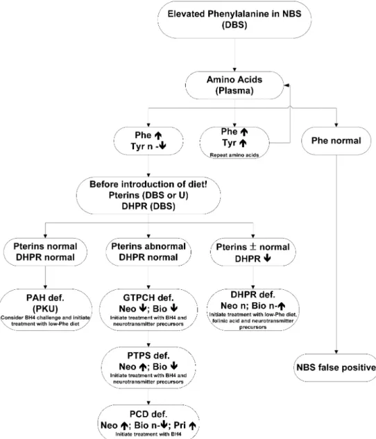
Table1and a diagnostic flow-chart following an abnormal newborn screening for elevated Phe is shown in Fig. 1. Patients with PTPS deficiency show clearly increased concentrations of neopterin, nearly no biopterin, and very low percentage of biopterin. Patients suffering from GTPCH deficiency are characterized by very low concen-trations of both biopterin and neopterin. In contrast, most patients suffering from DHPR deficiency have normal neopterin and biopterin levels in DBS (Fig.2).
Table 1 Reference values for neopterin and biopterin [nmol/L or nmol/g Hb] as well as %biopterin in patients with hyperphenylalani-nemia (HPA) and 6-pyruvoyl-tetrahydropterin synthase (PTPS) deficiency, GTP cyclohydrolase I (GTPCH) deficiency, and dihy-dropteridine reductase (DHPR) deficiency. Reference values for
biopterin [nmol/L] and biopterin per gram hemoglobin were not significantly different between the two age groups (median, 5 – 95 percentiles, * 25–75 percentile, recovery corrected). Neo = neopterin, Bio = biopterin, Pte = pterin, Hb = hemoglobin, %Bio = 100*Bio/ (Neo + Bio)
Neo [nmol/L] Bio [nmol/L] Neo [nmol/g Hb] Bio [nmol/g Hb] Bio [%]
HPA (n=271) 5.94 (1.7– 25.87) 2.75 (0.67– 8.87) 1.35 (0.35– 4.62) 0.53 (0.15– 1.68) 27.71 (11.16– 61.71) < 10 years HPA (n=27) 2.79 (0.91– 10.13) 2.75 (0.67– 8.87) 0.39 (0.15– 1.88) 0.53 (0.15– 1.68) 51.05 (12.97– 73.64) > 10 years PTPS (n=27) 23.96 (2.22– 75.37) 0 (0– 0.44) 4,46 (0.55– 9.55) 0 (0– 0.07) 0 (0– 3.08) GTPCH (n=7) 0.56 (0– 1.03)* 0 (0– 0.7)* 0.13 (0– 0.15)* 0 (0– 0.09)* 0 (0– 40.3)* DHPR (n=30) 3.78 (1.22– 33.56) 4.59 (0.40– 14.08) 0.80 (0.26– 6.17) 0.81 (0.2– 4.31) 49.41 (12.67– 77.56)
Test sensitivity
Although in most cases a clear diagnosis of a BH4
deficiency is possible by the analysis of neopterin and biopterin in DBS, the data revealed an overlap between pathologically decreased values in BH4-deficient patients
and the lower normal range in HPA patients (Fig.2). This is critical for a diagnosis of PTPS and GTPCH deficiency. To
clearly define the sensitivity and to improve test interpre-tation, test sensitivities all neopterin and biopterin results were analyzed. In addition sensitivity for urinary neopterin and biopterin analysis for PTPS and GTPCH deficiencies were calculated as well. Since the diagnosis of BH4
deficiencies always requires a combined interpretation of neopterin, biopterin, and percentage of biopterin, test sensitivity for these two pterins was also calculated.
Fig. 1 Diagnostic algorithm following abnormal newborn screening (NBS) for PKU. Every positive test for PKU needs to be confirmed by a quantitative analysis of phenylalaine (Phe) and tyrosine (Tyr) in plasma. Samples normal in the quantitative Phe and Tyr test need no further testing for hyperphenylalaninemia (HPA) variants. Samples with both elevated Phe and Tyr need another quantitative testing in order to confirm/exclude causes of HPA other than PKU or tetrahydrobiopterin (BH4) deficiency. Patients with confirmed HPA
(high Phe and normal or low Tyr) need to be tested for the cofactor BH4deficiency. This should be done before introducing the diet and at
high blood Phe levels. Dried blood spots (DBS) or random urine (U)
can be used for the differential diagnosis and depending on the profile of neopterin (Neo), biopterin (Bio), and primapterin (Pri) and dihydropteridine reductase (DHPR) activity in DBS, diagnosis of following BH4deficiencies can be established: GTP cyclohydrolase I
(GTPCH) deficiency (low or no detectable neopterin and biopterin), 6-pyruvoyl-tetrahydropterin synthase (PTPS) deficiency (high neopterin and low or no detectable biopterin), dihydropteridine reductase (DHPR) deficiency (normal neopterin and normal or elevated biopterin and no DHPR activity), and pterins-4a-carbinolamine dehydratase (PCD) deficiency (elevated neopterin, low-normal biopterin, and elevated primapterin). n: normal
Percentage of biopterin is an additional important parameter for the correct interpretation of results.
Interpretation of the test sensitivity for GTPCH deficiency was limited due to the low number of cases (n = 7). Nevertheless, test sensitivity for neopterin and biopterin reaches 100% when expressed per gram Hb, while for biopterin + pterin/g Hb a sensitivity of 85.7% is achieved. For neopterin and biopterin alone (expressed as nmol/L) sensitivity reaches 100% and 71.4%, respectively. The combined interpretation of biopterin and neopterin (both nmol/L and nmol/g hemoglobin) results in 100% sensitivity (Fig.3a). The same result was found for urine analyses where test sensitivity both for biopterin and neopterin reaches 100%. In patients with PTPS deficiency (n=27) a single parameter analysis of either neopterin or biopterin is less sensitive. Best results are achieved for biopterin both per liter or gram hemoglobin (85.5% and 96.3%, respectively). The increase of neopterin concentration in PTPS patients, compared to controls, is less sensitive (40.7% and 44.4 %; see Fig.3b).
The best test sensitivity in PTPS-deficient patients is achieved by a combined interpretation of neopterin and biopterin related to hemoglobin content (96.3%, Fig. 3b), followed by the concentration of neopterin and biopterin per liter (92.6%, Fig. 3b). Additional evaluation of biopterin + pterin either per liter or per gram hemoglobin does not further improve the sensitivity of the test. Urine analysis was possible in 20 patients with PTPS and showed for neopterin 95% test sensitivity while for the decreased concentration of biopterin 100% sensitivity is reached. Several PTPS-deficient presented normal pterin concentrations (data not shown). Thus, the calculation of biopterin + pterin may result in too high values and in contrast to the previous publication (Zurflüh et al.2005) should be not used.
Stability to light exposure
Stability of neopterin and biopterin to light exposure was investigated by analyzing a DBS from one healthy adult
a
b
%biopterin nmol/g Hb nmol/L extreme cases 75% percentile median 25% percentile minimum maximum outliers * * O Oc
Fig. 2 Pterin concentrations in DBS of patients with
hyperphenylalaninemia (HPA; < 10 years), and 6-pyruvoyl-tetrahydropterin synthase (PTPS) deficiency, GTP cyclohydrolase I (GTPCH) deficiency, and dihydropteridine reductase (DHPR) deficiency. The upper part (a) shows the pterins concentrations in nmol/L, the lower part (b) concentrations in nmol/g hemoglobin. Part (c) shows %biopterin. (Boxplot with median, quartiles, outliers (cases with values between 1.5 and 3 box lengths from the upper or lower edge of the interquartile range) and extremes (cases with values more than 3 box lengths from the upper or lower edge of the interquartile range). %biopterin=100*biopterin/ (neopterin + biopterin). K=controls
kept during 14 days either at day-light or in dark. This time frame was chosen to be sufficient to send a DBS by regular mail to the laboratory. Neopterin and biopterin were analyzed right after the blood draw (t0) and after one, five,
seven and 14 days.
Neopterin concentration from the DBS kept under light was decreased by around 6% after one day compared to DBS kept in the dark. No further difference was observed during the following 7 days but after 14 days neopterin was found to be 22% lower in DBS under light exposure. Comparably, biopterin levels under exposure to light were around 11% lower after one day. After 14 days biopterin levels were around 13% lower compared to levels form DBS kept in dark.
Hemoglobin quantification
Hemoglobin was measured in all 362 DBS. The median concentration was 8.0 g/dl. 95% of values were higher than 4.0 g/dl (5% percentile) and the minimal concentration was 1.0 g/dl.
Discussion
Blood collection on filter paper (Guthrie cards) currently plays a major role in the diagnosis of metabolic diseases. Apart from newborn screening by tandem mass-spectrometry, filter paper DBS are used for the measure-ment of a number of metabolites in serum or plasma including amino acids, acylcarnitines, sterols or for enzy-matic activities like DHPR (Arai et al. 1982) or lysosomal enzymes (Duffey et al.2010; Paglia et al.2010). The major advantage in DBS on filter paper is explained by the practical application of this medium. Samples can be transported over a long distance at room temperature and only a minimal sample volume is needed.
In children detected in the newborn screening with HPA (phenylalanine levels higher than 120 μmol/L), neopterin and biopterin analysis in urine was used for many years as a differential diagnosis of classical phenylketonuria (PKU) and defects in BH4 biosynthesis or recycling (Blau et al. 2010). A recently developed method for the measurement of neopterin and biopterin in DBS demonstrated a good
Fig. 3 Test sensitivity in seven patients with GTP
cyclohydrolase I (GTPCH) deficiency (a) and 27 patients with
6-pyruvoyl-tetrahydropterin synthase (PTPS) deficiency
(b).┌─┐ indicates sensitivity of combined parameter
interpretation. Neo = neopterin; Bio=biopterin; Pte = pterin; Bio + Pte = biopterin + pterin; Hb = hemoglobin;
correlation between the pterins pattern in DBS and urine for 70 patients with HPA (Zurflüh et al.2005). The aim of our study was to re-evaluate the usefulness of this method in a large population of patients with HPA over longer period of time and to establish reference values for both BH4
-deficient and BH4-non-deficient HPA patients. In addition,
we tested the sensitivity for each parameter and assessed the value of expressing neopterin and biopterin values to hemoglobin in the DBS sample.
As has been previously shown in urine, concentrations of neopterin and biopterin in DBS are higher in younger patients and show with exception of biopterin, biopterin + pterin and biopterin per hemoglobin significant age-related differences. For this reason we established reference ranges for children below and above 10 years of age. In children younger than 10 years a broad variation of neopterin and biopterin concentration is observed most probably due to differences in the corresponding blood phenylalanine concentration (Ponzone et al.1993). However, one should keep in mind that any activation of immune system results in an elevation of urinary neopterin, but not biopterin (Dale et al.2009; Murr et al.2002). This may result in a relative low percentage of biopterin in blood or urine.
So far only values for neopterin and biopterin of four patients with BH4 deficiency investigated by dried blood
spots were published (Zurflüh et al.2005). In our study 27 patients with PTPS deficiency, seven patients with GTPCH deficiency and 30 patients with DHPR deficiency were diagnosed. The pattern of DBS neopterin and biopterin in patients with BH4disorders was identical with that found in
urine. Pterins pattern allows a diagnosis and differentiation of patients with PTPS and GTPCH deficiency. As has been found with urinary determinations, reliable diagnosis by neopterin and biopterin analysis alone is not possible in DHPR deficiency. Here the reduced enzyme activity is decisive, and thus DHPR activity measurement in DBS is essential in every child with HPA. Reference ranges for BH4-deficient patients younger than 10 years were calculated
In order to optimize test interpretation, sensitivity for results of individual pterins was investigated in PTPS and GTPCH deficient patients. Best sensitivities were reached in GTPCH-deficient patients by the marked reduction of neopterin (at elevated blood Phe concentrations) both per liter and per hemoglobin content. Interestingly, for the reduced biopterin concentrations in GTPCH patients test sensitivity differs between the results per hemoglobin and per liter. Only biopterin concentrations expressed per hemoglobin detected all patients.
In PTPS-deficient patients the single parameter analyses yielded more equivocal results. The decreased concentra-tion of biopterin proved to be more relevant for the diagnosis than the increased concentration of neopterin.
However, in contrast to normal controls, PTPS-deficient patients always have extremely low %biopterin. Also, neopterin and biopterin calculated per hemoglobin content are more discriminative than those expressed as nmol/l per liter. Even with the combined interpretation of neopterin with biopterin or biopterin + pterin per liter, the sensitivity remains below the results of biopterin/g Hb alone (92.6% or 81.5% vs. 96.3%). For that reason determination of hemoglobin concentration in the sample and expression of neopterin and biopterin results according to hemoglobin concentration must be part of the test procedure. In our population 95% of samples had a hemoglobin concentration of higher than 4 g/dl. Although a DBS with only 1 g/dl hemoglobin revealed reliable results, we suggest a careful evaluation of each result with hemoglobin concentrations below 4 g/dl. Analysis of urinary neopterin and biopterin in PTPS-deficient patients reached a test sensitivity of 100%. It is important to note that in our study no patients with pterin-4a-carbinolamine dehydratase (PCD) deficiency were detected by pterins analysis from DBS. However, in the same test period we did not detect any PCD-deficient patients by urinary pterins either. PCD deficiency is characterized by the presence of low concentrations of primapterin in urine. HPA is present only in the early neonatal period in these patients. PCD deficiency is considered as a benign disease, although one patient was reported as having mild neurological signs including tremor in the neonatal period and with tendency to hyper- or hypotonia and motor delay (Dhondt2006).
In summary, our data confirm the usefulness of DBS for the screening for BH4 deficiency. Although, the
measure-ment of urinary neopterin and biopterin is more sensitive due to higher concentrations of pterins in urine, the DBS method is reliable and safe. No false negative tests were reported in the 5-year period. Phenylalanine, tyrosine, pterins (neopterin and biopterin) and DHPR activity can be measured from one single DBS, which facilitates significantly sample handling and transport costs. It is important to note that neopterin and biopterin in body fluids are sensitive to temperature, oxygen, and light (Zurflüh et al. 2005). We showed that both neopterin and biopterin levels show a tendency to be lower under light exposure, but this difference is not statistically significant and has no impact on the differential diagnosis. Nevertheless, exposure of DBS samples to sunlight should be avoided. In conclusion, this method, although not superior in analytical performance, is a practical, less expensive, and safe substitute for analysis of neopterin and biopterin in urine and a useful tool for the differential diagnosis of BH4deficiencies.
Acknowledgements This work was supported by the Swiss National Science Foundation (to N.B. and B.T.) grant no. 31003A-119982.
References
Arai N, Narisawa K, Hayakawa H, Tada K (1982) Hyper-phenylalaninemia due to dihydropteridine reductase deficiency: diagnosis by enzyme assays on dried blood spots. Pediatrics 70:426–430
Blau N, Bonafé L, Blaskovics M (2005) Disorders of phenylalanine and tetrahydrobiopterin metabolism. In: Blau N, Duran M, Blaskovics M, Gibson KM (eds) Physician' Guide to the Laboratory Diagnosis of Metabolic Disease. Springer, Heidelberg, pp 89–106
Blau N, Thöny B (2008) Pterins and related enzymes. In: Blau N, Duran M, Gibson KM (eds) Laboratory Guide to the Methods in Biochemical Genetics. Springer-Verlag, Berlin Heidelberg, pp 665–702
Blau N, Thöny B, Cotton RGH, Hyland K (2001) Disorders of tetrahydrobiopterin and related biogenic amines. In: Scriver CR, Beaudet AL, Sly WS, Valle D, Childs B, Vogelstein B (eds) The Metabolic and Molecular Bases of Inherited Disease. McGraw-Hill, New York, pp 1725–1776
Blau N, Van Spronsen FJ, Levy HL (2010) Phenylketonuria. Lancet 376:1417–1427
Dale RC, Brilot F, Fagan E, Earl J (2009) Cerebrospinal fluid neopterin in paediatric neurology: a marker of active central nervous system inflammation. Dev Med Child Neurol 51:317–323
Dhondt JL (2006) Laboratory diagnosis of phenylketonuria. In: Blau N (ed) PKU and BH4: Advances in Phenylketonuria and Tetrahydrobiopterin. SPS, Heilbronn, pp 161–179
Duffey TA, Bellamy G, Elliott S et al. (2010) A tandem mass spectrometry triplex assay for the detection of Fabry, Pompe, and mucopolysaccharidosis-I (Hurler). Clin Chem 56:1854–1861 Murr C, Widner B, Wirleitner B, Fuchs D (2002) Neopterin as a marker
for immune system activation. Curr Drug Metab 3:175–187 Niederwieser A, Matasovic A, Kuster T, Staudenmann W, Pfleiderer W,
Scheibenreiter S (1986) Catabolism of tetrahydrobiopterin in man. In: Cooper BA, Whitehead VM (eds) Chemistry and Biology of Pteridines 1986. Walter de Gruyter, Berlin, pp 305–308
Paglia G, D'Apolito O, Gelzo M, Dello Russo A, Corso G (2010) Direct analysis of sterols from dried plasma/blood spots by an atmospheric pressure thermal desorption chemical ionization mass spectrometry (APTDCI-MS) method for a rapid screening of Smith-Lemli-Opitz syndrome. Analyst 135:789–796 Ponzone A, Guardamagna O, Spada M et al. (1993)
Hyperphenyla-laninemia and pterin metabolism in serum and erythrocytes. Clin Chim Acta 216:63–71
Zorzi G, Thöny B, Blau N (2002) Reduced nitric oxide metabolites in CSF of patients with tetrahydrobiopterin deficiency. J Neuro-chem 80:362–364
Zurflüh M, Giovannini M, Fiori L et al. (2005) Screening for tetrahydrobiopterin deficiencies using dried blood spots on filter paper. Mol Genet Metab 86:96–103
![Table 1 Reference values for neopterin and biopterin [nmol/L or nmol/g Hb] as well as %biopterin in patients with hyperphenylalani-nemia (HPA) and 6-pyruvoyl-tetrahydropterin synthase (PTPS) deficiency, GTP cyclohydrolase I (GTPCH) deficiency, and dihy-d](https://thumb-eu.123doks.com/thumbv2/123doknet/14839153.624069/3.892.76.817.884.1053/reference-neopterin-biopterin-hyperphenylalani-tetrahydropterin-deficiency-cyclohydrolase-deficiency.webp)