ORIGINAL ARTICLE
Evaluation of [
18
F]-choline PET/CT for staging
and restaging of prostate cancer
Daniela B. Husarik&Raymond Miralbell&Markus Dubs&Hubert John&Olivier T. Giger&
Albert Gelet&Tibor Cservenyàk&Thomas F. Hany
Received: 6 November 2006 / Accepted: 25 July 2007 / Published online: 10 October 2007
# Springer-Verlag 2007
Abstract
Purpose To evaluate the accuracy of [18F]-choline (FCH) positron emission tomography/computed tomography (PET/ CT) for staging and restaging of prostate cancer.
Methods FCH PET/CT was performed in 111 patients with prostate cancer using 200 MBq FCH: 43 patients [mean age 63 years; mean prostrate specific antigen (PSA) 11.58μg/l] were examined for initial staging, and 68 patients (mean age 66.4 years) were examined for restaging (mean PSA 10.81 μg/l). FCH PET/CT results were correlated to histopathology, bone scan, morphology as revealed by
magnetic resonance imaging (MRI) and CT, PET/CT follow-up and PSA follow-up after therapy.
Results FCH PET/CT scans at initial staging correctly showed no metastases in 36/38 patients undergoing radical surgery, as confirmed by PSA levels <0.1 μg/l 6 months postoperatively. Lymphadenectomy was performed in 24 of these patients, revealing four false FCH-negative lymph nodes (LN). In one patient, only lymphadenectomy was performed since a FCH-positive LN was confirmed by histology. Four patients showed FCH-positive bone metas-tases, as proven by bone scan. FCH PET/CT scans at restaging correctly revealed local recurrence in 36 patients. No pathological FCH uptake was observed in 11 patients with biochemical recurrence. Twenty-three patients showed FCH-positive LN. Twenty LN were surgically removed in seven patients. Histopathology verified metastases in all LN, but revealed two additional metastastic, FCH-negative LN. Seventeen patients showed FCH-positive bone metastases, as proven by bone scan or MRI. Sensitivity to detect recurrent disease was 86%.
Conclusion The results obtained using FCH PET/CT scans for initial N-staging were discouraging, especially in terms of its inability to detect small metastases. Recurrent disease can be localized reliably in patients with PSA levels of >2μg/l. Keywords [18F]-Choline . Initial staging . PET-CT . Prostate cancer . Recurrence
Introduction
In Western Europe and Northern America, Prostate cancer (PC) is the most common tumour in men and the second most common cause of death from cancer in North American men [1]. Prostrate specific antigen (PSA) D. B. Husarik (*)
:
T. Cservenyàk:
T. F. HanyDepartment of Nuclear Medicine, University Hospital of Zurich, C NUK 8a, Raemistrasse 100,
8091 Zürich, Switzerland e-mail: [email protected] R. Miralbell
Department of Radiation Oncology, University Hospital Geneva, Geneva, Switzerland
M. Dubs
Department of Urology, Hospital of Uster, Uster, Switzerland
H. John
Department of Urology, Klinik Hirslanden, Zurich, Switzerland
O. T. Giger
Department of Pathology, University Hospital Zurich, Zurich, Switzerland
A. Gelet
Department of Urology and Transplantation, Edouard Herriot Hospital,
measurement in combination with digital rectal examination is often used for the early diagnosis of PC, with subsequent confirmation by transrectal biopsy. Magnetic resonance imaging (MRI) and transrectal ultrasound are performed to assess the T stage, while computed tomography (CT) and bone scan are additionally used for N and M staging.
Depending on the clinical stage, different recommended treatment options exist, consisting of watchful waiting, radical prostatectomy, radiotherapy, hormonal therapy or a combination of these [2]. Following the initial treatment with curative intent, patients are followed up by serum PSA measurement supplemented by digital rectal examination (DRE). Following radical prostatectomy, two consecutive PSA values of 0.2 μg/l and above are considered to represent recurrent cancer. After initial radiation therapy, three consecutive increasing PSA values above the previous PSA nadir measured at 3-month intervals represent recur-rent disease [2].
At this level, however, the sensitivity and specificity of conventional imaging techniques are limited, especially in terms of localizing the site of recurrence. However, even more advanced imaging techniques, such as [18 F]fluoro-deoxyglucose (FDG) positron emission tomography (PET), have been found to be of only limited value for staging and restaging of PC [3]. Recently, PET using [18F]- or [11 C]-labeled choline analogues as well as [11C]-acetate have demonstrated promising results in the localization of recurrent PC [4–10]. The aim of our study was to further evaluate the accuracy of [18F]choline for the initial staging and localizing recurrent disease of PC.
Materials and methods
The institutional review board approved the study protocol, and written informed consent was obtained from all patients before imaging. The studies were performed between July 2003 and May 2006. The inclusion criteria were: (1) initial staging: histological proven PC scheduled to undergo surgical resection; (2) recurrent disease: previously histological proven PC after treatment with curative intent (surgery, radiation treatment) and current proven biochemical recurrence. Patients at initial staging
Forty-three patients (mean age 63 years; range 51–74 years; SD 6.21 years) with bioptically proven PC eligible for surgery were examined for initial staging. Biopsy of the prostate was performed in all of these patients at least 14 days prior to the PET/CT scan (stage information: one, T1a; two, T1c; 15, T2a; four, T2b; 17, T2c; one, T3; one, T4). The mean free serum PSA level in these patients was 11.58μg/l (range 0.6–162 μg/l, SD 24.74), with an average
interval of 56 days (range 0–163 days) between PSA measurement and PET/CT. The mean Gleason score was 6.79 (range 5–10). Of the 43 patients, 30 were high-risk patients (high risk defined by PSA >10μg/l and/or Gleason ≥7). Only one patient was already receiving antihormonal treatment (PSA 0.6μg/l at time of PET/CT). The PET/CT findings were correlated to the histopathological findings of sampled lymph nodes in 25 patients: 115 lymph nodes; mean number of removed lymph nodes, 4.6; range, 1–11. An evaluation of the primary tumour regarding extent was not performed. PSA follow-up measurements were per-formed in all 38 patients undergoing radical prostatectomy. In four of the five patients who did not undergo surgery, a bone scan (n=4) and MRI (n=1) were performed. In one nodal positive patient, radical surgery was not performed. Patients with recurrent disease
Sixty-eight patients (mean age 66.4 years; range 46–81; SD 7.01) were examined due to biochemical recurrence. The mean free serum PSA level was 10.81 μg/l (range 0.36– 100 μg/l; SD 17.69 μg/l). PSA testing was performed in different laboratories at various locations prior to referral by the attending physician. In 53 patients, tumour stage at initial diagnosis ranged between pT2a and pT4. The initial tumour stage was not available for 15 patients. The mean Gleason Score at diagnosis was 7.2 (range 4–10) in 43 patients; for 25 patients the Gleason Score at initial diagnosis was not available. At the time of the PET/CT scan, 13 patients were under antihormonal treatment with goserelin (Zoladex) and/ or bicalutamide (Casodex) (n = 11) or had undergone orchiectomy (n=2). Previous therapy is summarized in Table1. The interval between the initial treatment and the PET/CT was 9–115 months. Four patients were scanned twice after 4, 7, 11 and 18 months, respectively. PET/CT findings were correlated to PSA follow-up after radiother-apy, histopathology, MRI, bone scan, clinical follow-up, follow-up by FCH PET/CT scans (interval 4–18 months) or apparent changes on the CT part of the PET/CT procedure. Synthesis of FCH
[18 F]-Fluorocholinefluoromethyldimethyl-2-hydroxyethyl-ammonium (FCH) was produced in-house using the method as described previously by Schmid et al. [11].
Imaging protocol
The patients were scanned with an integrated PET/CT scanner (Discovery LS; GE Healthcare) that consists of a full-ring PET scanner with a 14.6-cm transverse field of view (FOV) and an in-plane resolution of 4.8 mm full width at half maximum at the centre of the FOV and a
multisection CT scanner. A low-dose, unenhanced CT scan (80 mA, 0.5 s per rotation, 140 kV, 4.25-mm reconstructed section thickness) was obtained from the head to the pelvic floor. After the CT scan had been completed, the table position was moved axially to the initial position for the PET scanning so that the first FOV started at the ischiadic tuberosity, covering the pelvic floor and encompassing the urinary bladder. No indwelling urinary catheter was used because by scanning the bladder in the first FOV within 5 min of the intravenous injection of FCH, no activity is found in the bladder, whereas the activity in the prostate reaches a maximum in this 5-min window immediately following the injection, as shown by DeGrado et. al. [7]. The PET scan was initiated 120 s after the intravenous injection of a standard dose of 200 MBq FCH. Six to seven cradle positions, depending on the size of the patient, with an acquisition time of 3 min for emission per position were scanned. CT data were used for attenuation correction of the PET data and for image fusion. To obtain minimal attenuation artefacts, a previously described breathing protocol was used during the CT acquisition [18]. The PET images were reconstructed with a standard iterative reconstruction ordered-subset expectation maximization iterative algorithm (two iterative steps) and were reformat-ted into transverse, coronal and sagittal views.
Image interpretation
Image analysis was performed in consensus by two nuclear medicine physicians, one of whom also has a board certificate in radiology, reading on screen by using PET/CT review software (XELERISver. 1.0728; GE Healthcare), which allows
simultaneous scrolling through the corresponding PET, CT and fused sections in all three planes. Images were interpreted as either normal/probably normal if the FCH uptake was equal to the background or abnormal if the FCH uptake in a lesion was higher than the background. The maximum standardized uptake (SUVmax) values were determined for lesions rated as abnormal. SUVmax values of the primary tumour in the prostate were not measured at initial staging as it has been shown that prostate cancer can not be differentiated from benign prostatic hyperplasia and prostatitis [11].
Histopathological correlation
In 23 patients who underwent lymphadenectomy, PET/CT images were compared to histopathological findings. The surgically resected lymph nodes were fixed in formalin and embedded in paraffin, and the sections were then stained with hematoxylin and eosin. A pathologist determined the exact size of metastases independently.
In patients with a suspicion of recurrent disease, biopsy of the local recurrence (n=14) or surgical resection of lymph nodes (n=7) were performed.
Results Initial staging Overall results
Pathological FCH uptake in the prostate was observed in 42 of the 43 patients with histological proven PC examined for initial staging. In only one patient with newly diagnosed PC, who had already received antihormonal treatment, no pathological FCH accumulation was delineated in the prostate. Additional FCH uptake was observed in the lymph nodes in two patients, in osseous structures in three patients and in the lymph nodes and osseous structures in one patient. FCH-positive lymph nodes
In three patients, FCH PET/CT showed FCH-positive lymph nodes suggestive of lymph node metastases (mean SUVmax 5.04; range 4.9–5.2; SD 0.21).
In one case with a FCH-positive, pathologically enlarged obturator lymph node, lymphadenectomy of only this node was performed, and histopathology revealed a metastasis greater than 1 cm. The patient did not receive curative therapy due to concomitant bladder cancer, local progressive prostate cancer and a history of ear, nose and throat (ENT) cancer.
One patient with a slight FCH accumulation in an iliac extern lymph node (SUVmax 1.9) underwent radical Table 1 Initial therapy of patients with recurrent disease
Number of patients Previous therapy
26 RP 4 RP, HT 11 RP, RT 3 RP, RT, HT 8 RT 6 RT, HT 1 Ablatherm 1 HT 1 Hyperthermia 1 RT, HT, Ablatherm 1 RT, HT, RT of a bone metastasis 1 RT, HT, LAD recurrence 1 TURP, HT 1 RP, RT, HT, LAD, Samariumtherapy 2 RP, LAD recurrence
RP, Radical prostatectomy; HT, antihormonal therapy; RT, radiother-apy external beam or brachytherradiother-apy; LAD, lymphadenectomy; TURP, transurethral resection of the prostate
prostatectomy and lymphadenectomy in the obturator region. Histopathology of these lymph nodes did not reveal metastases. The PSA dropped below 0.04 μg/l within 3 months and remained below 0.04μg/l for 12 months after the operation, indicating that the FCH-positive lymph node did not contain prostate cancer cells and likely represented instead a reactive lymph node. As such, this lymph node was defined as a false FCH-positive lymph node in terms of metastasis.
One patient with bone metastases proven by bone scan showed FCH accumulation in two pathological enlarged obturator and iliac extern lymph nodes. Histopathological correlation was not available. No follow-up FCH PET/CT was performed in this patient; however, a follow-up bone scan demonstrated progressive osseous metastases. Correlation with lymphadenectomy
Histopathological work-up was performed on 115 lymph nodes sampled from 25 patients. Only one of these lymph nodes showed pathological FCH accumulation (see above) and was proven to be a metastasis measuring more than 1 cm.
Four lymph nodes that did not show FCH accumulation turned out to contain metastatic cells, with an overall tumor load measuring less than 0.5 cm. All other lymph nodes did not contain malignant cells. (Tables2,3).
Bone metastases
FCH accumulation in osseous lesions was found in four patients. Bone scan (n=4) and MRI (n=1) verified metastases in all patients (mean SUVmax 6.3; range 1.8–13.9; SD 4.21). Correlation with PSA follow-up
Postoperative PSA follow-up was performed in all 38 patients that underwent radical prostatectomy. None of these patients showed FCH accumulation suggestive of metastatic disease. Of these 38 patients, 36 showed no measurable PSA 6 months after surgery, demonstrating the absence of remaining prostatic tissue and thereby verifying the absence of metastatic disease. The remaining two patients presented with a detectable PSA 6 months after surgery. One of these was a patient who had had metastatic disease proven by
histopathology in one of the three lymph nodes that had been removed; he had a PSA of 0.35μg/l 6 months after surgery (pT3a; Gleason 4+5=9). The other patient had a PSA level of 0.36 μg/l 3 months after R1 resection of the prostatic tumour (pT2c), explaining the measurable PSA.
Recurrent disease/restaging Overall results
Pathological FCH accumulation was seen in 57/68 patients that presented with biochemical recurrence of PC and examined with FCH PET/CT. The location of pathological FCH accumulation is summarized in Table4. FCH PET/CT was unable to demonstrate the site of recurrent disease in 11 patients, although a biochemical recurrence was present. Only two of the patients that had a negative PET/CT scan were under antihormonal therapy. The details of patients with negative FCH PET/CT scans are summarized in Table5.
Local recurrence
Local recurrence was FCH positive in a total of 36 patients (mean SUVmax 2.9; range 1.2–9.7; SD 1.7 ). It was confirmed by biopsy in 14 patients (Fig. 1) and by a PSA drop during radiotherapy of the prostatic bed in eight patients; no antihormonal therapy was given to any patient. In two patients, the MRI scans were also positive for local recurrence. Local recurrence was not evaluated in the remaining 12 patients due to distant metastases and no clinical significance to the local situation.
Table 2 Histopathology of lymph nodes (initial staging)
Metastasis No metastasis
FCH positive 1 0
FCH negative 4 110
FCH, [18 F]-Fluorocholinefluoromethyldimethyl-2-hydroxyethyl-ammonium
Table 3 Patients with lymphadenectomy (initial staging)
Metastasis No Metastasis
FCH positive 1 0
FCH negative 2 22
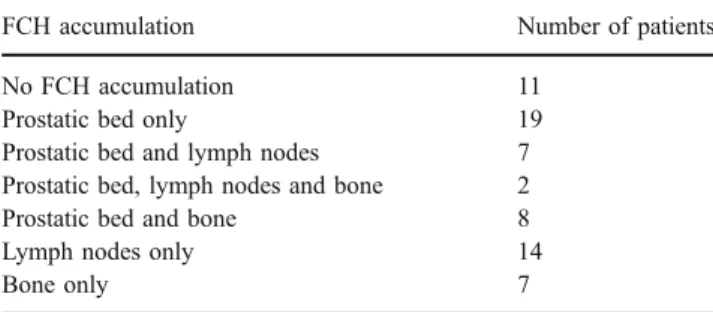
Table 4 FCH accumulation on PET/CT in recurrent disease
FCH accumulation Number of patients
No FCH accumulation 11
Prostatic bed only 19
Prostatic bed and lymph nodes 7 Prostatic bed, lymph nodes and bone 2
Prostatic bed and bone 8
Lymph nodes only 14
Bone only 7
Lymph node metastases
Pathological FCH accumulation in lymph nodes was found in 23 patients (mean SUVmax 4.3; range 1–14; SD 2.3). Lymph node metastases were confirmed by histopathology in seven patients (total sampled lymph nodes, n = 20; histopathology positive for cancer, n=20). The FCH PET/ CT was positive in 18 of these 20 lymph nodes. In two of these seven patients, histopathology revealed one addi-tional lymph node harbouring prostate cancer cells not seen on FCH PET/CT (2/20 false negative lymph nodes). In one of the seven, the patient’s PSA remained undetectable for 16 months after lymphadenectomy. However, after 18 months, a biochemical recurrence occurred (PSA 1.26μg/l), and FCH PET/CT showed several FCH-positive, pathological enlarged lymph nodes, compatible with meta-static disease. Overall, following lymphadenectomy, all of the patients showed PSA progression at some point in time. In three patients, lymph node metastases were confirmed by progressive FCH accumulation at the follow-up FCH PET/CT taken under conditions of a rising PSA level (Fig. 2). In seven cases, the FCH-positive lymph nodes were clearly enlarged and met malignancy criteria on CT and/or MRI imaging. In one patient with a FCH-positive lymph node, a decreasing PSA level during radiotherapy (RT) of the positive lymph node confirmed metastatic disease. Lymph node metastases could not be confirmed in five patients.
Bone metastases
Pathological FCH accumulation in osseous structures was seen in 17 patients (mean SUVmax 6.7; range 1.7–12; SD
3.04). Metastases to the bone was confirmed by bone scan (n= 11) (Fig.3), MRI (n=3) (Fig.4) and CT morphology (n=3). FCH PET/CT in patients with PSA <2 μg/l
Fourteen patients with PSA levels≤2 μg/l were examined. Of these 14 patients, four were receiving antihormonal therapy at the time of the FCH PET/CT scan. Of the 14 FCH PET/CT scans, ten demonstrated pathological accumulation, whereas PET/CT could not demonstrate recurrent disease in the remaining four cases. Only one of the four patients with a negative PET/CT scan at PSA <2 μg/l was receiving antihormonal therapy. The overall sensitivity of FCH PET/ CT imaging in patients with PSA levels <2 μg/l in our limited number of cases was 71%, in patients not receiving antihormonal therapy, 70% and in patients receiving anti-hormonal therapy with a measurable PSA <2 μg/l, 75%. Patient details are presented in Table 6. The overall sensitivity of FCH PET/CT at any PSA level was 86%, and at PSA levels of >2 μg/l, 87%. (Sensitivity without antihormonal therapy at any PSA level was 83%, and at PSA >2μg/l without antihormonal therapy, 86%. Sensitiv-ity at any PSA level in patients with HT was 84% and at >2μg/l, 88%).
FCH PET/CT in patients receiving antihormonal therapy Of the 13 patients examined with FCH PET/CT who were receiving antihormonal therapy, only two examinations did not show any pathological FCH accumulation even though PSA levels were pathological. In the other 11 patients, at least one site of pathological FCH uptake was confirmed to be metastatic disease or local recurrence (Fig. 5). Patient Table 5 Patients without FCH accumulation (n.a. not available)
Age (years) PSA Previous therapy Time after initial treatment TNM GS HT Verification
46 0.36 RP 9 months pT2c 7 N PSA drop after RT (FN LR)
63 0.5 RP 3 years, 2 months pT2a 6 N MRI (FN LR)
69 0.52 RP, RT 2 years, 3 months pT2b cN0 6 N PSA drop after RT (FN LR)
69 1.9 RP, RT 6 years, 4 months n.a. n.a. Y Under HT
67 2.1 RP 8 years, 5 months pT3a R1 n.a N Local biopsy positive
74 2.12 RT, HT,
Ablatherm
5 years, 3 months pT2b 5 N Local biopsy positive
68 3.41 RP, RT 7 years, 10 months pT2a n.a. N no PSA drop after RT
(FN distant disease)
65 3.5 RP, HT 7 years, 1 months pT3a pN0 8 Y PSA before HT 13
72 4.4 RP 5 years, 10 months pT4 7 N PSA drop after RT (FN LR)
76 4.77 RP 8 years, 10 months pT3a 5 N PSA drop after RT (FN LR)
74 10.69 RP 7 years, 3 months pT2b 6 N Very high PSA
PSA, Prostrate specific antigen; GS, Gleason score; RP, radical prostatectomy; HT, antihormonal therapy; RT, radiotherapy external beam or brachytherapy, LAD, lymphadenectomy; FN, false negative; MRI, magnetic resonance imaging; TNM, cancer staging system; LR, local recurrence
details are presented in Table 7. In general, pathological FCH uptake was seen when the tumour became hormone resistant, i.e., when the PSA started rising again. In the two cases with no pathological FCH accumulation, the patients had just started receiving antihormonal treatment and their PSA levels were dropping.
Discussion
When assessing the accuracy of FCH PET/CT imaging in prostate cancer, the patient population has to be split into two major subgroups: (1) patients with newly diagnosed prostate cancer and (2) patients with biochemical recurrence of prostate cancer after initial treatment.
On the basis of our data, FCH PET/CT imaging seems to have a limited value for initial staging of prostate cancer. The patients we examined with FCH PET/CT for initial staging all had proven prostate cancer and localizing the
tumour within the prostate was not the main goal in our study. In a previous study we showed that FCH PET/CT is not appropriate for initial T staging due to the limited spatial resolution of PET that does not enable the assessment of capsular infiltration, which in turn defines T3 tumours [11]. Additionally, pathological FCH accumu-lation occurs not only in prostate cancer, but also in benign prostatic hyperplasia and prostatitis. In some studies the authors showed that it was possible to localize cancer within the prostate [12,13].
When the SUVmax values obtained in our population are compared to previously published data on physiological uptake in various body regions, malignant and benign lesions clearly demonstrate higher uptake values [11]. SUVmax values measured in lymph node metastases at initial staging (mean 5.04; range 4.9–5.2; SD 0.21) and at restaging (mean 4.3; range 1–14; SD 2.3) were higher than those in subcutaneous fat (mean 0.2 ± 0.1; range 0.1–0.3). Interestingly, SUVmax values of bone metastases at initial Fig. 1 a MIP imaging, b top to
bottom computed tomography (CT), positron emission tomog-raphy (PET), fusion, all axial. Images are of a 70-year-old patient with a history of surgery 8 years prior and radiotherapy 5 years prior. [18 F]-Fluorocho- linefluoromethyldimethyl-2-hydroxyethyl-ammonium (FCH) PET/CT shows local recurrence (bold arrow) proven by biopsy (Gleason 7). Note the FCH in the veins of the right arm after insufficient flushing after FCH injection (thin arrow)
staging (mean SUVmax 6.3; range 1.8–13.9; SD 4.21) and at restaging (mean SUVmax 6.7; range 1.7–12; SD 3.04) were higher than those of lymph node metastases and physiological uptake in bone of the lumbar spine (mean 1.3 ± 0.5; range 0.2– 1.9). In proven local recurrence, SUVmax values (mean 2.9; range 1.2–9.7; SD 1.7) were lower than those in lymph node metastases and higher than those in subcutaneous fat (mean 0.2 ± 0.1; range 0.1–0.3) or in the bladder (mean 0.4 ± 0.1; range 0.2–0.7). However, no cutoff can be given since almost all detected lesions were due to malignancy, and only one single lesion was due to inflammation (false positive FCH uptake in one lymph node, SUVmax 1.9).
FCH PET/CT can be helpful to guide biopsy if a patient has persistent elevated PSA levels but the biopsies remain negative for tumour. One major limitation of FCH PET/CT for initial staging is the general inability of PET imaging to detect micrometastases. The identification of occult lymph node metastases appears to have an impact on the outcome of patients with prostate cancer [14]. Patients with occult lymph node metastases show significantly increased risk of prostate cancer recurrence and death. Another study suggests that more extensive pelvic lymphadenectomy in patients
under-going radical prostatectomy could improve the accuracy of staging and have a positive impact on disease-free survival and, perhaps, on overall-survival [15]. Often, only the obturator lymph nodes are resected. Schumacher et al. showed in their study that by resecting lymph nodes along the internal iliac artery, a larger amount of metastases was found. The proportion of patients with nodes either exclusively in this area or in combination with another location was 59% [16]. The sentinel lymph node concept might be the solution for accurate initial nodal staging in prostate cancer patients [17]. The method is widely accepted in melanoma, breast cancer and ENT tumours.
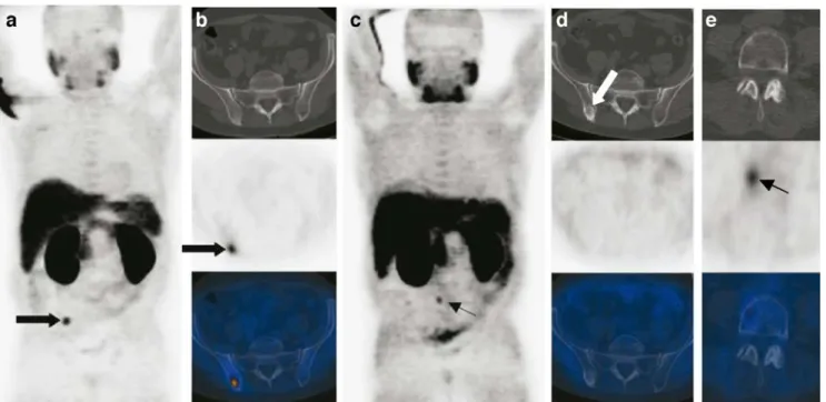
FCH PET/CT is well able to depict bone metastases of prostate cancer; nevertheless, [18F]fluoride PET has a greater sensitivity [18]. A possibility to enhance the detection rate of bone metastases on FCH PET/CT scans is late imaging at 65–200 min p.i. [10]. Whereas FCH accumulation in bone metastases on late imaging rises compared to early imaging (5–15 min p.i.), the time point of imaging has no influence on FCH accumulation in local recurrence or lymph node metastases. One could consider imaging the region of the prostate soon after the injection of Fig. 2 a MIP imaging, b top to bottom CT, PET, fusion, all axial, c
MIP, d top to bottom CT, PET, fusion, all axial (c, d 11 months later). Images of a 58-year-old patient, with a history of surgery 2 years, 4 months previously; PSA 1.07μg/l. Scan was initially evaluated as normal. In retrospect, a normal-sized lymph node (bold arrow) shows
increased FCH activity. FCH in the ureter on the left side (thin arrow). c, d 11 months later, PSA is now 5.25μg/l. FCH PET/CT shows one clearly enlarged and FCH-active lymph node in the iliac extern region on the right side (bold arrow)
FCH in order to evaluate this region without a radiotracer in the bladder and performing whole body images approxi-mately 1 h later. The disadvantage of this method would be that the patient has to be positioned on the scanner twice.
FCH PET/CT shows promising results in localizing recurrent PC. PSA doubling time is currently the best predictor of tumour progression. Nonetheless, there is no consensus on diagnostic work-up that should be performed. A PSA level of >0.2μg/l has been described as a definition for recurrent PC after prostatectomy [19]. Curative salvage therapy is most likely to succeed when realized in patients with PSA levels of <2 μg/l [2]. In the limited number of patients in our study that were examined at PSA levels of <2 μg/l, the overall sensitivity for detecting recurrent disease was 71%. The overall sensitivity in detecting recurrent disease at any PSA level in our study was 86%. No definite PSA cut-off level can be given– i.e. from when on FCH PET/CT can reliably detect recurrent disease. The PSA level is not the only factor determining whether pathological FCH accumulation will be found or not. Factors such as PSA doubling time, time to recurrence after initial therapy and PSA and the Gleason score at initial therapy seem to have an influence on the accumulation of FCH as well. In our study in patients with recurrent PC, we
found several normal-sized lymph nodes with minimally increased FCH accumulation above the background that turned out to be metastases, proven by histopathology or increasing FCH-uptake on follow-up PET/CT (Fig.2).
As bowel, ureter and lymph nodes can be very close together in the pelvis and abdomen, coregistration with CT is of great importance. The detection of small lymph node metastases with only minimal radiotracer accumulation on FCH PET alone is impossible. Pelvic MRI with surface coils and the use of superparamagnetic particles provide the sensitivity and the specificity which have never been obtained by the sole measurement of node size of the lymphatic chains draining the prostate gland and might be the future of lymph node imaging in prostate cancer [20,21]. According to Sella et al., MRI with endorectal coil depicts a high proportion of local recurrence after prostatectomy [22]. The advantage of PET/CT is, on one hand, the combination of functional and anatomical imaging of a large FOV and, on the other hand, an all-in-one examination.
In terms of FCH PET/CT imaging of patients receiving antihormonal therapy, Heinisch et al. showed that FCH PET/CT can be positive in these patients, even at very low PSA levels [23]. Pathological accumulation will occur in patients with hormone-resistant tumour tissue, as indicated Fig. 3 a Bone scan, b MIP, c
top to bottom CT, PET, fusion, all axial. Images of a 76-year-old patient with a history of surgery 17 years prior (staging pT3b), radiotherapy of recur-rence 16 years prior and orchi-ectomy 1 year, 1 month prior. FCH PET/CT confirms multiple osseous metastases also seen on the bone scan
by a rising PSA under therapy. On the other hand, patients with hormone-dependent tumours receiving antihormonal therapy will not show pathological FCH accumulation [24]. In the case of distant metastases, current treatment options include only therapies with palliative intent, such as radiotherapy to palliate bone pain and/or prevent impending fracture, palliative radionuclide therapy, ortho-pedic surgery to prevent or repair fractures, analgesics and bisphosphonates, all of which can significantly reduce the
risk of skeletal complications and delay their onset. If only systemic therapy is intended, the value of imaging is questionable. In such a case, measurement of PSA and patient questioning might be sufficient to evaluate therapy response. On the other hand, if focused therapy, such as radiotherapy of lymph node metastases or bone metastases, is intended, FCH PET/CT can nicely demonstrate therapy response (Fig. 4). Another indication for FCH PET/CT in recurrent disease can be the exclusion of distant metastases Fig. 4 a MIP imaging, b top to bottom CT, PET, fusion, all axial, c
MIP, d top to bottom CT, PET, fusion, all axial, e top to bottom CT, PET, fusion, all axial (c–e 1 year later). Images of a 59-year-old patient with a history of radiotherapy 3 years prior. Initial stage T2c, PSA 41μg/l, Gleason 8. PSA is now 2.00 μg/l. FCH PET/CT reveals
bone metastasis (bold arrow) verified by MRI in addition to a known local recurrence. One year later (c–e) the PSA had dropped to 0.7 μg/l but started rising again. PSA is now 1.5μg/l. FCH PET/CT shows no active tumour in the radiated bone metastasis (bold arrow). New metastasis in L 4 (thin arrow) in addition to the still existing local recurrence
Table 6 Patients with PSA <2μg/l
Age (years) PSA PET/CT Verification HT Therapy
79 0.35 LR, BM Bone scan Yes HT
46 0.36 No PSA drop No RP
66 0.4 LR, LN PSA drop Yes RP
63 0.5 No MRI LR No RP
69 0.52 No PSA drop No RP, RT
62 0.7 LR PSA drop No RP
68 0.81 BM CT morphology No RP, HT
81 1.2 LR MRI Yes (Orchiectomy) RP, RT, HT
61 1.26 LN CT morphology No RP, LAD
75 1.29 LR PSA drop RP
60 1.5 LR, BM Biopsy, MRI No RT HT RT bone met
69 1.9 No PSA Yes RP, RT
63 1.96 LR, BM Bone scan No RP
60 2.0 LR, BM Biopsy, MRI No
in patients with bioptically proven local recurrence of PC and planned local salvage therapy, such as radiotherapy or high-intensity focused ultrasound therapy [25].
In our opinion, the optimal diagnostic work-up after the initial diagnosis of PC consists of a bone scan or [18 F]-fluoride PET/CT to rule out bone metastases combined with sentinel lymph node dissection to attain accurate lymph node staging [26]. FCH PET/CT does not seem to be the ideal imaging modality for initial staging of PC. In the case of biochemical recurrence, however, we believe that FCH PET/CT imaging is of great help. The sensitivity of 71% in localizing recurrent disease at PSA levels <2μg/ is not very high, but at the moment there is no other imaging modality that performs better.
The considerable limitations of the study include surgical strategies at initial staging as well as the inclusion of a heterogeneous group of patients with possible recurrent disease. At staging surgery, a more extensive lymph node dissection procedure would have been preferred; however, dissection limited to the obturator fossa is the procedure of choice for many urological surgeons. Therefore, possible involved lymph nodes outside the obturator fossa region could have been missed. Further, an added contrast-enhanced CT part could possibly have improved the diagnostic accuracy. However, all missed lymph nodes were smaller than 1 cm and as such do not meet CT criteria for malignancy. The heterogeneity in treatment and time to biochemical recurrence of patients with recurrent prostate cancer reflects the typical referral pattern.
Conclusion
[18F]-Choline PET/CT does not seem to be suitable for the initial staging of prostate cancer due to its low sensitivity in detecting lymph node metastases. At the present time, [18F]-choline PET/CT as a single-step exam seems to be the most accurate imaging modality to identify the location of
Fig. 5 Top to bottom CT, PET, fusion, all axial. Images of a 66-year-old male with a history of surgery 3 years, 5 months prior. The patient was receiving antihormonal treatment. Staging PT2b pN1, Gleason 7, PSA 4.02 μg/l. The PSA is now 0.4 μg/l. FCH PET/CT revealed lymph node metastasis (bold arrow) (verified by PSA drop during radiotherapy of the lymph node)
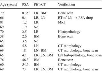
Table 7 Patients receiving antihormonal treatment Age (years) PSA PET/CT Verification
79 0.35 LR, BM Bone scan 66 0.4 LR, LN RT of LN→ PSA drop 81 1.2 LR MRI 69 1.9 No 70 2.5 LR Histopathology 58 2.6 BM Bone scan 65 3.5 No 66 5.8 LN CT morphology
69 18 LN, BM CT morphology, bone scan 65 42 LR, LN, BM LN histopathology, bone scan
76 46.3 BM Bone scan
60 54.6 BM CT morphology
69 73 LR, LN, BM CT morphology, bone scan+ LR, local recurrence; BM, bone metastasis; LN, lymph node metastasis; No, no FCH accumulation.
recurrent disease, even though the sensitivity rate is rather moderate. A possible future indication for FCH PET/CT might be therapy assessment of novel therapeutic strategies.
References
1. Jemal A, Murray T, Ward E, Murray T, Xu J, Smigal C, Thun MJ. Cancer statistics 2005. CA Cancer J Clin 2005; 55:10–30. 2. Aus G, Abbou CC, Bolla M, Heidenreich A, Schmid HP, van Poppel
H. EAU guidelines on prostate cancer. Eur Urol 2005; 48:546–51. 3. Gambhir SS, Czernin J, Schwimmer J, Silverman DH, Coleman
RE, Phelps ME. A tabulated summary of the FDG PET literature. J Nucl Med 2001; 42:1S–93S.
4. Hautzel H, Muller-Mattheis V, Herzog H, Roden W, Coenen HH,. Ackermann R, et al. The (11C) acetate positron emission tomography in prostatic carcinoma. New prospects in metabolic imaging. Urologe A 2002; 41:569–76.
5. de Jong IJ, Pruim J, Elsinga PH, Jongen MM, Mensink HJ, Vaalburg W. Visualisation of bladder cancer using (11)C-choline PET: first clinical experience. Eur J Nucl Med Mol Imaging 2002; 29:1283–8.
6. Hara T, Kosaka N, Kishi H. Development of (18)F-fluoroethyl-choline for cancer imaging with PET: synthesis, biochemistry, and prostate cancer imaging. J Nucl Med 2002; 43:187–99.
7. DeGrado TR, Coleman RE, Wang S, Baldwin SW, Orr MD, Robertson CN, Polascik TJ, Price DT. Synthesis and evaluation of 18F-labeled choline as an oncologic tracer for positron emission tomography: initial findings in prostate cancer. Cancer Res 2001; 61:110–7.
8. Kotzerke J, Prang J, Neumaier B, Volkmer B, Guhlmann A, Kleinschmidt K, et al. Experience with carbon-11 choline positron emission tomography in prostate carcinoma. Eur J Nucl Med 2000; 27:1415–9.
9. Hara T, Kosaka N, Kishi H. PET imaging of prostate cancer using carbon-11-choline. J Nucl Med 1998; 39:990–5.
10. Cimitan M, Bortolus R, Morassut S, Canzonieri V, Garbeglio A, Baresic T, Borsatti E, Drigo A, Trovo MG. [18]F-fluorocholine PET/CT imaging for the detection of recurrent prostate cancer at PSA relapse: experience in 100 consecutive patients. Eur J Nucl Med Mol Imaging 2006; 33:1387–1398.
11. Schmid DT, John H, Zweifel R, Cservenyak T, Wetsera G, Goerres GW, et al. Fluorocholine PET/CT in patients with prostate cancer: initial experience. Radiology 2005; 235:623–8.
12. Martorana G, Schiavina R, Corti B, Farsad M, Salizzoni E, Brunocilla E, et al. [11]C-choline positron emission tomography/ computerized tomography for tumor localization of primary prostate cancer in comparison with 12-core biopsy. J Urol 2006; 176:954–60. 13. Reske SN, Blumstein NM, Neumaier B, Gottfried HW. et al. Imaging prostate cancer with 11C-Choline PET/CT. J Nucl Med 2006; 47:1249–54.
14. Pagliarulo V, Hawes D, Brands FH, Groshen S, Cai J, Stein P, Lieskovsky G, Skinner DG, Coteet RJ. Detection of occult lymph node metastases in locally advanced node-negative prostate cancer. J Clin Oncol 2006; 24:2735–42.
15. Joslyn SA, Konety BR. Impact of extent of lymphadenectomy on survival after radical prostatectomy for prostate cancer. Urology 2006; 68:121–5.
16. Schumacher MC, Burkhard FC, Thalmann GN, Fleischmann A, Studer UE. Is pelvic lymph node dissection necessary in patients with a serum PSA<10 ng/ml undergoing radical prostatectomy for prostate cancer? Eur Urol 2006; 50:272–9.
17. Weckermann D, Goppelt M, Dorn R, Wawroschek F, Harzmann R. Incidence of positive pelvic lymph nodes in patients with prostate cancer, a prostate-specific antigen (PSA) level of≤10 ng/mL and biopsy Gleason score of ≤6, and their influence on PSA progression-free survival after radical prostatectomy. BJU Int 2006; 97:1173–8.
18. Langsteger W, Heinisch M, Fogelman I. The role of fluorodeox-yglucose, dihydroxyphenylalanine, choline, and 18F-fluoride in bone imaging with emphasis on prostate and breast. Semin Nucl Med 2006; 36:73–92.
19. Freedland SJ, Sutter ME, Dorey F, Aronson WJ. Defining the ideal cutpoint for determining PSA recurrence after radical prostatectomy. Prostate-specific antigen. Urology 2003; 61:365– 9.
20. Harisinghani MG, Barentsz J, Hahn PF, Deserno WM, Tabatabaei S, et al. Noninvasive detection of clinically occult lymph-node metastases in prostate cancer. N Engl J Med 2003; 348:2491–9. 21. Cornud F, Bellin MF, Portalez D. MRI and staging evaluation of
prostate cancer. J Radiol 2006; 87:228–43.
22. Sella T, Schwartz LH, Swindle PW, Onyebuchi CN, Scardino PT, Scher HI, Hricak H. Suspected local recurrence after radical prostatectomy: endorectal coil MR imaging. Radiology 2004; 231:379–85.
23. Heinisch M, Dirisamer A, Loidl W, Stoiber F, Gruy B, Haim S, Langsteger W. Positron emission tomography/computed tomogra-phy with F-18-fluorocholine for restaging of prostate cancer patients: meaningful at PSA < 5 ng/ml? Mol Imaging Biol 2006; 8:43–8.
24. Price DT, Coleman RE, Liao RP, Robertson CN, Polascik TJ, DeGrado TR. Comparison of [18 F]fluorocholine and [18 F] fluorodeoxyglucose for positron emission tomography of andro-gen dependent and androandro-gen independent prostate cancer. J Urol 2002; 168:273–80.
25. Poissonnier L, Chapelon JY, Rouviere O, et al. Control of prostate cancer by transrectal HIFU in 227 patients. Eur Urol 2006; 51:381– 387.
26. Hacker A, Jeschke S, Leeb K, Prammer K, Ziegerhofer J, Sega W, et al. Detection of pelvic lymph node metastases in patients with clinically localized prostate cancer: comparison of [18F]fluoro-choline positron emission tomography-computerized tomography and laparoscopic radioisotope guided sentinel lymph node dissection. J Urol 2006; 176:2014–18; discussion 2018–19.