ORIGINAL ARTICLE
Marginal seal stability of one bottle adhesives in Class V
vs. Class I cavities
Juan R. Mayoral&Ladislav Gregor&
Edson A. Campos&Miguel Roig&Ivo Krejci
Received: 27 September 2009 / Accepted: 7 December 2009 / Published online: 5 January 2010 # Springer-Verlag 2009
Abstract The aim of this study was to test the influence of two different cavity configurations on marginal stability of recent one bottle “etch & rinse” and “self-etch” adhesives in Class V vs. Class I cavities, before and after thermo-mechanical loading under simulation of dentinal fluid. Forty human upper molars were selected and assigned to five experimental groups. Intrapulpal pressure was main-tained during cavity preparation, restoration placement, finishing and stressing. Standardized Class I and V-Shaped Class V cavities were prepared on each tooth. Half of the margins of Class V cavities were located in enamel and half in dentin. All cavities were restored with different adhesives systems and a nano-hybrid composite. Materials were light-cured using a LED unit. Restored teeth were loaded in a computer-controlled chewing machine with 1.2 million mechanical occlusal cycles simultaneously with 3,000 thermal cycles (5–50–5°C). Impressions were made
with polyvinylsiloxane of each restoration before and after loading. Gold-coated epoxy replicas were prepared for SEM examination at ×200 magnification. Significant differ-ences between materials were found both before and after loading (Kruskal–Wallis, Bonferroni, p<0.05). Significant differences were also found between Class I and V restorations (Wilcoxon Matched-Pairs Signed-Rank Test, p<0.05). Even before thermo-mechanical loading, none of the groups had 100% continuous margin. Marginal seal stability of recent one bottle“etch & rinse” and “self-etch” adhesives are significantly different and susceptible to cavity configuration.
Keywords Marginal adaptation . C-factor . Dental adhesives . Pulpal pressure
Introduction
Several new one-bottle adhesives systems were recently introduced into the market. These systems have been developed as a result of the improvements in dental adhesion, in order to simplify the bonding procedures and make them less time-consuming [1]. Although there is a tendency toward adhesives with simplified application procedures, simplification does not necessarily guarantee equal or improved bonding effectiveness [2]. Based on the underlying adhesion strategy, adhesives can be classified in “etch & rinse” and “self-etch” [3]. The “etch & rinse” involves a separate etch-and-rinse phase before the appli-cation of the adhesive components: in the most common configuration, an acid (mostly 30–40% phosphoric acid) is applied and rinsed off. The one bottle “etch & rinse” adhesive systems thus combine the functions of primer and adhesive in one liquid [2,4] which follows the etching step,
J. R. Mayoral
:
M. RoigDepartment of Restorative Dentistry, International University of Catalonia, Barcelona, Spain
J. R. Mayoral
:
L. Gregor:
I. KrejciDivision of Cariology and Endodontology, School of Dentistry, University of Geneva,
Geneva, Switzerland E. A. Campos
Department of Restorative Dentistry, UNIFEB, Universitary Center of Barretos,
Barretos, Brazil
J. R. Mayoral (*)
Department of Restorative Dentistry, International University of Catalonia,
Josep Trueta, s/n, 08195 Sant Cugat del Vallès, Barcelona, Spain
often realized by the application of a gel etchant. The “self-etch” approach is based on the use of non-rinse mixtures of a weak acid and hydrophilic monomers or on acidic monomers that simultaneously condition and prime dentin [5]. One-bottle “self-etch” systems are based upon the simultaneous etching, priming, and bonding of the dental tissue using one single solution [6]. These systems seem to reduce postoperative sensitivity and are said to be less technique-sensitive than conventional adhesives [7].
Durable adhesion to tooth substrates is indispensable for clinical success of dental restorative materials. When direct resin composites are bonded to tooth structures using dental adhesives, the initial and residual polymerisation stresses that are present along the cavity walls may result in gap formation, leakage, recurrent caries, and pulpal irritation [8]. The stress generated at the adhesive interface by polymerizing resin composite is not only dependent on the curing contraction of the material per se, but also on cavity size and geometry, expressed as the relationship between the ratio of the free and restrained composite surface area of a dental restoration [9], and on application, processing, curing techniques as well as mechanical characteristics of the resin composite [10]. Studies have assessed the influence of C-factor and cavity type on marginal gap formation/microleakage, in particular, the influence of the confinement conditions imposed on the composite, the restorations volume and the compliance of the bonding substrate [11,12].
The effect of different adhesives on dentin-bonding performance and the influence of different cavity types is still not completely understood [13]. In certain cavity configurations, shrinkage stresses may become higher than the bond strengths, even with the most effective adhesive systems, leading to partial delamination of the adhesive system from the tooth surface. If this happens in the marginal region, marginal gaps and/or enamel fractures are the consequences [14], Shirai et al. [15] concluded that one-step self-etch adhesives appeared very sensitive to cavity configuration. Gaps between restoration and tooth are prone to microleakage, marginal discoloration, postoperative sensitivity, and secondary caries [16]. Therefore, assessing marginal adaptation of restorative materials is an important parameter to be tested when predicting its long-term behavior. Thus, the in vitro evaluation of the marginal adaptation under thermo-mechanical loading may be an important factor to consider for the prediction of clinical potential of a material [17].
This in vitro study investigated the influence of two different cavity configurations on the marginal stability of very recent one bottle etch & rinse and self-etch adhesives in Class V vs. Class I cavities, before and after thermo-mechanical loading under the simulation of dentinal fluid. Quantitative scanning electron microscopy (SEM) analysis
based on replicas was used to evaluate marginal adaptation. The null hypothesis tested was that there is no influence of the cavity configuration, the type of the adhesive system and the thermal and mechanical loading on the marginal adaptation and its stability.
Materials and methods
Forty caries-free human upper molars were selected and stored in water until use. The teeth were cleaned and randomly assigned to five experimental groups (Table 1): Group 1 (One Coat 7.0, Coltène Whaledent, Altstätten, Switzerland), Group 2 (Xeno V, Dentsply De Trey, Konstanz, Germany), Group 3 (XP Bond, Dentsply De Trey, Konstanz, Germany), Group 4 (Peak LC Bond, Ultradent, South Jordan, UT, USA), and Group 5 (Optibond FL, Kerr, Orange, CA, USA) as control group. All the specimens were mounted on a custom made specimen holders with their roots in the center using a cold-polymerizing resin (Technovit 4071, Kerr, Orange, CA, USA). Before mounting of each specimen, the apices were sealed using an adhesive system (Optibond FL, Kerr, Orange, CA, USA). A cylindrical hole was drilled into the pulpal chamber at approximately the middle third of the root and a metal tube of 1.2 mm in diameter was then adhesively luted using an adhesive system (Optibond FL, Kerr, Orange, CA, USA). Thereafter, the tube was connected using a flexible silicone hose to an infusion bottle. In order to simulate dentinal fluid, the infusion bottle was filled with horse serum diluted to a 1:3 ratio with PBS under normal hydrostatic pressure of about 25 mmHg. One day before starting the cavity preparations, the pulp chambers were evacuated with a vacuum pump using a three-way valve and subsequently filled with bubble-free diluted horse serum. The intrapulpar pressure was main-tained during the cavity preparation, restoration placement, finishing, and stressing.
Standardized Class I cavities were prepared at the occlusal surfaces and V-shaped Class V cavities on the buccal surfaces of each tooth. Half of the restoration margins of the Class V cavities were located in enamel and half in dentin. The Class I cavities were prepared with the use of cylindrical 80μm diamond burs and the Class V with the use of flame shape diamond burs (Intensiv SA, Grancia, Switzerland) under continuous water cooling. Each bur was replaced with a new one after four cavity preparations. The dimensions of the Class I cavities were 4.0–4.5 mm in height and 6.0–6.5 mm in length, for the Class V cavities were 3.0 – 3.5 mm in diameter, 2.5 – 3.0 mm in height and 1.5 mm in depth. The enamel margins for all the Class I and Class V cavities were beveled. All the cavities were finished using 40μm finishing diamond burs.
Eight teeth were randomly assigned to each experimental group.
The adhesive systems were applied following manufac-turers recommendations and all the restorations were restored using a nano-hybrid composite (Tetric EvoCeram, Ivoclar-Vivadent, Schaan, Liechtenstein, shade A2, Lot: K31914). Both adhesive and composite were light-cured using a powerful LED-curing unit (L.E.D. Demetron II, Kerr, Orange, CA, USA, Serial No: 792026758) with a relative output intensity of at least 1.200 mW/cm2 (L.E.D Demetron Radiometer, Kerr, Orange, CA, USA, Serial No: 79300278). Composite was inserted into the Class I cavities in three increments and on the Class V cavities in two increments: for the Class I cavities one increment was placed on the pulpar floor, the second on the vestibular wall, and the third on the lingual wall. In the Class V cavities, the first increment was placed cervically up to one
half of the cavity and the second increment occlusally, filling the other half of the cavity. All increments were light-cured for 40 s each. Immediately after polymerization, the restorations were finished and polished by using flexible aluminum oxide disks (Sof-Lex Pop-On, 3MEspe, St. Paul, MN, USA) with decreasing grit sizes. Final polishing was conducted under a stereomicroscope under ×12 magnification.
After storage for at least 1 week in water at 37°C in the dark, the restored teeth were loaded for 10 days in a computer-controlled chewing machine [18, 19]. Thermal and mechanical loading were applied simultaneously. Thermal cycling was performed in flushing water with temperatures changing 3.000× from 5°C to 50°C with a dwell time of 2 min each. The mechanical stress comprised in total 1.2 million load cycles transferred to the center of the occlusal surface at a frequency of 1.7 Hz. A maximal
Table 1 Description of the experimental groups, composition, and handling procedure of the adhesives systems tested
Adhesive system Composition Handling procedure
Self-etch
1 Steps One Coat 7.0
(Coltène Whaledent, Altstätten, Switzerland)
Hydroxyethyl methacrylate, Photoinitiators,
Ethanol. pH: 2.8
Shake the bottle well before use and dispense one drop into the dispensing well. Massage using a disposable brush for 20 s onto dentin and enamel. Gently dry for 5 s using oil free compressed air. Light cure for 10 s.
Lot: 0137063 Xeno V
(Dentsply De Trey, Konstanz, Germany)
Bifunctional Amides Acrylic,
Acidic Acrylic Amide, Functionalized Phosphoric Acid Ester, Acrylic Acid, Water, Tertiary Butanol, Initiator, Stabilizer. pH: 0.7
Dispense 1 or 2 drops twice, wetting all cavity surfaces uniformly with each application. Then gently agitate the adhesive for application. Agitate the adhesive for 20 s on the cavity surface. Evaporate solvent by thoroughly blowing with air from air syringe for at least 5 s.
Cure for at least 20 s. Lot: 0706000878
Etch & rinse
2 Steps XP Bond
(Dentsply De Trey, Konstanz, Germany)
Etchant: 35% H3PO4 Etch: Apply Etchant on E for 15 s and D: <_15 s,
rinse and dry. Apply adhesive, wet all cavity surface uniformly, leave the surface undisturbed for 20 s, evaporate solvent by thoroughly blowing air for at least 5 s, light cure for 10 s.
Lot: 0701000807 TCB Resin, Phosphoric acid modified
acrylate resin, TEGMA, HEMA, Butylated
Benzinediol (Stabilizer).Ethyl–
4-Dimethylamio Benzoate. Caphorquinone. pH:2.5
Peak LC Bond
(Ultradent, South Jordan, UT, USA)
Etchant: 35% H3PO4
Ethyl Alcohol Solvent, Methacrylic Acid, HEMA. pH: 1.18
Etch: Apply Etchant for 15 seconds on E and D, rinse for 5 s with air/water spray, leave D moist, Bond: Apply uniform coat with a brush tip. Brush gently for 10 s, Light cure for 20 s.Etch: Apply Etchant on E and D for 15 s, Rinse with water until complete removal for 15 s, Air dry without desiccating D. Dispense prime into disposable mixing well, apply material over E and D surfaces with a light scrubbing motion for 15 s, air dry for 5 s, dispense and apply adhesive over E and D uniformly creating a thin coating. Blow to margin to thin if necessary using a slight application of air, light cure for 20 s.
Lot: B2PSF
3 Steps Optibond FL
(Kerr, Orange, CA, USA)
Etchant: 35% H3PO4
Primer: HEMA,
GPDM, MMEP, Ethanol, Water, initiators
Primer: Lot: 453452 Bonding agent: Bis-GMA, HEMA, GPDM,
barium-aluminum borosilicate glass, disodium hexafluorosilicate, fumed silica (48% filler) pH: Prime: 1.9, Adhesive: 6.9 Adhesive: Lot: 2722726
E Enamel, D Dentin, TCB resin carboxylic acid modified dimethacrylate, TEGMA urethane bimethacrylate, HEMA 2-hydroxyethylmethacrylate, Bis-GMA bisphenol A-glycidyl methacrylate, GPDM glycerol phosphate dimethacrylate, MMEP methacryloxyethyl phthalate
load of 49 N was applied by using a natural lingual cusp taken from an extracted human molar. Pulpal pressure was maintained throughout the loading procedure. Impressions with a polyvinylsiloxane material (President light body, Coltène-Whaledent AG, Altstätten, Switzerland) were made of each restoration before and after loading. Gold-coated epoxy replicas were prepared for the computer-assisted quantitative margin analysis in a scanning electron micro-scope (XL20, Philips, Eidhoven, The Netherlands) at ×200 magnification. The marginal quality, expressed as percentages of“continuous margins”, was reported for the total marginal length in Class I and Class V cavities, and separately for the enamel and dentin margins in Class V cavities.
Differences in the percentages of “continuous margin” were statistically analyzed at the 95% confidence level using the NCSS-PASS® statistical software. The values of marginal adaptation for each group were not normally distributed (Shapiro–Wilk w test). For this reason, a Kruskal–Wallis and Bonferroni test was made for the initial and terminal values among groups and the Wilcoxon signed-rank test for the comparison of initial/terminal values within a group.
Results
The total percentages of “continuous margins” in Class I cavity type before and after thermal and mechanical loading for the five original groups are listed in Table2. The etch & rinse groups XP Bond, Peak LC Bond, and Optibond FL performed similarly before and after thermal and mechanical loading among groups (Kruskal–Wallis and Bonferroni post hoc test). Before loading, the one component etch & rinse Peak LC Bond showed the highest percentage of “continu-ous margins”, attaining [84.4 (12.8)] of “continuous mar-gins”, the lowest value was observed for the one component self-etch Xeno V [60.9 (11.8)]. After loading, Peak LC Bond showed the best performance [72.5 (9.3)]. The lowest marginal adaptation after loading was observed on Group 1 with the self-etch adhesive One Coat 7.0 [23.2 (12.9)].
Table 3 presents the values of continuous margin at enamel margin length in Class V cavity type, before and after thermal and mechanical loading. There is a trend for a better performance for etch & rinse adhesives, before and after mechanical loading on Class V enamel margins. The highest percentages of “continuous margins” before and after loading were found for the Group 5 Optibond FL [98.6 (2.2)] and [89.5 (7.9)]. The lowest marginal adapta-tion was observed for the“self-etch” Xeno V before [74.0 (12.8)] and after thermal and mechanical loading [35.7 (19.8)].
In Class V dentin margins (Table 3), the self-etch adhesive Xeno V performed with similar percentages of “continuous margins” to etch & rinse adhesives groups XP Bond and Optibond FL. The lowest mean values in dentin before loading were found for the“self-etch” One Coat 7.0 [64.7 (15.2)] with a significant difference after loading [20.2 (11.6)]. After thermal and mechanical loading an equally high marginal degradation on dentin was observed for all groups meaning that no significant differences (p< 0.05) could be detected between all groups.
At the total margin length in Class V (Table 3), better percentages of“continuous margins” were found at etch & rinse adhesives groups before and after thermal and mechanical loading, with the highest values for Optibond FL before [98.8 (1.4)] and after loading [72.4 (20.6)]. For the self-etch groups, the lower “continuous margins” percentages were observed for One Coat 7.0 showing higher marginal gaps before [72.3 (12.2)] and after loading [(29.0 (10.4)]. For all groups, it was observed that Class V cavity type was more favorable for preserving continuous margins in respect of the total margin length compared to the Class I cavity configuration. Thermal and mechanical loading significantly influenced the percentage of marginal adaptation (p<0.05, Wilcoxon matched-pairs signed-rank test) for all tested groups.
Figures1 and2shows some representative SEM micro-graphs of the marginal qualities expressed in “continuous margins” and “non continuous margins” on enamel and
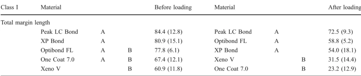
Table 2 Percentages of continuous margins at the total margin length in Class I cavities before and after thermal and mechanical loading
Class I Material Before loading Material After loading
Total margin length
Peak LC Bond A 84.4 (12.8) Peak LC Bond A 72.5 (9.3)
XP Bond A 80.9 (15.1) Optibond FL A 58.8 (5.2)
Optibond FL A B 77.8 (6.1) XP Bond A 54.0 (18.1)
One Coat 7.0 A B 67.4 (12.1) Xeno V B 31.5 (14.4)
Xeno V B 60.9 (11.8) One Coat 7.0 B 23.2 (12.9)
Differences among groups were statistically evaluated with Kruskal–Wallis and Bonferroni's test (p<0.05). Groups not connected by the same
dentin bonded interfaces of the different Class I and Class V restored groups observed in the investigation.
Discussion
Marginal adaptation is still a genuine problem in clinical dentistry and affects the longevity of adhesive restorations [16], is not directly correlated to clinical longevity or recurrent caries. This might be explained by the fact that in the clinical situation other parameters may be more important than marginal seal such as individual caries risk. However, it is obvious that the higher the percentage of continuous margin, the better is the adhesion, thus reflect-ing the quality of the adhesive technique and increasreflect-ing reliability [20]. The simulation of oral conditions might be crucial for a better evaluation and understanding of the performance of adhesive materials [18]. For this purpose, a chewing machine comprising simultaneous thermal cycling and cyclic occlusal mechanical loading, together with the simulation of dentinal fluid was used in this investigation [18, 19, 21]. This fatigue test may provide a better understanding of the behavior of dental adhesives under load [9]. Materials or interfaces normally fail because of the stresses and repeated loading, the most common observa-tion is a gap formaobserva-tion between the resin composite and enamel or dentin, resulting from polymerization shrinkage
that occurs before the restoration has been loaded, or after the application of repeated stresses [21]. Investigations have demonstrated the sensitivity of various bonding systems to pulpal pressure, dentinal fluid flow has a detrimental effect on the sealing ability of dentinal adhesives [22]. In this respect, the presence of pulpal pressure may be an important variable during bonding procedures with the intention of simulating in vivo conditions [23].
For the evaluation of marginal adaptation, a replica-based, computer-assisted quantitative SEM margin analysis was performed before and after loading. The method based on replicas has several advantages [18, 24, 25], it is quantitative, non-destructive and highly discriminative allowing to express the quality of the adaptation as percentages of “continuous margin” among the entire tooth/restoration interface to be assessed before and after exposure of stressing. Therefore, the combination of thermal and mechanical fatigue tests and SEM marginal analysis may provide relevant information when in vivo behavior of dentin-bonding agents is to be predicted on the basis of in vitro tests [26–28]. The sealing ability of the adhesive system itself relies on many factors such as bond strength, hydrophilicity, chemical stability, and the nature of the solvent [29,30]. The elastic modulus of the composite, its shrinkage, water uptake, and the coefficient of thermal expansion, among other factors, are important determinants that could influence the final performance of the
restora-Table 3 Percentages of continuous margins at the total margin length, in enamel and in dentin in Class V cavities before and after thermal and mechanical loading
Class V Material Before loading Material After loading
Total margin length
Optibond FL A 98.8 (1.4) Optibond FL A 72.4 (20.6)
XP Bond A B 93.6 (5.3) Peak LC Bond B 59.7 (17.1)
Peak LC Bond A B 84.6 (11.2) XP Bond A C 44.5 (14.6)
Xeno V B C 79.5 (8.4) Xeno V B C 39.7 (11.2)
One Coat 7.0 C 72.3 (12.2) One Coat 7.0 B C 29.0 (10.4)
Enamel margins
Optibond FL A 98.6 (2.2) Optibond FL A 89.5 (7.9)
Peak LC Bond A 97.5 (3.0) Peak LC Bond A 78.4 (22.1)
XP Bond A B 94.1 (4.9) XP Bond A C 66.8 (16.6)
One Coat 7.0 B 85.0 (8.3) One Coat 7.0 B C 45.5 (6.6)
Xeno V C 74.0 (12.8) Xeno V B 35.7 (19.8)
Dentin margins
Optibond FL A 98.6 (3.3) Optibond FL A 55.1 (36.9)
XP Bond A B 91.9 (10.1) Xeno V A 44.8 (15.8)
Xeno V A B 86.6 (12.5) Peak LC Bond A 40.4 (27.0)
Peak LC Bond B C 74.0 (21.3) XP Bond A 22.9 (25.5)
One Coat 7.0 C 64.7 (15.2) One Coat 7.0 A 20.2 (11.6)
Differences among groups were statistically evaluated with Kruskal–Wallis and Bonferroni's test (p<0.05). Groups not connected by the same
tions [31]. Because different results may be generated when an adhesive is tested with composites from different manufacturers, a composite of the same manufacturer was used in this investigation [32].
In this study, the cavity type significantly affected the mean values of“continuous margins” in all the adhesives tested. Even before thermo-mechanical loading, none of the groups had 100% continuous margin. As shown in Table2, the percentages of continuous margin for Class I restora-tions before thermal and mechanical loading were lower than the values for Class V restorations also before loading (Table3). The percentages of“continuous margins” for the Class I vs. Class V cavity types on enamel margins was not correlated, this would be an important parameter for consider testing adhesives systems in every cavity type. Polymerization contractions per se and contraction stress are important factors influencing the forces acting on the tooth-restoration interface [11]. This phenomenon is espe-cially pronounced in a Class I cavity type with a C-factor of around five, thus high shrinkage stresses may induce gaps between cavity wall/floor [15]. The sealing performance of adhesive resins is likely to be affected by cavity
configu-ration, dimensional changes of the restorative material, such as polymerization shrinkage, occlusal stresses, and the bonding capacity of the adhesive resins [1]. The most desirable method to eliminate the possibility of contraction stress induced marginal failure would be to eliminate shrinkage of the polymerizing restorative material [33]. Besides variations of the cavity type, there were also variations in the amount of restorative material used in each situation (Class I and Class V). According to some authors, the mass of restorative material used in each cavity type could also affect the results and must be considered [12, 13].
There were significant differences in the mean values of continuous margins between etch & rinse and self-etch adhesives for both Class I and Class V cavity type in enamel and dentin as well. Some studies indicated that phosphoric acid-etching remains a reliable mode of pre-treatment in obtaining better bonding [9,34,35], the use of etch & rinse technique have proven their effectiveness for achieving marginal seal [36]. It is known, that all-in-one adhesives and self-etching primers are intrinsically
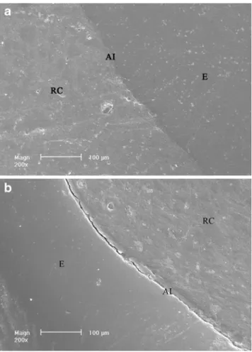
hydro-Fig. 2 Representative scanning electron microscopy micrographs
images (original magnification ×200) of “continuous margins” (a)
and “non continuous margins” (b) at the dentin-composite bonded
interface (E enamel, RC resin composite, AI adhesive interface)
Fig. 1 Representative scanning electron microscopy micrographs
images (original magnification ×200) of “continuous margins” (a)
and“non continuous margins” (b) at the enamel-composite bonded
philic because of the presence of acidic highly polar functional groups substituted on methacrylates. They rapidly absorb water, which results in polymer swelling and weakening of the polymer network [37]. Water absorption is assumed to be directly related to the hydro-philicity of these polymers with the consequence of lowering the mechanical properties [38]. Manufacturers have reformulated dentin adhesives to make them more compatible for bonding to intrinsically moist acid-etched dentin by adding 2-hidroxyethyl methacrylate and other hydrophilic resin monomers. When primers are mixed with adhesives in two-step single-bottle adhesives and self-etching primers, the adhesives are more permeable to water and hence absorb more water, contributing to the degrada-tion of resin–dentin bond strength over time [39]. Another disadvantage of one-component self-etch adhesives is seen in their relatively high water uptake, resulting in the formation of water trees at the interface [40].
Respecting the Class V cavity type, the self-etching adhesives systems showed more decreasing values of “continuous margins” after loading at dentin compared to enamel margins (Table3). Some studies have reported that self-etch adhesives bond less effectively to enamel than etch & rinse [41]. It is reported also that some self-etch adhesives interact less effectively with dentin compared to etch & rinse adhesives [36,42]. Because simulated dentinal fluid were used in this study, fluid penetration into the resin–dentin interface from dentinal tubules may occur during bonding and/or after bonding, and water diffusion from dentinal tubules into all-in-one adhesives and the resin–dentin interface may hasten hydrolytic degradation of resin components within the hybrid layer and/or adhesives, followed by hydrolysis of the naked collagen fibrils, contributing to the failure of resin–dentin bond [43]. Hashimoto et al. [44] reported that when bonding was carried out under hydrostatic pressure, the amount of water movement across the resin–dentin interface increased after polymerization compared with no pressure.
There was a different behavior between the two self-etch adhesive systems used in the study on the percentages of “continuous margins” showed on Class V cavity-type before and after loading in enamel and dentin substrates. For Group 2 (Xeno V), better mean values were obtained in dentin before loading (86.6±12.5 %) than in enamel (74.0±12.8 %), in Group 1 (One Coat 7.0) inferior marginal adaptation before loading was showed in dentin (64.7±15.2%) compared to enamel (85.0±8.3 %). These results may be in part related to the pH of these materials, One Coat 7.0 has a pH of 2.8 and Xeno V has a pH of 0.7. The study of Sensi et al. [45] found enhanced bond strength to dentin of self-etching adhesives systems related to low pH values. Basically, two types of self-etch adhesives can be distinguished: “mild” and “strong”. Strong self-etch adhesives have a very low pH
(<1) and “mild” self-etch pH (±2), the “mild” self-etch adhesives do not completely remove the smear layer, but do form a submicron hybrid layer [2]. The differences in pH values may not be the only important parameter to justify differences, as it may also depend on other factors, such as the dissociation constant (pKa), the chemical structure of the
adhesive components (which may be more or less chelating), the solubility of the formed salts and its application time and many others [7, 46]. A study of Van Landuyt et al. [47] indicated that not only the class into witch an adhesive can be classified is an important parameter for bonding effec-tiveness, but also the composition of the adhesive must be adapted to the application procedure.
In summary, within the limitations of this in vitro study, it was concluded that the marginal seal stability of recent one bottle etch & rinse and self-etch adhesive systems was significantly different and susceptible to cavity configuration and thermal and mechanical loading. None of the adhesives systems tested within this study was able to eliminate the formation of marginal defects even before thermal and mechanical loading, neither in enamel nor in dentin. The results require the rejection of the null hypothesis that there is no influence of the cavity configuration, the type of the adhesive system and the thermal and mechanical loading on the marginal adaptation and its stability.
Conflict of Interest Statement The authors declare that they have
no conflict of interest.
References
1. Abo T, Uno S, Sano H (2004) Comparison of bonding efficacy of an all-in-one adhesive with a self-etching primer system. Eur J
Oral Sci 112:286–292
2. Van Meerbeek B, De Munck J, Yoshida Y, Inoue S, Vargas M, Vijay P, Van Landuyt K, Lambrechts P, Vanherle G (2003) Buonocore memorial lecture. Adhesion to enamel and dentin:
current status and future challenges. Oper Dent 28:215–235
3. Van Meerbeek B, Vargas M, Inoue S, Yoshida Y, Peumans M, Lambretchts P (2001) Adhesives and cements to promote
preservation dentistry. Oper Dent 26:S119–S144
4. Lopes GC, Baratieri LN, de Andrada MA, Vieira LC (2002) Dental adhesion: present state of the art and future perspectives. Quintessence Int 33:213–224
5. De Munck J, Van Landuyt K, Peumans M, Poitevin A, Lambrechts P, Braem M, Van Meerbeek B (2005) A critical review of the durability of adhesion to tooth tissue: methods and
results. J Dent Res 84:118–132
6. Proenca JP, Polido M, Osorio E, Erhardt MC, Aguilera FS, Garcia-Godoy F, Osorio R, Toledano M (2007) Dentin regional bond strength of self-etch and total-etch adhesive systems. Dent
Mater 23:1542–1548
7. Knobloch LA, Gailey D, Azer S, Johnston WM, Clelland N, Kerby RE (2007) Bond strengths of one- and two-step self-etch
8. Frankenberger R, Tay FR (2005) Self-etch vs etch-and-rinse adhesives: effect of thermo- mechanical fatigue loading on marginal quality of bonded resin composite restorations. Dent
Mater 21:397–412
9. Giachetti L, Scaminaci Russo D, Bambi C, Grandini R (2006) A review of polymerization shrinkage stress: current techniques for posterior direct resin restorations. J Contemp Dent Pract 7:79–88 10. Kurokawa R, Finger WJ, Hoffmann M, Endo T, Kanehira M, Komatsu M, Manabe A (2007) Interactions of self-etch adhesives
with resin composites. J Dent 35:923–929
11. Watts DC, Satterthwaite JD (2008) Axial shrinkage-stress depends
upon both C-factor and composite mass. Dent Mater 24:1–8
12. Braga RR, Boaro LC, Kuroe T, Azevedo CL, Singer JM (2006) Influence of cavity dimensions and their derivatives (volume and 'C' factor) on shrinkage stress development and microleakage of
composite restorations. Dent Mater 22:818–823
13. Nikolaenko SA, Lohbauer U, Roggendorf M, Petschelt A, Dasch W, Frankenberger R (2004) Influence of c-factor and layering technique
on microtensile bond strength to dentin. Dent Mater 20:579–585
14. Krejci I, Stavridakis M (2000) New perspectives on dentin
adhesion—differing methods of bonding. Pract Periodontics
Aesthet Dent 12:727–732, quiz 734
15. Shirai K, De Munck J, Yoshida Y, Inoue S, Lambrechts P, Suzuki K, Shintani H, Van Meerbeek B (2005) Effect of cavity configuration and aging on the bonding effectiveness of six adhesives to dentin. Dent Mater 21:110–124
16. Bortolotto T, Ferrari M, Tay F, Krejci I (2007) Degradation of thermo-mechanically loaded adhesive Class V restorations after
18 months of water storage. Am J Dent 20:83–89
17. Bortolotto T, Ferrari M, Onisor I, Tay F, Krejci I (2005) Marginal adaptation of contemporary dentin bonding agents in enamel and
dentin under the simulation of dentinal fluid. Dent S Africa 7:46–58
18. Krejci I, Reich T, Lutz F, Albertoni M (1990) An in vitro test procedure for evaluating dental restoration systems. 1. A computer-controlled mastication simulator. Schweiz Monatsschr
Zahnmed 100:953–960
19. Krejci I, Kuster M, Lutz F (1993) Influence of dentinal fluid and stress on marginal adaptation of resin composites. J Dent Res
72:490–494
20. Heintze SD (2007) Systematic reviews: I. The correlation between laboratory tests on marginal quality and bond strength. II. The correlation between marginal quality and clinical outcome. J Adhes Dent 9(Suppl 1):S77–S106
21. Frankenberger R, Strobel WO, Kramer N, Lohbauer U, Winterscheidt J, Winterscheidt B, Petschelt A (2003) Evaluation of the fatigue
behavior of the resin–dentin bond with the use of different methods. J
Biomed Mater Res B Appl Biomater 67:712–721
22. Ozok AR, Wu MK, Ten Cate JM, Wesselink PR (2004) Effect of dentinal fluid composition on dentin demineralization in vitro. J
Dent Res 83:849–853
23. Sauro S, Pashley DH, Montanari M, Chersoni S, Carvalho RM, Toledano M, Osorio R, Tay FR, Prati C (2007) Effect of simulated pulpal pressure on dentin permeability and adhesion of self-etch
adhesives. Dent Mater 23:705–713
24. Friedl KH, Schmalz G, Hiller KA, Mortazavi F (1997) Marginal adaptation of composite restorations versus hybrid ionomer/
composite sandwich restorations. Oper Dent 22:21–29
25. Gladys S, Van Meerbeek B, Inokoshi S, Willems G, Braem M, Lambrechts P, Vanherle G (1995) Clinical and semiquantitative marginal analysis of four tooth-coloured inlay systems at 3 years. J Dent 23:329–338
26. Abdalla AI, Davidson CL (1993) Comparison of the marginal integrity of in vivo and in vitro Class II composite restorations. J
Dent 21:158–162
27. Braem M, Lambrechts P, Vanherle G (1994) Clinical relevance of
laboratory fatigue studies. J Dent 22:97–102
28. da cunha Mello FS, Feilzer AJ, de Gee AJ, Davidson CL (1997) Sealing ability of eight resin bonding systems in a Class II
restoration after mechanical fatiguing. Dent Mater 13:372–376
29. Tay FR, Pashley DH (2003) Water treeing—a potential
mecha-nism for degradation of dentin adhesives. Am J Dent 16:6–12 30. Manhart J, Trumm C (2009) Marginal adaptation of an
etch-and-rinse adhesive with a new type of solvent in class II cavities after artificial aging. Clin Oral Investig. Nov 24. [Epub ahead of print] 31. Hasegawa T, Itoh K, Koike T, Yukitani W, Hisamitsu H, Wakumoto S, Fujishima A (1999) Effect of mechanical properties of resin composites on the efficacy of the dentin bonding system.
Oper Dent 24:323–330
32. Krejci I, Hausler T, Sagesser LF (1994) New adhesives in Class V restorations under combined load and simulated dentinal fluid.
Dent Mater 10:331–335
33. Armstrong SR, Keller JC, Boyer DB (2001) The influence of water storage and C-factor on the dentin-resin composite micro-tensile bond strength and debond pathway utilizing a filled and
unfilled adhesive resin. Dent Mater 17:268–276
34. Inoue S, Vargas MA, Abe Y, Yoshida Y, Lambrechts P, Vanherle G, Sano H, Van Meerbeek B (2003) Microtensile bond strength of eleven contemporary adhesives to enamel. Am J Dent 16:329–334
35. De Munck J, Van Meerbeek B, Satoshi I, Vargas M, Yoshida Y, Armstrong S, Lambrechts P, Vanherle G (2003) Microtensile bond strengths of one- and two-step self-etch adhesives to bur- cut
enamel and dentin. Am J Dent 16:414–420
36. Pradelle-Plasse N, Nechad S, Tavernier B, Colon P (2001) Effect
of dentin adhesives on the enamel–dentin/composite interfacial
microleakage. Am J Dent 14:344–348
37. Yiu CK, King NM, Carrilho MR, Sauro S, Rueggeberg FA, Prati C, Carvalho RM, Pashley DH, Tay FR (2006) Effect of resin hydrophilicity and temperature on water sorption of dental
adhesive resins. Biomaterials 27:1695–1703
38. Hosaka K, Tagami J, Nishitani Y, Yoshiyama M, Carrilho M, Tay FR, Agee KA, Pashley DH (2007) Effect of wet vs. dry testing on the mechanical properties of hydrophilic self-etching primer
polymers. Eur J Oral Sci 115:239–245
39. Tay FR, Pashley DH (2003) Have dentin adhesives become too hydrophilic? J Can Dent Assoc 69:726–731
40. Moszner N, Salz U, Zimmermann J (2005) Chemical aspects of self-etching enamel–dentin adhesives: a systematic review. Dent Mater 21:895–910
41. Torii Y, Itou K, Nishitani Y, Yoshiyama M, Ishikawa K, Suzuki K
(2003) Effect of self-etching primer containingN-acryloyl aspartic
acid on enamel adhesion. Dent Mater 19:253–258
42. Tay FR, Pashley DH (2001) Aggressiveness of contemporary self-etching systems. I: Depth of penetration beyond dentin smear
layers. Dent Mater 17:296–308
43. Hosaka K, Nakajima M, Yamauti M, Aksornmuang J, Ikeda M, Foxton RM, Pashley DH, Tagami J (2007) Effect of simulated pulpal pressure on all-in-one adhesive bond strengths to dentine. J Dent
35:207–213
44. Hashimoto M, Ito S, Tay FR, Svizero NR, Sano H, Kaga M,
Pashley DH (2004) Fluid movement across the resin–dentin
interface during and after bonding. J Dent Res 83:843–848
45. Sensi LG, Lopes GC, Monteiro S Jr, Baratieri LN, Vieira LC (2005) Dentin bond strength of self-etching primers/adhesives. Oper Dent 30:63–68
46. Sarr M, Mine A, De Munck J, Cardoso MV, Kane AW, Vreven J, Van Meerbeek B, Van Landuyt KL (2009) Immediate bonding effectiveness of contemporary composite cements to dentin. Clin Oral Investig. Aug 25. [Epub ahead of print]
47. Van Landuyt KL, Peumans M, De Munck J, Lambrechts P, Van Meerbeek B (2006) Extension of a one-step self-etch adhesive