DOI 10.1007/s00402-009-0865-1
A R T H R O S C O P Y A N D S P O R T S M E D I C I N E
A new conservative-dynamic treatment for the acute ruptured
Achilles tendon
Felix Neumayer · Elyazid Mouhsine · Yvan Arlettaz · Gérald Gremion · Michael Wettstein · Xavier Crevoisier
Received: 6 January 2009 / Published online: 2 April 2009
© Springer-Verlag 2009
Abstract
Introduction There is a trend towards surgical treatment of acute ruptured Achilles tendon. While classical open surgi-cal procedures have been shown to restore good functional capacity, they are potentially associated with signiWcant complications like wound infection and paresthesia. Modern mini-invasive surgical techniques signiWcantly reduce these complications and are also associated with good functional results so that they can be considered as the surgical treat-ment of choice. Nevertheless, there is still a need for conser-vative alternative and recent studies report good results with conservative treatment in rigid casts or braces.
Patients/method We report the use of a dynamic ankle brace in the conservative treatment of Achilles tendon rupture in a prospective non-randomised study of 57 con-secutive patients. Patients were evaluated at an average fol-low-up time of 5 years using the modiWed Leppilahti Ankle Score, and the Wrst 30 patients additionally underwent a clinical examination and muscular testing with a Cybex isokinetic dynamometer at 6 and 12 months.
Results We found good and excellent results in most cases. We observed Wve complete re-ruptures, almost exclusively in case of poor patient’s compliance, two
partial re-ruptures and one deep venous thrombosis compli-cated by pulmonary embolism.
Conclusion Although prospective comparison with other modern treatment options is still required, the functional outcome after early ankle mobilisation in a dynamic cast is good enough to ethically propose this method as an alterna-tive to surgical treatment.
Keywords Achilles tendon rupture · Conservative treatment · Functional
Introduction
The acute rupture of the Achilles tendon occurs most com-monly in moderately sportive adults in their thirties and for-ties, but is also seen in younger athletes [23]. Eventually it occurs also in the elderly athletes. There is an ongoing dis-cussion whether a recently ruptured Achilles tendon is best treated by open or mini-invasive suture or conservatively by a casting or bracing technique. Both surgical and conserva-tive treatments have been reported to obtain good and excel-lent results in most cases. Meta-analyses have shown wound problems, infection, or paresthesia to be signiWcant compli-cations associated with surgical treatment [14, 31], even though percutaneous techniques seem to reduce the rate of complications [7]. On the other hand, the conservative treat-ment is more often complicated by re-rupture of the Achilles tendon [14, 16, 31]. Recent surgical and conservative proce-dures favour a functional bracing to rigid casting [3, 11, 19, 20, 24–26, 29]. To the best of our knowledge, all reports published about conservative treatment were using a rigid bracing type. Our aim was to evaluate a new functional con-servative treatment of the recently ruptured Achilles tendon, using an articulated dynamic ankle brace.
F. Neumayer · E. Mouhsine · Y. Arlettaz · M. Wettstein · X. Crevoisier
Department of Orthopaedics and Trauma Surgery, University Hospital (CHUV), Lausanne, Switzerland
G. Gremion
Department of Sports Medicine,
University Hospital (CHUV), Lausanne, Switzerland
X. Crevoisier (&)
CHUV Site Hôpital Orthopédique, Pierre-Decker 4, 1011 Lausanne, Switzerland
Materials and methods
Between 1998 and 2005, 57 consecutive patients who attended our outpatient clinic underwent a functional and conservative treatment for an acutely ruptured Achilles ten-don. The rupture was diagnosed by a positive Thompson test (calf squeeze test) in all cases, but in four cases conWr-mation was sought by ultra sound. We included patients of any age and any mechanism of injury with a Wrst episode of an acute rupture of the Achilles tendon (less than 10 days old). The risks and beneWts of both surgical and conserva-tive treatments were carefully explained to the patients, who then gave informed consent for this new treatment option. Professional athletes and patients with a re-ruptured Achilles tendons were excluded.
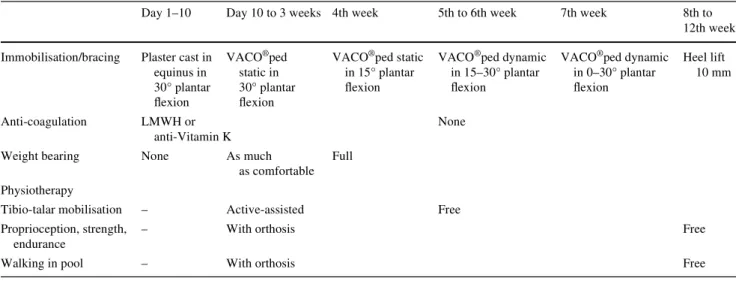
The bracing and rehabilitation procedure was as follows (reproduced also on Table1): after immobilisation with a cast in equinus for 10 days, the patients were authorised to walk with full weight bearing, protected by a commercial orthosis (VACO®ped, OPED; Fig.1). The equinus angle was set at 30° plantar Xexion until the end of week 3, and at 15° until the end of week 4. At the Wfth week the system was unlocked to allow ankle mobilisation of 30°–15°–0°, and at 30°–0°–0° at the seventh week. The orthosis was removed after 8 weeks. After removal of the orthosis, the patients wore a 10 mm heel lift for another 4 weeks. Phys-iotherapy started during the second week with endurance and proprioceptive training with the orthosis in place and active-assisted mobilisation of the tibio-talar joint without the brace. Patients continued physiotherapy for up to 3 months after the accident. Deep vein thrombosis prophy-laxis was given for 4 weeks either by low molecular weight heparins or by vitamin K antagonists.
All patients had follow-up examinations up to 12 months after the trauma. The Wrst 30 patients additionally underwent
muscular testing with a Cybex isokinetic dynamometer at 6 and 12 months. In June 2006 all patients were contacted and received a questionnaire. Subjective opinion of the out-come on a visual analogue scale from 0 to 10, any change in sporting activities and eventual late complications were investigated. We used a scoring system which was modiWed from the Leppilahti Ankle Score [17] by van der Linden-van der Zwaag et al. [18] (Table2). The Leppilahti Ankle Score has been used by several other authors [21, 29], but has not yet been validated.
Table 1 Re-education and bracing procedure using a dynamic orthosis (VACO®ped)
Day 1–10 Day 10 to 3 weeks 4th week 5th to 6th week 7th week 8th to 12th week
Immobilisation/bracing Plaster cast in equinus in 30° plantar Xexion VACO®ped static in 30° plantar Xexion
VACO®ped static in 15° plantar Xexion
VACO®ped dynamic in 15–30° plantar Xexion
VACO®ped dynamic in 0–30° plantar Xexion Heel lift 10 mm Anti-coagulation LMWH or anti-Vitamin K None
Weight bearing None As much
as comfortable Full
Physiotherapy
Tibio-talar mobilisation – Active-assisted Free
Proprioception, strength, endurance
– With orthosis Free
Walking in pool – With orthosis Free
Fig. 1 The orthosis used from day 10 to the eighth week, permitting
Results
Our study group included 12 women and 45 men with an average age of 45 years (24–73). Most of the injuries were due to indirect trauma during sport activities. Soccer, tennis and squash were most frequent but some ruptures happened during long distant running, ski or snowboard activities. Some patients took part in sport up to six times a week, while some did not do any (mean 1.9x/week). Two patients already had a history of Achilles tendopathy on the rup-tured side. None had had local inWltrations. No patient was under Xuoroquinolone antibiotic treatment. Three patients were under low dose corticoid treatment for obstructive pulmonary diseases. One patient had suVered from previous spontaneous pulmonary embolism.
Dynamometric follow-up evaluation demonstrated that the aVected side had regained 70% speed power, 75%
maximal power, and 70% endurance power at 6 months when compared to the healthy side. At 12 months these val-ues were 92%, 90%, and 90%, respectively (Fig.2).
We observed Wve complete re-ruptures and two partial re-ruptures. The partial re-ruptures were diagnosed by MR imaging as well as three out of the Wve complete re-rup-tures. The two other complete re-ruptures were diagnosed clinically. All re-ruptures happened during the Wrst 5 months after the accident. One re-ruptured tendon was sutured and another patient did not come for follow-up. Three patients with a re-rupture and the partial re-ruptures were treated conservatively by prolonged bracing; all healed with fair to good results. Five out of the seven patients with partial or complete re-ruptures had been showing poor compliance to the treatment protocol or caused the re-rupture when falling over. One patient devel-oped a deep venous thrombosis complicated by pulmonary embolism while he was wearing the orthosis; this patient was already known for a previous spontaneous pulmonary embolism. No relationship was found between the occur-rence of re-rupture and the use of low dose corticoid treat-ment. We observed few minor skin complications as superWcial ulcerations or complaints about dry skin.
We were able to collect 46 questionnaires from the 57 patients (82.5%). Two patients had died due to non-related causes, nine patients could not be located. The average fol-low-up time for the questionnaires was 5 years (15 months to 8 years). Two-thirds of the questioned patients had returned to their previous level of sporting activity. Of the remaining third, who had reduced or changed their sporting activity, only half did so because of actual pain or calf weakness, while the other half did so because of apprehen-sion.
The average overall satisfaction with the outcome was 8.1 § 2. The average of the modiWed Leppilahti Ankle Score was 56.8 § 14.9. For patients without major compli-cations the average overall satisfaction was 8.8 § 1.4 and the modiWed Leppilahti Ankle Score 60.6 § 14.4.
Table 2 ModiWed Leppilahti Ankle Score [23]
ModiWed by van der Linden-van der Zwaag et al. [18]
Clinical factor Scores
(max. 70 pts)
Pain
None 15
Mild, no limited recreational activities 10 Moderate, limited recreational activities,
but not daily activities
5
Severe, limited recreational and daily activities 0 StiVness
None 15
Mild, occasional, no limited recreational activities 10 Moderate, limited recreational activities,
but not daily activities
5
Severe, limited recreational and daily activities 0 Calf muscle weakness (subjective)
None 15
Mild, no limited recreational activities 10 Moderate, limited recreational activities,
but not daily activities
5
Severe, limited recreational and daily activities 0 Foot wear restrictions
None 10
Mild, most shoes tolerated 5
Moderate, unable to tolerate fashionable shoes,
modiWed shoes tolerated 0
Subjective result
Very satisWed 15
SatisWed with minor reservations 10 SatisWed with major reservations 5
DissatisWed 0
Fig. 2 Dynamometric results in comparison to the healthy side
(=100%) at 6 and at 12 months after the accident (N = 30) 70% 92% 75% 90% 70% 90% 0% 20% 40% 60% 80% 100% Speed Power Maximal power Endurance power 6 months 12 months
Discussion
Post-surgical and conservative “functional” treatment is understood by many authors as a rigid cast or brace immo-bilisation in full weight bearing. Some allow early ankle mobilisation, taking oV the orthosis [11–13, 29, 30]. After an initial period of 2 weeks of below-knee cast immobilisa-tion in full equinus, Saleh et al. [25] were using a splint which immobilised the ankle but allowed movements at the metatarsophalangeal joints. McComis et al. [20] used a brace for a total period of 16 weeks which permitted pro-gressive dorsal extension. When the patient was walking no ankle movement was allowed, but during exercise sessions active ankle Xexions were performed. After 4 weeks of rigid immobilisation in gravity equinus, Roberts et al. [24] dynamised their initial below-knee cast by removing a part anteriorly, in order to allow full extension of the ankle. Active ankle Xexion was encouraged during exercises, but since the cast stayed rigid it could not enhance ankle move-ments while walking. While gait analysis has shown the advantage of dynamic bracing in comparison to rigid ortho-sis in terms of more physiological ambulation [15], to our knowledge, there are no reports published on a conservative treatment of Achilles tendon ruptures with a dynamic orthosis allowing free active ankle movement when walk-ing in full weight bearwalk-ing.
Dynamic orthosis application, however, has been reported after surgical treatment. In a prospective random-ised study, Möller et al. [21] used a functional dynamic brace for their surgically treated patients, while the conser-vative group was immobilised in a rigid plaster cast. In this study, the surgical treatment was found to be superior, because of a particularly high incidence of re-ruptures in the conservative group (20.8%). The operative group had functional rehabilitation, while the conservative treatment consisted of traditional plaster casting; therefore the study does not allow any conclusions about conservative func-tional treatment of Achilles tendon rupture. On the other hand, it seems to conWrm the beneWt of early ankle mobili-sation and weight bearing for tendon healing. Calder and Saxby [6] were also using a dynamic brace for the
postop-erative period in a prospective study of mini-invasive Achilles tendon suturing; they achieved good and excellent results.
It has been demonstrated in animal models that mechan-ical stress in frequent exercise is an important factor for the quality of tendon healing [8, 9, 22, 28]. This is conWrmed by the superior results of functional conservative or post-surgical treatment procedures in comparison to non-weight bearing plaster cast immobilisation [14, 31]. Therefore, it seems logical that early ankle mobilisation in a dynamic cast should promote better functional results than a rigid immobilisation technique.
The mean modiWed Leppilahti Ankle Score in our study was comparable to two recent studies on conservative treat-ment procedures who were using the same scoring system [18, 30] (Table3). Our functional results regarding force and endurance at 12 months were very encouraging. Pro-spective randomised trials, however, will have to be carried out, to Wnd out if there is a statistically signiWcant diVerence to functional treatment in rigid orthosis.
According to a cross-sectional study from 2002, after conservative treatment an average of 62% of patients were able to return to their previous level of sports participation [16]. More recent studies report 49–75.2% [11, 13, 18]. Our results concur with these reports (Table3). A high percent-age of patients, did not return to their former sports level, because they feared re-rupture. This underlines the neces-sity, to reassure and accompany the patients until tendon healing has occurred completely and while restarting sport-ing activities.
Our clinical follow-up period was 12 months for all patients and for the questionnaire the minimum was 18 months. In the meta-analysis of Besch et al. [4], 16 stud-ies were identiWed who indicated the time interval of their re-ruptures. All appeared during the Wrst 6 months after removing of the cast, only one happened at 9 months due to a new trauma [29]. Therefore, our follow-up time should be suYcient to detect all re-ruptures.
A mean rate of re-rupture of 12.1% was found in conser-vative treatment in Kocher’s et al. cross-sectional study [16], while newer studies report only 2.1–7% [12, 13, 18,
Table 3 Comparison of results of recent studies on conservative treatment procedures
Wallace et al. [30] van der Linden-van der der Zwaag et al. [18]
Ingvar et al. [12] Hufner et al. [11] Present study
Number of patients 140 80 196 125 57
Return to sport at the same level
37% 49% – 75.2% 67.4%
ModiWed Leppilahti Score 61.4 59 – – 60.6
Re-rupture rate (partial) 2.1% (3.6%) 5% 7% 6.4% 9% (3.5%)
Other complications 2 DVT 1 temporary dropfoot
1 DVT + pulm. embolism 7 DVT 1 pulm. embolism
3 DVT 1 DVT + pulm. embolism
29] (Table3). Hufner et al. [11] pre-selected their patients by ultrasound examinations: inclusion criteria was a com-plete apposition of the tendon ends in 20° of plantarXexion. They still reported 6.4% re-rupture in their conservative functional treatment group. In our study, although we did not apply such strict patients’ selection, the rate of re-rupture was only slightly higher (9%). Furthermore, re-ruptures almost exclusively occurred in patients with poor compli-ance. Nevertheless, we shall need a larger number of patients to demonstrate that the early ankle mobilisation in full weight bearing in our treatment procedure is not a risk factor for re-rupture.
Functional results of operative and conservative treat-ment seem to be more or less equivalent [3, 21, 27, 30], although young athletes are thought to beneWt more from operative techniques [23]. Risk factors for post-operative complications after Achilles tendon suture are tobacco and steroid use, diabetes and female sex [5]. Especially in patients with more than one risk factor, functional conser-vative treatment should be the treatment of choice. In patients unable to follow a rehabilitation program or non-compliance, conservative plaster cast immobilisation can be considered, if functional results are of little concern [2].
Classic open surgery for acutely ruptured Achilles ten-don seems to progressively disappear from the recent litera-ture. Inversely, there is a trend towards mini-open techniques [1, 3, 6, 7, 10] and the interest in conservative treatment has also been renewed, based on recent studies reporting the eYciency of this therapeutic solution [11–13, 18, 29]. Nevertheless, to our knowledge, there is no exist-ing study reportexist-ing prospective comparison between these two most recommended treatment options. Both strategies have been recently applied on relatively large numbers of patients and were associated with equivalent good results. We still need prospective randomised controlled studies to compare functional results and gait. We also need to further investigate re-rupture rates and surgical risks and complica-tions. It would be important to analyse if functional and subjective results depend only on the treatment procedure or also on patient age and sports activity. In the meantime we are ethically in the position to propose either of the treatment options to the patient: mini-invasive surgery or conservative functional management.
There are limitations in the present study. Cybex dyna-mometric testing was no more available at our institution in the second half of the study so that only the Wrst 30 patients could undergo this speciWc assessment. Nevertheless, we decided to include the further 27 patients since the dynamo-metric assessment represents only one part of the study. A follow-up rate of 82.5% at a mean time of 5 years (and up to 8 years) might be considered as low. However, it appears quite acceptable when compared to the rates of 53–74% reported in other recent studies [11, 13, 18].
Conclusion
This is the Wrst published report about the use of a dynamic foot and ankle brace for the conservative treatment of acute Achilles tendon rupture. The present treatment resulted in good to excellent functional results in most cases and, there-fore, demonstrates that conservative functional treatment in the acute rupture of the Achilles tendon can be ethically pro-posed as an alternative to surgical treatment in most cases. It requires patient compliance and active participation, as well as a systematic medical follow-up during the Wrst 6 months. The re-rupture rate of 9% is slightly higher than in recently published series about conservative treatment but, consider-ing that no patients’ selection was applied and also that re-ruptures occurred almost exclusively in non-compliant patients, it is acceptable. Our results are consistent with recent reports that emphasise the advantages of early ankle mobilisation in aiming good functional results.
References
1. Amlang MH, Christian P, Heinz P, Zwipp H (2005) Percutaneous technique for Achilles tendon repair with the Dresden: instruments and results. Unfallchirurg 108:529–536
2. Arlettaz Y, Chevalley F, Gremion G, Leyvraz PF (1998) Les ruptures fraîches du tendon d’Achille–A propos de 14 cas traités conservativement. Swiss Surg 4:75–81
3. Assal M, Jung M, Stern R, Rippstein P, Delmi M, HoVmeyer P (2002) Limited open repair of Achilles tendon ruptures: a tech-nique with a new instrument and Wndings of a prospective multi-center study. J Bone Joint Surg Am 84:161–170
4. Besch S, Peyre M, Rodineau J, Dupre JP (2006) Traitement con-servateur des ruptures du tendon d’Achille. J Traumatol Sport 23:12–24
5. Bruggemann NB, Turner NS, Dahm DL, Voll AE, Hoskin TL, Jacofski DJ, Haidukewych GJ (2004) Wound complications after open Achilles tendon repair. An analysis of risk factors. Clin Orthop Relat Res 427:63–66
6. Calder JD, Saxby TS (2005) Early, active rehabilitation following mini-open repair of Achilles tendon rupture: a prospective study. Br J Sports Med 39:857–859
7. Cretnik A, Kosanovic M, Smrkolj V (2005) Percutaneous versus open repair of the ruptured Achilles tendon. A comparative study. Am J Sports Med 33:1369–1379
8. Enwemeka CS, Spielholz NI, Nelson AJ (1988) The eVect of early functional activities on experimentally tenotomized Achilles tendons in rats. Am J Phys Med Rehabil 67:264–269
9. Gelbermann RH, Menon J, Gonsalves M, Akeson WH (1980) The eVects of mobilization on the vascularization of healing Xexor tendons in dogs. Clin Orthop 153:283–289
10. Gorschewsky O, Pitzl M, Putz A, Klakow A, Neumann W (2004) Percutaneous repair of acute Achilles tendon rupture. Foot Ankle Int 25:219–224
11. Hufner TM, Brandes DB, Thermann H, Richter M, Knobloch K, Krettek C (2006) Long-term results after functional nonoperative treatment of Achilles tendon rupture. Foot Ankle Int 27:167–171 12. Ingvar J, Tägil M, Eneroth M (2005) Nonoperative treatment
of Achilles tendon rupture. 196 consecutive patients with a 7% re-rupture rate. Acta Orthop 76:597–601
13. Josey RA, Marymont JV, Varner KE, Borom A, O’Connor D, Oates JC (2003) Immediate, full weigthbearing cast treatment of acute Achilles tendon ruptures: a long-term follow-up study. Foot Ankle Int 24:775–779
14. Khan RJK, Fick D, Keogh A, Crawford J, Brammar T, Parker M (2005) Treatment of acute Achilles tendon ruptures. A meta-analysis of randomised, controlled trials. J Bone Joint Surg Am 87:2202–2210
15. Kitaoka HB, Crevoisier XM, Harbst K, Hansen D, Kotajarvi B, Kaufman K (2006) The eVect of custom-made braces for the ankle and the foot kinematics and ground reaction forces. Arch Phys Med Rehabil 87:130–135
16. Kocher MS, Bishop J, Marshall R, Briggs KK, Hawkins RJ (2002) Operative versus nonoperative management of acute achilles ten-don rupture. Expected-value decision analysis. Am J Sorts Med 30:783–788
17. Leppilahti J, Forsman K, Puranen J, Orava S (1998) Outcome and prognostic factors of Achilles rupture repair using a new scoring method. Clin Orthop Relat Res 346:152–161
18. van der Linden-van der Zwaag HMJ, Nelissen RGHH, Sintenie JB (2004) Results of surgical versus non-surgical treatment of Achil-les tendon rupture. Int Orthop 28:370–373
19. MaVulli N, Tallon C, Wong J, Lim KP, Bleakney R (2003) Early weightbearing and ankle mobilization after open repair of acute midsubstance tears of the Achilles tendon. Am J Sports Med 31:692–700
20. McComis GP, Nawoczenski DA, DeHaven KE (1997) Functional bracing for rupture of the Achilles tendon. Clinical results and analysis of ground-reaction forces and temporal data. J Bone Joint Surg Am 79:1799–1808
21. Möller M, Movin T, Granhed H, Lind K, Faxén E, Karlsson J (2001) Acute rupture of tendo Achillis. A prospective, randomised study of comparison between surgical and non-surgical treatment. J Bone Joint Surg Br 83:843–848
22. Palmes D, Spiegel HU, Schneider TO, Langer M, Stratmann U, Budny T, Probst A (2002) Achilles tendon healing: long-term bio-mechanical eVects of postoperative mobilization and immobilisa-tion in a new mouse model. J Orthop Res 20:939–946
23. Rettig AC, Liotta FJ, Klootwyk TE, Porter DA, Mieling P (2005) Potential risk of rerupture in primary Achilles tendon repair in ath-letes younger than 30 years of age. Am J Sports Med 30:119–123 24. Roberts CP, Palmer S, Vince A, Deliss LJ (2001) Dynamised cast
management of Achilles tendon ruptures. Injury 32:423–426 25. Saleh M, Marshall PD, Senior R, MacFarlane A (1992) The
SheYeld splint for controlled early mobilisation after rupture of the calcaneal tendon. A prospective, randomised comparison with plaster treatment. J Bone Joint Surg Br 74:206–209
26. Suchak AA, Spooner C, Reid DC, Jomha NM (2006) Postopera-tive rehabilitation protocols for Achilles tendon ruptures: a meta-analysis. Clin Orthop Relat Res 445:216–221
27. Thermann H, Zwipp H, Tscherne H (1995) Funktionelles Behandlungskonzept der frischen Achillessehnenruptur. Zweijahresergebnisse einer prospektiv-randomisierten Studie. Unfallchirurg 98:21–32
28. Tipton CM, Matthes RD, Maynard JA, Carey RA (1975) The inXuence of physical activity on ligaments and Tendons. Med Sci Sports 7:165–175
29. Wallace RGH, Traynor IER, Kernohan WG, Eames MHA (2004) Combined conservative and orthotic management of acute rup-tures of the Achilles tendon. J Bone Joint Surg Am 86:1198–1202 30. Weber M, Niemann M, Lanz R, Müller T (2003) Nonoperative treatment of acute rupture of the Achilles tendon. Results of a new protocol and comparison with operative treatment. Am J Sports Med 31:685–691
31. Wong J, Barrass V, MaVulli N (2002) Quantitative review of oper-ative and nonoperoper-ative management of Achilles tendon ruptures. Am J Sports Med 30:565–575
![Table 2 ModiWed Leppilahti Ankle Score [23]](https://thumb-eu.123doks.com/thumbv2/123doknet/14852959.630675/3.892.71.431.110.685/table-modiwed-leppilahti-ankle-score.webp)
