Effect of Antiretroviral Therapy on Apoptosis
Markers and Morphology in Peripheral Lymph
Nodes of HIV-Infected Individuals
S. Ehrhard, M. Wernli, G. Kaufmann, G. Pantaleo, G. P. Rizzardi, F. Gudat, P. Erb, M. Battegay
Abstract
Background: CD4+ T cell depletion and destruction and the involution of the lymphoid tissue are hallmarks of HIV infection. Although the underlying mechanisms are still un-clear, apoptosis appears to play a central role. The objective of this study was to investigate the effect of antiretroviral therapy on the lymph node tissue, particularly with respect to morphology and apoptosis.
Patients and Methods: Between 1997 and 1999, two inguinal lymph nodes were excised from 31 previously un-treated individuals who were in an early stage of HIV infec-tion, the first one prior to treatment and the second after 16 to 20 months of treatment. Paraffin sections were investi-gated for lymph node architecture, distribution of cellular and viral markers, apoptosis, and expression of apoptotic key molecules which indirectly reflect apoptotic processes. Results: After 16–20 months of antiretroviral therapy, a significant decrease in highly activated HIV-driven immune response was observed in the lymph node tissue as a marked reduction in follicular hyperplasia, a normalization of the follicular dendritic cell network, a significant increase in the number of CD4+ T cells, and a significant decrease in the number of CD8+ T cells. The expression of several pro-apoptotic (Fas, TRAIL, and active caspase 3) and anti-apoptotic (Bcl-2 and IL-7Ra) molecules that were reconsti-tuted in the tissues during therapy resembled their expres-sion in lymph nodes of HIV-negative individuals. Limitations of the study are (a) the lack of untreated patients in the late stages, (b) for ethical reasons, the lack of a control group with untreated patients, and (c) for methodological reasons, the restriction of sequential measurements of apotpotic markers to one-third of the patients.
Conclusion: Antiretroviral therapy initiated in the early stages in HIV infection may halt the irreversible destruction of the lymph node tissue and may partially normalize apoptotic processes
Infection 2008; 36: 120–129 DOI 10.1007/s15010-008-7368-9
Introduction
Potent antiretroviral therapy in HIV-infected patients leads to a suppression of the HIV load and to a
con-comitant increase in the number of CD4+ T cells [1–3] in the peripheral blood. However, the regulatory processes that are important for immune recovery take place in the lymphoid tissue which acts as a viral reservoir and major site of HIV replication. HIV preferentially replicates in CD4+ T lymphocytes, and virus production is 5 to 10 times higher in the lymphoid tissue than that in the peripheral blood lymphocytes [4–6]. Therefore, we com-pared lymph nodes that were excised from 31 previously untreated HIV-infected patients before therapy and after 16–20 months of antiretroviral therapy with respect to lymph node architecture, the distribution of cellular and viral markers, and the expression of apoptotic key mole-cules.
The persistence of HIV replication leads to chronic immune stimulation [7] and to typical alterations in the morphology of the lymphoid tissue [8]. Early in HIV-infection, follicular hyperplasia develops and germinal centers become infiltrated by cytotoxic CD8+ T cells. This overactive, massive immune reaction leads to a progres-sive destruction of follicular dendritic cells and their net-work within the germinal centers [9, 10]. This and the parallel depletion of CD4+ T cells lead to hypoplasia and the involution of the lymphoid tissue, resulting in a breakdown of the immune system in the advanced stage of HIV infection [11].
The underlying pathogenic mechanisms that are responsible for CD4+ T cell depletion in untreated HIV
S. Ehrhard, M. Wernli, P. Erb
Institute for Medical Microbiology, University of Basel, Basel, Switzerland
G. Kaufmann, M. Battegay (corresponding author)
Division of Infectious Diseases and Hospital Epidemiology, University Hospital-Basel, Basel, Switzerland; Phone: (+41/61) 265-5072, Fax: -3198, e-mail: mbattegay@uhbs.ch
G. Pantaleo
Service d’immunologie et d’allergie, University Hospital Lausanne, Lausanne, Switzerland
G. P. Rizzardi
San Raffaele Vita-Salute University, Milan, Italy F. Gudat
Institute of Pathology, University Hospital Basel, Basel, Switzerland
Received: September 20, 2007 Æ Revision accepted: December 4, 2007 Published online: March 31, 2008
infection are not completely understood [12–14]. During early infection, the cell destruction involves primarily CCR5+ effector memory T cells and occurs predomi-nantly in the mucosal tissue, particularly in the intestinal immune system [15, 16]. Disordered apoptosis and an in-creased rate of activation-induced cell death (AICD) lead to destabilization and progressive change of the homeo-stasis of resting naive and memory T cell populations [14, 17–20]. It is unclear, and even controversial, which of pro-apoptotic (e.g. Fas, Fas-ligand, TRAIL, and TRAreceptors) and anti-apoptotic (e.g. Bcl-2, c-Flip, and IL-7Ra) molecules are involved and to what extent.
Patients and Methods Study Design
Between 1997 and 1999, 31 previously untreated, adult HIV-1-infected patients, 27 men and 4 women, with a mean age of 35 ± 10 years were enrolled in this study. Twenty-four patients were treated with dual combination therapy that included one nucleoside reverse transcriptase and one protease inhibitor; seven patients were treated with a triple combination therapy that in-cluded one nucleoside reverse transcriptase and two protease inhibitors.
In all 31 patients, one inguinal lymph node was excised before the onset of therapy and a second node was excised after 16–20 months of therapy. The lymphoid tissue was fixed in for-malin, embedded in paraffin and grouped according to the classification of the European Study Group [21]. At the time of each excision, peripheral blood was also drawn from the patient to obtain CD4+ and CD8+ T cell counts. The viral load was determined in plasma (detection limit = 500 copies/ml) by using the HIV Amplicor Monitor (Roche Switzerland).
The project was approved by the Ethical Committee of the University Hospital Lausanne. The written informed consent was obtained from all participants.
Immunostaining of the Lymph Node Tissue
Paraffin sections of the lymph node tissue were immunohisto-chemically stained for the following antigens: CD4 (Novocastra, clone 1F6, dilution 1:20), CD8 (Dako Cytomation Copenhagen Denmark, clone, C8/144B), CD21 (Cell Marque, clone 2G9), p24 (Dako, clone Kal-1,1:4), Bcl-2 (Dako, clone 124), IL-7Ra (Santa Cruz Biotechnologies, 1:100), c-Flip [22] (1:400), Fas (Novocas-tra, clone GM30; 1:20), FasL [22] (1:80), TRAIL (Santa Cruz Biotechnologies, K-18, 1:75), and active caspase 3 (R&D; 1:1,500). The sections were stained according to the manufac-turers’ instructions; horse radish peroxidase (HRP)-conjugate (Envision + system, Dako) and AEC substrate (Dako) were used except for the CD4 stains, in which the ABC-Elite-Complex (Vector laboratories Inc, Burlingame, CA, USA) and DAB substrate (Dako) were used.
Apoptotic cells were detected by using the TUNEL technique with the In Situ Cell Detection Kit (Roche Diagnostics) according to the manufacturer’s protocol. Control slides, which were incu-bated only with the conjugates, did not show any staining.
The sections were counterstained with Mayer’s hematoxylin (Fluka, Buchs, Switzerland) and then mounted with crystal mount (Biomeda Corp., Foster City, CA, USA).
Lymph nodes that had been excised from five
HIV-Morphometry and Quantification
Morphometric measurements prior to and following therapy were focused on the secondary follicles because these best reflect the stage of HIV infection. Photographs were taken by using a Zeiss Axioplan 2 microscope (Carl Zeiss AG, Switzerland) that was equipped with a Hamamatsu camera and then analyzed with the KS300 image analysis software (Carl Zeiss AG, Switzerland). Regions of interest were segmented and the number of pixels/ segment was converted into mm2 by using defined areas of a Neubauer counting chamber as the reference.
The total active lymph node area (the cortex – including the germinal centers, the paracortex, and the medullary cords, but without medullary sinus and cicatrice tissue) was determined. The number and size of germinal centers were quantified per total active lymph node area on sections that were stained for Bcl-2.
The infiltration of germinal centers by CD8+ T cells was evaluated on immunohistochemically stained sections by sequentially measuring the area of up to 10 germinal centers. The proportion of stained CD8+ cells as determined by their hue, luminescence, and saturation (HLS) was taken as the grade of infiltration by CD8+ T lymphocytes.
For p24, the staining was graded only as positive or negative because the variation between follicles was too high for statistical analysis.
To quantify apoptosis in lymph node slides, the number of apoptotic cells within all visible germinal centers using the TUNEL technique was counted independently by two persons.
To quantify TRAIL, regions of interest were selected and in two to four of such regions which were converted into mm2, the
number of TRAIL-positive cells was counted independently by two persons.
For all other cellular (CD4, interfollicular CD8, CD21) and apoptosis-related antigens (Bcl-2, c-Flip, IL-7Ra, Fas, FasL, active caspase 3), the gray level intensity was determined by computer-assisted image analysis using the Openlab program (Improvision, Coventry, UK) as described by Bachmann et al. [22].
Statistical Analysis
Data were analyzed for differences before and after therapy by using StatView (StatView, 1998, Abacus Concepts Inc., Berke-ley, CA, USA). Wilcoxon’s rank sum test was used for analysis of differences between paired observations for nonparametric dis-tribution, and paired Student’s t-test was used for differences in normally distributed variables. Results were considered to be significant when p < 0.05. Values were expressed as the mean or median together with the standard deviation (SD) or range.
Results
Peripheral Blood Measurements
The viral load decreased significantly (p < 0.0001) in all 31 patients after a mean time of 18 months of therapy (Table 1) and was below the detection limit in 28 of 31 cases. The median CD4+ T cell count in all patients increased signi-ficantly (Table 1). The overall median CD8+ T cell count did not change significantly under therapy (Table 1).
Alterations in peripheral blood values, lymph node architecture, and apoptosis markers under ART in patients with follicular hyperplasia (with or without fragmentation; FH) or follicular involution/depletion (F-Inv/Depl).
Before ART 16–20 m of ART p-value
n Baseline values at first LN excision
n Values at second LN excision Peripheral blood
HIV-1 RNA (lg10copies/ml) 31 4.6 ± 0.45 31 2.8 ± 0.16 < 0.0001
FH 20 4.5 ± 0.49 20 2.7 ± 0.15 < 0.0001 F-Inv/Depl 11 4.6 ± 0.35 11 2.8 ± 0.19 < 0.0001 CD4+ T cells (cells/ll) 31 667 (349–1,213) 31 1,011 (278–2,458) 0.0004 FH 20 625 (349–1,088) 20 986 (278–2,458) 0.0068 F-Inv/Depl 11 742 (403–1,213) 11 1,055 (441–1,900) 0.03 CD8+ T cells (cells/ll) 31 1,162 (452–3,154) 31 1,004 (275–2,826) NS FH 20 1,171 (463–3,154) 20 1,086 (275–2,826) NS F-Inv/Depl 11 1,147 (452–1,775) 11 1,240 (349–2,340) NS Lymph node architecture
Reactive LN area (mm2)/section
31 19.5 ± 7 31 12.1 ± 6 < 0.0001 FH 20 19.4 ± 7 9 13.8 ± 8 0.002 F-Inv/Depl 11 19.7 ± 5 22 11.4 ± 6 0.003 Mean GC size (mm2) 31 1.8 ± 2 31 0.28 ± 0.7 0.0003 FH 20 2.7 ± 2 9 0.7 ± 1.2 < 0.0001 F-Inv/Depl 11 0.26 ± 0.3 22 0.11 ± 0.15 NS GC number/section 31 12 ± 8 31 6 ± 5 0.0004 FH 20 16 ± 7 9 11 ± 4 0.0004 F-Inv/Depl 11 5 ± 6 22 4 ± 4 NS CD8+T infiltration of GC (mm2) 26 0.013 ± 0.01 18 0.001 ± 0.003 < 0.0001 FH 19 0.015 ± 0.01 7 0.003 ± 0.005 0.0015 F-Inv/Depl 7 0.006 ± 0.01 11 0.001 ± 0.001 NS
Markers in lymph nodes CD4 (interfollicular)a 15 1,738 ± 166 15 1,939 ± 143 < 0.0001 FH 9 1,749 ± 189 2 2,101 ± 30 0.008 F-Inv/Depl 6 1,722 ± 139 13 1,914 ± 137 0.03 CD8 (interfollicular)a 16 1,736 ± 88 16 1,516 ± 81 < 0.0001 FH 11 1,726 ± 83 3 1,474 ± 47 0.003 F-Inv/Depl 5 1,759 ± 105 13 1,526 ± 85 0.04 CD21 (follicular)a 12 845 ± 110 12 1,017 ± 100 0.008 FH 10 868 ± 103 6 988 ± 75 0.02 F-Inv/Depl 2 727 ± 61 6 1,047 ± 120 NS Bcl-2 (mantle zone)a 13 1,218 ± 142 13 1,480 ± 198 < 0.0001 FH 10 1,217 ± 132 6 1,504 ± 242 0.005 F-Inv/Depl 3 1,218 ± 207 7 1,460 ± 169 NS IL-7Ra(follicular)a 8 875 ± 128 8 947 ± 89 0.01 FH 8 875 ± 128 6 965 ± 91 0.01 F-Inv/Depl 0 2 892 ± 80 TRAILb 21 19 ± 12 21 9 ± 5 0.0015 FH 15 23 ± 12 7 13 ± 7 0.003 F-Inv/Depl 6 10 ± 6 14 8 ± 3 NS
Active caspase 3 (sinusoidal)a
14 2,013 ± 71 14 2,151 ± 74 < 0.0001
FH 10 1,998 ± 78 4 2,159 ± 55 0.005
F-Inv/Depl 4 2,051 ± 27 10 2,148 ± 83 NS
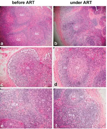
follicular hyperplasia (8 without and 12 with fragmenta-tion; representative examples in Figure 1a, c). Ten lymph nodes showed follicular involution and one follicular depletion (Table 2). Primary follicles were detected in only two lymph nodes. The interfollicular T cell area was hyperplastic in most patients (Figure 1e). The medulla was rich in plasma cells, and the sinuses showed a prom-inent immature sinus histiocytosis. None of the lymph
nodes showed histological signs of opportunistic infec-tions.
After 16–20 months of therapy, the reactive lymph node area and the mean size of the germinal center de-creased significantly in all patients (Tables 1, Figure 1b). The number of patients who developed follicular involu-tion or depleinvolu-tion increased after therapy (Table 1 and 2). Of the 20 patients who showed follicular hyperplasia prior to treatment, nine patients still showed this grade after therapy; follicular involution was evident in eight; and follicular depletion in three (Table 2). The lymph nodes of the 11 patients who had follicular involution/depletion initially remained at this histological stage during therapy (Table 2). The number of germinal centers per represen-tative section decreased significantly after therapy only in the group of patients with initial follicular hyperplasia (Table 1). Germinal centers disappeared in the lymph nodes of five patients. Primary follicles reappeared in lymph nodes of 17 patients. The interfollicular T cell area remained cell rich (Figure 1f). The number of CD8+ T cells that had infiltrated the germinal centers had de-creased significantly following therapy in the group that had initially shown follicular hyperplasia (Table 1, Fig-ures 1d, 2h).
Altogether, after 16–20 months of therapy, the lymph nodes for the majority of patients showed less follicular hyperplasia and more involution/depletion, and the nodes appeared less activated when compared to the corre-sponding baseline lymph node measurements.
Lymph Node Immunostaining
Not all stains on slides could be evaluated because the number of germinal centers either decreased or disap-peared following therapy or because of tissue damage.
HIV p24-, CD4+ T cell-, CD8+ T cell-, and CD21-Immunostaining
Viral p24 antigen was found only within germinal centers (Figure 2n, o). Prior to treatment, germinal centers in 21 of the 27 investigated patients were p24 positive, and in 6 patients negative. Following therapy, germinal centers Table 1
Continued
Before ART 16–20 m of ART p-value
n Baseline values at first LN excision n Values at second LN excision FH 9 13 ± 10 6 9 ± 8 NS F-Inv/Depl 2 8 ± 6 5 9 ± 4 NS
LN: lymph node, GC: germinal center, FH: patients with follicular hyperplasia (with or without fragmentation) before therapy and under ART, F-Inv/Depl: patients with follicular involution or depletion before therapy and under ART, NS: not significant, m: months, n: number of patients studied; Peripheral blood: median (range); viral load, lymph node architecture, and markers: mean ± SD (all paired comparisons);
a
Gray level intensity as evaluated by computer assisted image analysis,bmean TRAIL+cell count/mm2,cmean apoptotic cell count/GC
Figure 1. Lymph node architecture in an HIV-infected patient before and after antiretroviral therapy (ART). Follicular hyperplasia (FH) decreased after antiretroviral therapy (a, b). Germinal centers (GC) were destroyed by infiltrating cells before therapy and then recovered after antiretroviral therapy (c, d). Note vanishing of
Table 2
Histological grading of lymph nodes before and after antiretroviral therapy.
Lymph node grading before antiretroviral therapy n (%) Lymph node grading after 16–20 months of antiretroviral therapy
n
Follicular hyperplasia 8 (26%) Follicular hyperplasia 6 Follicular involution 2 Follicular hyperplasia with fragmentation 12 (39%) Follicular hyperplasia 1 Follicular hyperplasia with fragmentation 2 Follicular involution 6 Follicular depletion 3 Follicular involution 10 (32%) Follicular involution 9 Follicular depletion 1
Follicular depletion 1 (3%) Follicular depletion 1
n: number of patients
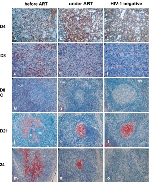
Figure 2. Immunohistochemical localization of CD4 (brown DAB reaction product), CD8, CD21, and p24 (red AEC reaction product) in a lymph node of a patient before and after antiretroviral therapy (ART) and in a lymph node of a HIV-negative individual. The CD4+ T cell number interfollicularly increased after antiretroviral therapy (a, b), whereas the number of CD8+ T cells decreased in the T cell area (TCA) (d, e) and within the germinal centers (g, h). The follicular dendritic cell marker CD21 increased after antiretroviral therapy (j, k). Note fragmentation of the red dendritic network by infiltrating lymphocytes (asterisk). p24 persisted in the germinal centers in some of the patients (m, n). The staining in HIV-negative lymph nodes is shown as controls (c, f, i, l, o). Hematoxylin counterstain, magnifications – a–f: 40·; g–p: 20·.
were p24 negative in the residual lymphoid tissue. In the remaining 14 patients, p24 persisted in the germinal centers after a median time of 18 months post-treatment. The initial six patients with p24-negative germinal centers remained negative. All lymph nodes from HIV-negative controls were HIV-negative for p24 staining (Fig-ure 2p).
In the interfollicular area of the lymph nodes, the numbers of CD4+ T cells increased significantly after
treated patients (Table 1, Figure 2d, e, g, h), yielding staining comparable to lymph nodes of HIV-negative controls (Figure 2c, f, i).
CD21, a marker for the follicular dendritic network, was weakly distributed over the germinal centers prior to therapy (Figure 2k) and condensed significantly after therapy in 11 of the 12 patients tested (Table 1, Figure 2l), resembling the staining in the germinal centers of HIV-negative controls (Figure 2m). A significant CD21 in-Figure 3. Expression of apoptosis
and pro-apoptotic and anti-apoptotic markers (AEC red reaction product) in the lymph node of a patient before and after antiretroviral therapy (ART) and in the node of an HIV-negative individual. Bcl-2 positive cells increased in the mantle zone (MZ) after antiretroviral therapy (a, b). IL-7Ra expression increased in germinal centers after
antiretroviral therapy (d, e). Active caspase 3 increased in the sinuses (g, h), and Fas slightly increased in the germinal centers after antiretroviral therapy (j, k). TRAIL was localized in the sinuses and decreased after antiretroviral therapy (m, n). Apoptosis measured by the TUNEL technique (red stain) was manifested primarily in the germinal centers. In higher magnifications (not shown here), apoptosis was mainly identified as tingible bodies in starry–sky macrophages and as single, scattered apoptotic bodies (p, q). The staining of the same markers is shown in HIV-negative lymph nodes (c, f, i, l, o, r). Hematoxylin counterstain, magnifications: a–c, k–o: 20·; d–i: 40·; p–r: 10·.
Apoptosis in Germinal Centers
Apoptosis as measured in paired lymph nodes obtained from 11 patients was preferentially detected in germinal centers; only a few apoptotic cells were found interfolli-cularly (Figure 3q, r), very similar as in the lymph nodes of uninfected individuals (Figure 3s). Apoptosis was manifested primarily as tingible bodies in starry sky macrophages as well as in single, scattered apoptotic bodies. Following therapy, a significant decrease in apoptosis was detected in lymph nodes on eight patients (p = 0.02), but a slight increase was found in three pa-tients; this yields no statistical significance when all 11 patients were considered together as a group (Table 1).
Immunostaining of Pro-apoptotic and Anti-apoptotic Molecules
The anti-apoptotic Bcl-2 molecules expressed mainly in B lymphocytes of the mantle zone had significantly increased in all 13 patients who could be evaluated after therapy (Table 1, Figure 3a, b). Only a few Bcl-2 positive cells were found in the germinal centers and in the in-terfollicular T cell area before and after therapy. IL-7Ra, which was expressed mainly in centrocytes and centro-blasts of the germinal centers, increased significantly after therapy (Table 1, Figure 3d, e). Active caspase 3 showed a significant increase in the sinuses of the lymph nodes (Table 1, Figure 3k, i) but not in the germinal centers after therapy (not shown). After therapy, staining for these three apoptotic molecules (Figure 3b, e, h) were very similar in intensity and distribution to the staining in HIV-negative control sections (Figure 3c, f, i).
TRAIL staining was detectable in polynucleated granulocytes, in mononuclear histiocytes, and in scattered granular cells that resembled basophils. TRAIL-positive cells were found primarily within vessels and sinuses, some within the interfollicular stroma, and only a few in the germinal centers (Figure 3n, o). The number of TRAIL-positive cells decreased significantly after therapy in the patients who were tested (Table 1). Only a few TRAIL-positive cells were found in the lymph nodes of HIV-negative individuals (Figure 3p).
The increase in Bcl-2, IL-7Ra, and active caspase 3 and the decrease in TRAIL were significant only in the group of patients that originally showed follicular hyper-plasia in their lymphoid tissue.
The staining of the anti-apoptotic cFlip and the pro-apoptotic Fas (Figure 3k, l) and FasL antigens did not change markedly following therapy and looked similar to staining in the lymph node tissue from HIV-negative individuals (Figure 3m, data not shown).
Discussion
The patients of this study were in the early stages of HIV infection as indicated by the still high CD4+ T cell counts in the blood (> 500 cells/ll) and the absence of advanced follicular destruction and atrophy in the lymph nodes. We
show that in these patients, antiretroviral therapy, which lowers the viral load and thus reduces the excessively high antigenic stimulation, markedly diminishes the HIV-dri-ven, highly activated immune response in lymph nodes. This is manifested by:
• A marked reduction of follicular hyperplasia: In early HIV infection, follicular hyperplasia is considered to be a physiological reaction of humoral antiviral immunity. The reduction of germinal centers in size and number after therapy fits well with the observed reduction of the viral antigen p24.
• A reconstitution of the follicular dendritic cell net-work: The disruption and fragmentation of dendritic network and involution of germinal centers are hall-marks of HIV pathology brought about by the abnor-mal infiltration with CD8+ T cells and their destructive activity on these cells known to carry the virus. The reconstitution of the network corresponds very well with the observed CD8+ T cell reduction and prom-inent although not always complete loss of antigen on these target cells.
• A reconstitution of Bcl-2-positive mantle zone lym-phocytes: The B cell system is affected by HIV infection also at the level of bone marrow derived mantle cells, probably due to the lack of helper factors from CD4 T helper cells. The observed sign of recovery of this committed stem cell compartment indicates a regeneration of the B cell system.
• The appearance of primary follicles: Their presence indicates a revival of primary B immune responses which are obviously exhausted by an exaggerated secondary immune response with germinal center formation before therapy.
• A significant increase in the CD4+ T cell number in the interfollicular area in association with a strong reduction of the CD8+ T cell number in the same area and in the germinal centers. This shift within the two T cell populations in all likelihood reflects the central effect of therapy by reducing viral effects on CD4 T cells and reducing the generation of cytotoxic CD8 cells directed against the former.
• A normalization of the apoptotic markers as evidenced by a distribution of the expression of pro-apoptotic and anti-apoptotic molecules that is similar to that of the uninfected lymph node tissue.
Prior to therapy, the majority of lymph nodes showed reactive follicular hyperplasia, often with giant germinal centers and/or pathologic fragmentation of germinal centers. After 16 to 20 months of therapy, the lymph nodes that were excised from these same patients showed a marked reduction in lymph node and germinal center size and also a significant decrease in the number of germinal centers. Primary germinal centers, which were virtually absent prior to therapy, reappeared in the lymph
nodes of a majority of the patients. In addition, the structure of the germinal centers normalized in most patients – some becoming very small, presumably as a consequence of the reduction of germinal center cells and of CD8+ T cells that had infiltrated the germinal centers and that may have had induced the damage. Because the interfollicular area in these patients remained cell rich, this decrease was likely due to a reduction in immune stimulation rather than to progression to a virus-induced degenerative stage.
HIV persists in the germinal centers throughout the entire course of HIV infection [23], and some viruses remain trapped on the surface of follicular dendritic cell. The antiviral response directly or indirectly (via bystander killing) leads to the destruction of the follicular dendritic cells network as the HIV infection progresses [11]. Zhang et al. [11] and Kuster et al. [24] reported that with present therapeutic options, complete abolition of viral replica-tion within the lymphoid tissue was not achieved, and Fischer et al. [25] showed residual HIV-1 RNA in tonsil biopsy specimen obtained from patients receiving anti-retroviral therapy indicating attenuated viral transcription in HIV infected cells that lack virion production. In our study, from the initial 21 patients with p24 positive lymph nodes, only 7 became p24 negative during therapy, while the plasma viral load decreased significantly in all 31 pa-tients.
In agreement with results of other studies [11, 26, 27], the follicular dendritic cell network recovered after ther-apy, as shown by a significant increase in CD21. In most of our patients, however, the follicular dendritic cell network was not completely disrupted prior to therapy, presum-ably because the patients were still in an early stage of HIV infection.
CD8+ T cells are the key effectors of the antiviral defense, thereby mediating the destruction taking place in the lymph nodes. Blackburn et al. [28] reported that the extent of anti-HIV activity by CD8+ T cells in the lym-phoid tissue correlated with the viral burden. We found a high number of CD8+ T cells in the interfollicular area and within the germinal centers before treatment, fol-lowed by a significant decrease after treatment. Consistent with other studies [27, 29, 30], the CD4+ T cell numbers in the lymph nodes increased significantly after therapy.
The lowering of the highly activated immune re-sponse is also reflected by the normalization of some important pro- and anti-apoptotic markers. The expres-sion of Bcl-2, IL-7Ra, TRAIL, and active caspase 3 was different in the lymph nodes that were excised 16– 20 months after treatment and markedly resembled expression in non-infected individuals. Moreover, in 8 of the 11 patients who were tested for these markers, apoptosis decreased after therapy, which indicated that apoptotic processes may have normalized with therapy.
the sinuses but not in germinal centers indicates a caspase-independent cell death pathway in germinal centers. In-deed, it has been shown that productive HIV infection can lead to caspase independent cell death [31].
Only a few other investigators have looked for apoptotic markers and apoptosis in lymph nodes; most have looked for them in peripheral blood lymphocytes. Interestingly, cellular distribution of several molecules seems to differ in lymph node lymphocytes and periph-eral blood lymphocytes, although this may be in part due to technical reasons. The peripheral blood markers were determined by FACS analysis, while the markers in the lymph nodes were identified by immunohistochemistry. For example, although Bcl-2 was found to be expressed in peripheral T-lymphocytes and strongly correlated with spontaneous T cell apoptosis [32–35], in lymph nodes, Bcl-2 was detected primarily in the B (mantle zone of the germinal centers) and not in the T cell area. Simi-larly, IL-7Ra was found to be expressed on peripheral T and B cells, and peripheral CD4+ T cell depletion cor-related well with the down-regulation of IL-7Ra [36, 37]. In lymph nodes, IL-7Ra which increased after therapy was detectable only in the germinal centers and not in the T cell area. The absence of IL-7Ra expression in the T cell area resembles those peripheral T cells in which a strong association of decreased IL-7Ra expression with low Bcl-2 expression has been found [36, 37]. In lymph nodes, a significant down-regulation of TRAIL was found after therapy due primarily to the reduction of cells of the phagocytic type, whereas TRAIL was not identifiable in B or T lymphocytes. In peripheral blood, TRAIL has been discussed as a major death-ligand in HIV infection [38]. Thus, TRAIL, but not FasL-depen-dent AICD, was found in peripheral CD4+ T cells that were isolated from HIV-infected individuals [39, 40]. Whether the CD4+ T cell recovery in lymph nodes after therapy is the consequence of the marked decrease of TRAIL-positive cells is unknown. When the apoptotic and histological markers were correlated, significant differences before and after therapy were found only in the group of patients with follicular hyperplasia. The failure to demonstrate significant differences before and after therapy in the follicular involution/depletion group may be due to the less numbers of patients tested in this study.
This study has some limitations: (a) Untreated patients who were in the early stages of HIV infection were selected for this study, but we have no comparison group of untreated patients in an advanced stage of HIV infection. (b) For ethical reasons, there is no matched control group of untreated HIV patients in early stages of HIV infection. (c) For technical reasons, apoptosis could only be followed sequentially in the lymph nodes of one-third of the patients in the study, and the apoptotic
However, because histological assessments were done in parallel, we believe that the results of the study are valid. The strength of the study is the considerable num-ber of patients from whom sequential lymph node tissue samples have been investigated, together with the analysis of a comprehensive number of immunological, virological, and histological parameters.
In conclusion, two processes might be involved in the current therapeutic setting of these patients: (a) a pro-gression of degenerative changes in the germinal centers as part of the virus-induced pathology in spite of therapy, and (b) the physiological reduction of a hyperimmune response as a result of therapy. Conventional histology indicates that viral damage prevails after involution has been established. The arrest of viral pathology and nor-malization of follicular hyperplasia appears to prevail in the stage of follicular hyperplasia. However, even in cases of persistent involution, there are signs of restoration, such as repopulation of the mantle zone, restoration of the dendritic net, normalization of apoptotic markers, and the appearance of primary follicles.
Our data are consistent with observations made in patients with primary HIV infection who were treated with antiretroviral therapy and cyclosporin A [41]. Rapid shutdown of T cell activation by cyclosporin A in the early phase of HIV infection proved to be beneficial for both immunological and virological measures by preventing ongoing infection of new target cells in the lymphoid tis-sue [2, 42]. Recent reports have shown that immune reconstitution in the intestinal lymphoid tissues did not take place after antiretroviral therapy in acute and early stage HIV infection [43, 44]. Because the intestinal lym-phoid system, as opposed to the inguinal lymph nodes, is constantly exposed to a huge number of different anti-gens, the continuously overactivated immune response may be more prone to the damaging effect of HIV infection. Therefore, HIV pathogenesis may be different in the intestinal lymphoid tissue and inguinal lymph node tissue.
Acknowledgments
This work was supported by the ‘Stiftung Infektionskrankheiten’ and the Swiss National Science Foundation 3138-051092.97 (Tandem) and 3100AO-111368/1. We thank Corine Felber and Ursula Du¨rmu¨ller for technical assistance.
References
1. Li TS, Tubiana R, Katlama C, Calvez V, Ait Mohand H, Autran B: Long-lasting recovery in CD4 T-cell function and viral-load reduction after highly active antiretroviral therapy in advanced HIV-1 disease. Lancet 1998; 351: 1682–1686.
2. Rizzardi GP, De Boer RJ, Hoover S, Tambussi G, Chapuis A, Halkic N, Bart PA, Miller V, Staszewski S, Notermans DW, Perrin L, Fox CH, Lange JM, Lazzarin A, Pantaleo G: Predicting the duration of
antiviral treatment needed to suppress plasma HIV-1 RNA. J Clin Invest 2000; 105: 777–782.
3. Battegay M, Nuesch R, Hirschel B, Kaufmann GR: Immunological recovery and antiretroviral therapy in HIV-1 infection. Lancet Infect Dis 2006; 6: 280–287.
4. Embretson J, Zupancic M, Ribas JL, Burke A, Racz P, Tenner-Racz K, Haase AT: Massive covert infection of helper T lymphocytes and macrophages by HIV during the incubation period of AIDS. Nature 1993; 362: 359–362.
5. Pantaleo G, Graziosi C, Demarest JF, Butini L, Montroni M, Fox CH, Orenstein JM, Kotler DP, Fauci AS: HIV infection is active and progressive in lymphoid tissue during the clinically latent stage of disease. Nature 1993; 362: 355–358.
6. Schacker T, Little S, Connick E, Gebhard K, Zhang ZQ, Krieger J, Pryor J, Havlir D, Wong JK, Schooley RT, Richman D, Corey L, Haase AT: Productive infection of T cells in lymphoid tissues during primary and early human immunodeficiency virus infection. J Infect Dis 2001; 183: 555–562.
7. Pantaleo G, Fauci AS: New concepts in the immunopathogen-esis of HIV infection. Annu Rev Immunol 1995; 13: 487–512. 8. Chadburn A, Metroka C, Mouradian J: Progressive lymph node
histology and its prognostic value in patients with acquired immunodeficiency syndrome and AIDS-related complex. Hum Pathol 1989; 20: 579–587.
9. Laman J, Claassen E, Van Rooijen N: Immune complexes on follicular dendritic cells as a target for cytolytic cells in HIV. AIDS 1989; 30: 543–548.
10. Baroni C, Vitolo D, Uccini S: Immunohistopathogenesis of per-sistent lymphadenopathy in HIV-positive patients. Ric Clin Lab 1990; 20: 1–10.
11. Zhang ZQ, Schuler T, Cavert W, Notermans DW, Gebhard K, Henry K, Havlir DV, Gunthard HF, Wong JK, Little S, Feinberg MB, Polis MA, Schrager LK, Schacker TW, Richman DD, Corey L, Danner SA, Haase AT: Reversibility of the pathological changes in the follicular dendritic cell network with treatment of HIV-1 infection. Proc Natl Acad Sci USA 1999; 96: 5169–5172. 12. Badley AD, Pilon AA, Landay A, Lynch DH: Mechanisms of
HIV-associated lymphocyte apoptosis. Blood 2000; 96: 2951–2964. 13. McCune JM: The dynamics of CD4+ T-cell depletion in HIV
dis-ease. Nature 2001; 410: 974–979.
14. Grossman Z, Meier-Schellersheim M, Sousa AE, Victorino RM, Paul WE: CD4+ T-cell depletion in HIV infection: are we closer to understanding the cause?. Nat Med 2002; 8: 319–323.
15. Brenchley JM, Schacker TW, Ruff LE, Price DA, Taylor JH, Beilman GJ, Nguyen PL, Khoruts A, Larson M, Haase AT, Douek DC: CD4+ T cell depletion during all stages of HIV disease occurs pre-dominantly in the gastrointestinal tract. J Exp Med 2004; 200: 749–759.
16. Mehandru S, Poles MA, Tenner-Racz K, Horowitz A, Hurley A, Hogan C, Boden D, Racz P, Markowitz M: Primary HIV-1 infection is associated with preferential depletion of CD4+ T lymphocytes from effector sites in the gastrointestinal tract. J Exp Med 2004; 200: 761–770.
17. Gougeon ML, Montagnier L: Apoptosis in AIDS. Science 1993; 260: 1269–1270.
18. Ameisen JC, Estaquier J, Idziorek T, De Bels F: Programmed cell death and AIDS pathogenesis: significance and potential mechanisms. Curr Top Microbiol Immunol 1995; 200: 195–211. 19. Finkel TH, Tudor-Williams G, Banda NK, Cotton MF, Curiel T,
Monks C, Baba TW, Ruprecht RM, Kupfer A: Apoptosis occurs predominantly in bystander cells and not in productively in-fected cells of HIV- and SIV-inin-fected lymph nodes. Nat Med 1995; 1: 129–134.
20. Haase AT, Henry K, Zupancic M, Sedgewick G, Faust RA, Melroe H, Cavert W, Gebhard K, Staskus K, Zhang ZQ, Dailey PJ, Balfour HH Jr., Erice A, Perelson AS: Quantitative image analysis of HIV-1 infection in lymphoid tissue. Science 1996; 274: 985–989. 21. Ost A, Baroni CD, Biberfeld P, Diebold J, Moragas A, Noel H,
Pallesen G, Racz P, Schipper M, Tenner-Racz K, et al. Lymphad-enopathy in HIV infection: histological classification and stag-ing. Acta Pathol Microbiol Scand 1989; 97: 7–15.
22. Bachmann F, Buechner SA, Wernli M, Strebel S, Erb P: Ultraviolet light downregulates CD95 ligand and trail receptor expression facilitating actinic keratosis and squamous cell carcinoma for-mation. J Invest Dermatol 2001; 117: 59–66.
23. Orenstein JM, Feinberg M, Yoder C, Schrager L, Mican JM, Sch-wartzentruber DJ, Davey RT Jr, Walker RE, Falloon J, Kovacs JA, Miller KD, Fox C, Metcalf JA, Masur H, Polis MA: Lymph node architecture preceding and following 6 months of potent anti-viral therapy: follicular hyperplasia persists in parallel with p24 antigen restoration after involution and CD4 cell depletion in an AIDS patient. Aids 1999; 13: 2219–2229.
24. Kuster H, Opravil M, Ott P, Schlaepfer E, Fischer M, Gunthard HF, Luthy R, Weber R, Cone RW: Treatment-induced decline of hu-man immunodeficiency virus-1 p24 and HIV-1 RNA in lymphoid tissue of patients with early human immunodeficiency virus-1 infection. Am J Pathol 2000; 156: 1973–1986.
25. Fischer M, Joos B, Hirschel B, Bleiber G, Weber R, Gunthard HF: Cellular viral rebound after cessation of potent antiretroviral therapy predicted by levels of multiply spliced HIV-1 RNA encoding nef. J Infect Dis 2004; 190: 1979–1988.
26. Voltersvik P, Dyrhol-Riise AM, Bostad L, Rosok BI, Olofsson J, Asjo B: Changes in tonsillar tissue in early HIV-1 infection and during 3 years of antiretroviral therapy. Apmis 2000; 108: 539–550. 27. Alos L, Navarrete P, Morente V, Garcia F, Garrido M, Plana M,
Mozos A, Lopez A, Gil C, Pumarola T, Caballero M, Blanch JL, Fumero E, Miro JM, Gallart T, Gatell JM, Campo E: Immunoar-chitecture of lymphoid tissue in HIV-infection during antiret-roviral therapy correlates with viral persistence. Mod Pathol 2005; 18: 127–136.
28. Blackbourn DJ, Mackewicz CE, Barker E, Hunt TK, Herndier B, Haase AT, Levy JA: Suppression of HIV replication by lymphoid tissue CD8+ cells correlates with the clinical state of HIV-in-fected individuals. Proc Natl Acad Sci USA 1996; 93: 13125–13130. 29. Rizzardi GP, Tambussi G, Bart PA, Chapuis AG, Lazzarin A, Pan-taleo G: Virological and immunological responses to HAART in asymptomatic therapy-naive HIV-1-infected subjects according to CD4 cell count. Aids 2000; 14: 2257–2263.
30. Dyrhol-Riise AM, Voltersvik P, Rosok BI, Olofsson J, Asjo B: Normalization of CD4+ cell numbers and reduced levels of memory CD8+ cells in blood and tonsillar tissue after highly active antiretroviral therapy in early HIV type-1 infection. AIDS Res Hum Retrovir 2000; 16: 191–201.
31. Petit F, Arnoult D, Lelievre JD, MoutouhdeParseval L, Hance AJ, Schneider P, Corbeil J, Ameisen JC, Estaquier J: Productive HIV-1 infection of primary CD4+T cells induces mitochondrial mem-brane permeabilization leading to a caspase-independent cell death. J Biol Chem 2002; 277: 1477–1487.
32. Boudet F, Lecoeur H, Gougeon ML: Apoptosis associated with ex vivo down-regulation of Bcl-2 and up-regulation of Fas in po-tential cytotoxic CD8+ T lymphocytes during HIV infection. J Immunol 1996; 156: 2282–2293.
33. Regamey N, Harr T, Battegay M, Erb P: Downregulation of Bcl-2, but not of Bax or Bcl-x, is associated with T lymphocyte apop-tosis in HIV infection and restored by antiretroviral therapy or by interleukin 2. AIDS Res Hum Retrovir 1999; 15: 803–810. 34. Airo P, Torti C, Uccelli MC, Malacarne F, Palvarini L, Carosi G,
Castelli F: Naive CD4+ T lymphocytes express high levels of Bcl-2 after highly active antiretroviral therapy for HIV infection. AIDS Res Hum Retrovir 2000; 16: 1805–1807.
35. Balestrieri E, Grelli S, Matteucci C, Minutolo A, d’Ettorre G, Di Sora F, Montella F, Vullo V, Vella S, Favalli C, Macchi B, Mastino A: Apoptosis-associated gene expression in HIV-infected pa-tients in response to successful antiretroviral therapy. J Med Virol 2007; 79: 111–117.
36. Koesters S, Alimonti J, Wachihi C, Matu L, Anzala O, Kimani L, Embree J, Plummer F, KR F: IL-7Ralpha expression on CD4+ T lymphocytes decrease with HIV disease progression and in-versely correlates with immune activation. Eur J Immunol 2006; 36: 336–344.
37. Rethi B, Fluur C, Atlas A, Krzyzowska M, Mowafi F, Grutzmeier S, De Milito A, Bellocco R, Falk KI, Rajnavolgyi E, Chiodi F: Loss of IL-7Ralpha is associated with CD4 T-cell depletion, high interleu-kin-7 levels and CD28 down-regulation in HIV infected patients. Aids 2005; 19: 2077–2086.
38. Miura Y, Koyanagi Y: Death ligand-mediated apoptosis in HIV infection. Rev Med Virol 2005; 15: 169–178.
39. Katsikis PD, Garcia-Ojeda ME, Torres-Roca JF, Tijoe IM, Smith CA, Herzenberg LA: Interleukin-1 beta converting enzyme-like pro-tease involvement in Fas-induced and activation-induced peripheral blood T cell apoptosis in HIV infection: TNF-related apoptosis-inducing ligand can mediate activation-induced T cell death in HIV infection. J Exp Med 1997; 186: 1365–1372. 40. Jeremias I, Herr I, Boehler T, Debatin KM:
TRAIL/Apo-2-ligand-induced apoptosis in human T cells. Eur J Immunol 1998; 28: 143–152.
41. Rizzardi GP, Harari A, Capiluppi B, Tambussi G, Ellefsen K, Ciu-ffreda D, Champagne P, Bart PA, Chave JP, Lazzarin A, Pantaleo G: Treatment of primary HIV-1 infection with cyclosporin A coupled with highly active antiretroviral therapy. J Clin Invest 2002; 109: 681–688.
42. Cohen OJ, Fauci AS: Benchmarks for antiretroviral therapy. J Clin Invest 2000; 105: 709–710.
43. Mehandru S, Poles MA, Tenner-Racz K, Jean-Pierre P, Manuelli V, Lopez P, Shet A, Low A, Mohri H, Boden D, Racz P, Markowitz M: Lack of Mucosal Immune Reconstitution during Prolonged Treatment of Acute and Early HIV-1 Infection. PLoS Med 2006; 3: e484.
44. Veazey RS, Lackner AA: Impact of antiretroviral therapy on intestinal lymphoid tissues in HIV infection. PLoS Med 2006; 3: e515.