HAL Id: tel-01557493
https://tel.archives-ouvertes.fr/tel-01557493
Submitted on 6 Jul 2017
HAL is a multi-disciplinary open access
archive for the deposit and dissemination of sci-entific research documents, whether they are pub-lished or not. The documents may come from teaching and research institutions in France or abroad, or from public or private research centers.
L’archive ouverte pluridisciplinaire HAL, est destinée au dépôt et à la diffusion de documents scientifiques de niveau recherche, publiés ou non, émanant des établissements d’enseignement et de recherche français ou étrangers, des laboratoires publics ou privés.
Study of circular code motifs in nucleic acid sequences
Karim El Soufi
To cite this version:
Karim El Soufi. Study of circular code motifs in nucleic acid sequences. Bioinformatics [q-bio.QM]. Université de Strasbourg, 2017. English. �NNT : 2017STRAD004�. �tel-01557493�
DOCTORALE SCHOOL «Mathematics, Information Sciences and Engineering»
ICube
Thesis
presented by:Karim EL SOUFI
defended on : 24 January 2017
to obtain:
Doctor of the university of Strasbourg
Discipline / Speciality
: Computer science
Study of circular code motifs in nucleic
acid sequences
THESIS supervised by :
MICHEL Christian Professor, University of Strasbourg
RAPPORTEURS :
COMET Jean-Paul Professor, University of Nice Sophia Antipolis
FIMMEL Elena Professor, Hochschule Mannheim
OTHER MEMBERS OF THE JURY :
COLLET Pierre Professor, University of Strasbourg, jury president
STRÜNGMANN Lutz Professor, Hochschule Mannheim
ÉCOLE DOCTORALE «Mathématique, Sciences de l'Informatique et de
l’Ingénieur»
ICube
THÈSE
présentée par :Karim EL SOUFI
soutenue le : 24 Janvier 2017
pour obtenir le grade de:
Docteur de l’université de Strasbourg
Discipline / Spécialité
: Informatique
Étude des motifs de code circulaire
dans les séquences d'acides nucléiques
THÈSE dirigée par :
MICHEL Christian Professeur, Université de Strasbourg
RAPPORTEURS :
COMET Jean-Paul Professeur, Université Nice Sophia Antipolis
FIMMEL Elena Professeur, Hochschule Mannheim
AUTRES MEMBRES DU JURY :
COLLET Pierre Professeur, Université de Strasbourg, président du jury
STRÜNGMANN Lutz Professeur, Hochschule Mannheim
Abstract
Le travail effectué dans cette thèse présente une nouvelle approche de la théorie du code circulaire dans les gènes qui a été initiée en 1996. Cette approche con-siste à analyser les motifs construits à partir de ce code circulaire. Ces motifs particuliers sont appelés motifs de code circulaire. Ainsi, nous avons développé des algorithmes de recherche pour localiser les motifs de code circulaire dans les séquences d’acides nucléiques afin de leurs trouver une signification bioinforma-tique. En effet, le code circulaire X identifié dans les gènes est un ensemble de trinucléotides qui a la propriété de retrouver, synchroniser et maintenir la phase de lecture. De plus, il possède la propriété d’autocomplémentarité C : les trin-ucléotides de X sont complémentaires entre eux, c’est-à-dire X = C(X). Enfin, il a la propriété C3 : les ensembles de trinucléotides P(X) et P2(X) par per-mutation de X d’un et de deux nucléotides, respectivement, sont également des codes circulaires et de plus complémentaires entre eux, c’est-à-dire C(X1) = X2 et C(X2) = X1 .
Notre travail de recherche s’est donc intéressé à la recherche de motifs du code circulaire X dans des séquences d’ADN ou d’ARN. Nous avons commencé notre analyse avec le centre de décodage du ribosome (ARNr) qui est une région ma-jeure dans le processus de traduction des gènes aux protéines. Puis, nous avons étendu les résultats obtenus avec le ribosome aux ARN de transfert (ARNt) pour étudier les interactions ARNr-ARNt. Enfin, nous avons généralisé la recherche de motifs de code circulaire X dans l’ADN aux chromosomes d’eucaryotes complets. La théorie du code circulaire a contribué à l’analyse du centre de décodage du ribosome, en particulier à sa structure primaire. De fa con surprenante, les nucléotides universellement conservés A1492 et A1493 dans tous les ARNr de bactéries, d’archées, d’eucaryotes nucléaires et de chloroplastes appartiennent à
des motifs de code circulaire mAA(X). Le nucléotide conservé G530 dans les
ARNr des bactéries et des archées est également inclus dans des motifs de code
circulaire mG(X). Le développement d’un algorithme de recherche des motifs
de code circulaire associé à l’alignement global des séquences multiples permet d’identifier les motifs de code circulaire mG(X) dans les ARNr nucléaires et chloroplastes, résultat qui ne peut pas être obtenu part des méthodes classiques
Directeur de thèse: Prof. MICHEL Christian EL SOUFI Karim de bioinformatique. Par ailleurs, un nouveau motif de code circulaire m(X) uni-versellement conservé dans les sept organismes étudiés est identifié grˆace à la théorie du code circulaire. La visualisation spatiale des trois motifs mAA(X),
mG(X) et m(X) montrent qu’ils appartiennent au centre de décodage du
ribo-some dans tous les ARNr étudiés de bactéries, d’archées, d’eucaryotes nucléaires et de chloroplastes. En conclusion, la fonction biologique du centre de décodage du ribosome qui a été attribuée à un nombre restreint de nucléotides, précisé-ment les nucléotides A1492, A1493 et G530, peut maintenant être associée à des motifs de code circulaire comportant au moins deux et jusqu’à cinq trinucléotides successifs.
Nous identifions également de nouvelles propriétés de cette théorie du code circulaire avec des analyses statistiques de motifs du code circulaire X de grande taille dans les génomes des eucaryotes. Pour la première fois, les régions non-codantes (en dehors des gènes) sont étudiées avec cette théorie du code circulaire. Les motifs du code circulaire X de grande taille de longueurs l ≥ 15 trinucléotides et de cardinalité (composition) supérieure à 10 trinucléotides ont la plus grande fréquence d’apparition dans les génomes des eucaryotes par rapport (i) aux 23 motifs de codes circulaires bijectifs de grande taille, (ii) aux deux motifs de codes circulaires permutés de grande taille ; (iii) aux motifs aléatoires de grande taille obtenus avec des codes aléatoires (non circulaires). Les plus longs motifs du code circulaire X sont identifiés dans les génomes des eucaryotes, par exemple un motif
X dans une région non-codante du génome Solanum pennellii avec une longueur
de 155 trinucléotides (465 nucleotides) associé à une probabilité d’occurrence
de 10−71 , deux motifs X dans des régions non-codante du génome Salmo salar
avec des longueurs de 118 trinucléotides (354 nucléotides) avec une probabilité d’occurrence de 10−52 , etc. Le plus longs motif du code circulaire X dans le génome humain se trouve dans une région non-codante du chromosome 13 avec une longueur de 36 trinucléotides et une probabilité d’occurrence de 10−11.
Les motifs du code circulaire X dans les régions non-codantes des génomes sont probablement des vestiges évolutifs de gènes primitifs utilisant le code cir-culaire pour la traduction des gènes aux protéines. Cependant, les études statis-tiques réalisées dans ce travail de thèse montrent que les motifs X apparaissent préférentiellement dans les gènes avec une proportion de motifs X (de longueurs l supérieure à 10 trinucléotides et de cardinalité supérieure à 5 trinucléotides) égal à 8 dans les gènes / régions non-codantes pour 138 génomes eucaryotes complets. Ce facteur de 8 est également retrouvé avec les motifs X dans les gènes / régions non-codantes des 24 chromosomes humains. D’un point de vue biologique, cette
propriété peut s’expliquer par le fait que les mutations (substitution, insertion et délétion de nucléotides) sont plus fréquentes dans les régions non-codantes par rapport aux gènes.
L’existence du code circulaire X dans les gènes est un problème ouvert depuis sa découverte en 1996. Nous montrons dans la suite de notre travail que le con-cept de code circulaire dans les régions d’ADN de faible complexité existe égale-ment avec les codes circulaires unitaires (UCC) de dinucléotides, trinucléotides et tétranucléotides générant des motifs UCC de dinucléotides répétés (Di+ mo-tifs), de trinucléotides répétés (motifsTri+) et de tétranucléotides répétés (motifs
Tetra+ ) dans les génomes d’eucaryotes. Plus précisément, 12 codes UCC de din-ucléotides sont ”strong comma-free” et quatre d’entre eux {AT }, {CG}, {GC} et {T A} sont auto-complémentaires. Egalement, 48 codes UCC de trinucléotides sont ”strong comma-free” et 12 codes UCC de trinucléotides sont ”comma-free”. Enfin, 180 codes UCC de tétranucléotides sont ”strong comma-free”, 60 codes UCC de tétranucléotides sont ”free” et 12 codes ”strong comma-free” {AAT T }, {ACGT }, {AGCT }, {CAT G}, {CCGG}, {CT AG}, {GAT C}, {GGCC}, {GT AC}, {T CGA}, {T GCA} et {T T AA} sont en plus autocomplé-mentaires. Ainsi, les motifs Di+, Tri+ et Tetra+ permettent de retrouver une phase de lecture modulo 2, modulo 3 et modulo 4, respectivement, dans les ré-gions non-codantes des eucaryotes. De plus, les propriétés C2, C3et C4 permettent également de retrouver les phases décalées et la propriété d’autocomplémentarité permet l’appariement des deux brins de l’ADN dans les régions non codantes des eucaryotes. Un motif UCC et son motif UCC complémentaire ont la même distri-bution dans les génomes des eucaryotes, à la fois selon leur nombre d’apparition et leur nombre total de nucléotides. Cette propriété est observée avec les motifs
Di+, Tri+ et Tetra+ . De plus, pour les motifs Tri+ et Tetra+ , un motif UCC et son motif UCC complémentaire ont des occurrences croissantes inversement proportionnel à leur nombre de liaisons hydrogène.
De manière surprenante, on observe une rareté des trinucléotides répétés (mo-tifs Tri+) dans les grands génomes d’eucaryotes par rapport aux motifs Di+ et
Tetra+ . Ce résultat statistique est obtenu avec des mesures de moyenne et de médiane, et confirmé par deux tests statistiques (un test paramétrique t de Stu-dent pour échantillon apparié et un test non-paramétrique W de Wilcoxon signé). Ainsi, les codes circulaires unitaires de trinucléotides associés aux trinucléotides répétés dans les génomes des eucaryotes pourraient avoir contribué à la forma-tion du code circulaire X dans les gènes. Une conséquence d’une telle hypothèse serait la persistance de certaines propriétés statistiques des trinucléotides répétés
Directeur de thèse: Prof. MICHEL Christian EL SOUFI Karim du code circulaire X. De fa con inattendue, des paires de trinucléotides identiques (14 trinucléotides parmi 20) du code circulaire X sont préférentiellement utilisées dans les gènes des eucaryotes. Pour la première fois depuis 20 ans, la théorie du code circulaire dans les gènes est étendue aux génomes dans ce travail de thèse. Ainsi, le code circulaire pourrait être une structure mathématique des gènes mais également des génomes.
Acknowledgments
Firstly, I would like to express my sincere gratitude to my supervisor Prof. Christian Michel for his patience, motivation, and immense knowledge in the field. His guidance helped me in all the time of research and writing of this thesis.
My regards to the jury members, Prof. Jean-Paul Comet and Dr. Julie Thompson, for showing interest in my work, and Profs. Pierre Collet, Elena Fimmel and Lutz Strüngmann for their continued interest. The valuable feed-back that they offered was instrumental to the enhancement of my work.
I would like to thank the Lebanese Association for Scientific Research (LASeR) for their help in financing my thesis and my team Complex Systems and Trans-lational Bioinformatics (CSTB) for their support. A special gratitude to my friends and loved ones who offered assistance when I needed it most.
Last but not the least, I would like to thank my parents for whom without I would not be here. Therefore, I dedicate this thesis to them.
Contents
Introduction 1 Circular code . . . 1 Biological context . . . 2 Thesis structure . . . 3 1 Biological environment 5 1.1 Introduction . . . 61.2 Nucleic acid sequence . . . 6
1.3 Ribosome . . . 8 1.3.1 Translation . . . 8 1.3.2 Biomolecular structure . . . 9 1.4 Biological databases . . . 11 1.4.1 Crystallographic database . . . 12 1.4.2 Sequence database . . . 13 1.5 Summary . . . 15 2 Code Theory 17 2.1 Introduction . . . 18 2.2 History of codes . . . 18 2.2.1 Diamond code . . . 18 2.2.2 Comma-free code . . . 19 2.2.3 Genetic code . . . 21 2.3 Circular code . . . 21
2.3.1 Circular code motifs . . . 25
2.3.2 Unitary circular codes of dinucleotides, trinucleotides and tetranucleotides . . . 26
2.4 Summary . . . 29
3 Data and Methods 31 3.1 Introduction . . . 33
3.2 Data acquisition . . . 33
3.2.1 Ribosomal data . . . 33 iii
3.2.2 Genomic database . . . 34
3.3 Circular code motifs . . . 36
3.3.1 Search algorithms . . . 36
3.3.1.1 Biinfinite word . . . 36
3.3.1.2 Sets algorithm . . . 37
3.3.1.3 Frames algorithm . . . 41
3.3.2 Definition of random code motifs . . . 42
3.3.3 Definition of the 23 bijective circular codes motifs . . . 43
3.3.3.1 Bijective transformation circular codes . . . 43
3.3.3.2 Main properties of the 23 bijective transformation circular codes . . . 47
3.3.4 Statistical analysis of X circular code motifs . . . 47
3.3.4.1 Coverage of X motifs in tRNA . . . 47
3.3.4.2 Expectation of the occurrence number of a motif . . 48
3.3.4.3 Ratio of X motifs in coding and non-coding regions . 48 3.4 Ribosome study tools . . . 50
3.4.1 Multiple sequence alignment . . . 50
3.4.2 Molecule viewer . . . 50
3.5 Repeated motifs . . . 52
3.5.1 Dinucleotide unitary circular code motifs . . . 52
3.5.2 Trinucleotide unitary circular code motifs . . . 52
3.5.3 Tetranucleotide unitary circular code motifs . . . 53
3.5.4 Statistical analysis of repeated motifs . . . 53
3.5.4.1 Occurrence number of unitary circular code motifs . 53 3.5.4.2 Base number of unitary circular code motifs . . . 55
3.5.4.3 Total base number of unitary circular code motifs . . 55
3.6 Occurrence number of trinucleotide pairs . . . 56
3.7 Summary . . . 59
4 Results and Discussion 61 4.1 Introduction . . . 63
4.2 X circular code motifs in the ribosome . . . 63
4.2.1 X circular code motifs in the ribosomal decoding center . . . 63
4.2.1.1 The conserved A1492 and A1493 nucleotides . . . 63
4.2.1.2 The conserved G530 nucleotide . . . 68
4.2.2 Spatial study of X circular code motifs near the ribosomal decoding center . . . 73
center . . . 73 4.2.2.2 Conserved X motifs in the rRNA of prokaryotes . . . 74 4.2.2.3 Conserved X motifs in the rRNA of eukaryotes . . . 78 4.2.3 X circular code motifs in prokaryotic tRNAs . . . 80 4.2.3.22 Summary of X circular code motifs in tRNA sequences 99 4.2.3.23 Coverage of X circular code motifs in prokaryotic
tRNAs . . . 100 4.3 Analysis of X circular code motifs in eukaryotic genomes . . . 100 4.3.1 Occurrence of large randoms code motifs in eukaryotic genomes101 4.3.2 Occurrence of large motifs from X, X1, X2 and the 23
bijec-tive transformations of X in eukaryotic genomes . . . 102 4.3.3 Largest X motifs in eukaryotic genomes . . . 105 4.3.4 Largest X motifs in Homo sapiens . . . 108 4.3.5 X motifs in coding regions versus non-coding regions in
eu-karyotic genomes . . . 108 4.3.6 X motifs in coding regions versus non-coding regions in
Homo sapiens . . . 110
4.4 Analysis of unitary circular code motifs in eukaryotic genomes . . . 111 4.4.1 Occurrence of repeated dinucleotides in eukaryotic genomes . 112 4.4.2 Occurrence of repeated trinucleotides in eukaryotic genomes . 112 4.4.3 Occurrence of repeated tetranucleotides in eukaryotic genomes114 4.4.4 Largest repeated motifs in eukaryotic genomes . . . 116 4.4.5 Scarcity of repeated trinucleotides in eukaryotic genomes . . 119 4.5 Identical trinucleotide pairs of the X circular code in eukaryotic
gene sequences . . . 120 4.6 Summary . . . 123
5 Conclusion 125
References 130
List of Figures
1.1 Nucleotide. . . 6
1.2 Phosphodiester bond. . . 7
1.3 DNA structure. . . 7
1.4 Ribosome during translation. . . 9
1.5 Decoding center of the ribosome. . . 10
1.6 Partial 3D structure of E. coli ribosome. . . 11
1.7 Complete 3D structure of E. coli ribosome. . . 12
1.8 Protein Data Base (PDB) growth. . . 13
2.1 Diamond code. . . 19
2.2 Circular code definition. . . 23
2.3 Circular code example. . . 23
2.4 Circular code window. . . 24
3.1 ClustalX output. . . 50
3.2 Modified multiple sequence alignment output. . . 51
3.3 Jmol. . . 51
4.1 3D structure of the ribosome of Escherichia coli. . . 65
4.2 3D structure of the ribosome of Thermus thermophilus. . . 66
4.3 3D structure of the ribosome of Pyrococcus furiosus. . . 67
4.4 3D structure of the ribosome of Saccharomyces cerevisiae. . . 68
4.5 3D structure of the ribosome of Triticum aestivum. . . 69
4.6 3D structure of the ribosome of Homo sapiens. . . 71
4.7 3D structure of the ribosome of Spinacia oleracea. . . 72
4.8 Conserved X motifs in Escherichia coli. . . 75
4.9 Conserved X motifs in Thermus thermophilus. . . 75
4.10 Conserved X motifs in Pyrococcus furiosus. . . 77
4.11 Conserved X motifs in Saccharomyces cerevisiae. . . 77
4.12 Conserved X motifs in Triticum aestivum. . . 78
4.13 Conserved X motifs in Homo sapiens. . . 78 vii
4.14 Occurrence numbers of the large motifs from X, X1, X2 and the 23 bijective transformations of X. . . 102 4.15 Occurrence numbers, by length, of the large motifs from X, X1, X2
and the top six bijective transformations of X. . . 103 4.16 Occurrence numbers, by cardinality, of the large motifs from X,
X1, X2 and the top six bijective transformations of X. . . 104 4.17 Base ratio of coding/non-coding regions and base ratio of X motifs
in the 138 eukaryotic genomes. . . 105 4.18 Base ratio of coding/non-coding regions and base ratio of X motifs
in Homo sapiens genome. . . 108 4.19 Occurrence numbers of repeated dinucleotides in eukaryotic genomes.113 4.20 Base numbers in repeated dinucleotides in eukaryotic genomes. . . 113 4.21 Occurrence numbers of repeated trinucleotides in eukaryotic genomes.114 4.22 Base numbers in repeated trinucleotides in eukaryotic genomes. . . 115 4.23 Occurrence numbers of repeated tetranucleotides in eukaryotic
genomes. . . 116 4.24 Base numbers in repeated tetranucleotides in eukaryotic genomes. . 117 4.25 Preference of identical trinucleotide pairs in gene sequences. . . 121
List of Tables
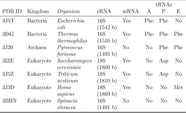
2.1 Trinucleotide occurrences. . . 22
2.2 Circular code statistics. . . 25
3.1 Protein Data Base (PDB) entries. . . 34
3.2 Genomes characteristics. . . 35
3.3 Bijective transformation circular codes . . . 46
4.1 X motifs containing the A1492 and A1493 nucleotides. . . 64
4.2 X motifs containing the G530 nucleotide. . . 70
4.3 Conserved motif in the decoding center. . . 73
4.4 Prokaryotic conserved motifs near the decoding center. . . 74
4.5 Eukaryotic conserved motifs near the decoding center. . . 79
4.27 The coverage of X motifs in tRNA sequences. . . 101
4.28 Top 20 largest X motifs in the 138 eukaryotic genomes. . . 106
4.29 Largest X motifs in Homo sapiens chromosomes. . . 107
4.30 Base ratio of coding/non-coding regions and base ratio of X motifs in the 138 eukaryotic genomes. . . 109
4.31 Base ratio of coding/non-coding regions and base ratio of X motifs in the Homo sapiens genome. . . 110
4.32 Largest repeated motifs in eukaryotic genomes. . . 117
4.33 Correlation matrix of the base number of all the repeated trinu-cleotides in eukaryotic genomes. . . 118
4.34 Scarcity of repeated trinucleotides. . . 119
Introduction
We offer here a general introduction of this thesis, realized at The Engineering Science, Computer Science and Imaging (ICube) laboratory. Created in 2013, the laboratory brings together researchers of the University of Strasbourg, the CNRS (French National Center for Scientific Research), the ENGEES and the INSA of Strasbourg in the fields of engineering science and computer science. With around 580 members, ICube is a major driving force for research in Strasbourg. The work done in this thesis presents a new direction for circular code iden-tified in 1996 by analysing the motifs constructed from circular code. These particular motifs are called circular code motifs. We applied search algorithms to locate circular code motifs in nucleic acid sequences in order to find biological significance. We start with an overview of circular code and its property of cod-ing frame retrieval for genes. Afterwards we present the biological environment in which we applied our work. Finally, we show the structure in which this thesis is presented.
Circular code
The genetic code consists of 64 trinucleotides {AAA, . . . , T T T }, called codons. Each codon encodes for one of the 20 amino acids used in the synthesis of proteins (translation). Most of the amino acids are encoded by more than one codon. Some of the codons have a special purpose. The ATG codon serves as the starting point of translation while also encoding the amino acid methionine at the same time. While the following three trinucleotides {T AA, T AG, T GA}, called stop codons, are an exception, they do not encode for an amino acid they signal the end of a translation process.
The genetic code can be expressed as either ribonucleic acid (RNA) codons or deoxyribonucleic acid (DNA) codons. RNA codons occur in messenger RNA (mRNA) and are the codons that are actually read during translation. Each mRNA molecule acquires its sequence of nucleotides by transcription from the corresponding gene. Genes are DNA sequences which are read modulo 3 let-ters among the three possible frames. As such, only one frame, called reading
frame, which begins with a start codon and ends with a stop codon, codes the corresponding protein sequence according to the genetic code.
However, this does not mean a translation starts at every AT G codon, even though it accounts for most start codons, it could be one of the following {T T G, GT G, CT G}. Additional requirements need to be present when assign-ing a start codon, such as the ribosome bidassign-ing side, the shine-delgarno sequence. This short sequence needs to be located 7 to 13 bases upstream of the start codon. Add to that the importance of maintaining the correct reading frame.
All this indicates that the procedure of maintaining the correct reading frame of genes is far more complex. It was theorized that there are sets of trinucleotides called circular codes X which have the property of reading frame retrieval, syn-chronization and maintenance (Michel, 2012). Furthermore, there are circular codes which have in addition the C self-complementary property, i.e. the trinu-cleotides of X are complementary to each other, i.e. X = C(X). Finally, there are self-complementary circular codes X which have in addition the C3 property, i.e. the permuted trinucleotide sets P(X) and P2(X) of X by one and two nu-cleotides, respectively, are also trinucleotide circular codes and complementary to each other, i.e. C(X1) = X2 and C(X2) = X1. In 1996, a C3 self-complementary trinucleotide circular code X has been identified in genes (reading frame of mR-NAs) simultaneously in eukaryotes and prokaryotes (Arquès and Michel, 1996). Biological context
Our work revolves around searching for X circular code motifs in DNA or RNA sequences. A circular code motif is basically a sequence of nucleotides where its trinucleotides belong to the circular code. In our biological environment we approached the matter by addressing the very specific and narrow, then moving to the more general and broader look. The first part of the study was confined to the ribosome, more specifically, we started with the decoding center of the ri-bosome. A region that is very important to the translation process. Afterwards, we included the transfer RNA in the ribosome in the study, in order to give a wider look at the circular code motifs in the ribosome and whether a possible interaction exist. Later in our work, to address the presence of X circular code motifs in DNA, we searched the entire database of complete eukaryotic chromo-somes. This involved a huge amount of data and computation. Our results from circular code were thoroughly compared to those of random generated codes in
Thesis structure
This thesis is structured into an introduction, four chapters for the main body and a conclusion.
The first chapter will present the biological environment we are working in. Our main focus lies in the nucleic acid sequence, we will be explaining what is a sequence made of. We worked in two different biological contexts, the first was the ribosome, in order to highlight the importance of our findings we explaining briefly the translation process and shed some light on the 3D structure of the ribosome. The second phase of our work involved the sequence of complete eukaryotic chromosomes, which was acquired from the RefSeq database.
The second chapter serves as an introduction of circular code, beginning with the root that lead to its discovery. This takes us back to the race of cracking the genetic code, as many code theories fell in light of reality once the genetic code was finally cracked. We will explain how the circular code came to be after the discovery of the genetic code, its features and significance. Finally, we present what are circular code motifs, which serve as the main study course of this thesis. The data and methods are presented in the fourth chapter. We present the data obtained from the databases mentioned before. A new algorithm was written to extract motifs from sequences, this algorithm is versatile as it can take any code as a parameter. Multiple sequence alignments were modified to allow us to switch the attention onto circular code motifs, this combination allowed us to find interesting results in the ribosome. The chapter also explains how the huge data from the complete eukaryotic chromosomes was approached. We show the various codes we used and the statistical methods employed to compare them.
Our results are presented, detailed and discussed in the fourth chapter. The amount of data that we extracted was just huge. This extensive chapter shows the discoveries found in the decoding center of the ribosome, while also raising some questions about several interesting motifs found in the ribosome and offer an intriguing take on the structure of the tRNA. We also present an deep statistical analysis of the significance and uniqueness of the X circular code, discovered in
1996 (Arquès and Michel, 1996), when compared to other codes, whether they
are circular, bijective transformation or randomly generated.
Finally, we give the conclusions from our various findings while providing 3
some theories, as our work raises questions on the importance and role of the circular code while highlighting new properties.
1
Biological environment
1.1 Introduction . . . 6 1.2 Nucleic acid sequence . . . 6 1.3 Ribosome . . . 8 1.3.1 Translation . . . 8 1.3.2 Biomolecular structure . . . 9 1.4 Biological databases . . . 11 1.4.1 Crystallographic database . . . 12 1.4.2 Sequence database . . . 13 1.5 Summary . . . 15 5
6 1.1. INTRODUCTION 1.1 Introduction
In this chapter we will define the biological environment in which we are working. This thesis can be divided into two different sub studies with respect to the biological environment. In the first half of this study we searched for circular code motifs in ribosomes, the cellular protein factory. The study focused on the presence of circular code motifs in important areas of the ribosome. To accomplish this the data used includes the 3D structure of the ribosome and a spatial examination of these motifs. The second part of our work involves a more general study and a wider scope. The sequences of complete eukaryotic chromosomes were retrieved and searched for circular code motifs.
We will give brief description of the nucleic acid sequence, which constitute our base target of research, and then introduce the ribosome while explaining its function during translation. Finally, we explain the nature of the data we are using and its source.
1.2 Nucleic acid sequence
Nucleosides that have one or more phosphate groups attached to the sugar are called nucleotides, those containing ribose (OH) are known as ribonucleotides while those containing deoxyribose (H) are known as deoxyribonucleotides (Fig-ure 1.1). Nucleobases are nitrogen-containing rings linked to a sugar within a nucleoside, historically called simply as bases. These bases are grouped into two families depending on strong resemblance, Cytosine (C), Thymine (T ), and Uracil (U) are called pyrimidines while guanine (G) and adenine (A) are purines. Each nucleotide is named after the base it contains.
Figure 1.1: Structural elements of a nucleotide: nucleoside in green, bases in blue (with their groups) and the
Nucleotides are linked together by the formation of covalent bonds between the phosphate group attached to the sugar of a nucleotide and the hydroxyl group on the sugar of the next nucleotide (Figure 1.2). These links will form a nucleic acid polymer. Nucleotides are therefore responsible for the storage and retrieval of biological information.
Nucleic acids differ in the type of sugar contained in their sugar backbone. Those based on the sugar ribose are known as ribonucleic acids (RNA), and contain the bases A, G, C, and U. They are generally a single-stranded polynu-cleotide chain. While based on deoxyribose are known as deoxyribonucleic acids (DNA), and contain the bases A, G, C, and T (T is chemically similar to the U in RNA). They are a double helix composed of two polynucleotide chains that run in opposite directions and are held together by hydrogen bonds between the bases of the two chains (Figure 1.3).
Figure 1.2: An example of
phosphodiester bonds (PO3−4 )
between Thymine (T) and two molecules of Adenine (A). (By G3pro [Public domain], from Wikimedia Commons).
Figure 1.3: Chemical structure of DNA. The two chains run in
op-posite directions. (By Madeleine Price Ball [Public domain], via Wikimedia Commons).
These two types of nucleic acid hold the genetic information in all life forms, but they differ in roles. On one hand the DNA is more stable because of the double helix structure making it ideal for long term keeping, on the other hand
8 1.3. RIBOSOME the RNA is a transient carrier of information.
1.3 Ribosome
The ribosome, a cellular organelle, is a complex macromolecule consisting of RNAs and proteins. It is responsible for the synthesis of the cell protein by translating specific genetic information that is encoded in the deoxyribonucleic acid (DNA) of the cell genome and transferred to the ribosome by messenger RNA (mRNA).
A ribosome is composed of two subunits, a large subunit and a small subunit. Each subunit is an assembly of ribosomal RNAs (rRNAs) and ribosomal proteins. The small subunit is responsible for initiation, identification of the correct reading frame and encoding of the genetic code. The main chemical reaction of protein synthesis, peptide bond formation, occurs in the large subunit. A ribosome contains three transfer RNA (tRNA) binding sites: A-site (aminoacyl), P-site (peptidyl), and E-site (exit).
1.3.1 Translation
Translation is the process of adding one amino acid after the other to the growing polypeptide chain until the protein synthesis in done. Initially the two before mentioned subunits of the ribosome come together on an mRNA at it’s 5’ end.
At first, the aminoacyl tRNA carrying the amino acid binds to the A-site where the decoding center containing the universally conserved nucleotides G530, A1492 and A1493 of the smaller rRNA subunit is tasked with distinguishing cog-nate from non-cogcog-nate tRNAs by anticodon-codon interactions with the mRNA
codon (Wilson, 2014). After the aminoacyl-tRNA binds to the corresponding
codon on the mRNA, a peptide-bond forms between the carboxyl end of the polypeptide chain at the P-site and the new arrived amino-acid at the A-site. This reaction is catalysed by an enzymatic site in the large subunit. Conse-quently, the larger subunit shifts relatively to the smaller subunit, thus moving the tRNAs in the A and P sites to the P and E sites respectively. Following this, the small subunit will move exactly three nucleotides along the mRNA, aligning itself with the large subunit. This will incur a reset, where the tRNA at the E-site is ejected while the A-site is empty for a new tRNA. Given that the mRNA is being translated in the direction of the 3’ from the 5’, means that the protein is formed first from its N-terminal end all the way to its C-terminus.
Figure 1.4: The mechanics of ribosome during translation. The start of the aminoacyl tRNA at the A-site, then
the transition to the P-site and the exit through the E-site. Showing the position of mRNA with respect to the smaller ribosomal subunit and the various tRNAs. (By LadyofHats [Public domain], via Wikimedia Commons).
When synthesis of the protein is finished, the two subunits of the ribosome separate. Translation speed varies between domains, between six and nine amino acids per seconds for eukaroytes, while it is between seventeen and twenty-one for prokaryotes (Reuveni et al., 2011).
1.3.2 Biomolecular structure
Understanding the functionality of the ribosome was accomplished by determin-ing its three-dimensional structure. While several methods exist to determine the structure of a protein, two are most commonly used, with a third method steadily on the rise (Figure 1.8).
The first of which is X-ray crystallography, where the protein is purified and crystallized. Given the tiny wavelength of X-rays (0.1 nanometre), scientists can then probe the structure of very small objects at the atomic level. Interpreting the resulting map of electron density determines the location of each atom. The second method is Nuclear Magnetic Resonance (NMR) spectroscopy, where a protein is purified, placed in a strong magnetic field then subjugated to a blast of radio waves. These steps will align the atoms according to the magnetic field,
10 1.3. RIBOSOME
Figure 1.5: (a), (b), The overall conformations of universally conserved G530, A1492 and A1493 of 16S rRNA
in the cognate structures are identical to those in the near-cognate models when the mismatches are at the first (a) or second (b) codon-anticodon positions. (c), differences between the position of the first uridine in the
U U Ucodon base-paired to theGAGanticodon of tRNA2Leufrom the 70S structure and from the 30S model.
(d), Comparison of the anticodon loops of tRNATyrin the cognate (red) and near-cognate (cyan) states. (e),
Rearrangements of rRNA helices h44 and H69 in the near-cognate state upon binding of the aminoglycoside
paromomycin (PAR). The near-cognate structures with tRNATyrare shown. (f), Magnified view of the changes
in the A1493 phosphate position (Demeshkina et al.,2012).
which the radio waves will then disturb briefly before returning to their aligned position. This will allow scientist to study the relative position of these atoms in a protein. The third method is Electron Microscopy (EM), where a beam of electrons is used to image the molecule directly. Typically, EM experiments are combined with information from X-ray crystallography or NMR spectroscopy for atomic details mainly due for present limitation of EM.
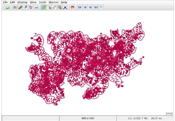
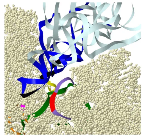
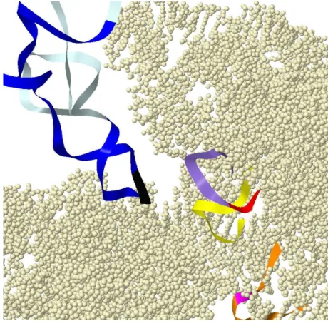
Figure 1.6: Crystallographic structure of the smaller subunit of Escherichia coli ribosome. The 3D positioning
of mRNA in green, the three tRNAs in different shades of blue and the smaller ribosomal subunit (Generated using Jmol).
Determining the structure of a protein doesn’t rely on these methods only, in most cases prior knowledge is necessary. Such as amino acid sequence and preferred geometry of atoms.
1.4 Biological databases
We will be presenting now the different biological databases used to retrieve the data on which the studies were conducted. For the first part of the study we used crystallographic data to examine if what we are searching for is in significant regions of the ribosome. In the second part of the study, we used large quantity of data of genomes to thoroughly examine the circular code in eukaryotic organisms.
12 1.4. BIOLOGICAL DATABASES
Figure 1.7: Crystallographic structure of the Escherichia coli ribosome. The 3D positioning of mRNA in green,
the three tRNAs in different shades of blue, the smaller ribosomal subunit in light brown and the large subunit in orange (Generated using Jmol).
1.4.1 Crystallographic database
Crystallographic databases are created with the goal of collecting information about the structure of molecules and crystals. The protein data bank (PDB) archive is such a database (www.rcsb.org). Structural biologists use methods such as X-ray crystallography, NMR spectroscopy, and cryo-electron microscopy to construct a 3D structure of proteins. This information is deposited in the PDB, which is then annotated and publicly released into the archive by the worldwide protein data bank (wwPDB), an organization that is responsible with maintaining the archive.
PDB holds structures for proteins and nucleic acids, such as ribosomes, onco-genes, drug targets and complete viruses. Multiple structures can exist for the same molecule depending on the test conducted or the scope of the study. The files found in the database consist in principle of, the atoms in each protein and their three-dimensional coordinates, a header that summarizes the input and the experiments in which this structure was acquired.
10 1000 1976 1978 1980 1982 1984 1986 1988 1990 1992 1994 1996 1998 2000 2002 2004 2006 2008 2010 2012 2014 2016 Year Number of Ne w Str uctures P er Y ear EM NMR Xray
Rate of Protein Structure Determination (Log Scale)
Figure 1.8: The increase of crystallographic structure submissions to the PDB according to methods. X-ray
crystallography (blue) and Nuclear Magnetic Resonance (green) were the dominant methods while Electron Microscopy (red) is becoming more popular with the years (numbers provided by PDB).
Molecular graphics software are available to visualize these files in 3D, such as Jmol, RasMol, Swiss PDB viewer, ...etc, with additional features such as measuring distances and bond angles. These software allow us to carefully study and structure and identify interesting structural features.
1.4.2 Sequence database
The National Center for Biotechnology Information (NCBI) boasts an array of biological database (www.ncbi.nlm.nih.gov), bioinformatic tools and services. Particularly we are interested in Reference Sequence (RefSeq) and Genbank databases.
RefSeq (www.ncbi.nlm.nih.gov/refseq) is a large multi-species,
non-redundant, curated sequence database consisting everything from transcripts and translation products to whole genomes. While the database is non-redundant, alternate assemblies of the same sequence can exist. RefSeq employs a strict curative process where a record may be an essentially unchanged, validated copy of the original submission, or include updated or additional information supplied
14 1.4. BIOLOGICAL DATABASES by collaborators or NCBI staff.
GenBank (www.ncbi.nlm.nih.gov/genbank), on the other hand, a
redun-dant archival database is an annotated collection of all publicly available nu-cleotide sequences and their protein translations submitted directly by individ-ual laboratories, as well as from bulk submissions from large-scale sequencing centres. GenBank continues to grow at an exponential rate, doubling every 10 months.
1.5 Summary
In this chapter we presented the nucleic acid sequences (DNA and RNA) and their composition. Then, we preceded to explain the translation process inside the ribosome and how a protein is synthesised inside a cell. Finally, we shed light on the bimolecular structure of a ribosome. This biological introduction is to help us better understand how we study circular code in a nucleic acid sequence. The 3D structure of a ribosome is vital for our first part of the study, where we focus on the decoding center of the ribosome and then move on to enlarge the scope of the study to examine the area around the decoding center and possible interactions with the tRNAs present in a ribosome at the time of translation.
We mentioned as well the nature of data we are working with. Crystallo-graphic structures were an essential part when it came to figure out the working mechanics of a ribosome. As such, crystallographic databases were vital for us to understand and study circular code motifs presence in a ribosome. Finally, the sequence database RefSeq that houses a huge amount of complete chromosomes proved excellent for us to conduct a study on the entire set of eukaryotic genomes published at the time.
In the next chapter, we will start with a brief history of codes, and how the discovery of DNA structure ushered the race to crack the genetic code. After-wards, we will present the circular code, how it was discovered and why it is an interesting study due to the many properties it has.
2
Code Theory
2.1 Introduction . . . 18 2.2 History of codes . . . 18 2.2.1 Diamond code . . . 18 2.2.2 Comma-free code . . . 19 2.2.3 Genetic code . . . 21 2.3 Circular code . . . 21 2.3.1 Circular code motifs . . . 25 2.3.2 Unitary circular codes of dinucleotides, trinucleotides andtetranucleotides . . . 26 2.4 Summary . . . 29
18 2.1. INTRODUCTION 2.1 Introduction
In this chapter we will be explaining what is circular code, how it came to be, and in order to do that we will begin with a brief history of how code theory began and its purpose. We will mention codes that were very important at the time of their inception and why they were dismissed eventually. Finally, we explain what are circular code motifs while shedding light a subclass called unitary circular codes that are interesting for us.
2.2 History of codes
The discovery of the double-helix structure of DNA (Watson and Crick, 1953)
raised the question of how to translate a 4 letter alphabet into 20 words? The first deduction was it cannot be a one-to-one mapping, and a two letter word results in 16 words, still short of 20. Therefore, the representation could not be smaller than a three letter word (trinucleotide). But that would also give 64 words, which is an excess to the 20 amino acids. Scientist shortly rushed to crack the secret of genetic expression. This lead to the publication of several codes hoping to answer this question.
2.2.1 Diamond code
The first to propose a coding scheme following the Watson-Crick structure was George Gamow, better known for his work on the Big Bang theory.
Called the diamond code (Gamow, 1954), it suggested that double-stranded
DNA acted directly as a template for assembling amino acids into proteins. Gamow envisioned the grooves in the double helix as holes that would fit the side chains of amino acids in a ”key-and-lock” fashion (Figure 2.1). Even though the diamond have four corners, only three of them are utilized because the paired bases on the horizontal diagonal are complementary. This is essence makes the diamond code a triplet code. Figure 2.1 show that there is 20 distinct holes.
Gamow reasoned that 12 holes are symmetrical, these diamonds could be flipped end-for-end or flopped side-to-side without changing their meaning. This allowed the possibility of several triplets coding for the same amino acid, thus dealing with the problem of having 64 trinucleotides and 20 amino acids.
This code had another feature that lead to its eventual dismissal. Each nucleotide was simultaneously present in three trinucleotides, for example, a
base sequence ACGT AA would result in four overlapping trinucleotides: ACG,
CGT, GT A, GT A. Which was proven to be wrong.
Figure 2.1: George Gamow's diamond code used the key-and-lock between the grooves found in DNA and the
amino acids (Gamow,1954).
2.2.2 Comma-free code
Proposed by Crick, Griffith, and Orgel, 1957, comma-free code was an
effort to study the encoding of three nucleotides {AAA, AAC, . . . , T T T } into amino acids. By excluding the four periodic permuted trinucleotides {AAA, CCC, GGG, T T T } and distributing the 60 remaining trinucleotides into 20 groups of three trinucleotides such that each group contains the set of trinu-cleotides that can be permuted from each other following the circular permuta-tion map (Definipermuta-tion 2.1). Based on this we can deduce that a comma-free code can have only one trinucleotide from each group, therefore a set contains at most 20 trinucleotides.
Notation 2.1. The nucleotides define the genetic alphabet B = {A, C, G, T }.
The set of non-empty words (words, respectively) over B is denoted by B+ (B∗, respectively). The set of the 64 words of length 3 (trinucleotides or triletters) on B is denoted by B3 = {AAA, . . . , T T T }. Let x1 · · · xn be the concatenation of the words xi for i = 1, . . . , n, the symbol ” · ” being the concatenation operator.
20 2.2. HISTORY OF CODES
Notation 2.2. In genes, there are three frames f. By convention here, the
reading frame f = 0 is established by a start codon {AT G, GT G, T GT, T T G} and the frames f = 1 and f = 2 are the reading frame f = 0 shifted by one and two nucleotides in the 5’-to-3’ (left to right) direction, respectively.
Definition 2.1. The trinucleotide circular permutation map P : B → B is
defined by P(l0 · l1· l2) = l1 · l2 · l0 for all l0, l1, l2 ∈ B, e.g. P(AT G) = T GA. The second iterate of P is denoted as P2, e.g. P2(AT G) = GAT. By extension to a trinucleotide set S, the set circular permutation map P : P(B3) → P(B3), P being the set of all subsets of B3, is defined by P(S) =
{
v|u, v ∈ B3, u ∈ S, v = P(u)}, i.e. a permuted trinucleotide set P(S) is obtained by applying the circular permutation map P to all its trinucleotides, e.g. P({ACG, AGT }) =
{CGA, GT A} and P2({ACG, AGT }) = {GAC, T AG}.
Despite having an identical number of trinucleotides as amino acids, no trin-ucleotide comma-free code was identified in genes. Early in the sixties it was discovered that the trinucleotides T T T , an excluded trinucleotide in comma-free code, in fact codes phenylalanine (Nirenberg and Matthaei, 1961), this in turn would lead to the abandonment of comma-free code.
Definition 2.2. A set S ⊂ B+ of words is a code if, for each x
1, . . . , xn, y1, . . . , ym ∈ S, n, m ≥ 1, the condition x1· · · xn = y1· · · ym implies n = m and xi = yi for i = 1, . . . , n, e.g. B3 = {AAA, ..., T T T } is a code, where as
X = {A, GC, AGC}is not a code as there are two decompositions A·GC = AGC.
Definition 2.3. (Fimmel, Michel, and Strüngmann, 2016) Let X ⊆ Bm, m ∈ N with m ≥ 2, be an m-nucleotide code. The directed graph G(X) = (V (X), E(X)) associated with X has a set of vertices V (X) and a set of edges E(X) defined as follows: V(X) = {N1. . . Ni, Ni+1. . . Nm : N1. . . Nm ∈ X, 1 ≤ i ≤ m − 1} E(X) = {[N1. . . Ni, Ni+1. . . Nm] : N1. . . Nm ∈ X, 1 ≤ i ≤ m − 1}. (2.1)
Theorem 2.1. (Fimmel, Michel, and Strüngmann, 2016) Given an m-nucleotide code X ⊆ Bm, m ≥ 2, the following statements are equivalent:
1. The code X is comma-free.
Theorem 2.2. (Fimmel, Michel, and Strüngmann, 2016) Given an m-nucleotide
code X ⊆ Bm, m ≥ 2, the code X is strong comma-free if the maximal length
of a path in G(X) is 1. 2.2.3 Genetic code
In 1961, Marshall Nirenberg and Heinrich Matthaei managed to crack the first word of the genetic code. They performed an experiment which showed that a chain of the repeating bases U (Uracil) forced a protein chain made of one repeating amino acid, phenylalanine. This was a breakthrough experiment which proved that the code could be broken.
After the initial discovery the team grew in size to replicate the poly-U ex-periment model to other amino acids. Using 20 test tubes for each amino acid, respectively, they experimented with the different 64 combination of nucleotides to form three letter words.
By 1966 the genetic code was complete, all the mappings between the 64 codons and the 20 amino acids were established. As it turns out, there was no pattern in the code. Some amino acids were represented by one or two codons, some by more, ignoring all mathematical approaches to solving this coding mys-tery.
2.3 Circular code
In 1996, a statistical analysis of occurrence frequencies of the 64 trinucleotides was conducted in the three frames (Definition 2.2), of genes of both prokaryotes and eukaryotes. The study showed that the trinucleotides are not uniformly dis-tributed in the three frames (Arquès and Michel,1996). By convention here, the frame zero is the reading frame in a gene, and the frames 1 and 2 are the reading frame 0 shifted by 1 and 2 nucleotides in the 5’-to-3’ direction, respectively. By excluding the four periodic permuted trinucleotides {AAA, CCC, GGG, T T T } and by assigning each trinucleotide to a preferential frame (frame of its highest occurrence frequency), three subsets X = X0, X1, and X2 of 20 trinucleotides each, were assigned to frame 0, 1 and 2, respectively (Table 2.1). The analysis was based on the large gene populations (protein coding regions) of eukaryotes (26,757 sequences, 11,397,678 trinucleotides) and prokaryotes (13,686 sequences, 4,709,758 trinucleotides) (Arquès and Michel, 1996). The following circular code
22 2.3. CIRCULAR CODE
Table 2.1: Frequency of trinucleotides occurrences according to frame 0, 1 and 2 in genes sequences from
eukaryotes and prokaryotes (Arquès and Michel,1996). The table shows the occurrences of the first seven
trinucleotides fromB3, with their preferred frame in bold.
Frequency(%)
Trinucleotide Frame 0 Frame 1 Frame 2
AAA 3.38 2.75 2.44 AAC 2.18 1.59 1.38 AAG 1.98 3.21 0.81 AAT 2.17 1.37 1.69 ACA 1.22 1.91 1.11 ACC 2.09 1.60 0.79 ACG 1.30 2.49 0.68 ... ... ... ...
X = {AAC, AAT, ACC, AT C, AT T, CAG, CT C, CT G, GAA, GAC,
GAG, GAT, GCC, GGC, GGT, GT A, GT C, GT T, T AC, T T C}. (2.2)
The two sets X1and X2, of 20 trinucleotides each, in the shifted frames 1 and 2 of genes can be deduced from X by the circular permutation map (Definition 2.1), i.e. X1 = P(X) and X2 = P2(X).
Definition 2.4. The nucleotide complementarity map C : B → B is defined by
C(A) = T, C(C) = G, C(T ) = A and C(G) = C. According to the property
of the complementary and anti-parallel double helix, the trinucleotide
comple-mentary map C : B3 → B3
4 is defined by C(l0 · l1 · l2) = C(l2) · C(l1) · C(l0) for all l0, l1, l2 ∈ B, e.g. C(AT G) = CAT . By extension to a trinucleotide
set S, the set complementarity map C : P(B3) → P(B3), is defined by
C(S) = {v | u, v ∈ B3, u∈ S, v = C(u)}, i.e. a complementary trinucleotide set C(S)is obtained by applying the complementarity map C to all its trinucleotides, e.g. C({ACG, AGT }) = {ACT, CGT }.
Definition 2.5. A trinucleotide code X ⊂ B3 is circular code if, for each x1, . . . , xn, y1, . . . , ym ∈ X, n, m ≥ 1, r ∈ B∗, s∈ B+, the conditions sx2· · · xnr =
y1· · · ym and x1 = rs imply n = m, r = ε (empty word) and xi = yi for
i = 1, . . . , n (Figure 2.2 for a graphical representation of the circular code defi-nition).
Theorem 2.3. (Fimmel, Michel, and Strüngmann, 2016) Given an m-nucleotide code X ⊆ Bm, m ∈ N with m ≥ 2, the following statements are equivalent:
1. The code X is circular. 2. The graph G(X) is acyclic.
Definition 2.6. An m-nucleotide unitary circular code X ⊆ Bm (UCC) is a code with a unique word.
Figure 2.2: Graphical representation of the circular
code definition (Arquès and Michel,1996).
Figure 2.3: The following code
{AAT, AT G, CCT, CT A, GCC, GGC}
is not circular, since(AT G, GCC, CT A)can
be read differently if we shift frames showing us(AAT, GGC, CCT )(Michel,2012).
Remark 2.1. A trinucleotide code C containing either one periodic permuted
trinucleotide P P T = {AAA, CCC, GGG, T T T } or two non-periodic permuted trinucleotides NP P T = {t, P(t)} for a trinucleotide t ∈ B3\P P T cannot be circular. Thus, the two trinucleotide codes B3 and B3\P P T are not circular.
Remark 2.2. The fundamental property of a circular code is the ability to retrieve
the reading (original or construction) frame of any sequence generated with this circular code. A circular code is a set of words over an alphabet such that any sequence written on a circle (the next letter after the last letter of the sequence being the first letter) has a unique decomposition (factorization) into words of the circular code (Michel, 2012) (Figure 2.3 for an example). The reading frame in a sequence (gene) is retrieved after the reading of a certain number of letters (nucleotides), called the window of the circular code. The length of this window
24 2.3. CIRCULAR CODE for retrieving the reading frame is the letter length of the longest ambiguous word, not necessarily unique, which can be read in at least two frames, plus one letter (Figure 2.4).
Figure 2.4: An example of how a window can determine in which frame the circular code motif is, if size is
sufficient (13 nucleotides). Retrieval of the reading frame of the wordw= . . . AGGT AAT T ACCAG . . .
of the trinucleotide circular codeX(Equation 2.2). Among the three possible factorizationsw˜0,w˜1andw˜2,
only one factorizationw˜1into trinucleotides ofXis possible leading to. . . A·GGT ·AAT ·T AC ·CAG·. . .
Thus, the first letterAofwis the 3rd letter of a trinucleotide ofX(Michel,2012).
Remark 2.3. At window length l > 12 nucleotides there is no ambiguous word of
the circular code X.
Definition 2.7. A trinucleotide set X1 = P(X) of a trinucleotide circular code X is permuted if, for each x ∈ X, P(x) ∈ P(X). The permuted trinucleotide set X2 = P2(X)is defined similarly.
Definition 2.8. A trinucleotide circular code X ⊂ B3 is maximal if for each x∈ B3, x∈ X, X ∪ {x}/ is not a trinucleotide circular code.
Definition 2.9. A trinucleotide circular code X is self-complementary if, for
each x ∈ X, C(x) ∈ X.
Definition 2.10. An m-nucleotide circular code X ⊆ Bm is Cn if the m
permuted m-nucleotide codes X1 = P(X), . . . , Xm = Pm(X) are circular.
An m-nucleotide comma-free X ⊆ Bm (strong comma-free, respectively) is
CFm (SCFm, respectively) if the m permuted m-nucleotide codes X1 =
P(X), . . . , Xm = Pm(X) are comma-free (strong comma-free, respectively). A trinucleotide circular code X ⊂ B3 is C3 (m = 3) if the permuted trinucleotide sets X1 = P(X) and X2 = P2(X)are trinucleotide circular codes.
Definition 2.11. A trinucleotide circular code X ⊂ B3 is C3 self-complementary if X, X1 = P(X)and X2 = P2(X)are trinucleotide circular codes satisfying the following properties X = C(X), C(X1) = X2 and C(X2) = X1.
Table 2.2: The number of sets that can be circular for each class of trinucleotide (Arquès and Michel,1996;
Michel and Pirillo,2010; Michel, Pirillo, and Pirillo,2012).
Circular code (maximal) Number
Potential 3 486 784 401
Identified 12 964 440
Self-complementary 528
C3 221 328
C3 Self-complementary 216
A circular code containing 20 codons is designated as maximal (Definition 2.8). The number possible maximal circular codes is 320 out of 6420, i.e. prob-ability of 10−27, only 12 964 440 are effectively circular codes. Which makes finding these three circular code sets in genes quite intriguing. These three trinucleotide sets posses several strong mathematical properties. They have the fundamental property to always retrieve the reading frame in any position of any sequence generated with the circular code. Initiation and stop trinucleotides, as well any frame signals are not necessary to define the reading frame. A win-dow of 13 nucleotides allows to retrieve the reading frame for all the ambiguous words generated with X. Therefore, circular codes are less constrained than the comma-free codes. Moreover, the X0 code is in particular very interesting since it is symmetric under the complementary transformation. In other words, we exchange every trinucleotide in the set with its complement (Definition 2.4), the
set remains unchanged. While the X1 and X2 sets are complementary to each
other.
2.3.1 Circular code motifs
A circular code motif is a phrase composed by words, in this case trinucleotides, from a circular code. Consequently, an X motif can be found in any DNA or RNA sequence, where we have successive trinucleotides from the X. As mentioned above, a window of 13 nucleotides is sufficient to distinguish a circular code motif in any given frame. Let us examine the following example:
Example 2.1.
Sequence: AGT CAGT AGCT GAGGCAGCT CGAAAT T CGT
26 2.3. CIRCULAR CODE
Consider the DNA sequence in example 2.1. If we read this
se-quence in frame 0, we can see the four following consecutive trinu-cleotides {CAG, CT C, GAA, AT T } belonging to X (Equation 2.2). Therefore,
CAGCT CGAAAT T is an X motif that starts at nucleotide 16 and ends at
nucleotide 27 according to the sequence.
Definition 2.12. An m-nucleotide unitary circular code motif (UCC motif)
generated by an m-nucleotide unitary circular code (Definition 2.6), is a con-catenation of n words w of size m denoted wn. The class of the motifs wn for all n is denoted by m+.
Definition 2.13. Two UCC motifs w+
1 and w2+are said equivalent if w1+ and w2+ are related by the circular permutation map P (Definition 2.1).
2.3.2 Unitary circular codes of dinucleotides, trinucleotides and tetranucleotides
The 42 − 4 = 12 dinucleotide unitary codes Di = {l
1l2} with l1l2 ∈ B and l1 ̸= l2 are circular and C2 . The 43 − 4 = 60 trinucleotide unitary codes
Tri = {l1l2l3} with l1, l2, l3 ∈ B and l1l2 ̸= l2l3 are also circular and C3. The 44−16 = 240tetranucleotide unitary codes Tetra = {l
1l2l3l4}with l1, l2, l3, l4 ∈ B and l1l2 ̸= l3l4are also circular and C4(excluding {l1l2l1l2}since it is not circular, (l1l2)2 is a dinucleotide repeat of {l1l2}). We describe some additional stronger combinatorial properties for the codes Di, Tri and Tetra. From Theorem 2.2, an m-nucleotide unitary code {li. . . li} with li ∈ B, i.e. starting and ending by the same letter, cannot be strong comma-free.
2.3.2.1 Unitary circular codes of dinucleotides
The 12 dinucleotide unitary codes {l1l2} and {P (l1l2)} = {l2l1} with l1, l2 ∈ B and l1 ̸= l2, i.e. {AC}, {AG}, {AT }, {CA}, {CG}, {CT }, {GA}, {GC}, {GT }, {T A}, {T C} and {T G}, are strong comma-free (SCF ) by Theorem 2.2. Thus, a dinucleotide unitary strong comma-free code {l1l2}has a permuted code {l2l1} which is also strong comma-free (SCF2 property, see Definition 2.10). Further-more, the four dinucleotide unitary strong comma-free codes {AT }, {CG}, {GC} and {T A} are self- complementary.
2.3.2.2 Unitary circular codes of trinucleotides
The 24 trinucleotide unitary codes {l1l2l3}, {P(l1l2l3)}= {l2l3l1}and {P2(l1l2l3)} = {l3l1l2} with l1, l2, l3 ∈ B and l1 ̸= l2 ̸= l3 , i.e. {ACG}, {ACT }, {AGC}, {AGT }, {AT C}, {AT G}, {CAG}, {CAT }, {CGA}, {CGT }, {CT A}, {CT G}, {GAC}, {GAT }, {GCA}, {GCT }, {GT A}, {GT C}, {T AC}, {T AG}, {T CA}, {T CG}, {T GA} and {T GC}, are strong comma-free (SCF ) by Theorem 2.2. Thus, a trinucleotide unitary strong comma-free code {l1l2l3}has two permuted codes {l2l3l1}and {l3l1l2}which are also strong comma-free (SCF3 property, see Definition 2.10).
The 24 trinucleotide unitary codes {l1l1l2} and {P2(l1l1l2)} = {l2l1l1} with
l1, l2 ∈ B and l1 ̸= l2 , i.e. {AAC}, {AAG}, {AAT }, {ACC}, {AGG},
{AT T }, {CAA}, {CCA}, {CCG}, {CCT }, {CGG}, {CT T }, {GAA}, {GCC}, {GGA}, {GGC}, {GGT }, {GT T }, {T AA}, {T CC}, {T GG}, {T T A}, {T T C} and {T T G}, are strong comma-free (SCF ) by Theorem 2.2. The 12 trinucleotide unitary codes {P(l1l1l2)} = {l1l2l1} with l1, l2 ∈ B and l1 ̸= l2 , i.e. {ACA}, {AGA}, {AT A}, {CAC}, {CGC}, {CT C}, {GAG}, {GCG}, {GT G}, {T AT },
{T CT } and {T GT }, are comma-free (CF ) by Theorem 2.1. Thus, a
trinu-cleotide unitary strong comma-free code {l1l1l2} has one permuted code {l2l1l1} which is also strong comma-free and one permuted code {l1l2l1}which is comma-free code. Corollary, a trinucleotide unitary comma-comma-free code {l1l2l1} has two permuted codes {l1l1l2} and {l2l1l1}which are strong comma-free.
2.3.2.3 Unitary circular codes of tetranucleotides
The 24 tetranucleotide unitary codes {l1l2l3l4}, {P(l1l2l3l4)} = {l2l3l4l1}, {P2(l
1l2l3l4)} = {l3l4l1l2} and {P3(l1l2l3l4)} = {l4l1l2l3} with l1, l2, l3, l4 ∈ B and l1 ̸= l2 ̸= l3 ̸= l4 are strong comma-free (SCF ) by Theorem 2.2. Thus, a tetranucleotide unitary strong comma-free code {l1l2l3l4} has three permuted codes {l2l3l4l1}, {l3l4l1l2}and {l4l1l2l3}which are also strong comma-free (SCF4 property, see Definition 2.1). The 48 tetranucleotide unitary codes {l1l2l1l3}, {P(l1l2l1l3)}= {l2l1l3l1}, {P2(l1l2l1l3)}= {l1l3l1l2}and {P3(l1l2l1l3)}= {l3l1l2l1} with l1, l2, l3 ∈ B and l1 ̸= l2 ̸= l3 are strong comma-free (SCF ) by Theorem 2.2. Thus, a tetranucleotide unitary strong comma-free code {l1l2l1l3}has three per-muted codes {l2l1l3l1}, {l1l3l1l2}and {l3l1l2l1} which are also strong comma-free (SCF4 property).
28 2.3. CIRCULAR CODE {P3(l
1l1l2l3)} = {l3l1l1l2} with l1, l2, l3 ∈ B and l1 ̸= l2 ̸= l3 are strong comma-free (SCF ) by Theorem 2.2. The 24 tetranucleotide unitary codes {P(l1l1l2l3)} = {l1l2l3l1}with l1, l2, l3 ∈ B and l1 ̸= l2 ̸= l3 are comma-free (CF ) by Theorem 2.1. Thus, a tetranucleotide unitary strong comma-free code {l1l1l2l3} has two permuted codes {l2l3l1l1} and {l3l1l1l2} which are also strong comma-free and one permuted code {l1l2l3l1} which is comma-free. Corollary, a tetranucleotide unitary comma-free code {l1l2l3l1} has three permuted codes {l1l1l2l3}, {l2l3l1l1} and {l3l1l1l2} which are strong comma-free.
The 24 tetranucleotide unitary codes {l1l1l1l2} and {P3(l1l1l1l2)}= {l2l1l1l1} with l1, l2 ∈ B and l1 ̸= l2 are strong comma-free (SCF ) by Theorem 2.2. The 24 tetranucleotide unitary codes {P(l1l1l1l2)} = {l1l1l2l1} and {P2(l1l1l1l2)} = {l1l2l1l1}with l1, l2 ∈ B and l1 ̸= l2 are comma-free (CF ) by Theorem 2.1. Thus, a tetranucleotide unitary strong comma-free code {l1l1l1l2} has one permuted code {l2l1l1l1}which is also strong comma-free and two permuted codes {l1l1l2l1} and {l1l2l1l1}which are free. Corollary, a tetranucleotide unitary comma-free code {l1l1l2l1}has one permuted code {l1l2l1l1}which is also comma-free and two permuted codes {l1l1l1l2} and {l2l1l1l1} which are strong comma-free. The 12 tetranucleotide unitary codes {l1l1l2l2} and {P2(l1l1l2l2)} = {l2l2l1l1} with l1, l2 ∈ B and l1 ̸= l2 are strong comma-free (SCF ) by Theorem 2.2.
The 12 tetranucleotide unitary codes {P(l1l1l2l2)} = {l1l2l2l1} and {P3(l
1l1l2l2)} = {l2l1l1l2} with l1, l2 ∈ B and l1 ̸= l2 are comma-free (CF ) by Theorem 2.1. Thus, a tetranucleotide unitary strong comma-free code {l1l1l2l2} has one permuted code {l2l2l1l1} which is also strong comma-free and two per-muted codes {l1l2l2l1}and {l2l1l1l2}which are comma-free. Corollary, a tetranu-cleotide unitary comma-free code {l1l2l2l1} has one permuted code {l2l1l1l2} which is also comma-free and two permuted codes {l1l1l2l2} and {l2l2l1l1}which are strong comma-free.
Furthermore, the 12 tetranucleotide unitary strong comma-free codes {AAT T }, {ACGT }, {AGCT }, {CAT G}, {CCGG}, {CT AG}, {GAT C}, {GGCC}, {GT AC}, {T CGA}, {T GCA} and {T T AA} are self-complementary. There is no tetranucleotide unitary comma-free code which is self-complementary.
2.4 Summary
We briefly gave the history of the genetic code, starting from its root with the discovery of the structure of DNA, through the various code theories, such as diamond code and comma-free code, that were proposed to help solve the mystery of nucleotides coding for amino acids. This shows us how circular code was discovered in 1996, why it is still relevant and an interesting study subject even though the genetic code has been already discovered and proven. We presented the circular code with its features and properties. We defined the unitary circular code, explaining how simple repeats are circular code. This allows us to study the large amount of sequences that has low-complexity DNA.
In the next chapter we will present the data that was retrieved for ribosomal RNA and eukaryotic genomes. For each environment we developed different algorithms to extract the results we need. Furthermore, several methods were adopted to help use analyse the results obtained. For this we divided our context into: (i) X circular code motifs in ribosomal RNA, (ii) X circular code motifs in eukaryotic genomes (iii) unitary circular codes (simple repeats) in eukaryotic genomes and (iv) trinucleotide pairs in gene sequences.
3
Data and Methods
3.1 Introduction . . . 33 3.2 Data acquisition . . . 33 3.2.1 Ribosomal data . . . 33 3.2.2 Genomic database . . . 34 3.3 Circular code motifs . . . 36 3.3.1 Search algorithms . . . 36 3.3.1.1 Biinfinite word . . . 36 3.3.1.2 Sets algorithm . . . 37 3.3.1.3 Frames algorithm . . . 41 3.3.2 Definition of random code motifs . . . 42 3.3.3 Definition of the 23 bijective circular codes motifs . . . 43 3.3.3.1 Bijective transformation circular codes . . . 43 3.3.3.2 Main properties of the 23 bijective transformation
circular codes . . . 47 3.3.4 Statistical analysis of X circular code motifs . . . 47 3.3.4.1 Coverage of X motifs in tRNA . . . 47 3.3.4.2 Expectation of the occurrence number of a motif . . 48 3.3.4.3 Ratio of X motifs in coding and non-coding regions . 48 3.4 Ribosome study tools . . . 50 3.4.1 Multiple sequence alignment . . . 50 3.4.2 Molecule viewer . . . 50 3.5 Repeated motifs . . . 52
32
3.5.1 Dinucleotide unitary circular code motifs . . . 52 3.5.2 Trinucleotide unitary circular code motifs . . . 52 3.5.3 Tetranucleotide unitary circular code motifs . . . 53 3.5.4 Statistical analysis of repeated motifs . . . 53 3.5.4.1 Occurrence number of unitary circular code motifs . 53 3.5.4.2 Base number of unitary circular code motifs . . . 55 3.5.4.3 Total base number of unitary circular code motifs . . 55 3.6 Occurrence number of trinucleotide pairs . . . 56 3.7 Summary . . . 59
3.1 Introduction
Previously, we explained the biological context in which we are working and the type of data needed for our study. Now we will show the actual data, how it was obtained, processed and dealt with it.
An extensive collection of classes and algorithms were written to handle our data, from reading PDB, alignments and huge FastA files, to searching sequences for motifs, all the way to the statistical tests applied to the found motifs. Our code has been refined and optimized to handle huge flat files with fast processing time.
We grouped together the search algorithms and statistical analysis tools de-pending on topic. We have a group that handles X circular code motifs and its comparison with various other codes. While another group aim at the study of unitary circular codes, otherwise known as simple repeats. Two sections handle the tools used for ribosome examination and another that studies the trinu-cleotide pairing in gene sequences.
3.2 Data acquisition
The used in this work can be divided along two different studies. The first focused on spatial interaction of circular code motifs, for which it required structural data of ribosomes from PDB archive. The second study concerned the search of circular code motifs on a genomic scale, which were retrieved from RefSeq database.
3.2.1 Ribosomal data
To study structural significance of circular code motifs, we collected ribosomal
data from the before mentioned PDB archive in 2014 (Section 1.4.1, www.rcsb.
org/pdb). The selected PDB entries have necessarily a bacterial 16S rRNA or a eukaryotic 18S rRNA. An entry from each organism having this criteria was chosen, with preference going towards entries having an mRNA and all or any tRNAs. This emphasize allows us to better study the spatial interaction of X motifs from rRNA, mRNA and tRNA (Section 4.2).
The studied PDB crystallographic structures are two bacterial entries:
Es-cherichia coli (Brilot et al., 2013) and Thermus thermophilus (Jenner et al.,
2010); one archaea: Pyrococcus furiosus (Armache et al., 2013); three nuclear