HAL Id: tel-01292379
https://tel.archives-ouvertes.fr/tel-01292379
Submitted on 23 Mar 2016
HAL is a multi-disciplinary open access
archive for the deposit and dissemination of
sci-entific research documents, whether they are
pub-lished or not. The documents may come from
teaching and research institutions in France or
abroad, or from public or private research centers.
L’archive ouverte pluridisciplinaire HAL, est
destinée au dépôt et à la diffusion de documents
scientifiques de niveau recherche, publiés ou non,
émanant des établissements d’enseignement et de
recherche français ou étrangers, des laboratoires
publics ou privés.
UNIVERSITE PARIS-SUD
ÉCOLE DOCTORALE
Sciences et Technologie de l’Information, des Télécommunications
et des Systèmes
Laboratoire d’Imagerie par Résonance Magnétique Médicale et Multi-Modalité
DISCIPLINE : PHYSIQUE
THÈSE DE DOCTORAT
Soutenance le 22/09/2014
par
Pablo MILIONI DE CARVALHO
Low-Dose 3D Quantitative Vascular X-ray Imaging
of the Breast
Composition du jury :
Directeur de thèse : Serge MULLER Directeur de Recherche (GE Healthcare)
Rapporteurs : Martin YAFFE Professeur (Faculty of Medicine, Univ. of Toronto) Ioannis SECHOPOULOS Professeur Adjoint (Emory Univ. School of Medicine)
Examinateurs : Irène BUVAT Directeur de Recherche (IMNC, Université Paris Sud) Alain NOEL Chercheur (CRAN, Université de Loraine)
Luc DARRASSE Directeur de Recherche (IR4M, Université Paris Sud) Ann-Katherine CARTON Chercheur (GE Healthcare)
PhD Dissertation: Final Version
October 21, 2014
Acknowledgements
This PhD thesis research work was funded by GE Healthcare (Buc, France) in collaboration with
IR4M laboratory based at the Universit´e Paris Sud (Orsay, France), and with the financial
sup-port of the ANRT (Association Nationale de la Recherche Technique) under CIFRE convention n° 2010/756.
I am very thankful to everyone who has directly or indirectly helped me in the completion of my thesis. I would like to begin by thanking my thesis advisor, Dr. Serge Muller, as well my academic co-advisor, Dr. Luc Darrasse, for the opportunity of working under their supervision and for welcoming me into their remarkable research groups. Thank you Serge for all the confidence you put in me, from the old days as an intern until my recent official integration in the team. You always inspired me to go further than I thought possible. I wish to convey my deepest gratitude to Dr. Ann-Katherine Carton, who has been my daily scientific advisor, for her continuous encouragement and support, the never-ending scientific discussions as well as the valuable advices and contributions she made to this thesis. I am indebted to her more than she knows.
I would like to thank Dr. Martin Yaffe and Dr. Ioannis Sechopoulos who accepted to review this manuscript. Thank you for the precious time you spent reading this thesis and the valuable
comments you provided. I would like to extend my thanks to Dr. Ir`ene Buvat and Dr. Alain No¨el
for accepting being members of my jury.
I wish to express my gratitude to the other members of the BCARe team. Thanks to Dr. R˘azvan
Iordache and Dr. Sylvie Saab-Puong, close collaborators who provided important feedback on my
research work. Thanks also to Giovanni, Laurence, Nausika¨a, Anja and Zhijin for all the good
moments we shared during these last years, both inside and outside the office. I would like to extend my gratitude to the many colleagues at Buc. Thanks to Dr. Remy Klauz for the numerous discussions and helpful advices, fruits of a wide experience in X-ray imaging. I would like to express my gratitude to Drs. Cyril Riddell and Vincent Bismuth, who provided me with critical insight on 3D reconstruction and image processing, helping me progress in my investigations. Thanks to Dr. Lilane Ramus, Mathias Cisaruk and Matthieu Guillard, for helping to acquire the data on the interventional guided system. And thanks to Xavier, Matthieu, Yann, Colin, Fred, Richard, Heitor, Laure, Greg, and all others GE colleagues I did not mention. Thank you for your friendship and for making of Buc a wonderful work environment.
I would like to thank the members of IR4M for welcoming me in the lab. Thanks Drs. Xavier Maˆıtre and Luc Darrasse for the helpful guidance and feedback during these three years. Thank you Dr. Jean-Christophe Ginefri and Albine Pinseel for the administrative support at the university. Thanks my fellow PhD students at IR4M, in particular Marion and Maya. I owe special thanks to Dr. Clarisse Dromain, for her numerous clinical advices in contrast-enhanced imaging and breast
anatomy. I would like to thank Dr. Sandra Canal´e and Ariane Dunant, from Gustave Roussy
cancer institute, for their cooperation during the reader study.
I would like to express my gratitude to the members of the CatSim collaboration. In particular, I would like to thank Dr. Bruno de Man and Dr. Paul Fitzgerald from GE Global Research Center (GRC), who have maintained the CatSim project alive and cooperated with my work in improving the simulation platform. I would like to thank Dr. Jed Pack for his feedback on simulation aspects,
Summary
Background: Worldwide, breast cancer is the most common cancer and second deadliest can-cer in women. Diagnostic imaging techniques are a critical part for screening, diagnosis, tumor staging and cancer therapy of the breast, guiding clinicians towards a more effective treatment planning and resulting in a better health outcome for the patient. Contrast-Enhanced Magnetic Resonance Imaging (CE-MRI) is the current standard imaging technique allowing detection of abnormal vascular development and lesion contrast uptake. CE-MRI is however very costly and not widely available. Moreover, its spatial resolution might not be sufficient to depict certain types of lesions including microcalcifications, whose presence is an important diagnostic indicator. It has been demonstrated that in combination with an iodinated vascular contrast agent, contrast-enhanced X-ray imaging can also give morphological and functional images. The development of Contrast-Enhanced Spectral Mammography (CESM) has made the clinical use of intravenous con-trast enhancement with conventional mammography possible. However, CESM is a 2D projection technique and therefore presents limitations to depict the 3D internal structures of lesions and to provide accurate quantitative 3D functional information.
Contrast-Enhanced Digital Breast Tomosynthesis (CE-DBT) and dedicated Contrast-Enhanced Breast CT (CE-bCT) are two breast imaging modalities currently under investigation by academic and industrial research groups. It is however anticipated that the quantitative potential of CE-DBT is limited, due to the inherent low depth-resolution of limited opening angle CE-DBT modality. CE-bCT with quasi-isotropic spatial resolution and voxel signal intensity proportional to the linear attenuation coefficient is believed to offer more accurate quantitative information, though a low-dose operation is still a challenge.
Objectives: The purpose of this thesis has been to study the technical feasibility of CE-bCT and its potential to accurately depict and localize tumors, as well as to provide accurate quantitative morphological and functional imaging information about tumors, at low radiation dose levels. To understand the incremental value of CE-bCT over CE-DBT, the quantitative potential of both technologies have been compared. This investigation has been performed through computer simulations.
Methods: At first, a simulation platform capable of modeling various X-ray breast imaging techniques and providing radiographic images of simple and complex computational phantoms was developed and validated. Secondly, an optimization study of a CE-bCT technique based on a dual-energy approach was performed, aiming to maximize image quality of iodine-enhanced and morphological images. Finally, the quantitative potential of CE-bCT and CE-DBT was compared through the assessment of iodine-enhanced lesion detectability, characterization, localization and 3D extent measurement. In a human observer study, depiction and characterization of iodine-enhanced lesions of different sizes, shapes and iodine uptakes was compared between CE-bCT and CE-DBT using a mesh-based anthropomorphic breast phantom.
Results: Simulation results assuming ideal detectors showed that to obtain optimal iodine de-tectability in recombined dual-energy images, low energy (LE) and high energy (HE) spectra need to bracket the iodine K-edge and an approximate 50%-50% average glandular dose repartition-ing between the LE and HE is required. Comparison between different polychromatic spectral acquisition strategies revealed that iodine K-edge imaging with both energy-discriminating and
essentially round lesions, CE-DBT is more accurate and more precise in estimating lesion dimen-sions than CE-bCT, with image noise being the determinant factor in the comparison.
Conclusions: The simulation studies in this PhD thesis suggest that dual-energy iodine-injected CE-bCT could be a feasible technique for breast tumor depiction, localization and characterization, with dose levels comparable to standard mammography. While preliminary comparisons with CE-DBT suggests comparable depiction and characterization performance, the fully 3D information combined with high spatial resolution confirms CE-bCT as an interesting low-dose evolution of CESM toward 3D quantitative assessment of contrast uptakes and potential alternative to CE-MRI for some clinical indications.
Keywords: breast, computed tomography, simulation, dual-energy, spectral optimization, image quality, observer study
R´
esum´
e
Contexte : Le cancer du sein est le cancer le plus fr´equent et le deuxi`eme cancer le plus
mor-tel chez la femme. Les techniques d’imagerie constituent un ´el´ement essentiel pour le d´epistage,
le diagnostic, la stadification et le traitement du cancer du sein. Elles guident les cliniciens vers
une planification du traitement plus efficace, entrainant de meilleurs r´esultats sur la sant´e de la
patiente. L’imagerie par r´esonance magn´etique avec injection de produit de contraste (CE-MRI)
est actuellement la technique d’imagerie standard pour la d´etection du d´eveloppement vasculaire
anormal et des prises de contraste des l´esions mammaires. CE-MRI est cependant tr`es coˆuteuse et
peu disponible. De plus, sa r´esolution spatiale pourrait ˆetre insuffisante pour la d´etection de
cer-tains types de l´esions, et ne permet pas d’imager les amas de microcalcifications, dont la pr´esence
est un important indicateur diagnostic. Il a ´et´e d´emontr´e que l’imagerie du sein par rayons X en
combinaison avec l’injection d’un produit de contraste peut ´egalement fournir des images
morpho-logiques et fonctionnelles. Le d´eveloppement de l’angiomammographie double-´energie (CESM) a
permis l’utilisation des produits de contraste intraveineux en clinique avec des appareils
conven-tionnels de mammographie. Cependant, CESM est une technique de projection 2D et pr´esente,
par cons´equence, des limites pour d´ecrire la structure 3D interne des l´esions et pour fournir une
information fonctionnelle 3D pr´ecise.
La tomosynth`ese num´erique du sein avec injection de produit de contraste (CE-DBT) et le scanner
d´edi´e du sein avec injection de produit de contraste (CE-bCT) sont deux techniques d’imagerie
actuellement en investigation par des groupes de recherche acad´emiques et industriels. Il est
cepen-dant anticip´e que le potentiel quantitatif de la CE-DBT soit limit´e, en raison de la faible r´esolution
en profondeur due `a l’ouverture angulaire limit´ee de la DBT. CE-bCT, avec sa r´esolution spatiale
quasi-isotrope et son intensit´e de signal proportionnelle au coefficient d’att´enuation lin´eaire, est suppos´ee offrir une information quantitative plus pr´ecise, bien qu’une utilisation `a faible dose de radiation reste toujours un d´efi.
Objectifs : L’objectif de cette th`ese a ´et´e d’´etudier la faisabilit´e de la m´ethode CE-bCT et sa capacit´e `a d´etecter et localiser des tumeurs vascularis´ees, ainsi que d’offrir de l’information
morphologique et fonctionnelle pr´ecise sur les tumeurs. Pour comprendre la valeur ajout´ee de
la CE-bCT par rapport `a CE-DBT, le potentiel quantitatif des deux m´ethodes a ´egalement ´et´e
compar´e. Nos ´etudes ont ´et´e r´ealis´ees grˆace `a des simulations par ordinateur, valid´ees par des
mesures exp´erimentales.
M´ethodes : Dans un premier temps, une plateforme de simulation capable de mod´eliser diff´erentes
techniques d’imagerie du sein par rayons X, et fournissant des images radiographiques de fantˆomes
num´eriques simples et complexes, a ´et´e impl´ement´ee et valid´ee. Deuxi`emement, une ´etude d’op-timisation pour la technique CE-bCT bas´ee sur une approche double-´energie a ´et´e r´ealis´ee, dans
le but de maximiser la qualit´e des images ´equivalentes-iode ainsi que des images morphologiques.
Enfin, le potentiel quantitatif des m´ethodes CE-bCT et CE-DBT a ´et´e compar´e au travers de
l’´evaluation de la d´etectabilit´e, de la caract´erisation, de la localisation et de la mesure de l’´etendue 3D des l´esions iod´ees. Dans une ´etude impliquant des observateurs humains, la d´etectabilit´e et la caract´erisation des l´esions iod´ees de diff´erentes tailles, formes et concentrations ont ´et´e compar´ees
entre CE-bCT et CE-DBT, grˆace `a l’utilisation d’un fantˆome anthropomorphique num´erique du
sein.
En ce qui concerne la comparaison entre les m´ethodes CE-bCT et CE-DBT, une ´etude de
quan-tification a d´emontr´e que la CE-bCT est sup´erieure pour la quantification de la concentration de
l’iode, tandis qu’en CE-DBT les artefacts caus´es par la r´esolution limit´ee en profondeur empˆechent
toute quantification pr´ecise de la prise de contraste (au moins, sans avoir une connaissance a priori
des dimensions des l´esions selon les trois axes). Dans une ´etude avec des observateurs humains,
les r´esultats ont r´ev´el´e que la sensibilit´e et la sp´ecificit´e de d´etectabilit´e et de caract´erisation des l´esions prenant le contraste n’ont pas ´et´e statistiquement diff´erentes entre les deux m´ethodes, `a
l’exception de la caract´erisation du contour des l´esions pour laquelle CE-DBT a d´emontr´e une
sensibilit´e sup´erieure. Dans la mˆeme ´etude, les r´esultats ont ´egalement montr´e que pour les l´esions
essentiellement rondes, CE-DBT est plus pr´ecise dans l’estimation des dimensions des prises de
contraste, le bruit ´etant le facteur d´eterminant dans la comparaison.
Conclusions : Les ´etudes de simulation men´ees pendant cette th`ese sugg`erent que le scanner d´edi´e du sein avec injection de produit de contraste iod´e pourrait ˆetre une technique r´ealisable pour la d´etection, localisation et caract´erisation des tumeurs du sein, pour un niveau de dose comparable `a
une mammographie standard. Bien que les comparaisons pr´eliminaires avec CE-DBT sugg`erent une
performance comparable sur la d´etection et caract´erisation, l’information 3D compl`ete combin´ee
avec une haute r´esolution spatiale font de CE-bCT une ´evolution int´eressante de CESM vers une
´
evaluation quantitative 3D des prises de contraste, et une alternative potentielle `a CE-MRI pour
certaines indications cliniques.
Mots-cl´es : sein, tomodensitom´etrie, simulation, double-´energie, optimisation spectrale, qualit´e
Contents
Acknowledgements i
Summary iii
R´esum´e v
Table of Contents vii
List of Figures xi
List of Tables xv
List of Acronyms xvii
Introduction 1
1 Clinical Context 5
1.1 Breast Anatomy and Cancer Development . . . 5
1.2 Standard Vascular Imaging Techniques for Breast Cancer Treatment . . . 10
1.2.1 Contrast-Enhanced Magnetic Resonance Imaging . . . 10
1.2.2 Contrast-Enhanced Digital Mammography . . . 11
1.2.3 Other Vascular Imaging Techniques Used in Clinical Practice . . . 12
1.3 Novel X-Ray Techniques for Vascular Imaging of the Breast . . . 14
1.3.1 Contrast-Enhanced Digital Breast Tomosynthesis . . . 14
1.3.2 Contrast-Enhanced Dedicated Breast CT . . . 14
2 3D Breast X-ray Imaging Simulation: evolution of CatSim 19 2.1 Coordinate System and Acquisition Geometry Definition . . . 21
2.2 X-ray Source Modeling and Emitting Fluence Spectra . . . 23
2.2.1 X-ray Source Modeling . . . 23
2.2.2 Pre-patient X-ray Fluence Spectrum Calculation . . . 24
2.3 Computational Human Breast Phantoms . . . 27
2.3.1 Analytic Models . . . 27
2.3.2 Meshed Models . . . 28
2.3.3 Voxelization of Analytic and Meshed-Based Models . . . 29
2.4 X-ray Projection . . . 31
2.5 Monte Carlo Simulation of X-ray Photon Scattering . . . 34
2.5.1 Overview on the Hybrid Analytic-MC Method . . . 35
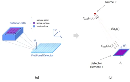
2.5.2 Detector Incident Scattered Photon Fluence Calculation . . . 37
2.6 Monte Carlo Simulation of Absorbed Radiation Dose . . . 38
3.2.3 Peak Scatter-to-Primary Ratio Values . . . 57
3.3 Glandular Dose Simulation Validation . . . 62
3.3.1 Conventional Mammography Geometry . . . 62
3.3.2 Breast CT Geometry . . . 64
3.4 Detector Model Validation . . . 67
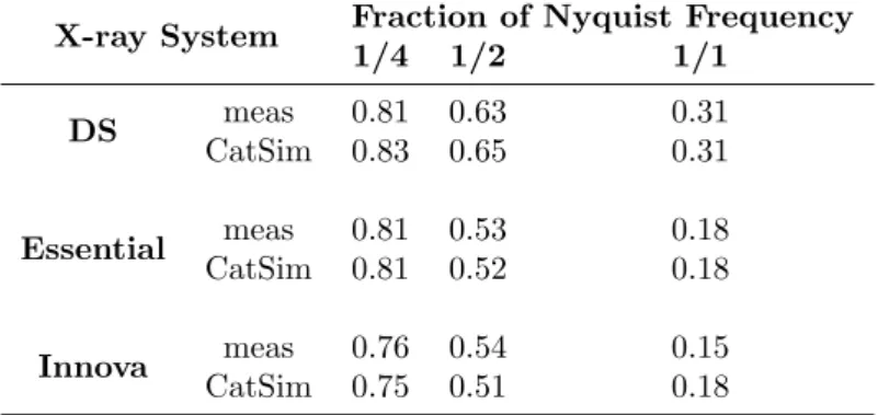
3.4.1 Modulation Transfer Function . . . 67
3.4.2 Noise Power Spectrum . . . 70
3.4.3 Image Signal Descriptive Statistics . . . 73
3.5 Conclusions . . . 81
4 Iodine-Enhanced Breast X-ray Imaging Framework 83 4.1 Basis Material Decomposition . . . 84
4.2 Spectral Imaging . . . 84
4.3 Acquisition Strategies to Obtain Dual-Energy Data . . . 89
4.4 Iodine-Enhanced Breast X-ray Imaging . . . 93
4.4.1 Dual-Energy Recombination for CE-DBT . . . 93
4.4.2 Dual-Energy Recombination for CE-bCT . . . 94
4.5 Discussion . . . 98
5 Spectral Optimization of Dual-Energy Contrast-Enhanced Breast CT 101 5.1 Optimization Critical-to-Quality Factors . . . 103
5.1.1 IQ Assessment in Recombined Iodine-Equivalent Images . . . 104
5.1.2 IQ Assessment in LE Morphologic Images . . . 105
5.2 Cone-Beam Breast CT Acquisition Geometry . . . 107
5.3 Monochromatic Spectra Optimization . . . 109
5.3.1 Optimization Method . . . 109
5.3.2 Research Space and Data Analysis . . . 110
5.3.3 Optimal Spectra and Dose Allocation . . . 111
5.3.4 Discussion . . . 115
5.4 Polychromatic Spectra Optimization . . . 116
5.4.1 Candidate Acquisition Strategies for DE-bCT . . . 116
5.4.2 Optimization Method . . . 117
5.4.3 Research Space and Data Analysis . . . 119
5.4.4 Optimal Acquisition Parameters for Iodine Uptake Depiction . . . 122
5.4.5 Discussion . . . 127
5.5 Performance Comparison Between Candidate Dual-Energy Acquisition Strategies . 129 5.5.1 Comparison Methodology . . . 129
5.5.2 Optimal Acquisition Parameters from Constrained Optimization . . . 130
Contents ix
5.5.4 Discussion . . . 134
5.6 Impact of CsI-Scintillator Thickness on Optimal Spectra . . . 137
5.6.1 Energy-Dependent Absorption Efficiency . . . 137
5.6.2 Optimization Framework . . . 138
5.6.3 Optimal Spectra and Dose Allocation for Iodine Uptake Depiction . . . 138
5.6.4 Discussion . . . 138
5.7 Conclusions . . . 141
6 Quantitative Comparison Between CE-bCT and CE-DBT 143 6.1 Iodine-Enhanced Lesion 3D Extent Estimation in CE-bCT vs CE-DBT . . . 144
6.1.1 Breast Phantoms and X-ray Image Simulation . . . 144
6.1.2 Lesion Extent Estimation and Quantitative Analysis Method . . . 145
6.1.3 Effect of CE-bCT vs CE-DBT Topologies . . . 146
6.1.4 Discussion . . . 148
6.2 Iodine Uptake Quantification in CE-bCT vs CE-DBT . . . 151
6.2.1 Breast Phantoms and X-ray Image Simulation . . . 151
6.2.2 Figure-of-Merit for Iodine Quantity Estimation . . . 151
6.2.3 Effect of CE-bCT vs CE-DBT Topologies on Quantification Accuracy . . . 152
6.2.4 Discussion . . . 152
6.3 Iodine-Enhanced Lesion Detectability and Characterization in CE-bCT vs CE-DBT: a Human Observer Study . . . 154
6.3.1 Image Database . . . 154
6.3.2 Observers . . . 156
6.3.3 Viewing Conditions . . . 158
6.3.4 Preference Study Questionnaire . . . 159
6.3.5 Reader Training . . . 160
6.3.6 Statistical Analysis . . . 160
6.3.7 Results - Part I: Preference Scale Usage and Interobserver Agreement . . . 162
6.3.8 Results - Part II: Detectability and Characterization in CE-bCT vs CE-DBT 165 6.3.9 Discussion . . . 169
6.4 Conclusions . . . 172
7 Towards Low-Dose Fully 3D Quantitative Breast X-ray Imaging 175 7.1 Anti-Correlated Noise Reduction . . . 175
7.2 Total-Variation Regularization . . . 179
7.3 Assessment of Iodine-Enhanced Lesion Detectability Improvement in CE-bCT . . . 181
7.4 Conclusions . . . 184
Conclusions and Perspectives 185 A Fundamentals of 3D X-ray Imaging 191 A.1 X-ray Production . . . 191
A.2 X-ray Interactions with Matter in Diagnostic Imaging . . . 192
A.3 X-ray Detection . . . 194
A.4 Acquisition Gantry . . . 197
A.5 Tomographic Reconstruction . . . 197
B Reference Apparatus 203 C Scattered Radiation Impact on Image Quality 205 C.1 Materials and Methods . . . 206
C.2 Results . . . 207
List of Figures
1-1 Illustration of the Breast Anatomy . . . 6
1-2 Incidence and mortality rates in France per cancer type in 2011 . . . 8
1-3 Evolution of breast cancer incidence and mortality rates in France, from 1980 to 2005 9 1-4 Clinical workflow for breast cancer treatment and main imaging techniques . . . . 11
2-1 Illustration of a cone-beam X-ray system geometry . . . 21
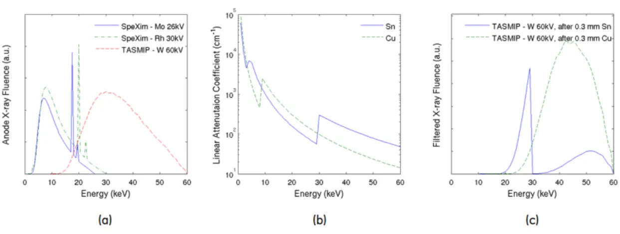
2-2 Example of X-ray fluence energy spectra generated by SpeXim and TASMIP models 25 2-3 Example bowtie filtration combining three materials (aluminum, graphite and copper) 25 2-4 Analytic phantom models for compressed and uncompressed breasts . . . 28
2-5 Meshed-based anthropomorphic breast model used in this study . . . 30
2-6 Example of simulated mammography, tomosynthesis and breast CT morphological images using mesh-based anthropomorphic breast model . . . 30
2-7 Illustration of primitive structure priorities in a multi-structure phantom and its consequence in total object thickness calculation . . . 32
2-8 Illustration of ray-tracing projection on two meshed primitive structures: a sphere and a torus . . . 32
2-9 Overview of hybrid MC-analytic scatter simulation . . . 37
2-10 A scheme of the Flat Panel Detector (FPD) model . . . 41
2-11 Simplified 5-steps cascade model for indirect-detection . . . 42
3-1 Experimental set-up for SpeXim model beam quality validation . . . 50
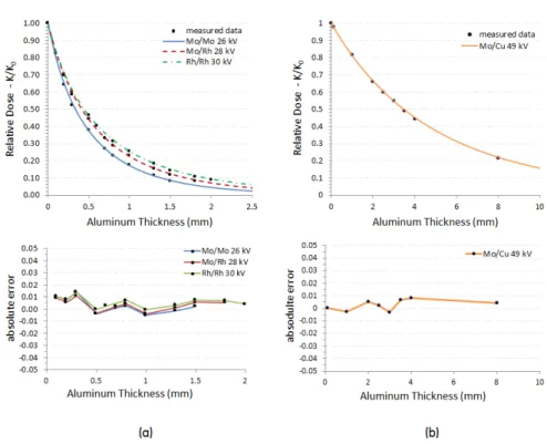
3-2 Relative dose values K(t)/K0 as function of the aluminum thickness, calculated using experimental and simulated data . . . 50
3-3 Simulation setup for scattered radiation PSF validation and illustration of scattered photons spatial distribution over the detector surface . . . 53
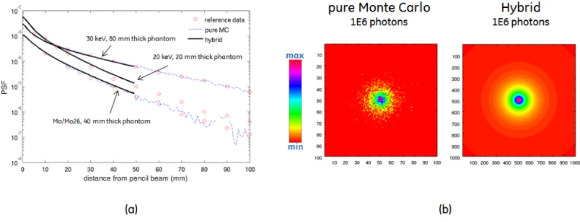
3-4 Results of scattered radiation PSF validation, for pure Monte Carlo and Hybrid approaches . . . 54
3-5 Simulated cone-beam geometries used for scatter simulation validation, according to Ref. [200], Ref. [110] and Ref. [113] . . . 56
3-6 Scatter intensity profile comparison with Ref. [200] . . . 56
3-7 SPR profiles comparison with Ref. [110] . . . 57
3-8 Scatter Intensity and SPR profile comparison with Ref. [113]: dependency with phantom diameter . . . 58
3-9 Scatter Intensity and SPR profile comparison with Ref. [113]: dependency with air gap . . . 59
3-10 Maximum SPR value comparison with Ref. [114] . . . 61
3-11 Scatter plot of reference vs simulated peak SPR values found in Figure 3-10 . . . . 61
3-12 Simulated geometry used to validate DgN simulation with CatSim using conven-tional mammography geometry . . . 63
3-13 Results of glandular dose simulation validation, for mammography geometry . . . . 63
3-22 Experimental setup illustration on GE Senographe DS used for SI validation . . . 75
3-23 Experimental setup illustration on GE Innova IGS620 used for SI, noise and SNR validation . . . 77
3-24 Measured primary and scatter signal profiles in GE Senographe DS and GE Innova IGS620 projection images . . . 78
3-25 Innova IGS620 X-ray images obtained from measurement and from simulations with-out and with blur from pre-sampling MTF . . . 78
3-26 Average signal intensity, noise σ and SNR as function of the tube voltage and for 60, 80 and 100 mA tube currents . . . 79
3-27 Measured vs simulated SNR for all simulated conditions and as function of the tube voltage . . . 79
4-1 Illustration of four main acquisition strategies to obtain dual-energy data . . . 90
4-2 Flowchart of image-based decomposition . . . 95
4-3 Flowchart of projection-based decomposition . . . 96
4-4 Phantom used in a priori calibration method for optimal dual-energy CE-bCT re-combination . . . 97
4-5 Illustration of calibration weighting for image and projection-based recombination 98 5-1 Optimization Critical-to-Quality (CTQ) factors . . . 103
5-2 Cone-Beam CT geometry used in dual-energy CE-bCT spectra optimization . . . . 107
5-3 Computational breast phantom used for monochromatic spectra optimization . . . 110
5-4 DgN coefficients as function of the incident monochromatic beam energy for the 10, 14 and 18 cm diameter phantoms . . . 111
5-5 Illustration of (a) CN RDiodine−bg, (b) CN RDµCal−glandand (c) CN RDgland−adipose as function of LE dose allocation ratio τ . . . 112
5-6 Monochromatic spectra optimization results for 10 cm diameter phantom . . . 112
5-7 Monochromatic spectra optimization results for 14 cm diameter phantom . . . 113
5-8 Monochromatic spectra optimization results for 18 cm diameter phantom . . . 113
5-9 Computational breast phantom used for polychromatic spectra optimization . . . . 118
5-10 Input spectra Snet(E) for W/Sn and W/Cu anode/filter combinations, for different tube voltages and filter thicknesses . . . 121
5-11 Polychromatic DgN coefficients as function of the tube voltage . . . 121
5-12 Illustration of iodine CNRD at optimal dose allocation for SS-DF acquisition strat-egy, as function of LE and HE tube voltages and filter thicknesses . . . 123
5-13 Optimization results for DS-DF technique as function of the LE and HE filter thick-nesses . . . 125
5-14 Optimization results for SS-DF technique as function of the LE and HE filter thick-nesses . . . 125
5-15 Optimization results for SS-FkV technique as function of the filter thickness . . . . 126
5-16 Optimization results for SS-PC technique as function of the tube voltage and filter thickness . . . 127
List of Figures xiii
5-17 Idealistic tube voltage waveform for each acquisition strategy and constant power
relationship between LE and HE tube voltage and current . . . 130
5-18 Optimization results for SS-FkV technique when constant power constraint is applied133 5-19 Optimization results for SS-PC technique when constant power constraint is applied 133
5-20 Results of the comparison between the four considered dual-energy strategies . . . 134
5-21 Absorption efficiency η(E) for 100, 250 and 600 µm CsI thicknesses . . . 137
5-22 Optimal LE, AGD allocation ratio τoptand CN RDiodine−bgas a function of the CsI
scintillator thickness . . . 139
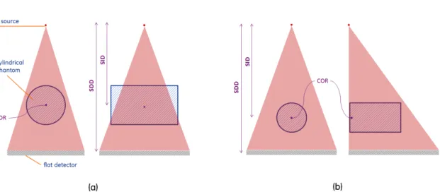
6-1 CE-bCT and CE-DBT topologies and phantom configurations for quantitative
com-parison studies of Sections 6.1 and 6.2 . . . 144
6-2 Illustration of 3D lesion extent estimation . . . 147
6-3 Method for automatic lesion extent assessment . . . 147
6-4 Reconstructed lesion magnification in the CE-DBT depth-direction, due to limited
angular span acquisition . . . 149
6-5 Results of 3D lesion estimation precision comparison between CE-bCT and CE-DBT 149
6-6 Measured vs true ellipsoidal lesions eccentricity in both CE-bCT and CE-DBT . . 150
6-7 Crel as a function of lesion diameter, for lesions positioned at different distances
from the chest wall . . . 152
6-8 Crel as function of the ellipsoid depth-direction eccentricity for bCT and
CE-DBT, for different x-z plane diameter values . . . 153
6-9 Illustration of the five iodine-enhanced lesion types considered in the human observer
study . . . 155
6-10 Illustration of VOI cropping to form CE-DBT and CE-bCT final review images . . 157
6-11 Examples recombined iodine-enhanced images for each topology and for each con-sidered lesion type . . . 158 6-12 Likert scale for Q1/Q2 (lesion existence) when Gold(Q1) is 0 (lesion not present)
and 1 (lesion present), per individual readers and per acquisition geometry . . . . 162
6-13 Sensitivity and Specificity of each reader in lesion detectability task (Q1), per
ac-quisition geometry . . . 162
6-14 Average Likert scale values for all questions, per individual reader, and
indepen-dently of the characterization answers . . . 163
6-15 Interobserver agreement in CE-DBT and CE-bCT data through assessment of
Co-hen’s Kappa coefficients, κC, and Proportion of Overall Agreement, po . . . 164
6-16 Bland-Altman plot of median measured extents among the three readers for CE-bCT
and CE-DBT . . . 169
7-1 Centered and normalized pixel values in material projections and in their respective
tomographic reconstructed images . . . 178
7-2 Detectability improvement in Iodine-equivalent slices of a textured phantom . . . . 183
7-3 Bar plot of Average Glandular Dose required to achieve minimal detectability as
function of the different denoising schemes . . . 183
A-1 X-ray Tube illustration and Tungsten anode X-ray intensity spectrum . . . 193
A-2 Probabilities of Photoelectric, Rayleigh and Compton interactions in water . . . . 195
A-3 Linear att. coefficients of fibroglandular and adipose tissues, hydroxyapatite and iodine . . . 195
A-4 Illustration of indirect and direct detection processes . . . 197
A-5 Typical mammography, tomosynthesis and dedicated breast CT geometries . . . . 198
A-6 Radon Transform and Central Slice Theorem . . . 199
A-7 Central Slice Theorem representation for limited angle tomosynthesis acquisition . 201
A-8 Tomosynthesis reconstruction and consequent geometric deformation in depth di-rection . . . 202
List of Tables
2.1 Definition and units of the main variables used along Chapter 2 . . . 22
2.2 Spectrum models used in this study . . . 23
3.1 X-ray spectra used in the SpeXim validation experiments and the expected HVL
and Tube Yield values, as required by GE Senographe DS Quality Control (QC) manual . . . 48
3.2 Experimentally assessed and simulated N-th value layers . . . 51
3.3 References for Scatter Intensity and Scatter-to-Primary Ratios profile validation . 54
3.4 Measured and simulated MTF values at 1/1, 1/2 and 1/4 fractions of the Nyquist
frequency . . . 71
3.5 Lorentzian parameters for the optical spreading function H(f ) models . . . 71
3.6 Measured and simulated average per-pixel SI for the Senographe DS experiment . 77
4.1 Characteristics, advantages and limitations of different acquisition strategies to
ob-tain dual-energy data . . . 92
5.1 Summary of optimal monochromatic acquisition spectra for different phantom
di-ameters . . . 114
5.2 Summary of input parameters for each acquisition technique considered in the
poly-chromatic spectra optimization . . . 120
5.3 Lower and upper limits of optimal input parameters and optimal Figures-of-Merit
for the unconstrained polychromatic spectra optimization . . . 123
5.4 Lower and upper limits of optimal input parameters and optimal Figures-of-Merit
for the constant power polychromatic spectra optimization . . . 131
5.5 Optimal input parameters and optimal Figures-of-Merit for LE and HE
polychro-matic spectra requiring 1 kW tube power . . . 135
6.1 Parameters used to simulate the CE-bCT and CE-DBT implementations in the
investigations of Sections 6.1 and 6.2 . . . 145
6.2 Total number of simulated lesion types for the human observer study, per acquisition
geometry . . . 155
6.3 Parameters used for CE-DBT and CE-bCT implementations in the human observer
study . . . 156
6.4 Review questionnaire based on ACR-BIRADS and used to assess the readers confidence159
6.5 Contingency table of reader answers as function of their true value, Gold(·) . . . . 160
6.6 Classification of reader agreement strength proposed by Landis and Koch [288], and
Fleiss et al. [290] . . . 161
6.7 Q1 (lesion existence): distribution of majority opinion answers vs Gold, for CE-DBT
and CE-bCT . . . 165
6.8 Q3 (mass/non-mass): distribution of majority opinion answers vs Gold, for CE-DBT
and CE-bCT . . . 165
6.9 Q5 (shape): distribution of majority opinion answers vs Gold, for CE-DBT and
CE-bCT . . . 166
6.10 Q5 Sensitivity analysis results using multivariable logistic model . . . 166
List of Acronyms
ACNR Anti-Correlated Noise Reduction
ACR American College of Radiology
AGD Average Glandular Dose
bCT breast CT
BI-RADS Breast Imaging-Reporting and Data System
CE-bCT Contrast-Enhanced breast CT
CE-DBT Contrast-Enhanced Digital Breast Tomosynthesis
CE-DM Contrast-Enhanced Digital Mammography
CE-MRI Contrast-Enhanced Magnetic Resonance Imaging
CESM Contrast-Enhanced Spectral Mammography
CE-US Contrast-Enhanced Ultrasound
CNR Contrast-to-Noise Ratio
CNRef f effective Contrast-to-Noise Ratio
CNRD Contrast-to-Noise Ratio normalized by Dose
CsI Cesium Iodide
CT Computed Tomography
DBT Digital Breast Tomosynthesis
DE Dual-Energy
DgN Normalized Glandular Dose Coefficient
DQE Detective Quantum Efficiency (M T F2/N P S)
DS-DF Dual-Source with Dual Filtration
EI Energy-Integrating
FBP Filtered BackProjection
FOM Figure-of-Merit
FOV Field-of-View
FPD Flat Panel Detector
HE High-Energy
HVL Half Value Layer
IGS Image Guided System
KERMA Kinetic Energy Released per unit Mass
LE Low-Energy
MC Monte Carlo
MRI Magnetic Resonance Imaging
MTF Modulation Transfer Function
MX Mammography
NNPS Normalized Noise Power Spectrum
NPS Noise Power Spectrum
PC Photon-Counting
PDF Probability Density Function
PMMA Poly Methyl MethAcrylate
PSF Point Spread Function
QDE Quantum Detection Efficiency (η(E))
Introduction
Worldwide, breast cancer is the most common non-skin cancer and second deadliest cancer in women, with nearly 1.7 million new cases diagnosed in 2012 [1]. In the United States, 12.3% percent of women will be diagnosed with breast cancer at some point during their lifetime. For
2013, approximately 232 000 new cases and 39 000 deaths are estimated1. In France, approximately
one woman in ten will develop a breast cancer during her lifetime. With 53 000 new cases and almost 11 500 deaths estimated in 2011 [2], breast cancer is the most common cancer among women in France and is the leading cause of cancer death.
In the light of these statistics, breast cancer is a major problem for global public health. In response, the government of various countries worldwide have adopted organized breast screening examination programs for an asymptomatic population satisfying certain eligibility criteria, e.g. age group, breast density, family history etc. Mammography is the only technique which has shown to reduce the mortality from breast cancer in a cost-effective way [3], and is today the standard technique for population-based screening. Ultrasound (U/S) and Magnetic Resonance Imaging (MRI) are complementary imaging techniques used mostly for dense breasts and high-risk women [4, 5].
During breast cancer development, the formation of new pathological blood vessels through angiogenic process provides irrigation for tumors beyond 1-2 mm and thus fuel their growth [6]. The new vascular network replenishes the tumors with nutrients, oxygen and enables them to eliminate metabolic wastes. In a breast cancer diagnosis, staging and therapy follow-up setting, imaging techniques combined with intravenous contrast agents have been developed to visualize breast tumor angiogenesis. When injected in the body, the contrast media enhances the visibility of abnormal vascular development and cancerous lesions, which could ultimately improve cancer-ous lesion detection, characterization and localization. Contrast-Enhanced Magnetic Resonance Imaging (CE-MRI) is the current standard imaging technique allowing detection of abnormal vas-cular development and lesion contrast uptake [7]. CE-MRI is however very costly and not widely available. Moreover, its spatial resolution might not be sufficient for certain types of lesions and does not allow microcalcifications depiction, whose presence is an important diagnostic indicator. Other techniques such as contrast-enhanced full-body Computed Tomography (CT) [8], Positron Emission Tomography (PET) combined with full body CT [9] and contrast-enhanced Doppler U/S (CE-US) [10] have also shown positive results in emphasizing breast tumor angiogenesis. However, these techniques each have their own limitations with respect to each technical aspects includ-ing spatial resolution, radiation dose, cost, operator dependent outcome and economical aspects including availability.
Contrast-enhanced X-ray imaging of the breast, as a potential less costly alternative, is cur-rently under investigation. With an iodinated vascular contrast agent, contrast-enhanced X-ray imaging can also provide morphological and functional images. Two imaging techniques have been proposed: temporal and dual-energy recombination. In the temporal technique, radiographic im-ages of the breast are acquired before and after intravenous administration of an iodinated contrast agent, using X-ray spectrum containing energies predominantly above the K-edge of iodine [11,12].
1National Cancer Institute, http://seer.cancer.gov/statfacts/html/breast.html (Accessed on April 4th, 2014)
of this technique was shown in 2003 [16] and became clinically available in 2010 with the
intro-duction of SenoBright application (GE Healthcare; Chalfont St Giles, UK). CESM is however a
2D projection technique of a 3D object, therefore limiting morphologic characterization of contrast uptakes when compared to 3D techniques. An alternative 3D X-ray imaging technique, with wide availability, low-cost, specifically designed for breast imaging and providing accurate quantitative position, morphologic and functional lesion information would be therefore a natural evolution of contrast-enhanced X-ray imaging.
Contrast-Enhanced Digital Breast Tomosynthesis (CE-DBT) and Contrast-Enhanced dedicated breast CT (CE-bCT) are two potential 3D evolutions of CESM and currently under investigation by academic and industrial research groups. It is however anticipated that the quantitative potential of CE-DBT is limited, due to the inherent low depth-resolution of limited opening angle DBT modality. CE-bCT with quasi-isotropic spatial resolution and voxel signal intensity proportional to linear attenuation coefficients is believed to offer more accurate quantitative information, though a low-dose operation could still be a challenge. Today, the incremental value of CE-bCT over CE-DBT in their ability to provide quantitative information at low radiation dose levels is still unknown. A complete evaluation on the quantitative performance of both technologies would be beneficial.
For the same reasons as CESM, both CE-DBT and CE-bCT techniques would benefit from iodine K-edge imaging techniques using dual-energy acquisitions. While some research effort has been oriented towards the assessment and optimization of dual-energy CE-DBT geometry, acquisi-tion parameters and protocols [17–26], little investigaacquisi-tion on dual-energy recombinaacquisi-tion techniques for iodinated contrast-agent and tumor angiogenesis enhancement in a dedicated breast CT setup was performed. As a consequence, before any comparison with CE-DBT, an optimization study aiming to reveal the dual-energy acquisition strategy and acquisition parameters maximizing the performance of CE-bCT in depicting contrast-agent uptake is imperative.
This PhD thesis research has been focused on two topics: 1) the optimization and assessment of an iodine-enhanced dual-energy breast CT technique and 2) its comparison with CE-DBT in their potential to accurately depict and localize tumors, as well as to provide accurate quantitative infor-mation on contrast uptake morphology and concentration, at radiation dose levels comparable to a two-view mammogram. A cone-beam geometry was considered both for CE-bCT and CE-DBT, since there is an interest in investigating techniques based on typical mammography geometries. This choice would allow for instance to provide different exams with the same equipment, reducing cost and improving accessibility. All investigations have been performed through realistic computer simulations. This theoretical approach allows quantifying the effect of each individual parameter in a complex imaging chain on image quality.
In Chapter 1, a short description on the breast anatomy, the development of cancerous tumors and its association to pathological angiogenesis is provided. An overview on established clinical vascular imaging techniques for breast cancer treatment is presented. Recently developed and commercially available techniques with increasing acceptance and usage are also discussed. Finally, CE-DBT and CE-bCT techniques are presented as two new potential candidates for vascular
Introduction 3
contrast-agent imaging. Details on technological aspects, and their individual advantages and limitations are provided.
In Chapter 2, a detailed description of the simulation platform software developed for our re-search is provided. This platform is based on CatSim, a computer software initially conceived at GE Global Research Center (CT and X-ray Laboratories, Niskayuna, NY, USA) to simulate 3rd generation CT projection images using analytical test objects [27]. As part of this PhD thesis research, modules for CatSim simulation chain have been developed to model X-ray projections of breast phantoms at a typical energy range used in breast imaging. The design of digital breast phantoms aiming to emulate the compressed and uncompressed breast anatomy is presented. Both simple phantoms, combining simplistic geometric shapes and complex mesh-based anthropomor-phic phantoms, providing more realistic allure of the breast anatomy, are considered.
In Chapter 3, in order to make sure that the implemented simulation chain is capable of emulating realistic physical phenomena underlying an X-ray breast imaging system, an extensive validation of the developed models with regard to previously published and experimentally obtained data is provided. The accuracies of X-ray spectrum modeling, Monte Carlo simulation of photon interactions with matter as well as signal and noise propagation in digital detectors are qualitatively and quantitatively evaluated.
In Chapter 4, the main existing spectral imaging acquisition techniques and the algorithms used to combine spectral images into exploitable functional information are discussed. A brief overview on the different methods allowing to obtain dual-energy data is also provided. A formulation of the dual-energy three-material decomposition allowing for iodine K-edge imaging in digital breast tomosynthesis and dedicated breast CT setups are discussed.
In Chapter 5, the optimization of dual-energy spectra and acquisition strategies for CE-bCT is performed, leveraging the implemented simulation platform and the dual-energy recombination
framework described in Chapters 2 and 4, respectively. The critical factors and optimization
criteria are defined, and the spectral optimization is performed using monochromatic hypothesis. Then, a new spectral optimization is performed for different polychromatic dual-energy acquisition strategies (dual-source, fast kVp switching, energy-discriminating detector). The performance of the different dual-energy acquisition strategies is subsequently compared. Finally, since columnar structured Cesium Iodide (CsI) scintillators have been widely used for digital breast X-ray imaging, a spectral optimization study considering different CsI layer thicknesses is performed.
In Chapter 6, using previously optimized dual-energy acquisition parameters for CE-bCT and CE-DBT, their quantitative potential is evaluated and compared through a series of experiments. Two preliminary studies evaluate the effect of CE-bCT and CE-DBT system topologies on lesion 3D extent estimation precision and iodine uptake quantification accuracy. For a more complete evaluation, a human observer study comparing iodine-enhanced lesion depiction and characteriza-tion in simulated CE-DBT and CE-bCT iodine-equivalent images is presented.
In Chapter 7, the optimized CE-bCT acquisition is discussed with particular focus on radiation dose requirements to depict iodine-enhanced lesions with minimal size and minimal uptake expected in clinical practice, while dose levels are compared to those expected for CE-DBT and current two-view standard mammography. Different post-processing denoising strategies are evaluated to improve iodine-enhanced lesion detectability in recombined CE-bCT images and potentially reduce the associated radiation dose.
Finally, we conclude on the main results and contributions of this PhD thesis research, and put forward research development perspectives for CE-bCT and CE-DBT applications.
Chapter 1
Clinical Context
1.1
Breast Anatomy and Cancer Development
Anatomy of the Healthy Breast
The human breast is mainly composed of fibroglandular and adipose tissues. The mammary gland is enveloped by a thin layer of connective and adipose tissues called Cooper’s ligaments. It provides a connection between the pectoral muscle and its deep surface, as well as between the skin and the superficial surface. The internal structure of the mammary gland is composed by a tree-like structure of lactiferous ducts, which originate at the nipple through lactiferous sinuses, and grow towards the pectoralis muscle. At the terminal part of the lactiferous ducts we encounter lobules regrouping milk-secreting glands, i.e. the alveoli or acinii. The glandular tissue refers to the aggregation of terminal ductal lobular units (0.5 to 2 mm diameter), each containing between ten and hundred alveoli (approximately 0.12 mm diameter). The latter are surrounded by dense fibrous connective tissues with adipose cavities [28–30], as well as epithelial cells which are responsible for milk production during the lactation period. The glandular and connective tissues together are known as Fibroglandular tissue. Beside the intraglandular adipose tissue, adipose tissue is further divided in two types based on their anatomical location: subcutaneous adipose compartments are positioned under the skin and retromammary adipose tissue is found near the chest wall. The proportion of adipose and fibroglandular tissue varies among individuals. After the menopause, the proportion of glandular tissues tends to decrease, being replaced by adipose tissue. Figure 1-1a illustrates a sagittal cross-section of the breast anatomy.
A dense vascular network carries nutrients and oxygen inside the breast. Arteries carry oxy-genated blood from the heart to the chest and the breasts, while the veins bring the deoxyoxy-genated blood to the heart. The axillary artery extends from the armpit and drains the outer half of the breast. The internal mammary artery leaves the neck down to the breast, draining its inner part. A lymphatic network is concentrated in the armpit area. Axillary lymphs located near the armpit drain 97% of lymphatic fluid in the breast. Another lymph chain extends along the median axis of the thorax (internal mammary chain) and drains the remaining 3% of lymphatic fluid. Figure 1-1b illustrates the lymphatic system network in the breast and adjacent regions.
Breast Cancer Development
Breast cancer is most commonly linked to cancerous development in the lactiferous ducts or the glandular lobules. There are many types of associated diseases, resulting in lesions most often classified into benign or malignant lesions. Benign lesions do not endanger the life of the patient while malignant lesions have the potential to lead to her death.
Among benign lesions we distinguish mainly the cysts, fibroadenomas, papillomas, abscess, hematoma, inflammation, radial sclerosis, ductal ectasia, lipomas and cytosteatonecrosis. Although
Figure 1-1: (a) Sagittal cross-section illustration of the breast anatomy and (b) lymphatic system network illustration
benign lesions are not life-threatening, they can cause symptoms and sometimes be linked with a higher risk of developing breast cancer in the future. Benign lesion are usually classified into 3 general groups of lesion, based on the cells growth (proliferative) and abnormality (atypia): non-proliferative lesions do not seem to affect cancer risk, non-proliferative lesions without atypia slightly increase cancer risk and proliferative lesions with atypia raise the risk of cancer.
Malignant lesions are primarily classified by their histological appearance (in situ or invasive) and their tissular origin (ducts or lobules). Carcinoma in situ is a form of pre-invasive cancer, characterized by a proliferation of cancerous cells within a particular tissue compartment without invading the surrounding tissue. Ductal Carcinoma In Situ, or DCIS, is associated to cancerous development within the mammary ducts, while Lobular Carcinoma In Situ, or LCIS, is associated with cancerous development in the lobules. On the other hand, invasive carcinoma does not confine itself to the initial tissue compartment. Three-quarters of invasive carcinomas are Invasive Ductal Carcinoma (IDC), in which tumor cells progress not only inside the ducts but also around them. The IDCs are often lobulated masses containing spicules radiating from their center. Around 10 to 15% [31] of invasive carcinomas are Invasive Lobular Carcinoma (ILC), in which tumor cells infiltrate the tissue filaments running along the ducts and vessels, thus preserving the original
architecture of the breast. Due to the small creation of new connective tissue, it is hard to
distinguish an ILC from the normal parenchyma.
The presence and proximity of lymph nodes are important factors influencing breast cancer spreading and the appearance of metastasis. Cancerous cells from the breast can infiltrate the lymph nodes and be transported to other areas of the body through the lymphatic system. Treat-ment and survival is often determined by whether or not breast cancer remains localized or spreads to other locations in the body. The latter case is usually associated with a dramatically decreases in a patient’s likelihood of survival.
There are several known physiological factors that can be associated with the risk of developing a breast cancer. Firstly, the gender, age and reproductive or hormonal factors such as early first period, late first pregnancy, late parity, low number of live-born children, oral contraceptive and hormone replacement therapy (HRT). Other factors to be considered are: radiotherapy through the chest wall during childhood, close family history of breast or ovarian cancers constituting a genetic predisposition to the development of breast cancer, the BRCA1 and BRCA2 gene mutations, the presence of diseases diagnosed as benign which can evolve to a cancer, smoking, obesity, increased alcohol consumption, etc.
1.1. Breast Anatomy and Cancer Development 7
malign lesions, we refer the reader to the work of A. Cooper [32].
Tumor Angiogenesis
Almost all tissues growing larger than a few millimeters in size develop a vascular network through a process known as angiogenesis. Once formed, the vascular network is a stable system that regen-erates slowly. The normal regulation of angiogenesis is governed by a fine balance between factors that induce the formation of blood vessels and those that halt or inhibit the process. When this balance is destroyed, it usually results in pathological angiogenesis which causes increased blood-vessel formation in diseases which depend on the angiogenesis to grow. Pathological angiogenesis, or the abnormal rapid proliferation of blood vessels, is implicated in over 20 diseases, including cancer.
During breast cancer development, formation of new pathological blood vessels will provide irrigation for tumors beyond 1–2 mm and thus fuel to their growth [6], replenishing them with
nutrients, oxygen and enabling them to eliminate metabolic wastes. Angiogenesis and tumor
growth is then accompanied by the disruption of the basement membrane and the cancer, until now in situ, becomes invasive.
Imaging techniques combined with intravenous contrast agents have been developed to highlight breast tumor angiogenesis. When injected in the body, the contrast media enhances the visibility of abnormal vascular development and cancerous lesions, which could ultimately improve lesion detection, characterization, localization and vascularization quantification. A brief description on the most widely used imaging techniques highlighting breast tumor angiogenesis is provided in Section 1.2.
For breast imaging, clinical studies have hypothesized that tumor angiogenesis could be as-sociated with the diagnostic outcome of breast cancer [33]. The calculation of the Microvessel Density (MVD), which consists in counting microvessels stained by immunohistochemistry and observed by optical microscopy, is commonly used to quantify angiogenesis associated with breast cancer. It could be an effective prognostic factor for invasive breast cancer [34–36]. Additionally, several studies have shown that vascular density is also associated with a more aggressive disease in patients with no lymph node invasion and in those with in situ [37–39] breast cancer.
Epidemiology
Worldwide, breast cancer is the most common non-skin cancer and second deadliest cancer in women than any other cancer, with nearly 1.7 million new cases diagnosed worldwide in 2012. This represents about 12% of all new cancer cases and 25% of all cancers in women [1]. In the United States, 12.3% percent of women will be diagnosed with breast cancer at some point during their lifetime. For 2013, approximately 232 000 new cases are estimated, representing 14.1% of all
new cancer cases in the U.S., and 39 000 deaths1.
In France, approximately one woman in ten will develop breast cancer in her life. With 53 000 new cases estimated in 2011 [2], breast cancer is the most common cancer among women, representing 33% of all new cancer cases in women. Breast cancer is also the leading cause of cancer death in France, with almost 11 500 deaths estimated in 2011, accounting for 18.3% of female cancer deaths.
Figure 1-2 shows the cancer incidence and mortality rates in France, per cancer type, as dis-closed by the Institute for Public Health Surveillance (Institut de Veille Sanitaire - InVS) and the National Cancer Institute (Institut Nationale du Cancer - INCa), in 2011 [2]. We can see that breast cancer leads both incidence and mortality rates, followed by colon rectum and lung cancers. Figure 1-3 illustrates the evolution of breast cancer incidence and mortality in France from 1980 to 2005, as disclosed by the InVS [41]. The incidence of breast cancer in France has increased
1National Cancer Institute, http://seer.cancer.gov/statfacts/html/breast.html (Accessed on April 4th, 2014)
(a) Hematological tumors are excluded from solid tumors (b) The skin cancers other than melanoma, are excluded.
(c) The shares of deaths from cervical cancer of the uterus and uterine cancer were estimated by a specific method [40] (d) Mortality estimates are not presented due to the uncertain quality of the data.
significantly and steadily between 1980 and 2005 with an incidence rate (standardized to the world population), which has almost doubled from 56.8 to 101.5 cases per 100 000 women. The mortality rate (standardized to the world population) decreased from 19.8 to 17.7 cases per 100 000 women from 1995 to 2005, thanks to screening and early detection, as well as to progress in treatment. Accordingly, and along with the substantial mortality rates described above, we may conclude that breast cancer remains today a major problem for global public health.
Early Breast Cancer Detection
Knowing that the probability of a tumor producing metastasis increases with its size, it has been shown that its early detection reduces mortality [42]. In response, the government of various countries worldwide have adopted organized breast screening examination programs for an asymp-tomatic population satisfying certain eligibility criteria, e.g. age group, breast density, family history etc. These criteria vary from one country to another, depending on public health policies and local statistics. In France, for example, since January 1st 2004, the female population between 50 and 74 years-old is invited every 2 years to participate in a free screening examination [43].
Mammography is the current standard for population-based screening. When applied in a screening setting, mammography is the only technique which has shown to reduce the rate of death from breast cancer in a cost-effective way [3], especially among women aged over 50 years [44]. Mammography is a low radiation x-ray projection technique providing morphological images of breast tissue. Clinical studies have shown a 25% to 30% reduction of the mortality associated with this disease in women aged 50 to 69 years and a reduction of approximately 16% for those aged between 40 and 49 years [45]. Ultrasound (U/S) and Magnetic Resonance Imaging (MRI) are complementary imaging techniques used mostly for dense breasts and high-risk women [4, 5].
In France, mortality which had remained stable since 1980, began to decrease, as illustrated in Figure 1-3. This increased survival, concurring with the results described in most Western
1.1. Breast Anatomy and Cancer Development 9
Figure 1-3: Evolution of breast cancer incidence and mortality rates in France (standardized to the world population), from 1980 to 2005. Data source: Belot et al., 2008 – Ref. [41], www.invs. sante.fr/surveillance/cancers/estimations_cancers/default.htm (Accessed 06 Dec 2013)
countries [1], can be explained in part by increasing the proportion cancers detected at an early stage in connection with the development of screening practices and secondly the major therapeutic progress in the early 2000’s [46].
the main benefits and limitations of each imaging technique, as discussed below.
1.2.1
Contrast-Enhanced Magnetic Resonance Imaging
In certain situations, such as when a woman has a very high risk of breast cancer and high breast density, Contrast-Enhanced MRI (CE-MRI) in combination with mammography is recommended as a screening tool [4, 5, 7, 48–51]. CE-MRI has the advantage that it does not involve exposure to ionizing radiation, for which high-risk women may exhibit an increased sensitivity [52–55]. It has been shown that CE-MRI can increase detection of breast cancer for certain groups of women [56, 57]. Using gadolinium chelates as a vascular contrast agent, CE-MRI does not only provide 3D images of breast morphology, but also functional images allowing detection of abnormal vascular development and lesion contrast uptake. The signal intensity of gadolinium-enhanced lesions in CE-MRI was shown to be correlated with the Microvessel Density (MVD) [58, 59]. Another important information in CE-MRI, complementary to the contrast-enhanced lesion morphology, is the kinetics of the contrast agent. Early studies have shown that malignant lesions tended to take an earlier and more pronounced contrast than the benign lesions [60, 61]. Hence, the shape of the curve of contrast enhancement can be used as an indication of the benign or malignant nature of the lesion. In a tumor staging and treatment setting, CE-MRI is currently the most recommended tech-nique by the European Society of Breast Imaging (EUSOBI) [7], the European Society of Breast Cancer Specialists (EUSOMA) [51] and the American College of Radiology (ACR) [7]. CE-MRI results have shown to strongly correlate with the extent of disease [62–64], which forms the basis for treatment decisions. CE-MRI is also recommended for monitoring early cancer response to chemotherapy so as to avoid unnecessary toxicity and cost without potential benefit from treat-ment and to decide whether or not to continue or change the therapy plan [65]. After neo-adjuvant chemotherapy, assessment of residual tumor [66] before surgery allows the surgical approach to be optimized to ensure negative margins and maximize breast conservation surgery.
Despite its great success, CE-MRI presents some important limitations. From a technical stand-point, the first limitation is the inability to rapidly image the whole breast volume while preserving a good spatial resolution. The speed of the MRI acquisition comes at the expense of its spatial resolution, which may negatively affect the morphological analysis of the contrast uptake. Another important limitation is the appearance of non-pathological contrast uptakes linked to hormonal fluctuations [67], surgery and radiotherapy [68]. In addition, some benign fibroadenomas may be-have identically to malignant tumors. From a socio-economic standpoint, two big disadvantages of CE-MRI are its high cost and limited availability [69, 70]. In France, a CE-MRI costs about
five times a bilateral mammography exam (293¿ for a CE-MRI exam, 66¿ for a bilateral
mam-mography exam [71]) and only 20% of prescribed CE-MRI exams are performed due to limited availability. In addition, as discussed above, CE-MRI has a high rate of false-positives [72], which may result in more unnecessary biopsies and other additional examinations. Other practical disad-vantages that should be highlighted are the discomfort encountered by claustrophobic patients and the safety issues for patients who posses metal implants and non-MRI-compatible foreign bodies.
1.2. Std Vascular Imaging Techniques for Breast Cancer Treatment 11
Figure 1-4: Clinical workflow for breast cancer management and the corresponding main imaging techniques associated to each step. The main limitations of each technique are summarized
1.2.2
Contrast-Enhanced Digital Mammography
The advent of digital mammography and the ability to develop post-processing techniques have stimulated interest in Contrast-Enhanced Digital Mammography (CE-DM). Two imaging tech-niques have been developed: temporal and spectral recombination. Both techtech-niques take advan-tage of the sudden increase in iodine attenuation coefficient (absorption edge) as a function of X-ray photon energy; just above its K-shell binding energy (33.2 keV), iodine attenuation is 5.5 time higher than just below its K-edge (cf. Annex A for more details in x-ray photon interaction with matter).
CE-DM using a temporal subtraction technique was pioneered in 2002 [11,12]. In this approach, radiographic images of the breast are acquired before and after intravenous administration of an iodinated contrast agent using a high-energy (HE) X-ray spectrum containing energies predomi-nantly above the K-edge of iodine. Logarithmic subtraction of the pre- and post-contrast images are used to obtain functional iodine enhancement images in which signal intensities are proportional to the quantity of iodine crossed by the X-ray beam. This technique feasibility was demonstrated in two clinical pilot studies [13, 73]. Later clinical studies have shown some potential of the tem-poral approach in highlighting tumor angiogenesis uptake and contrast-agent kinetics, ultimately improving the final diagnosis when it complements a standard mammography examination [13–15]. Although these clinical studies revealed the prognostic potential of the temporal approach, this technique has not been implemented in clinical practice. The main reason is that the temporal approach required two injections of iodine to image two breasts, entailing in longer examinations and increased iodine toxicity exposition. Additionally, Diekmann et al. [15] evidenced in a multi-center clinical study that the most important diagnostic information comes from the morphological analysis of the contrast enhancement and its intensity, rather than the contrast-agent kinetic in-formation. Moreover, substantial patient motion artifacts due to the extended time between pre-and post-contrast acquisitions was observed pre-and entailed in the loss of lesion morphologic detail.
To overcome the limitation of the temporal approach, dual-energy (DE) contrast-enhanced digital mammography, also named contrast-enhanced spectral mammography (CESM) has been proposed as an alternative solution [16]. In CESM, after iodinated contrast-agent injection, low-energy (LE) and high-low-energy (HE) image pairs are acquired at energies that closely bracket the iodine K-edge, simultaneously or in rapid successions. Iodine-enhanced images are then obtained by recombining the two images. GE Healthcare is currently the only company with a commercial CESM application as extension of the existing indications for diagnostic mammography with the
The feasibility of tomographic breast imaging using a standard whole body scanner was assessed in early 80’s by Muller et al. [74]. The authors concluded that although the system being technically capable of providing good images and the ability to detect distant metastases through incidental findings, the trade-off between the new diagnostic information and the high amount of radiation dose delivered to the breast and neighboring body tissues in the thorax was insufficient when compared to standard mammography. Moreover, the contemporaneous dedicated breast computed tomography (CT) scanners produced better results.
Later on, the development of multidetector CT (MDCT) scanners with faster acquisitions and improved resolution encouraged further research on breast imaging. Many clinical studies were conducted to evaluate MDCT when associated with the administration of iodinated contrast-agents as a tool for breast lesion diagnosis, cancer extent and conservation surgery planning [8,75–79]. For these applications, contrast-enhanced MDCT imaging of the breast has shown to provide excellent sensitivity (especially for patients with dense breast) but limited specificity [80]. Moreover, Inoue et al. [77] also noted that that contrast-enhanced MDCT could be used for dynamic CT and distinguish carcinoma from benign lesions. However, in all these studies radiation dose to the
patient was still substantially higher than standard mammography levels [78]. More recently
contrast-enhanced CT has been combined with Positron Emission Tomography (PET) imaging systems [81,82] in order to produce in order to generate additional functional information. However, a practical limitation is that nuclear imaging techniques have a relatively long implementation time and are very expensive [9].
Contrast-Enhanced Ultrasound
Breast Ultrasound (U/S) can be used to solve equivocal radiological signs detected on a single incidence of mammography, particularly in the differentiation of cystic lesions from solid masses. It also allows for a better appreciation of the lesion composition. Breast US imaging has however some limitations. Rather unusually, adipose tissues in the breast are hypoechoic when compared to the parenchyma. The majority of masses and breast cancers are also hypoechoic. As a consequence, a substantial number of cancers are difficult or impossible to depict, since they have the same echogenicity as the surrounding tissue. Normal breast structures may be confused with cancer, generating a high rate of false-positives.
Contrast-Enhanced Ultrasound (CE-US), combining the use of Doppler with gas-filled mi-crobubbles contrast agents (1 to 10 microns in diameter), have shown to improve the detection of blood vessels and down to diameters of the order of 40 microns [10]. As in CE-MRI, the assessment of contrast uptake kinetics, in particular the derived outputs such as the peak of uptake intensity, the mean transit time, the slope of the wash-in curve, the washout time and the area under the curve of contrast, can be important parameters to distinguish benign and malignant lesions [83,84]. In CE-US however, microbubbles contrast agents are rather unstable, due to their size, and con-sequently their lifetime inside the body is short (between 2 and 5 minutes). Hence, this time constraint might jeopardize the contrast uptake curve assessment and prevent the examination to cover the whole breast volume. From a more practical standpoint, another limitation of breast
1.2. Std Vascular Imaging Techniques for Breast Cancer Treatment 13
US is that the examination quality depends on the operator, and is therefore poorly reproducible. Moreover, the manipulation is highly difficult due the lack of anatomical landmarks to guide the review. Thus, it may still remain unclear whether the entire breast was imaged or not.
In summary, today the standard imaging techniques used for breast cancer diagnosis, staging and therapy follow-up is CE-MRI; it provides morphologic, topological and functional information of the tumor. CE-MRI is however very costly and is not widely available. Contrast-Enhanced full body CT is characterized with limited spatial resolution and a substantial radiation dose to the patient. Techniques based on U/S such as Doppler and contrast-enhanced Doppler have also shown positive results in emphasizing breast tumor angiogenesis. However, the examination quality is highly dependent on the operator. More recently, CE-DM has shown promising results, though its benefits compared to CE-MRI and CE-US are still to be demonstrated. CE-DM 2D projection nature may however limit the characterization of cancerous lesions when compared to 3D techniques.
In order to improve the accuracy of tumor diagnostics, staging and cancer therapy monitoring, with wider accessibility and better dose-cost effectiveness, an alternative technique specifically de-signed for breast imaging, providing accurate 3D quantitative position, morphologic and functional lesion information is definitely welcome. In the next section Contrast-Enhanced Digital Breast To-mosynthesis (CE-DBT) and Contrast-Enhanced Dedicated Breast CT (CE-bCT) are presented as two potential candidates.

![Figure 3-7: SPR profiles comparison with Ref. [110]. (a) Scatter Intensity over the detector surface using Hybrid method for W/Al 80 kV and 10 cm diameter phantom, monochromatic beam at 50 keV and 14 cm diameter phantom and W/Al 80 kV and 18 cm diameter ph](https://thumb-eu.123doks.com/thumbv2/123doknet/14740505.754996/80.892.183.723.147.433/profiles-comparison-scatter-intensity-detector-diameter-monochromatic-diameter.webp)