HAL Id: tel-01875026
https://tel.archives-ouvertes.fr/tel-01875026
Submitted on 16 Sep 2018HAL is a multi-disciplinary open access archive for the deposit and dissemination of sci-entific research documents, whether they are pub-lished or not. The documents may come from teaching and research institutions in France or abroad, or from public or private research centers.
L’archive ouverte pluridisciplinaire HAL, est destinée au dépôt et à la diffusion de documents scientifiques de niveau recherche, publiés ou non, émanant des établissements d’enseignement et de recherche français ou étrangers, des laboratoires publics ou privés.
regulation by phosphorylation and demonstration of its
role in cellular cohesion
Maria Isabel Acosta-Lopez
To cite this version:
Maria Isabel Acosta-Lopez. The E3 ubiquitin ligase HACE1 : characterization of its regulation by phosphorylation and demonstration of its role in cellular cohesion. Molecular biology. Université Côte d’Azur, 2017. English. �NNT : 2017AZUR4065�. �tel-01875026�
École Doctorale des Sciences de la Vie et de la Santé
Unité de recherche : Inserm 1065- C3M
THÉSE DE DOCTORAT
Présentée en vue de l’obtention du grade de Docteur en Sciences
de l’ Université Côte d’Azur – UFR Sciences
Mention : Interactions moléculaires et cellulaires
Défendue publiquement par
Maria Isabel ACOSTA-LOPEZ
The E3 ubiquitin ligase HACE1: Characterization of its regulation by
phosphorylation and demonstration of its role in cellular cohesion.
HACE1 E3 ubiquitine ligase : Caractérisation de sa régulation par
phosphorylation et mise en évidence de son rôle dans la cohésion cellulaire.
Dirigée par Emmanuel Lemichez, Directeur de Recherche, C3M
et co-dirigée par Orane Visvikis, Chargé de Recherche, C3M
Soutenue le 15 septembre 2017
Devant le Jury de soutenance composé de :
Dr. Anne DEBANT Directeur de Recherche Examinatrice et
Présidente du Jury
Dr Serge ROCHE Directeur de Recherche Rapporteur
Dr. René-Marc MEGE Directeur de Recherche Rapporteur
Dr Emmanuel LEMICHEZ Directeur de Recherche Directeur de thèse
Acknowledgements
I would like to thank Emmanuel for welcoming me into his team and giving me such an interesting project. For his time, interesting ideas and the impromptu scientific discussions we engaged in during this four years. I would like to thank all the current and previous members of the team that I had the pleasure to work with, specially Anne, Patrick and Daniel; who guided me at the beginning and were always available when I was puzzled and needed help.A big thank you goes to Orane, who was there day after day to discuss and decide the course of my project in her honest and straightforward manner. I appreciate all the time and energy she dedicated to teach me all sorts of thigs; from basic experimental techniques and data presentation to her method for neatly structuring my ideas. It was a pleasure to work with her, and I truly think her guidance is a big reason to why I have successfully reached this point.
I also want to thank all our collaborators; Serge Urbach and Jérôme Boudeau in Montpellier for all their advice and help with the mass-spectrometry part of my project, Edward Manser and Yue-Wai Ng in Singapore for their expert help on PAK, and Sophie Tartare-Deckert and Aude Mallavialle from team 11 for sharing their knowledge and tools on EMT.
I thank my friends here in Nice and fellow Signalife students for their cheerful energy and support during all this time. Their company has made my stay in Nice so much joyful! I specially thank Ramona, my ex-collocataire and writing buddy, who from the beginning of my stay in this city was a strong friend I could rely on.
I would like to thank Johan for his patience, love and support during my last year of thesis, for understanding the late hours and having dinner ready when I came home. I appreciate his attentive ear, and his tireless eyes, that helped me edit and proofread this entire manuscript!
Finally, I would like to thank my family in Colombia for their continuous support, love and understanding. I hope that this achievement will make them proud.
(ANR-11-LABX-0028-01)
FRM 4th year Doctoral grant
Table of Contents
List of Figures ... 1
List of Tables ... 3
List of Abbreviations ... 4
General Introduction ... 7
Chapter 1. Ubiquitin Signaling ... 9
1.1. The Ubiquitin system. ... 10
1.1.1. Ubiquitin ... 11
1.1.2. The writers: The enzymatic cascade of ubiquitin conjugation ... 12
1.1.3. The ubiquitin code: Signaling implications of ubiquitination ... 15
1.1.4. When ubiquitination goes awry: Implications in human diseases ... 18
1.2. E3 ubiquitin ligases: focus on the HECT family. ... 20
1.2.1. RING E3 ligases. ... 20
1.2.2. RBR E3 ligases. ... 21
1.2.3. HECT E3 ligases. ... 22
1.3. Regulation of HECT E3s. ... 26
1.3.1. Regulation of Nedd4 family ligases. ... 27
1.3.2. Regulation of HECT ligases outside the Nedd4 family. ... 36
1.3.3. Modulation of the activity of HECT E3s by small chemical compounds. ... 38
Chapter 2. Rho GTPases ... 41
2.1. The Rho GTPase family ... 41
2.2. Regulation of the activity of Rho GTPases. ... 43
2.2.1. Structural basis of Rho GTPase activity. ... 43
2.2.2. Regulation of the GDP-GTP cycle ... 44
2.2.3. Additional regulatory mechanisms ... 46
2.3. Molecular and cellular aspects of Rho GTPase signaling ... 47
2.3.1. From extracellular stimuli to effector proteins: Focus on PAK ... 47
2.3.2. General view of the cellular roles of Rho GTPases ... 52
2.3.3. Role of Rho GTPases in cytoskeleton reorganization ... 53
2.3.4. Role of Rho GTPases in adherens junctions ... 56
2.4. Rho GTPases and pathology. ... 60
2.4.1. Rho GTPases in neurological diseases ... 60
2.4.2. Rho GTPases and cancer ... 60
2.4.3. Rho GTPases and bacterial infection. ... 62
Chapter 3. Ubiquitination of Rho GTPases: Rac1 and its E3 ligase HACE1 ... 65
3.1. Regulation of Rho GTPases by ubiquitination ... 65
3.1.1. Ubiquitination of RhoA ... 66
3.2.2. Targets of HACE1: Rac1 and beyond ... 71
3.2.3. HACE1: a dual function protein involved in numerous cellular processes ... 75
Thesis objectives ... 85
Chapter 4. Results and Discussion ... 87
4.1. Research Article -imminent submission-: Group-I PAKs mediate the phosphorylation of HACE1 on serine 385 in order to regulate its oligomerization state and Rac1 ubiquitination ... 87
4.2. Implication of HACE1 in epithelial cell-cell adhesion ... 129
Chapter 5. General Discussion and Perspectives ... 143
5.1. Phospho-regulation of HACE1 by Rac1/Cdc42 and group I PAKs. ... 143
5.1.1. Regulation of HACE1 by phosphorylation of Ser-385 in vivo: Is an adaptor at play? ... 143
5.1.2. How can the conformational change upon Ser-385 phosphorylation affect HACE1 function? ... 147
5.1.3. PAK controls Rac1 ubiquitination by HACE1: signaling implications ... 149
5.2. HACE1 and epithelial cell-cell adhesion: keeping EMT at bay. ... 151
5.2.1. Does HACE1 have a role in the establishment and maintenance of AJ? ... 151
5.2.2. A parallel between HACE1 and PAK during EMT in cancer ... 152
Conclusions ... 155
Bibliography ... 157
List of Figures
Chapters 1-3
Figure 1.1. Ubiquitin structure characteristics. ... 12
Figure 1.2. Generalized enzymatic mechanisms of Ubiquitin (Ub) transfer between enzymes and ultimately to a target. ... 13
Figure 1.3. Classes of E3 ubiquitin ligases. ... 14
Figure 1.4. Physiological roles associated with individual chain. ... 16
Figure 1.5. The mammalian HECT E3 ligases. ... 24
Figure 1.6. Schematic showing the HECT E3 catalytic cycle. ... 26
Figure 1.7. Domain structure of the nine members of the Nedd4 family and their auto-inhibitory conformations. ... 28
Figure 1.8. Mechanisms of regulation of Nedd4-family ligases ... 29
Figure 1.9. Activation and cellular localization of Nedd4-2 by IP3 and Calcium ... 31
Figure 1.10. Schematic representation of how the Nedd4 family ubiquitin ligases self-regulate through autoubiquitination ... 33
Figure 1.11. Model of the conformational regulation of HUWE1 and the proposed mechanism of its inhibition by p14ARF ... 38
Figure 2.1. Domain architecture of the Rho GTPases. ... 42
Figure 2.2. Rho GTPase activity ... 44
Figure 2.3. Overview of Rho GTPase regulation. ... 47
Figure 2.4. Signaling of Rho GTPases. ... 48
Figure 2.5. Overview of PAK structure, activation and targets. ... 51
Figure 2.6. Rho, Rac and Cdc42 in cytoskeleton dynamics ... 55
Figure 2.7. Structural model of the core E-cadherin/catenin cell adhesion complex ... 57
Figure 2.8. Selected examples of bacterial virulence factors targeting various stages of Rho protein regulation. ... 64
Figure 3.1. Representation of HACE1 domain organization ... 69
Figure 3.2. 3D model of HACE1 Ankyrin repeats important for its interaction with Rac1 ... 74
Figure 3.3. Representation of the model for the role of Syn5 ubiquitination in p97/p47-mediated post-mitotic Golgi membrane fusion ... 77
Figure 3.4. HACE1 controls Rac1-dependent NADPH oxidases. ... 79
Figure 3.5. HACE1 is a central player in the control of cellular redox balance. ... 80
Chapter 4
Section 4.1.
Research Article
: Group-I PAKs mediate the phosphorylation of HACE1 on serine 385 in order to regulate its oligomerization state and Rac1 ubiquitination.Figure 1. CNF1 increases phosphorylation of HACE1 on Ser-385 ... 94
Figure 2. Activation of Rac1 or Cdc42 mediates phosphorylation of HACE1 on Ser-385 ... 96
Figure 3. Group I PAKs induce direct phosphorylation of HACE1 ... 98
Figure 4. HACE1(S385E) phospho-mimetic blocks Rac1 ubiquitination in cells ... 100
Figure 5. Ser-385 phosphorylation modulates HACE1 oligomerization ... 103
Sup Figure S1. CNF1 and Rac1 induce phosphorylation of Ser-385 ... 119
Sup Figure S2. Phosphorylation of HACE1 S385 is independent of mTOR signaling . 120 Sup Figure S3. Group I PAKs induce direct phosphorylation of HACE1 ... 120
Sup Figure S4. HACE1 does not induce its own ubiquitination in MCF12A ... 121
Sup Figure S5. In vivo homophilic interaction of HACE1 requires HECT binding to ANK+MID region. ... 122
Sup Figure S6. Phosphorylation of Ser-385 does not promote a specific association of HACE1 to 14-3-3 proteins ... 123
Sup Figure S7. Ser-385 is located within an Intrinsically Disordered Region ... 124
Section 4.2. Implication of HACE1 in cell-cell adhesion Figure 4.1. HACE1 interacts with α-catenin ... 130
Figure 4.2. Generation of shHACE1 cell lines ... 131
Figure 4.3. Loss of HACE1 disrupts the epithelial monolayer integrity ... 132
Figure 4.4. HACE1 loss results in the acquisition of an EMT-like signature ... 134
Figure 4.5. The EMT-like signature is not an immediate effect of HACE1 loss and is partially dependent on exogenous signals ... 137
Figure 4.6. HACE1 does not ubiquitinate α-catenin ... 138
Chapters 5
Figure 5.1. Representation of the possible relationships between p-HACE1S385, the binding to a cellular adaptor, the change in HACE1 oligomerization and the modulation of HACE1’s function.. ... 149List of Tables
Table 3.1. Cellular processes regulated by HACE1 ... 83
Sup Table S1. Plasmid List ... 125
Sup Table S2. Primer List ... 127
Sup Table S3. Antibody List ... 127
Table 5.1. Examples of common phenotypic effects of HACE1 depletion and PAK hyper-activity in cancer cells ... 154
AJ: Adherens Junctions AMP-Ub: Ubiquitin adenylate
CISK: Cytokine-independent survival kinase CNF1: Cytotoxic necrotizing factor 1 DUBs: Deubiquitinating enzymes EGF: Epidermal growth factor
EMT: Epithelial-mesenchymal transition ENaC: Epithelial sodium channels GAP: GTPase Activating Protein GDF: GDI Displacement Factor GDP: Guanosine Di-Phosphate
GEF: Guanin nucleotide Exchange Factor GTP: Guanosine Tri-Phosphate
HACE1: HECT-domain and Ankyrin-repeat containing E3 ubiquitin protein ligase 1
HECT: Homologous to E6AP C-terminus HGF: Hepatocyte growth factor
HRG: Heregulin IFN: Interferon
IKKβ: IkappaB kinase beta/ Nuclear Factor NF-Kappa-B Inhibitor Kinase Beta JNK: c-Jun N-terminal kinase
LIMK: LIM-motif containing kinase MAPK: mitogen-activated protein kinase MET: Mesenchymal-epithelial transition MLC: Myosin light chain
MLCP: MLC phosphatase
NADPH: nicotinamide adenine dinucleotide phosphate
Nedd8: Neural precursor cell-expressed, developmentally downregulated 8 NF-kB: Nuclear Factor kappa B.
PAK: p21-activated kinase
PDGF: Platelet-derived growth factor PKA: Protein kinase A
PTM: Post-translational modifications
Rac: Ras-related C3 botulinum toxin substrate RBR: ring between ring
Rho: Ras homologue
RhoGDI: Rho GDP Dissociation Inhibitor RING: Really interesting New gene ROS: Reactive Oxygen Species
SGK1: serum and glucocorticoid related kinase 1 Smurf: Smad ubiquitination-related factor SRF: Serum-response factor
βAR: β-Adernergic receptor
SUMO: small ubiquitin-related modifier Syn5: Syntaxin5
TGFβ: transforming growth factor beta TNF: Tumor necrosis factor
TNFR1: TNF receptor 1
TRAF: TNFR associated factor UBD: Ubiquitin binding domain UBEx: Ubiquitin binding exosite Ubls: Ubiquitin-like modifiers UBP: ubiquitin binding protein UBS: ubiquitin binding site
UPS: Ubiquitin Proteasome System
WASP: Wiskott-Aldrich syndrome protein
General Introduction
The post-translational modification of proteins by ubiquitination serves multiple cellular purposes. It modulates protein-protein interactions along diverse signaling pathways and targets proteins for degradation by the proteasome, thereby contributing to protein homeostasis and to the temporal dynamics of signaling networks. In 2002, our team demonstrated that small intracellular signaling proteins of the Rho GTPase family can be ubiquitinated and addressed to the ubiquitin proteasome system. The members of the Rho GTPase family are present in all eukaryotes and act as molecular switches best known to regulate cytoskeletal dynamics. In addition, they take part in the regulation of gene expression, cell cycle progression, as well as in the cellular response to pathogenic agents. Through the study of the regulation of Rho GTPase activity by ubiquitination, our team identified the first E3 ubiquitin ligase that directly ubiquitinates and promotes the proteasomal degradation of Rac1, the founding member of the Rho GTPase family. This E3 ligase is HACE1, a protein first found epigenetically silenced in sporadic Wilms’ tumor and then repeatedly found to be downregulated in numerous human diseases, including cancer, neurodegenerative diseases and developmental conditions. Despite the important role of HACE1 in the maintenance of cell homeostasis, nothing is known about the post-translational regulation of its activity. In this work, we aim to contribute to this knowledge gap by studying how HACE1 is regulated by phosphorylation and, in parallel, explore the role of HACE1 in the regulation of intercellular adhesion, a cellular process that heavily depends on Rho GTPase signaling.The first three chapters of this thesis comprises the introduction to my work. Chapter 1. aims to give a general view of the Ubiquitin field with particular emphasis on the characteristics and regulatory mechanisms of the E3 ubiquitin ligase family; Chapter 2. is dedicated to the Rho GTPases, their mechanisms of action, cellular functions and implications in human disease; and Chapter 3. gives a more detailed description of the current knowledge of HACE1.
Chapter 1.
Ubiquitin Signaling
Contents
1.1. The Ubiquitin system. ... 10
1.1.1. Ubiquitin ... 11
1.1.2. The writers: The enzymatic cascade of ubiquitin conjugation ... 12
1.1.3. The ubiquitin code: Signaling implications of ubiquitination ... 15
1.1.4. When ubiquitination goes awry: Implications in human diseases ... 18
1.2. E3 ubiquitin ligases: focus on the HECT family. ... 20
1.2.1. RING E3 ligases. ... 20
1.2.2. RBR E3 ligases. ... 21
1.2.3. HECT E3 ligases. ... 22
1.3. Regulation of HECT E3s. ... 26
1.3.1. Regulation of Nedd4 family ligases. ... 27
a. Release of auto-inhibition by adaptors ... 30
b. Modulation of auto-inhibition by PTM ... 32
c. Regulation of substrate interaction by adaptors. ... 34
d. Regulation of substrate interaction by PTM. ... 34
1.3.2. Regulation of HECT ligases outside the Nedd4 family. ... 36
a. E6AP/Ube3A ... 36
b. HUWE1 ... 36
1.3.3. Modulation of the activity of HECT E3s by small chemical compounds. ... 38
Ubiquitination is a dynamic and complex type of post-translational modification implicated in nearly all aspects of eukaryotic biology. Ubiquitin is a 8.5kDa protein that was first isolated from bovine thymus and was associated with the induction of lymphocyte differentiation (Goldstein et al. 1975). This new small protein was believed to be ubiquitously expressed (hence its name) from bacteria and yeast to animals and higher
plants. Moreover, the 76 amino acid long sequence of ubiquitin was found to be remarkably conserved among eukaryotes (Goldstein et al. 1975), which suggested a fundamental role. A role so vital that it imposed incredibly tight evolutionary constraints on ubiquitin’s structure. The first observation of covalently attached ubiquitin was made by H. Busch and collaborators: they described a protein, which they called “A24”, that curiously had one C-terminus and two N-termini (Goldknopf and Busch 1977). This particular protein was later found to be the histone H2A bound to ubiquitin by an isopeptide bond, which made H2A the first substrate of the ubiquitin pathway to be found (Hunt and Dayhoff 1977). However, it wasn’t until some years later, between 1978 and 1983 that the teams of A. Herschko and I. Rose discovered the central role of ubiquitin in non-lysosomal, ATP-dependent intracellular protein degradation (Ciehanover et al. 1978; Wilkinson et al. 1980). They isolated and described the enzymatic cascade composed of three enzymes (E1, E2, and E3) necessary for the activation and covalent attachment of ubiquitin onto protein substrates (Ciechanover et al. 1981; Hershko et al. 1981; Hershko et al. 1983), a work that granted them the Nobel prize in chemistry in 2004. Since then, it has been shown that the ubiquitin system is not only involved in the degradation of proteins by the proteasome (Finley 2009) but also in non-proteolytic processes such as membrane trafficking (Hicke and Dunn 2003), DNA repair (Jentsch et al. 1987; Cohn et al. 2007; Stewart et al. 2009) and chromatin dynamics (Wright et al. 2012). In this chapter, I will briefly describe the properties and mechanisms of action of the proteins involved in ubiquitination, explore the variety of signals ubiquitination can transduce, and finally focus on one of the key components of the ubiquitin system: the E3 ubiquitin protein ligases.
1.1. The Ubiquitin system.
The parallel between cell signaling and language is one often drawn, and with good reason. Both are communication systems that encode and relay precise signals that elicit specific actions. Within this allegory, one can say that the ubiquitin system is composed of words based on a common root (ubiquitin). These words form sentences that are written down
by the ubiquitin conjugating system (E1, E2, E3), are read by ubiquitin binding proteins (UBPs), and are erased by deubiquitinating enzymes (DUBs).
1.1.1. Ubiquitin
Ubiquitin is the founding and best-studied member of a family of ubiquitin-like modifiers (Ubls) encompassing nearly 20 proteins in eukaryotes that share 9-58% homology with ubiquitin. Notable examples of Ubls are SUMO (small ubiquitin-related modifier) and Nedd8 (Neural precursor cell-expressed, developmentally downregulated 8) (van der Veen and Ploegh 2012). In humans, ubiquitin precursors are encoded by four genes: UbB and UbC, which encode poly-ubiquitin peptides of three and nine units, respectively; and UBA52 and RPS27A/UBA80, which encode ribosomal subunits fused to the C-terminus of a ubiquitin monomer. Following translation, free ubiquitin is generated by the cleavage of the gene products by DUB proteases (Monia et al. 1989).
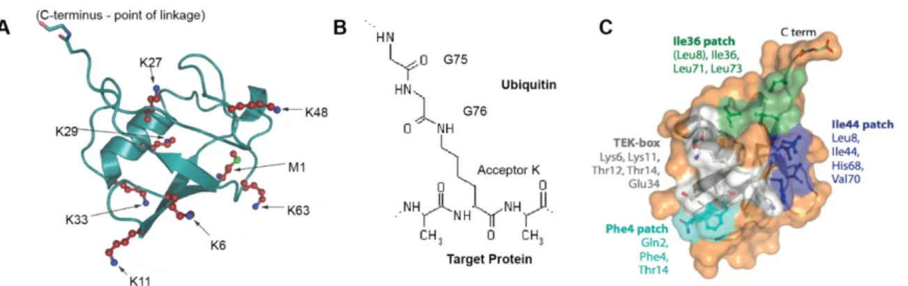
Much of the communication potential of the ubiquitin system is based on the unique properties of ubiquitin itself. Ubiquitin is a highly stable protein of 8.5kDa and 76 amino acids with a chain ball-like structure (Vijay-Kumar et al. 1987) whose surface presents several recognition patches that have been implicated in binding to E3 ubiquitin protein ligases, UBPs and DUBs (Dikic et al. 2009; Kamadurai et al. 2009; Cui et al. 2010; Ye et al. 2011) (Fig. 1.1.C). Ubiquitin is covalently conjugated onto substrate proteins by an isopeptide bond between the C-terminal glycine 76 of ubiquitin and usually the side chain amine of a lysine residue in the substrate protein (Fig. 1.1B). This reaction can lead to monoubiquitination or multi-monoubiquitination of a substrate protein. Moreover, ubiquitin itself disposes of seven lysines and its first methionine that can act as ubiquitin linking sites and enable the formation of ubiquitin chains of different topologies (Fig. 1.1A). The particular conformation of the chains is known to expose or restrict access to ubiquitin recognition patches, determining the type of ubiquitin-binding proteins that can be recruited and thus affecting the induced downstream signaling response (Komander and Rape 2012).
Figure 1.1. Ubiquitin structure characteristics. A. Structure of ubiquitin indicating that all its linking residues: seven lysine residues (red, with blue nitrogen atoms) and a methionine (with a green sulfur atom). The lysines are located on different surfaces of the molecule; M1 is the linkage point in linear chains, and is spatially close to K63. The C-terminal G75-G76 motif involved in isopeptide bond formation is indicated (red oxygen atoms, blue nitrogen atoms). B. Representation of the isopeptide bond between ubiquitin and a target protein. C. Representation of ubiquitin’s surface indicating four recognition patches in different colors. The name of each patch and the residues that form part of it are indicated in the same color as the patch’s surface. Adapted from (Komander 2009) and (Komander and Rape 2012)
1.1.2. The writers: The enzymatic cascade of ubiquitin conjugation
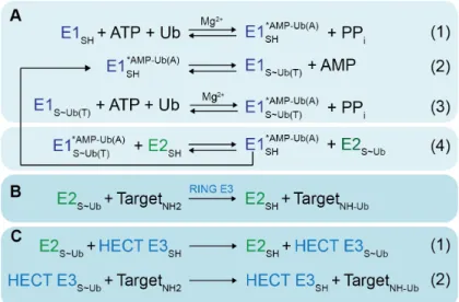
The writer system that leads to ubiquitin chain formation and conjugation onto substrates is composed of three types of enzymes that work in a hierarchical fashion (Hershko et al. 1983): E1s (ubiquitin-activating enzymes), E2s (ubiquitin-conjugating enzymes), and E3s (ubiquitin protein ligase enzymes). E1 enzymes begin the cascade by using the hydrolysis of ATP to catalyze the formation of a phosphodiester bond between the C-terminus of ubiquitin and the phosphate group of AMP, producing ubiquitin adenylate (AMP-Ub). Next, the sulfhydryl group of the E2 active cysteine attacks the AMP-Ub, forming a high-energy thioester bond with ubiquitin’s C-terminus and displacing AMP. The E1 then catalyzes for a second time the adenylation of a ubiquitin monomer and forms a non-covalent complex with it in its adenylation domain. The double-ubiquitin-loaded E1 can then facilitate the transfer of the thioester-bound ubiquitin from E1 to an active cysteine in E2 (Haas et al. 1982; Haas et al. 1983; Schulman and Harper 2009; Schäfer et al. 2014). Subsequently, the ubiquitin-charged E2 can bind to E3 enzymes that lack an active cysteine (RING family) or that possess an active cysteine (HECT and RBR families); in the first case the E2 transfers the charged ubiquitin directly to a substrate bound to the
E3, and in the second case the E2 transfers the ubiquitin to the active cysteine of an E3, which then conjugates the ubiquitin onto a substrate (Deshaies and Joazeiro 2009; Stewart et al. 2016) (Fig. 1.2).
Figure 1.2. Generalized enzymatic mechanisms of ubiquitin (Ub) transfer between enzymes and ultimately to a target. * refers to a noncovalent complex, ~ refers to a high-energy thioester bond, - refers to a covalent bond (phosphodiester in AMP-Ub or isopeptide in Target-Ub). A. Initial steps catalyzed by E1. (1) E1 binds Mg2+, ATP and a Ub, and catalyzes the acyl-adenylation of the Ub’s C-terminus. (2) E1 catalytic cysteine attacks the Ub~AMP intermediate, to form the covalent thioester-linked E1~Ub intermediate. (3) E1 then adenylates a 2nd Ub molecule, such that E1 binds 2 Ub molecules: Ub(T) is thioester- linked to E1’s catalytic cysteine; Ub(A) is associated noncovalently at the adenylation site. (4) Doubly-Ub-loaded E1 binds an E2 and Ub(T) is transferred from the E1 to the E2 catalytic cysteine. B. RING E3s enhance Ub transfer from E2 to a target. C. HECT and RBR E3s (represented by the HECT family in this figure) contain a catalytic cysteine, and (1) form a covalent thioester intermediate with a Ub prior to Ub ligation to a target lysine (2). Adapted from (Schulman 2011)
The human genome encodes 2 E1s, 37 E2s and more than 600 E3s (Li et al. 2008; Komander 2009), making the E3 enzymes the most diverse and evolutionarily refined actors of the cascade. E3 enzymes regulate target specificity and, together with E2 enzymes, direct bond formation.
E3 enzymes are classified in three categories based on the structure of their E2-binding domain and on their ubiquitin transfer mechanism (Fig. 1.3): (i) The Really Interesting
New Gene (RING) family catalyzes the single-step transfer of ubiquitin from the E2 to the substrate, while (ii) the Homologous to E6AP C-Terminus (HECT) and (ii) the RING-Between-RING (RBR) families ubiquitinate substrates in a two-step reaction where ubiquitin is first transferred from the E2 to an active cysteine within the E3 catalytic domain and then from the E3 to the substrate (Huibregtse et al. 1995; Deshaies and Joazeiro 2009; Smit and Sixma 2014). I will further expand on the mechanisms of action of these three E3 families in the section 1.2.
Figure 1.3. Classes of E3 ubiquitin ligases. Simplified representation of domain structure and the reactions catalyzed by three classes of E3 ubiquitin ligases. A. Schematic of Really Interesting New Gene (RING) E3-mediated catalysis. The RING domain binds E2~ubiquitin (~ indicates a thioester bond) and a substrate-binding domain recruits the substrate. Ubiquitin is transferred directly from the catalytic cysteine of E2 to a substrate lysine. B. Homologous to E6AP Carboxyl Terminus (HECT) E3 catalysis. The N-lobe of the HECT domain binds E2~ubiquitin and a substrate-binding domain recruits the substrate. Ubiquitin is transferred from E2 to the catalytic cysteine of the C-lobe of the HECT domain and subsequently to a substrate lysine. C. RING-between-RING (RBR) E3 catalysis. the RING1 domain binds E2~ubiquitin and ubiquitin is transferred from E2 to the catalytic cysteine of RING2 and then to a substrate lysine. Adapted from (Buetow and Huang 2016).
In addition to the previously described types of E3 ligases, which are present in eukaryotes; it has been shown that despite having no intrinsic ubiquitination machinery, pathogenic bacteria encode bacterial E3 ligases (BELs) that have the capacity to manipulate the host ubiquitin system during infection (reviewed in (Ashida and Sasakawa 2016) and (Ashida et al. 2014)). Some of these BELs structurally and functionally mimic host HECT-type and RING-type E3s (Maculins et al. 2016; Ashida and Sasakawa 2016). However, a third
any eukaryotic E3 ligases (Quezada et al. 2009). They are characterized by the presence of a C-terminus NEL catalytic domain and N-terminal Leucine-Rich Repeat (LRR) domains which are required for substrate binding and also inhibit the NEL domain in absence of a substrate (Chou et al. 2012).
1.1.3. The ubiquitin code: Signaling implications of ubiquitination
Ubiquitin can be singularly conjugated or attached to substrates as chains with different conformations determined by the residue that links one ubiquitin to the next. This can be seen as words (ubiquitin) that associate and form different sentences (ubiquitin chains), and constitutes the base of a system that D. Komander and M. Rape have called the “ubiquitin code” (Komander and Rape 2012). The panoply of ubiquitination chain types attached to substrates by ubiquitin conjugating enzymes can be recognized and disassembled by specific DUB enzymes, contributing to the dynamic regulation of the code (Mevissen and Komander 2017). To interpret the ubiquitin code, cells count on an array of UBPs with a variety of ubiquitin binding domains (UBDs) that specifically recognize specific types of ubiquitin chains and can then mediate particular cellular responses (Husnjak and Dikic 2012) (Fig. 1.4A).
It has been estimated that the majority of ubiquitin in mammalian cell lines is conjugated as a single ubiquitin (>60%), most likely due to monoubiquitination of histone H2A, which is one of the most abundant cellular components (Clague et al. 2015). Monoubiquitination of substrates is typically associated with alterations of intra- or intermolecular interactions that in turn affect their localization, complex formation or activity. After monoubiquitination, ubiquitin chains with K48- and K63- linkages are the second most abundant types of chains. K48-chains are mainly involved in the targeting of proteins for proteasomal degradation. While K63-chains often act as secondary messengers and scaffolds, allowing the formation of rapid and reversible signaling complexes involved in processes such as the activation of the Nuclear Factor kappa B (NF-kB) transcription factor, DNA repair, innate immune responses, clearance of damaged mitochondria, and protein sorting (Komander and Rape 2012).
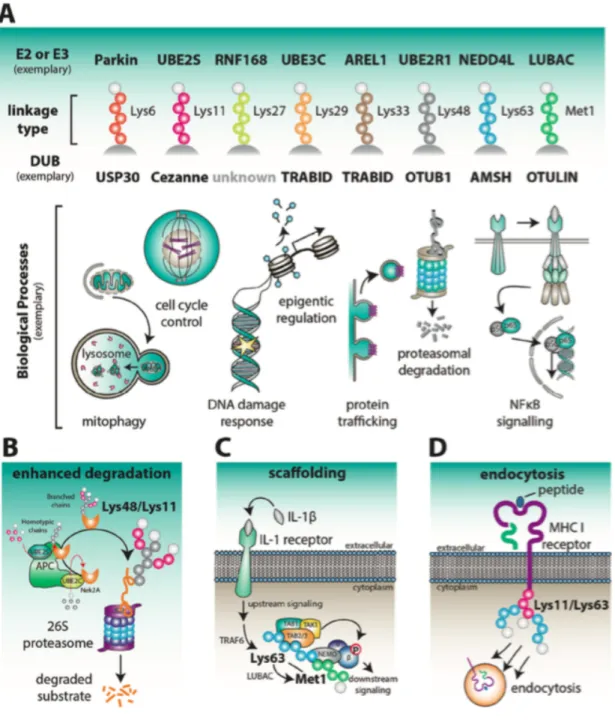
Figure 1.4 Physiological roles associated with individual chain types. A. Examples of E2 or E3 enzymes that assemble and DUBs that disassemble ubiquitin chains with linkage preferences is indicated. Below, illustrations show some of the biological processes that determined ubiquitin linkage types have been associated with. B. APC/C is active during early mitosis and modifies cell cycle regulators such as Nek2A with Lys48/Lys11-linked branched polyubiquitin. In this process, UBE2C first assembles short chains on the substrates, and these are then elongated on each ubiquitin by Lys11-linked polymers. Lys48/Lys11 branched chains enhance proteasomal degradation.
C. Mixed or branched Lys63/Met1-linked chains serve as protein scaffolds at immune receptors, such as IL-1 receptors, to promote NF-kB signaling. D. A viral E3 ligase initiates endocytic internalization of the MHC class I receptor through the attachment of mixed or branched Lys11/Lys63-linked ubiquitin chains. Taken from (Swatek and Komander 2016).
Proteomic studies have revealed that chains linked by the remaining 5 lysines (K6-, K11-, K27-, K-29-, K33-) and the first methionine (M1-) of ubiquitin also exist in cells but in much smaller proportions and are referred to as "atypical chains". In human cells, conjugated K11-linked chains increase in abundance during mitosis and early G1 phase and have, accordingly, been found to target cell cycle regulators for degradation by the proteasome, which is reminiscent of the function of K48-linked chains. K11 chains are produced by specific E3 enzymes like APC/C (anaphase promoted complex) in partnership with the E2 Ube2S. M1-linked chains are quickly synthetized following activation of inflammatory signaling cascades and are recognized by UBPs with linkage-specific UBDs, such as the ubiquitin binding in ABIN and NEMO (UBAN) domain (Yau and Rape 2016). Like K11 chains, M1-chains are assembled by particular E3 enzymes, like the Linear Ubiquitin Chain Assembly Complex (LUBAC), which play pivotal roles in immune signaling and NF-kB activation (Yau and Rape 2016). The four other types of lysine linkages of ubiquitin are much less characterized and seem to be assembled by E3 ligases that have mixed specificity. K6-linked chains are observed during the removal of damaged mitochondria from cells, K-27 chains seem to be involved in regulating DNA repair and autoimmunity, K29-chains are reported to have roles in proteasomal degradation, and K33-linked chains appear to regulate trafficking through the trans-Golgi network (Swatek and Komander 2016; Yau and Rape 2016).
So far, I have described homotypic chains, that is, chains where ubiquitin monomers are connected by a single type of linkage. However, heterotypic chains are also formed on substrates, and they can be either of mixed or branched nature. Mixed chains are composed of ubiquitin subunits connected to only one ubiquitin subunit at a time by various lysines (or M1), while branched chains have ubiquitin subunits conjugated to more than one lysine or the M1 residue at a time. These kinds of chains are proving to be functionally diverse, participating in numerous signaling cascades due to their ability to attract several UBPs in specific combinations and thus elicit unique reactions as exemplified in figure 1.4B-D (Swatek and Komander 2016).
increased enormously by recent discoveries showing that ubiquitin can be subjected to phosphorylation, acetylation, and modification by Ubls (like SUMO and Nedd8), adding another layer of regulation and of interaction with other PTM systems (Cui et al. 2010; Herhaus and Dikic 2015; Swatek and Komander 2016; Yau and Rape 2016).
Additionally, pathogenic bacteria strategically modify ubiquitin and Ubls to interfere with the host ubiquitination system and achieve successful infection. For example, the bacterial effector Cif (cycle inhibiting factor) family encoded by Burkholderia pseudomallei and by enteropathogenic Escherichia coli can deamidate glutamine 40 of ubiquitin and Nedd8, thereby inhibiting ubiquitin chain extension and hampering the Nedd8-dependent activation of CRLs (Cullin-RING Ligases) (Cui et al. 2010). Similarly, arginine phosphorybolisation of ubiquitin induced by SdeA produced by Legionella pneumophila has been shown to impair host ubiquitin-dependent processes (Bhogaraju and Dikic 2016).
1.1.4. When ubiquitination goes awry: Implications in human diseases
Considering the widespread implication of ubiquitination in cell signaling, it is not surprising that the deregulation of the ubiquitin system contributes to the development and progression of several pathologies including cancer, neurodegenerative diseases, autoimmunity, metabolic and inflammatory disorders, infection and muscle dystrophies (reviewed in (Popovic et al. 2014)).
Disease-associated perturbations in the ubiquitin system may occur at multiple levels: i) at any point during the multi-step process of ubiquitin conjugation, commonly via mutation or deletion of E1, E2, E3 enzymes, or of the substrate itself, ii) during ubiquitin recognition (by de-regulation of UBPs), or iii) during de-ubiquitination (by de-regulation of DUBs) (Popovic et al. 2014; Groen and Gillingwater 2015). One of the best-known examples of an E3 ligase whose perturbation leads to a pathology is E6AP (also known as Ube3a). Genetic alterations on E6AP that result in loss of function are known to cause Angelman syndrome, a rare neurogenetic disorder characterized by severe mental retardation, speech impairment, ataxia, seizure and frequent busts of laughter (among other symptoms) (Buiting et al. 2016). Moreover, several types of cancer and immune
pathologies present alterations in the canonical NF-kB pathway, which heavily relies on the conjugation and recognition of different types of ubiquitin chains (as described in section 1.1.2). For instance, it has been shown that patients with inherited deficiency in HOIL-1 (an E3-ligase present in the LUBAC complex that regulates NF-kB activation) suffer from chronic autoinflammation, muscular amylopectinosis and susceptibility to bacterial infections (Boisson et al. 2012). Another example is the DUB A20/TNFAIP3, which is considered a tumor suppressor due to its role in restraining exacerbated inflammation via the NF-kB pathway. Concordantly, mutations in A20/TNFAIP3 that decrease its expression or compromise its activity are commonly found in patients with lymphomas as well as in patients suffering from inflammatory conditions including rheumatoid arthritis, psoriasis, systemic lupus erythematosus, celiac disease, Crohn’s disease and diabetes (Hymowitz and Wertz 2010; Ma and Malynn 2012). Also associated with the NF-kB pathway is the UBP and autophagy adaptor Sequestosome 1 (SQSTM1, also known as p62). A mutation near the UBD of SQSTM1/p62 has been shown to cause Paget disease of bone, a common and chronic skeletal disorder (Laurin et al. 2002). In addition to inflammation, one of the key ubiquitin-dependent processes deregulated in cancer is genomic instability. For instance, FANCL, an E3 ligase whose activity is necessary for the correct localization of DNA repair factors (Garcia-Higuera et al. 2001), has been found to be mutated in hereditary ovarian and breast cancer as well as in Fanconi anemia, a rare cancer-prone genetic disease characterized by chromosomal instability (Peng et al. 2007; Xie et al. 2010).
Due to the increasingly recognized implication of the ubiquitin system in disease, great efforts have been made towards the development of inhibitors and agonists of the enzymes of the ubiquitin system (E1, E2, E3 and DUBs) for therapeutic applications (Huang and Dixit 2016). More about this topic, focusing on regulatory strategies of HECT E3 ligases, can be found in section 1.3.
1.2. E3 ubiquitin ligases: focus on the HECT family.
As seen briefly in section 1.1.2, the E3 ubiquitin ligases (E3s) are the main contributors of specificity in the ubiquitin conjugation system. In accordance with this role there is a great variety of these enzymes (>600 in humans), which are classified in three types: RING, RBR, and HECT. In this section I will expand on the characteristics of each family of E3s and their mechanisms of action with a special emphasis, at the end of the section, on the HECT family.
1.2.1. RING E3 ligases.
The great majority of E3 ligases belong to the RING family. Bioinformatic analyses estimate that there are around 600 members in humans (Li et al. 2008). Example members of this family are c-Cbl, which is essential for ubiquitination and lysosomal degradation of the epidermal growth factor receptor (EGFR) (Levkowitz et al. 1999), and APC/C, which promotes ubiquitination and proteasomal degradation of anaphase inhibitors, ensuring timely chromatid separation and mitotic exit (Craney et al. 2016).
RING E3s are characterized by a catalytic RING domain that requires the coordination of two zinc ions to fold correctly, or a U-box domain, which closely resembles RING domains in structure but does not coordinate zinc ions (Deshaies and Joazeiro 2009). RING E3 ligases are very diverse and can be active as monomers, homodimers, heterodimers or as part of large multi-subunit complexes (as reviewed in (Buetow and Huang 2016)). Examples of these large complexes are APC/C (Anaphase promoting complex/cyclosome) (Chang and Barford 2014) and the E3 Cullin-RING ligases (CRL), a large family of mutli-subunit RING E3s (Petroski and Deshaies 2005a; Lydeard et al. 2013). The prototypical CRL is the Skip/Cullin/F-box complex (SCF).
RING ligases simultaneously bind the substrate protein and the ubiquitin-loaded E2, and mediate the direct transfer of ubiquitin from E3 to substrate. It has been observed that the RING-mediated approximation of E2~Ub and Substrate is not sufficient to reach optimal transfer rates (Seol et al. 1999; Petroski and Deshaies 2005b; Saha and Deshaies
2008) and that the nature of the E2-RING interaction is important since not all E2-RING pairings lead to substrate ubiquitination (Brzovic et al. 2003; Ozkan et al. 2005). In 2012, the crystal structures of RNF4 and BIRC bound to E2~Ub were elucidated (Dou et al. 2012; Plechanovová et al. 2012) and they revealed that RING E3s are more than scaffolds: they prime ubiquitin for transfer by stabilizing the highly dynamic E2~Ub into a closed conformation that renders it more reactive towards transfer (Page et al. 2012; Dou et al. 2012; Pruneda et al. 2012; Soss et al. 2013). This mechanism has been recently expanded from homodimeric RINGs to monomeric RINGs (Dou et al. 2013; Buetow et al. 2015; Branigan et al. 2015), indicating that the mechanism might be universal to many other RING E3-E2 pairs.
After the first ubiquitin is transferred to a substrate, the formation of polyubiquitin chains often ensues. RING-E3 ligases can catalyze chain elongation much faster than chain initiation and in cooperation with a single E2 enzyme. For example, the E2 Cdc34 (also known as UbcH3 or UBE2R1) is 5 to 30 times faster at chain elongation than at initiation and is specific of Lys48 (Petroski and Deshaies 2005b). In other cases, chain initiation and elongation can be carried out by separate E2s. An example of this is APC/C-mediated polyubiquitination: the E2 UbcX (also known as UbcH10 or UBE2C) has a preference for monoubiquitination or short ubiquitin chains, whereas the E2 Ubc4 (also known as UbcH5 or UBE2D) usually assembles long polyubiquitin chains (Yu et al. 1996), the authors of this study proposed that they operate sequentially.
In RING E3- catalyzed ubiquitination, chain linkage specificity (chain topology) is thought to be determined by the E2 enzyme cooperating with the RING E3 ligase (Chen and Pickart 1990; Haas et al. 1991; Hofmann and Pickart 1999).
1.2.2. RBR E3 ligases.
The RBR E3 ligases are viewed as hybrids between RINGs and HECTs and were only recently defined as a distinct type of E3 ubiquitin ligases (Wenzel et al. 2011). They are characterized by a catalytic domain composed of two RING fingers (RING1 and RING2) and a central in-between RING (IBR) zinc-binding domain. The RING1 domain binds to
the loaded E2 and the RING2 domain contains an active cysteine residue capable of forming a reversible thioester intermediate with ubiquitin. Besides the common RING1-IBR-RING2 motif, RBR family members possess other domains, which gives diversity to the family (Wenzel et al. 2011; Stieglitz et al. 2012; Smit et al. 2012; Spratt et al. 2014).
There are 14 RBRs encoded in the human genome, and the three best-characterized are PARKIN, commonly mutated in Parkinson disease; HOIP (HOIL-1L interacting protein), the central E3 subunit of LUBAC (linear ubiquitin chain assembly complex) in NF-kB signaling; and HHARI (human homologue of Ariadne) (Kitada et al. 1998; Kirisako et al. 2006; Ikeda et al. 2011).
RBR proteins are commonly found in auto-inhibited conformations that are not competent for ubiquitin transfer. Release from the auto-inhibited state occurs once RBRs bind to E2~Ub, as observed by Lechtenberg and colleagues in a study where they solved the structure of the fully active human HOIP in complex with an E2~Ub (Lechtenberg et al. 2016). Moreover, they observed that contrary to RINGs, and similar to HECTs (see section below), HOIP stabilizes E2~Ub in an extended conformation where the E2~thioester bond is juxtaposed with the RING2 active site cysteine and is optimal for transfer. This is in agreement with the fact that RBR’s RING1 finger alone cannot promote ubiquitin transfer (Wenzel et al. 2011), as it does not activate E2~Ub like a canonical RING domain (in a closed conformation).
Little is known about how chain type is determined by RBRs. So far, the only well studied case is HOIP, where M1-chain specificity has been shown to depend on the presence of a linear ubiquitin chain-determining domain in HOIP (Smit et al. 2012; Riley et al. 2013).
1.2.3. HECT E3 ligases.
Members of this family of E3 ligases were among the first E3 enzymes to be cloned, and are the best functionally characterized among the thioester-forming E3s. The ubiquitin ligase function of the HECT family of proteins was first observed through studies of the degradation of the p53 tumor suppressor in cells infected by oncogenic forms of human
papillomavirus (HPVs) (Scheffner et al. 1990). Biochemical studies revealed that p53 degradation depended on the HPV E6 gene and a host protein named E6-AP (E6-associated protein), and that the complex of these proteins functioned as a p53-specific E3 {Scheffner:1993ur}. Further studies determined that E6-AP contained a conserved region of about 350 amino acids towards its C-terminus (Huibregtse et al. 1995) and that within this region, called the homologous to E6-AP C-terminus (HECT) domain, was a highly conserved cysteine located around 35 residues upstream of the C-terminus that is required for E6-AP activity (Scheffner et al. 1995).
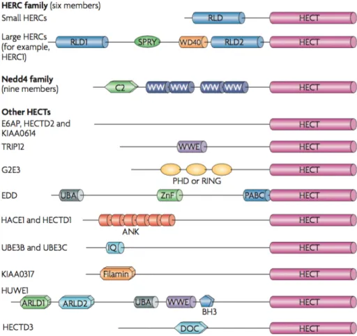
Most HECTs arose before the emergence of animals or very early in metazoan evolution (Marín 2010), and the human genome encodes 28 HECT E3 ligases (Rotin et al. 2009). All members of the HECT family are characterized by a HECT domain located at their C-terminus and most also contain a variety of protein-protein or protein-lipid interaction domains towards their N-terminus (Rotin et al. 2009). Based on their N-terminal domain architecture, the 28 human HECT E3s are commonly divided into three sub-families (Fig. 1.5): (i) the Nedd4 family has 9 members in humans and is characterized by C2 and WW domains that allow them to bind phospholipids and PY motifs in substrate proteins. This is the best-studied family, most of what is known about structure and enzymatic mechanisms of HECT E3s come from studies done with NEDD4 E3 ligases; (ii) the HERC family, with 6 human members, contain regulator of chromosome condensation 1 (RCC1)-like domains (RLDs) that are not well described. This family can be divided in two groups, the large HERCs (>500 kDa) and the small HERCs (around 100kDa), which have a single RLD; (iii) the “other” family, with 13 human members, comprises HECTs which contain protein-protein interaction domains different from the previously mentioned
Figure 1.5. The mammalian HECT E3 ligases. The 28 human HECT E3 ligases are often grouped into three families. Two of these, the Nedd4 family and the HERC family, can be clearly distinguished by their domain architecture. The HERC family members, which contain one or more regulator of chromosome condensation 1 (RCC1)-like domains (RLDs), can be divided into two groups — the small HERCs that carry a single RLD and the large HERCs that contain more than one RLD and additional domains, such as SPRY and WD40. Nedd4 family members are characterized by a unique domain architecture, with all members containing an N-terminal C2 domain and two to four WW domains. The remaining HECT proteins contain a myriad of domains (as shown). From (Rotin et al. 2009).
The details of the enzymatic mechanism by which HECT E3s catalyze the transfer of ubiquitin onto a substrate have proven to be elusive for a long time. Currently, this knowledge gap is rapidly closing thanks to the increasing abundance of structural information that captures the different stages of the two step-ubiquitin transfer reaction that HECTs catalyze (Buetow and Huang 2016; Zheng and Shabek 2017). The catalytic HECT domain has a bi-lobed structure, where the two lobes are connected by a flexible hinge loop. The N-terminal lobe (N-lobe) binds to E2~Ub and the C-terminal lobe
(C-lobe) contains the catalytic cysteine (Huang et al. 1999). The flexible hinge allows the two lobes to rotate, a characteristic that is necessary for ubiquitin transfer (Verdecia et al. 2003; Ogunjimi et al. 2005). Thanks to the elucidation of the structure of Nedd4-2 bound to the E2 UBE2D2 (UbcH5b) loaded with ubiquitin (Kamadurai et al. 2009), the current model of transthiolation is the following: Nedd4-2's N-lobe binds to E2~ub and upon rotation of the hinge, the C-lobe binds ubiquitin. Interestingly, this interaction stabilizes E2~Ub in an open conformation, contrary to the closed conformation induced by RING E3s. This arrangement brings together the two catalytic cysteine residues of E2 and E3 and promote the formation of the HECT E3~Ub intermediate. This mechanism is likely to be shared among other HECT E3s (Buetow and Huang 2016). Once the E2 leaves, the interaction between the Ub and the C-lobe remains the same, as evidenced in the crystal structure of Nedd4-1~Ub (Maspero et al. 2013). In the next step, however, Kamadurai and collaborators observed that the N-lobe of the HECT domain rotates almost 130º from its previous position (while bound to E2~ub or conjugated with Ub). This change juxtaposes the active cysteine bound to the Ub and the acceptor lysine of the substrate (in this case the yeast Nedd4 homolog Rsp5 and its target Sna3) (Kamadurai et al. 2013).
Not much is known about how HECT E3s catalyze chain elongation and determine linkage specificity. Kim and colleagues demonstrated that the linkage specificity of several HECT E3s depends on the particular sequence identity of the C-lobe of the ligase’s HECT domain and is, contrary to RING E3s, independent of the identity of the cooperating E2 enzyme (Kim and Huibregtse 2009). Nedd4 E3s have a UBD (aka ubiquitin-binding exosite hereafter referred as UBEx) within the N-lobe of their HECT domain. It has been shown that the UBEx is critical for polyubiquitin chain formation (Ogunjimi et al. 2005; French et al. 2009; Kim et al. 2011; Maspero et al. 2011; Maspero et al. 2013). Indeed, it has been reported that inhibiting the binding of ubiquitin to the UBEx ,either by mutation or by small chemical inhibitors, impairs polyubiquitin chain elongation but not E2-/E3- transthiolation nor the conjugation of the first ubiquitin to a substrate (Kim et al. 2011; Maspero et al. 2011; Kathman et al. 2015). On the other hand, Zheng and colleagues recently showed that occupation of the UBEx by ubiquitin variants in different members
of the Nedd4 family can influence many properties of the reaction both positively and negatively (Zhang et al. 2016).
Figure 1.6. Schematic showing the HECT E3 catalytic cycle. In the absence of any binding partner, the C-lobe can rotate relative to the N-lobe thanks to the hinge loop. Upon encountering E2~ubiquitin, the N-lobe binds E2 and the C-lobe rotates to bind ubiquitin, thereby juxtaposing the catalytic cysteine residues from E2 and E3. Upon ubiquitin transfer onto the catalytic cysteine of the C-lobe, E2 is released. E3 binds the substrate through its substrate-binding domain and the C-lobe undergoes rotation to juxtapose the catalytic cysteine of E3 and a substrate lysine for ligation. The actual order in which HECT E3 recruits E2~ubiquitin and substrate is not yet known. From (Buetow and Huang 2016)
The number of HECT E3 ligases represent less than 5% of all the E3 ligases found in humans, yet they have been shown to have important physiological roles in many biological processes such as, fetal growth and development, regulation of DNA damage and replication, modulation of immune responses, among other processes. Consequently, their de-regulation leads to development of different pathologies (reviewed in (Scheffner and Kumar 2014) and (Rotin et al. 2009)). Therefore, it is important to understand how this family of E3 ligases is regulated.
1.3. Regulation of HECT E3s.
The activity of E3 ligases are tightly regulated on a variety of levels including cooperation with E2 enzyme(s), E3 processivity and substrate recognition. The regulation of these processes is usually mediated by structural rearrangements triggered by interaction with auxiliary factors or by post-translational modifications (PTMs) such as phosphorylation and ubiquitination (Ogunjimi et al. 2005; Wiesner et al. 2007; Rotin et al. 2009; Maspero et al. 2011). Currently, most of what is known about the regulation of HECT E3 ligase
activity comes from studies focusing on members of the Nedd4 subfamily. Therefore, this section will mainly discuss the literature involving the regulation of this subfamily of HECT ligases, then mention a couple of recent studies involving two HECT ligases outside of the Nedd4 subfamily, and finally go through some examples of regulation of HECT ligase activity by small chemical inhibitors.
1.3.1. Regulation of Nedd4 family ligases.
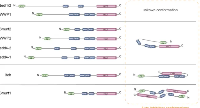
Nedd4 family ligases share a common domain structure and, at steady state, most of them favor inhibitory conformations that protects them and their targets from untimely ubiquitination (Fig. 1.7). Smurf2, Nedd4-1 (aka Nedd4), Nedd4-2 (aka Nedd4-L), and WWP2 are negatively regulated under basal conditions through intramolecular interactions involving the C2 and HECT domains (Wiesner et al. 2007; Mund and Pelham 2009; Wang et al. 2010a); (Bruce et al. 2008). In the case of Itch, auto-inhibitory conformation is mediated by its WW and HECT domains and is proposed to occur intra-molecularly (Gallagher et al. 2006; Riling et al. 2015). Despite the high degree of homology between Smurf1 and Smurf2, the cis interaction between the C2 and the HECT domains that inactivates Smurf2 is not found in Smurf1 due to the shorter linker region between its HECT and C2 domains (Wiesner et al. 2007). Instead, it has been reported that the full-length Smurf1 forms homodimers through intermolecular contacts mapped to a fragment containing the C2 and the WW domains of one molecule and the HECT domain of the partner (Wan et al. 2011).
The details of how these closed conformations block enzymatic activity is not yet clear for all the ligases mentioned above but, to date, the best characterized one is Smurf2. As previously mentioned, Smurf2 auto-inhibition is governed by the interaction between the C2 and HECT domains; specifically, the C2 domain binds the N-lobe of the HECT domain and restricts movement of the C-lobe, which makes the active cysteine inaccessible for an incoming E2~Ub and thereby precluding transthiolation (Wiesner et al. 2007; Mari et al. 2014). Moreover, the C2 domain partially buries the UBEx, which is essential for E3 processivity (Mari et al. 2014). Another enzymatic step affected by the auto-inhibitory
conformation of Smurf2 is the recruitment and binding of the its associated E2 (UbcH7); it has been shown that Smurf2 interacts very weakly with UbcH7, and that binding to the adaptor protein SMAD7 (disrupting its intra-inhibitory interactions) is necessary for a functional interaction between Smurf2 and UbcH7 (Ogunjimi et al. 2005).
Figure 1.7. Domain structure of the nine members of the Nedd4 family and their auto-inhibitory conformations. Smurf2, WWP2, Nedd4-2 and Nedd4-1 form intra-molecular interactions between their C2 and HECT domains, as exemplified with Smurf2; Itch auto-inhibitory conformation is similar, but the HECT domain interacts with WW domains instead of the C2 domain. Smurf1 presents inhibitory interactions between the C2 domain, a WW domain and the HECT domain in
trans instead of cis, forming an inactive homo-dimer.
Similar to Smurf2, nuclear magnetic resonance (NMR) and biochemical analysis have shown that in Nedd4-1 the C2 domain has the potential to regulate E3 activity by keeping the HECT domain in a low-activity state where its ability for transthiolation and non-covalent ubiquitin binding is impaired (Mari et al. 2014). Contrary to Smurf2, recent studies have shown that Itch can bind to E2s while in its auto-inhibitory conformation. However, the transfer of ubiquitin from E2 to the active cysteine of Itch is thwarted (Riling et al. 2015).
Figure 1.8. Mechanisms of regulation of Nedd4-family ligases. An increasing number of studies have shown that adaptors (orange) and PTMs such as ubiquitination and phosphorylation (kinases shown in mauve) can promote (lighter orange or mauve, and pointed arrow), inhibit (darker orange or mauve, and blunt arrow) or modulate (circle-ending line) one or more aspects of Nedd4-ligases activity; such as the ligase localization and interaction with substrates, its binding to E2 enzymes, and its intrinsic E3 catalytic activity. Details about how each adaptor and PTM modulates the different E3 ligases are discussed in the main text.
The last ten years have seen a boom in the number of studies exploring how the cell controls the activity of Nedd4 E3 ligases, and a multitude of adaptors and PTM events (mainly phosphorylation) have been shown to modulate one or more aspects of Nedd4-ligases activity. This includes the disruption or promotion of (i) their auto-inhibitory conformations, which affects their intrinsic catalytic activity; (ii) their cooperation with E2 enzymes; (iii) their cellular localization; and (iv) their substrate-affinity (Fig. 1.8). As shown in figure 1.8, some adaptors bind and regulate one or more E3 ligases by several mechanisms. Despite this, for practical purposes I will classify the PTMs and Adaptors into two groups: those which modulate the catalytic activity of the E3 ligase by affecting
their conformation, and those who modify the E3 ligase interaction with substrates by affecting its cellular localization and/or its affinity for its targets.
a. Release of auto-inhibition by adaptors
The Nedd4 family-interacting proteins (NDFIP) 1 and 2 have been described as activating adaptors targeting several Nedd-4 E3 ligases. Indeed, they promote the auto-ubiquitination of Itch, Nedd4-1, Nedd4-2, Smurf1, WWP1 and WWP2, as well as the ubiquitination of JunB, c-Jun and endophilin by Itch and Nedd4-1 (Mund and Pelham 2009). Mechanistically, it was shown that NDFIP1 binds multiple WW domains of Nedd4 ligases through its PY motifs and disrupts Nedd4 auto-inhibition (Riling et al. 2015). In contrast with NDFIP 1 and 2, most other adaptors reported to date activate only one or two E3 ligases.
Smurf2 is activated upon binding the adaptor protein SMAD7, whose expression is regulated by extracellular stimuli like the transforming growth factor beta (TGFβ) (Kee and Huibregtse 2007). SMAD7 PY motifs interact with the WW domains of Smurf2 and the N-terminus of SMAD7 interacts with the HECT domain of Smurf2, causing the release of the C2 domain (Wiesner et al. 2007; Aragón et al. 2012) and enabling Smurf2-mediated transthiolation. In addition, Smad7 activates Smurf2 in two other ways: first, it facilitates the recruitment of the E2 (Ogunjimi et al. 2005) and second, it directs Smurf2 to the plasma membrane, where it mediates its interaction with several substrates (Kavsak et al. 2000; Di Guglielmo et al. 2003; Izzi and Attisano 2004).
Smurf1 has been shown to be regulated by two adaptor proteins: CKIP-1 (casein-kinase-2 interacting protein -1) and with Cdh1 (Lu et al. 2008; Wan et al. 2011). Wan and colleagues showed that CKIP-1 and Cdh1 bind to Smurf1 and activate it by disrupting the formation of inhibitory Smurf1 dimers, thereby promoting Smurf1 autoubiquitination and ubiquitination of RhoA, one of Smurf1’s targets (Wan et al. 2011).
There are several examples of auxiliary proteins that activate Itch, notably Spartin and Numb. Spartin binds to Itch via its PY motifs and recruits it to lipid droplets. This
interaction increases Itch enzymatic activity at a specific cellular location and enables the ubiquitination of proteins present on the lipid droplets, such as adipohilin (Hooper et al. 2010). Di Marcotullio and colleagues showed that Numb activates Itch by disrupting inhibitory intramolecular interactions between its HECT and WW domains; moreover, Numb recruits Gil1 and mediates its interaction with Itch, leading to Gil1 ubiquitination and degradation (Di Marcotullio et al. 2011).
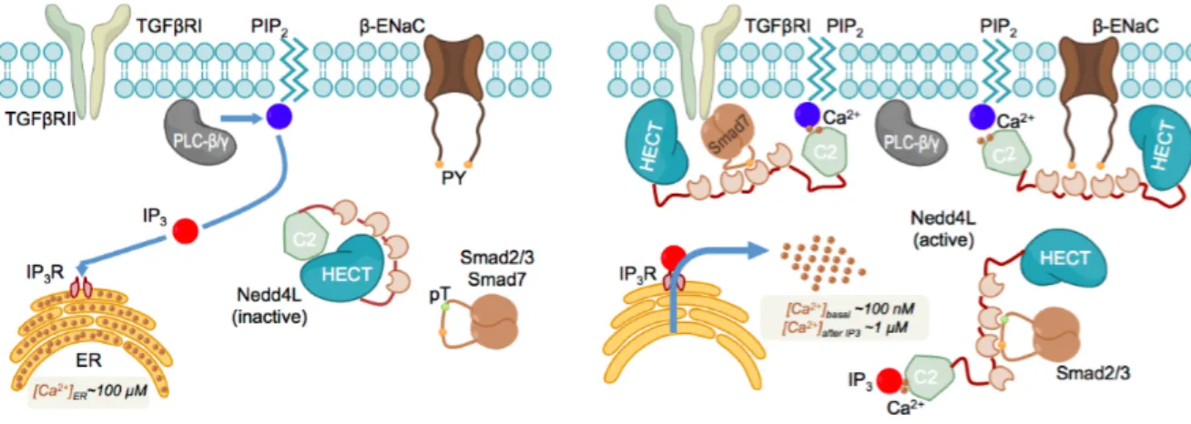
Figure 1.9. Activation and cellular localization of Nedd4-2 by IP3 and Calcium. IP3-induced Ca2+ delivery into the cytoplasm triggers the transition from the closed an inactive conformation of the ligase (left panel) to the active one (right panel). Once active, different WW domains in Nedd4-2 are able to recognize the ligase’s cytoplasmic targets including Smad2/3 and Smad7. The ligase is also able to relocate to the plasma membrane, where it possibly anchors to the IP3 head groups of the PIP2 lipids and targets the membrane receptors, such as TGFβRI and β- ENaC. From: (Escobedo et al. 2014).
Small cellular messengers can also act as activating adaptors that release the auto-inhibition of Nedd4 ligases. Wang and colleagues demonstrated that calcium ions release the C2 domain-mediated auto-inhibition in both Nedd4-1 and Nedd4-2 by disrupting the binding of the C2 domain to the HECT domain (Wang et al. 2010a). More recently, a study using NMR revealed that Ca2+ and inositol 1,4,5-triphosphate (IP3) bind to the C2 domain of Nedd4-2 using the same region that mediates the interaction with the HECT domain (Escobedo et al. 2014). Thus, the balance between the closed and open conformation of Nedd4-2 results from the competition between Ca2+, IP3, and the HECT
domain to bind the C2 domain. Therefore, the activity of Nedd4-2 depends on the intracellular levels of Ca2+ and IP3. IP3 is generated by hydrolysis of the membrane phospholipid phosphatidylinositol 4,5-biphsphate (PIP2) and is found in the cytosol and the endoplasmatic reticulum, where it binds to its receptor. Both IP3 and PIP2 can bind to Nedd4-2 in the presence of Ca2+, which enables Nedd4-2 to act either at the cytosol or at the membrane; where it can target specific substrates, including the cytosolic Smad7 (which makes Nedd4-2 an indirect regulator of Smurf2 activity) (Fig. 1.9) (Escobedo et al. 2014).
b. Modulation of auto-inhibition by PTM
Release of auto-inhibition by post-translational modifications has been less reported than activation via adaptors. Here, I will mention three cases where phosphorylation activates Nedd4 ligases and a recent study that shows that ubiquitination of Nedd4 and Rsp5 inhibits their activity in a proteasome-independent manner.
JNK1 kinase phosphorylates Itch at S199, S232, and T222. These modifications disrupt the auto-inhibited conformation of Itch mediated by its WW and HECT domains and therefore induces its activation (Gao et al. 2004; Gallagher et al. 2006). A second kinase, ATM, phosphorylates Itch in response to DNA damage and induces its activation (Santini et al. 2014). The authors of this study identify that phosphorylation of S161 is critical for ATM mediated activation of Itch and they propose that this modification disrupts Itch’s intra-inhibitory interactions, as reported for JNK induced phosphorylation. Following the same trend, Persaud and colleagues have described that following activation of FGFR1 or EGFR, the effector tyrosine kinase c-Src is activated, which then phosphorylates Nedd4-1 on Y43 (C2 domain) and Y585 (HECT domain). They demonstrate that phosphorylation of Nedd4-1 disrupts the C2-HECT interaction and thereby relieves inhibition of the E3 ligase, which results in the ubiquitination and degradation of substrates such as FGFR1 (Persaud et al. 2014).
Earlier this year, Attali and colleagues have shown that ubiquitination also affects Nedd4 ligases conformation and activity. Specifically, they demonstrated that ubiquitination of a
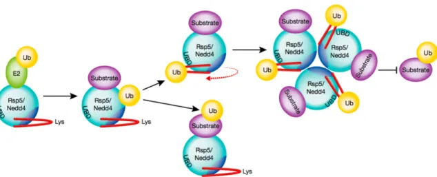
flexible alpha-helix in the HECT domain of Nedd4-1 and Rsp5 (Nedd4-1 homologue in Yeast) induces the formation of an inactive homo-trimer (Attali et al. 2017). The described mechanism relies on both ubiquitination of specific lysine residues located in the alpha-helix and on the presence of a UBEx within the HECT domain. Upon ubiquitination, the alpha helix rotates to approach its linked ubiquitin to the UBEx. This structural change clears a region that is required for oligomerization (Fig. 1.10). How this change in conformation inactivates NEDD4-1 and RSP5 is still unclear.
Figure 1.10. Schematic representation of how the Nedd4 family ubiquitin ligases self-regulate through auto-ubiquitination. Ubiquitin (Ub) is transferred from a ubiquitin-conjugating enzyme (E2) to either the Rsp5/Nedd4-1 active-site cysteine and then to E3-bound substrate, or onto a flexible α-helix on the E3. Upon auto-ubiquitination of the α-helix, the position of the helix rotates to interact with a conserved UBEx (“UBD” in the figure) opening access to an oligomerization domain and trimerization of Nedd4-1. Self-association renders the E3 inactive. From (Hill and Kleiger 2017).
Up to this point I have described adaptors and PTMs that directly influence E3 catalytic activity, and some that in addition modulate the localization of the E3 ligase, thereby dictating their interaction with a set of substrate proteins. In the next two sections I will show some examples of PTMs and auxiliary proteins that regulate substrate recognition, reportedly without affecting the E3 catalytic activity.
c. Regulation of substrate interaction by adaptors.
The 14-3-3 family members bind targets that have been phosphorylated at specific serine or threonine residues and are key regulators of a wide variety of cell signaling pathways mediated by phosphorylation (Muslin et al. 1996). 14-3-3 proteins have been shown to associate to Nedd4-2 following its phosphorylation by SGK1 (Serum- and Glucocorticoid-related Kinase 1) and PKA (Protein Kinase A). This association with 14-3-3 proteins inhibits the interaction between Nedd4-2 and its substrate ENaC (epithelial sodium channel) leading to reduced ENaC ubiquitination and subsequent degradation (Ichimura et al. 2005; Bhalla et al. 2005; Nagaki et al. 2006; Chandran et al. 2011).
Similarly, Oberst and colleagues demonstrated that Nedd4-binding partner 1 (N4BP1) binds to the second WW domain of Itch and inhibits its interaction with several substrates (Jun, p73 and p63) by binding competition, thereby preventing their ubiquitination (Oberst et al. 2007).
Auxiliary proteins can also promote enzyme-substrate interactions. Members of the α- and β-Arrestin families have been shown to bind to the WW domains of Itch, Nedd4-1 and Nedd4-2 and mediate their association with β2 adrenergic receptor, which leads to ubiquitination and recycling of the receptor (Shea et al. 2012; Han et al. 2013).
d. Regulation of substrate interaction by PTM.
Contrary to the activating effect of serine and threonine phosphorylation of Itch, tyrosine phosphorylation seems to negatively modulate the ability of Itch to selectively bind and ubiquitinate some of its targets, such as JunB and c-Jun. Gao and colleagues found that phosphorylation of a tyrosine within the PPXY motif of Itch by c-Abl inhibited its binding to c-Jun (Gao et al. 2006). Similarly, Fyn kinase phosphorylates Itch on Tyr-371, which inhibits its binding to JunB and therefore hampers JunB ubiquitination (Yang et al. 2006).
Numerous studies have shown that hormone-induced phosphorylation of Nedd4-2 by PKA, SGK and IKKβ inhibits Nedd4-2 interaction with ENaC, inhibiting its ubiquitination (reviewed in (Snyder 2009)). For instance, SGK1 phosphorylates Nedd4-2 at S221, S327
and T245 and increases the surface abundance of all three ENaC subunits (α-, β- and γ-), while Nedd4-2 phosphorylation by PKA at S221 and S327 specifically increases the abundance of α-ENaC (Ismail et al. 2014). These studies suggest that the pattern of phosphorylation on Nedd4-2, modulated by different kinases, controls its association with the different subunits of ENaC. Similarly, it has been shown that Nedd4-2 phosphorylation by SGK1 downstream mTORC2 activation results in reduced Nedd4-2-JunB interaction and increased JunB stability (Heikamp et al. 2014). In another study, it has been demonstrated that following their TGFβ-induced phosphorylation, SMAD2/3 interact with Nedd4-2 via its second WW domain (WW2), which results in their ubiquitination and degradation; and that SGK1 inhibits this interaction by phosphorylating two serine residues flanking Nedd4-2 WW2 domain (Gao et al. 2009).
Another remarkable case of modulation of substrate binding by phosphorylation was shown in a study by Cheng and colleagues. They find that PKA phosphorylates Smurf1 at Thr-306 and shifts Smurf1’s affinity for its substrates. Namely, phosphorylation at Thr-Thr-306 on Smurf1 reduces its affinity for Par6 but increases it towards RhoA (Cheng et al. 2011).
Finally, we have seen that ubiquitination of Nedd4 ligases can lead to inhibition of their catalytic activity by trimerization (Attali et al. 2017). Additionally, Woelk and collaborators reported an example of self-ubiquitination-dependent recruitment of substrates: they observed that self-catalyzed monoubiquitination of Nedd4-1 serves to recruit EPS15, which is subsequently monoubiquitinated by Nedd4-1 (Woelk et al. 2006).
In summary, Nedd4 ligases are regulated by auto-inhibition, either by intra-molecular interactions or by inter-molecular interactions, forming steady state homodimers like Smurf1 or ubiquitination-triggered trimers like Nedd4-1 and Rsp5. With the exception of these trimers, the auto-inhibitory conformations are stable at basal levels and upon a stimulus (phosphorylation or binding of an adaptor protein) they can be disrupted. Each particular mechanism of activation disrupts the same interaction but in response to different upstream signals and, as seen before, they can additionally dictate the subcellular localization of E3s, thereby influencing their access to substrates. In other cases, the