HAL Id: tel-02918052
https://tel.archives-ouvertes.fr/tel-02918052
Submitted on 20 Aug 2020HAL is a multi-disciplinary open access archive for the deposit and dissemination of sci-entific research documents, whether they are pub-lished or not. The documents may come from teaching and research institutions in France or abroad, or from public or private research centers.
L’archive ouverte pluridisciplinaire HAL, est destinée au dépôt et à la diffusion de documents scientifiques de niveau recherche, publiés ou non, émanant des établissements d’enseignement et de recherche français ou étrangers, des laboratoires publics ou privés.
Study of lymphocyte autophagy in normal and
autoimmune responses
Diane Murera Uwanyirigira
To cite this version:
Diane Murera Uwanyirigira. Study of lymphocyte autophagy in normal and autoimmune responses. Immunology. Université de Strasbourg, 2016. English. �NNT : 2016STRAJ068�. �tel-02918052�
École Doctorale des Sciences de la Vie et de la Santé (ED414)
THÈSE
Présentée par
Diane MURERA
Soutenue publiquement le
30 septembre 2016
Pour obtenir le grade de
Docteur de l’université de Strasbourg
Discipline :
Sciences de la vie et de la Santé
Spécialité :
Immunologie
THÈSE dirigée par :
Madame le Professeur Sylviane Muller Directrice de thèse
Institut de Biologie Cellulaire et Moléculaire (IBMC), Immunologie et Chimie Thérapeutique, CNRS UPR3572, Strasbourg, France
Membres du JURY :
Madame le Professeur Anna Katharina Simon Rapporteur Externe
Kennedy Institute of Rheumatology, University of Oxford, Oxford, UK
Madame le Docteur Julie Déchanet-Merville Rapporteur Externe
Unité CIRID, CNRS UMR 5164, Université de Bordeaux, Bordeaux, France
Monsieur le Docteur Philippe Kastner Rapporteur Interne
IGBMC, INSERM U964, CNRS UMR 7104, Université de Strasbourg, Ilkirch, France
Study of lymphocyte autophagy
in normal and autoimmune responses
Acknowledgements
First of all I would like to greatly thank the members of my Jury, Professor Anna Katharina Simon, Dr Julie Déchanet-Merville and Dr Philippe Kastner to have accepted to evaluate my work.
Je voudrais aussi remercier le Pr Sylviane Muller, ma Directrice de thèse et Directrice de l’Unité de recherche du CNRS UPR 3572, de m’avoir accueillie au sein de son laboratoire et de m’avoir permis d’effectuer mes travaux de thèse dans un environnement scientifique des plus favorable. Un grand merci aussi de m’avoir laissé une liberté et une autonomie qui ont grandement favorisé mon développement scientifique.
J’adresse mes remerciements au Fond National de la Recherche du Luxembourg pour le soutien financier apporté à ce projet de thèse.
Un grand merci au Dr Frédéric Gros sans qui cette thèse n’aurait véritablement pas été possible. Merci d’avoir consacré une (grande) partie de tes vacances à mes corrections. Promis c’est la dernière fois que je te demande ce service. Merci aussi de m’avoir fait confiance tout au long de ma thèse, de m’avoir aiguillé dans le bon sens quand il le fallait. Tu m’as donné la possibilité de faire évoluer ce projet mais aussi de m’épanouir aussi bien scientifiquement que personnellement. Malgré toutes tes responsabilités, tu as toujours su rester disponible à tout moment pour des échanges scientifiques et des échanges moins sérieux (je pense notamment à tes supers blagues et jeux de mots…) C’est vrai que niveau humour tu as aussi contribué à mon évolution.
En parlant d’humour je voudrais également dire merci au Dr Johan Arnold qui a aussi été un véritable mentor à ce niveau-là mais pas que… Pendant les 4 années que nous avons passé à la même paillasse, tu es devenu plus qu’un collègue. Tu es le grand frère que je rêvai d’avoir. D’ailleurs, je ne pensais pas qu’il serait aussi blond (je parle juste de la couleur de cheveux bien évidemment). Je suis un peu triste que tu partes aussi loin mais je sais qu’à Boston ça sera la totale éclate, alors profite mais n’oublie pas ta petite sœur « Luxembourgeoise ».
Merci à Florent l’autre membre de notre petite équipe. Je sais que je t’ai abandonné ces derniers temps mais je n’avais pas le choix, écriture de thèse oblige. Les joies de la rédaction t’attendent bientôt toi aussi, alors je te souhaite d’avance bien du courage. J’exagère ce n’est pas si terrible que ça (mais presque…).
Vielen Dank dir auch Nico. Du weißt es ja bestimmt schon aber du warst ein beispielhafter Lehrling. Eigentlich der Beste. Naja ich gebe es zu du warst auch der Einzige. Auf jeden Fall habe ich es genossen mit dir zu arbeiten. Ich wünsche dir alles Gute für dein Studium und wart’s ab bald bist du dann auch soweit wie ich, das heißt am Ende (im doppelten Sinne des Wortes)!
ski à ma soutanance de thèse, ça sera la fin de notre amitié). Mais on aura surtout bien rigolé tous ensemble. Sachez que j’ai apprécié chacun des moments passez avec vous. Merci donc à Adriano (alias The Nose ou C2L, au choix), Ben, Matthioush, Zaz, Pauline, Princess von Seifert, Fafa, Flora, Delphine, et Margot.
Merci beaucoup aussi à ma petite Maud et bientôt Docteur Wilhelm^^. Malgré les circonstances pas toujours des plus réjouissantes, ça fut un plaisir de partager le bocal avec toi. Même si nous avons débuté quasi en même temps à l’ICT (à un an près quoi), ces quelques mois passés à tes côtés, mon permis de te découvrir davantage et franchement tu es juste géniale! Comme-quoi on ne connait jamais véritablement une personne avant d’avoir partagé un bureau avec elle! Merci en tout cas à toi et à « Pumpkin » d’avoir été là pour partager les bonheurs de la rédaction.
Merci à Léa et Carole pour tous les supers moments passés au labo et en dehors. A défaut de découvertes scientifiques révolutionnaires, vous aurez été ma plus belle découverte de l’ICT. Vous êtes formidables les Girls et je sais que j’ai de la chance de vous avoir rencontré. Merci pour vos encouragements pour la dernière ligne droite de ma thèse et merci pour les moments de distraction qui m’ont permis de m’évader, pour un moment, du stress de la rédac.
Merci aux anciens Chichi, Julie, Max, Ben de m’avoir si bien intégrée dans le labo à mon arrivée.
Merci à toi Pauline pour ta gentillesse, ta bonne humeur et ta disponibilité pour discuter de sciences et bien plus encore. Et au fait, on repart ensemble en congrès quand tu veux!
Merci à Hayet, Fanny, Hélène et Astrid ! C’est peu de le dire mais vous avez été comme des secondes mères pour moi. Vos gâteaux au chocolat, les sachets de bonbon à côté de la machine à café, vos prêts de tenus de ski de vos enfants (n’est-ce pas Fanny), vos mots d’encouragement, bref votre gentillesse à mon égard pendant les 5 années passées à l’ICT (eh oui quand-même), m’ont vraiment beaucoup touché.
Je tiens aussi à remercier chaleureusement Isabelle pour sa force tranquille à toute épreuve. Tu as toujours su trouver une solution à mes petits problèmes de thésardes et tout ça en gardant le sourire.
Monique, Delphine, Itto, Karine et David merci pour votre efficacité et votre patience avec nous pauvres étudiants qui ne savons pas toujours ce que nous faisons à l’animalerie.
Un grand merci à tous les membres de l’ICT. Vous avez tous contribué de loin ou de près à mon épanouissement au labo et je vous en suis très reconnaissante.
De nombreuses personnes ont contribué de manière directe ou indirecte à la réussite (j’espère) de cette thèse et surtout à ce que je garde toujours le morale même pendant les moments de gros doute :
Merci à mes ex-colocs de oufs : Nica, Gisèle, Cindy et Lilly, la meilleure sœur au monde ! Merci d’avoir toujours cru en moi. Dir sidd die allerbescht Matbewunner wou en sech wënschen kann! Ech hun iech mega viel gären an vermessen et all Daag net mei mat iech ze wunnen. Maja falls dir rem loscht hutt sin ech rem voll dobei fir mat iech eng WG ze deelen.
Jess, Vanessa an Lindsay dir sidd natiirlech mat dobei fir d’WG. Op alle Fall, Merci daat dir mech net vergiess hutt och wann ech sou weit eweeg liewen an sou selten heem kommen (daat kennt leider dofun wann een eng Thèse mescht…). Dass weint iech daat ech mech emmer rem frëen am Land ze kommen an mech nach emmer dohem villen zu “Lëtz”. Kann sinn daat ech nach mei weit gin mais ech wees daat ech emmer op iech ziele kann fir mech welkomm ze fillen wann ech rem hem kommen.
Merci à mes BFFs de master Annie, Quentin, Méli, Marion et Delphine. Vous étiez là dès le but de cette aventure. On a su se soutenir mutuellement pendant toutes les étapes plus ou moins difficiles du master jusqu’à la thèse et au-delà ! Vous avez été de supers potes à tous points de vue et j’espère que vous le resterez encore très longtemps. Où que vous soyez sachez que je trouverai toujours un moyen de venir vous voir et faire la fête avec vous (surtout si c’est à Hawaï…)!
Merci aux accros du Quiz tordue : Carole, Benoit, Audrey, Patrick, Emmanuelle et Marie. Nos retrouvailles du jeudi soir ont été toujours les bienvenues, d’ailleurs il va falloir qu’on les reprenne très prochainement. Comme je serai la dernière à soutenir, je ne vous garantis pas d’être à vos soutenances respectives, mais je compte bien évidemment sur vous tous pour venir à la mienne.
Merci à Olivier pour m’avoir fait découvrir les meilleurs restos et bars de Stras (et oui c’est un vrai expert). Grâce à toi je peux éventuellement me réorienter en critique gastronomique si je change d’avis sur la science ou que la science change d’avis sur moi. Merci aussi d’avoir essayé de faire de moi une sportive aguerrie. Je n’y suis pas encore mais je m’y remets bientôt et tu vas voir je vais être une machine.
Un grand merci à Adrien qui fut d’une grande aide et d’un grand soutien morale pendant mes derniers jours de rédaction. Merci aussi pour ton sacrifice culinaire, sache qu’il ne sera pas oublié.
Et enfin, « last but not least », merci à ma famille de m’avoir toujours encouragé à faire ce que j’aime. Grâce à vous je sais aussi qu’il faut toujours persévérer dans ses entreprises et que l’effort fini par payer un jour ou l’autre. Le travail de cette thèse en est la preuve.
LIST OF FIGURES AND TABLES
ABBREVIATIONS
INTRODUCTION
1 THE AUTOPHAGIC PATHWAYS IN CELLULAR HOMEOSTASIS
5
1.1 AUTOPHAGY:CONCEPT AND CELLULAR FUNCTIONS 5
1.2 MICROAUTOPHAGY 7
1.3 CHAPERONE-MEDIATED AUTOPHAGY 9
1.4 MACROAUTOPHAGY 11
1.4.1 FROM INITIATION TO DEGRADATION:DISSECTION OF THE AUTOPHAGIC MACHINERY 13
1.4.2 NON-CANONICAL AUTOPHAGY 23
1.4.3 SELECTIVE AUTOPHAGY 26
1.4.3.1 Autophagy receptors and adaptor proteins 27
1.4.3.2 Macromolecules and organelle specific degradation 29
1.4.4 AUTOPHAGY-INDEPENDENT ROLES OF ATGS 36
2 AUTOPHAGY IN HEALTH AND DISEASE
40
2.1 AUTOPHAGY IN NEURODEGENERATIVE DISEASES 40
2.1.1 PARKINSON’S DISEASE (PD) 41
2.1.2 ALZHEIMER’S DISEASE (AD) 42
2.2 AUTOPHAGY AND CANCER:A DOUBLE EDGED SWORD 43
2.2.1 AUTOPHAGY AND CANCER CELL SUPPRESSION 43
2.2.2 AUTOPHAGY AS A PRO-SURVIVAL PATHWAY FOR CANCER CELLS 44
2.3 AUTOPHAGY AND AGING 45
3 AUTOPHAGY AND IMMUNITY
47
3.1 INVOLVEMENT OF AUTOPHAGY IN INNATE IMMUNE RESPONSES 47
3.1.1 AUTOPHAGY IN PATHOGEN CLEARANCE (XENOPHAGY) 47 3.1.2 AUTOPHAGY INDUCTION BY PATTERN RECOGNITION RECEPTORS 51 3.1.3 AUTOPHAGY IN THE REGULATION OF TYPE IIFN PRODUCTION 53
3.1.4 AUTOPHAGY IN THE REGULATION OF INFLAMMATORY RESPONSES 54
3.2 INVOLVEMENT OF AUTOPHAGY IN ADAPTIVE IMMUNE RESPONSES 56
3.2.1 AUTOPHAGY IN ANTIGEN PRESENTATION 57
3.2.1.1 MHC class I antigen presentation 57
3.3 LYMPHOCYTE HOMEOSTASIS AND ACTIVATION 60
3.3.1 AUTOPHAGY IN B LYMPHOCYTE HOMEOSTASIS 61
3.3.2 AUTOPHAGY IN T LYMPHOCYTE HOMEOSTASIS 64
3.3.2.1 Autophagy in T lymphocyte development: From Hematopoietic stem cells to memory T cells 64 3.3.2.2 Insights into T lymphocyte activation, their metabolism and the link with autophagy 74
4 AUTOPHAGY IN IMMUNE DYSFUNCTIONS
81
4.1 AUTOPHAGY IN INFLAMMATION-INDUCED METABOLIC DISORDERS 81
4.1.1 OBESITY 81
4.1.2 DIABETES 82
4.2 AUTOPHAGY IN AUTOINFLAMMATORY DISEASES 83
4.2.1 SPECIAL FOCUS ON CROHN’S DISEASE 83
4.3 AUTOPHAGY AND AUTOIMMUNITY 85
4.3.1 ROLE OF AUTOPHAGY IN SYSTEMIC LUPUS ERYTHEMATOSUS 88
4.3.1.1 SLE an immunological conundrum 89
4.3.1.2 SLE and autophagy 93
RESULTS
1 PUBLICATION 1
99
1.1 FORWORD 99
1.2 AUTOPHAGY IS DISPENSABLE FOR B-CELL DEVELOPMENT BUT ESSENTIAL FOR HUMORAL AUTOIMMUNE RESPONSES
100
2 PUBLICATION 2
115
2.1 FORWORD 115
2.2 AUTOPHAGY IS INTEGRAL TO CD4T CELL MEMORY MAINTENANCE 116
3 PROJECT 3
141
3.1 FORWORD 141
3.2 SIGNALING PATHWAYS INDUCING AUTOPHAGY IN RESPONSE TO TCR STIMULATION (PRELIMINARY RESULTS) 142
DISCUSSION AND PERSPECTIVES
1 DISCUSSION AND PERSPECTIVES
157
1.1 CONTEXT OF THE STUDY 157
1.2 AUTOPHAGY AND LONG-TERM HUMORAL IMMUNITY 158
ANNEX
1 PUBLICATION 3
171
LIST OF FIGURES AND TABLES
Figure 1: Overview of three types of autophagy. 6
Figure 2: Microautophagy. 8
Figure 3: CMA Step by step. 11
Figure 4: Insulin mediated Class I PI3K/AKT pathway. 14
Figure 5: An overview of the autophagic machinery from initiationto elongation. 16 Figure 6: The essential roles of UVRAG and and the PIK3CIII complex in autophagy. 18 Figure 7: Schematic representation of endosome, autophagsome maturation and fusion with lysosomes.
22 Figure 8: Schematic overview of autophagy receptor/adaptor protein and their targets. 29
Figure 9: Regulation of mitophagy. 32
Figure 10: Examples of pathogens degraded via the autophagic machinery. 51 Figure 11: Schematic representation of the regulation of the NLRP3 inflammasome by autophagy. 56 Figure 12: Antigen processing and presentation on MHC II molecules 60
Figure 13: Role of autophagy for thymic T cell development. 69
Figure 14: Signaling pathways induced after TCR activation. 76
Figure 15: Schematic representation of metabolic T cell regulation during an immune response. 79 Figure 16: Prouved and suspected implication of autophagy in autoimmune disease development. 88 Figure 17: Modelization of factors leading to SLE pathogenesis. 93
Table 1: Autophagy-independent roles of Atgs 39
Table 2: Mouse models of B cell specific deletion of autophagy 63
A
ACR: American College of Rheumatology AD: Alzheimer’s diseases
AIDS: Acquired immunodeficiency syndrome AIM2: Absent in melanoma 2
ALFY: Autophagy-linked FYVE ALS: Amyotrophic lateral sclerosis AMPK: AMP-activated protein kinase AP-1: Activator protein 1
ASC: Apoptosis-associated Speck-like protein containing a
Caspase-recruitment domain
ATM: Ataxia-telangiectasia mutated ATP: Adenosine tri-phosphate Aβ: Ameloid β
B
BAG3: BCL2-associated athanogene 3 BAT: Brown adipose tissue
BRCA1: Breast cancer 1
C
CA: Citrullinated antigen CaM: Calmodulin
CAMK4: Calcium/calmodulin-dependent protein kinase
type IV
CDS: Cytosolic DNA sensors CHOP: C/EBP homologous protein
CIIM: Class II compartment
Class III PI3K: Class III phosphatidylinositol 3-kinase CLIP: Class-II associated invariant chain peptide CMA: Chaperone-mediated autophagy CNS: Central nervous system
CoA: Acetyl coenzyme A
COX IV: Cytochrome c oxidase complex IV CRAC: Calcium release-activated channel CREM-α: cAMP response element modulator α
D
DAG: Diacylglycerol DC: Dendritic cell
DCFP1: Double FYVE containing protein 1 DENV: Dengue virus
DISC: Death-inducing signaling complex DN: Double negative T cell
DNA: Deoxyribonucleic acid Dnch1: Dynein heavy chain1
DRAM1: DNA-damage-regulated autophagy modulator 1 Drp1: Dynamin-related protein 1
DSS: Dextran sodium sulphate
E
EAE: Experimental induced autoimmune
encephalomyelitis
EBNA 1: Epstein Barr nuclear antigen 1 EBV: Epstein-Barr virus
EGO: Exit from rapamycin-induced growth arrest ERK: Extracellular signal-regulated protein kinase ERRα: Estrogen-related receptor α
ES cells: Embryonic stem cells
ESCRT: Endosomal sorting complex required for transport
F
FADD: Fas-associated death domain FFA: Free fatty acid
FIP200: Focal adhesion kinase [FAK] family-interacting
protein of 200 kDa
FYCO1: FYVE and coiled-coil (CC) domain–containing
protein 1
G
GABARAP: γ-amino-butyric acid receptor-associated
protein
GAP: GTPase-activating protein
GAPDH: Glyceraldehyde-3-phosphate dehydrogenase GAS: Group A Streptococcus
GATE: Golgi-associated ATPase enhancer
GcAVs: GAS-containing LC3-positive autophagosome-like
GFAP: Glial fibrillary acidic protein GI: Gastro-intestinal
GLUT1: Glucose transporter 1
GOPC: Golgi-associated PDZ and coiled-coil-containing Grp78: Glucose regulated protein 78
GVHD: Graft vs host disease
GWAS: Genome wide association studies
H
hCMV: Human cytomegalovirus HCV: Human hepatitis C virus HD: Huntington’s disease HDAC6: Histone deacetylase-6 HFD: High fat diet
HIF-1α: Hypoxia-inducible factor 1α HLA: Human leucocyte antigen
HOPS/class C Vps: Homotypic fusion and
protein-sorting/class C vacuole protein-sorting
Hsp90: Heat-shock protein of 90 kDa
HSPA8: Heat shock protein family A (Hsp70) member 8
I
IC: Immune complexes
IC-DNA: Immune complexes associated to DNA IFN: Interferon
iNKT: Invariant natural killer T cells IP3: Inositol 1,4,5-triphosphate
IP3R: Inositol triphosphate receptor IPS-1: Interferon-beta promoter stimulator 1 IRF: Interferon regulatory protein
IRS1: Insulin receptor-substrate 1 ITAM: Tyrosine-based activation motifs ITK: Inducible tyrosine kinase
J
JNK: c-Jun N-terminal protein kinase 1
K
KSHV: Kaposi’s sarcoma associated herpesvirus
L
LAMP2A: Lysosome-associated membrane protein type
2A
Lck: Lymphocyte protein tyrosine kinase LD: Lipid droplet
LMNB1: Lamin B1
M
MAMP: Microbial-associated molecular pattern
MAP1LC3: Microtubule-associated-protein 1 light chain 3 MAPK: Mitogen-activated protein (MAP) kinase
MCMV: Murine cytomegalovirus
MDA5: Melanoma differentiation associated gene 5 MDP: Muramyldipeptide
MEF: Mouse embryonic fibroblast MFN: Mitofusins
MHC: Major histocompatibility complex MOG: Oligodendrocyte glycoprotein MS: Multiple sclerosis
MTOC: Microtubule organization center mTOR: Mammalian target of rapamycin MVB: Multivesicular body
Myd88: Myeloid differentiation primary response 88
N
NBR1: Neighbor of BRAC1 gene 1 NDP52: Nuclear dot protein of 52KD NET: Neutrophil extracellular trap NFAT: nuclear factor of activated T cells NF-kB: Nuclear protein kB
NLR: Oligomerization domain receptors (NOD)-like
receptor
NLRP3: NOD-like receptor pyrin domain containing protein 3
NSCLC: Non-small-cell lung cancer NSM: Non selective microautophagy
O
OPTN: Optineurin
P
PBD: Peroxisome biogenesis disorders
PD: Parkinson’s disease PDI: Disulfide isomerase
PDK1: 3-phosphoinositide dependent protein kinase-1
PFKFB3: phosphofructo-2-kinase/fructose-2,
6-bisphosphatase 3
PI3K:
PINK1: PTEN-induced putative kinase 1 PINK1: PTEN-induced putative kinase 1 PIP2: Phosphatidylinositol 4,5,-biphosphate
PIP3: Phosphatidylinositol 3,4,5-triphosphate PKC: Protein kinase C
PLCγ1: Phospholipase Cγ1
PMN: Piecemeal microautophagy of the nucleus PYHIN: Pyrin and HIN domain family member
R
RA: Rheumatoid arthritis
Raptor: Regulatory-associated protein of mTOR Rheb: Ras homolog enriched in brain
RIG1: Retinoic acid inducible gene 1 RLIP: Rab-interacting lysosomal protein
RLR: Retinoic acid-induced gene (RIG)-I-like receptor RNA: Ribonucleic acid
RNase A: Ribonuclease A ROS: Reactive oxygen species RTK: Receptor tyrosine kinase
Rubicon: RUN domain and cysteine-rich domain
containing, Beclin 1-interacting protein
S
SCV: Salmonella-containing vacuole sdRNA: single stranded RNA SIN: Sindbis virus
siRNA: small interfering ribonucleic acid
SLAM1: Signaling lymphocytic activation molecule family 1 SLE: Systemic lupus erythematous
SLR: SQTSM1/p62-like receptors
SNARE: Soluble N-ethylmaleimide-sensitive factor
attachment protein receptor
secretion system
SYK: Spleen tyrosine kinase
T
TAP: Transporter associated with antigen processing TBK1: TANK binding kinase 1
TEC: Thymic epithelial cells TG: Triglyceride
TIID: type II diabetes TLR: Toll-like receptor
TOM: translocase outer mitochondrial TOR: Target of rapamycin
TRA: Tissue-restricted antigen TREX-1: 3 prime repair exonulease 1 TSC: Tuberous sclerosis complex
U
ULK1/2: Unc-51-like kinases 1 (ULK1) and 2 UPR: Unfolded protein response
UPS: Ubiquitin proteasome degradation system UV : Ultra-violet
UVRAG: UV resistance associated gene protein
V
VAMP: Vesicle-associated membrane protein v-ATPase: vacuolar H+-ATPase (
VDAC1: Voltage-dependent anion channel 1 VMP1: Vacuole membrane protein 1 VSV: Vesicular stomatis virus
W
WASH: Wiskott-Aldrich syndrome protein WAT: white adipose tissue
WIPI: WD-repeat PI3P effector protein WT: Wild type mice
Z
ZAP70: Zeta chain of T cell receptor associated protein
INTRODUCTION – The Autophagic Pathways in Cellular Homeostasis
1
THE AUTOPHAGIC PATHWAYS IN CELLULAR HOMEOSTASIS
1.1 Autophagy: Concept and Cellular Functions
The te autophag f o the G eek fo self-eating) was first encountered in the early 1960ties and is tightl li ked to Ch istia de Du e’s dis o e of the l soso e te ea s efo e (de Duve et al., 1955). The Belgian scientist was indeed the one who named this process (de Duve C, Ciba Foundation Symposium: Lysosome; Little, Brown, 1963) describing the digestion of intracellular content as a o t ast to hete ophag , hi h is the l soso al deg adatio of ate ial o igi ati g f o outside the cell. As a matter of fact, the degradation of intracellular structures had been observed first by Sam L. Clark in kidney cells from new born mice and by Alex Novikoff in starved liver cells (Clark, 1957; Novikoff et al., 1956). Later on, Antti Arstila and Benjamin Trump identified by electron microscopy that those structures displayed a double membrane and that they seemed to deliver cytoplasmic content to lysosomes for degradation (Arstila and Trump, 1968). Those were the findings that launched the autophagy era that has been growing and evolving for over five decades.
Years of research on autophagy have allowed to grasp the importance of this process for cellular homeostasis. The first investigations on the subject revealed the role of autophagy in cell metabolism. The fact that treatment by glucagon seemed to increase autophagic bodies as shown in liver cells by Thomas P. Ashfold and Keith R. Porter (Ashford and Porter, 1962), De Duve (Deter and de Duve, 1967) and others gave the first hint in that direction. Glucagon was at that time suspected to be involved in protein catabolism, which was skillfully demonstrated later by Micheal R. Charlton and colleagues (Charlton et al., 1996). Furthermore the studies by Ulrich Pfeifer showed that insulin, also a metabolic hormone, contrary to glucagon was able to inhibit the formation of autophagic vesicles. This and the fact that autophagy seemed to be induced by nutrient deprivation (Mortimore and Ward, 1976) and more specifically controlled by the concentration ofcertain amino acids, led to the definite conclusion that autophagy was a catabolic process involved in protein turnover (Mortimore et al., 1983) (Seglen et al., 1980). As already mentioned and often seen by electron microscopy, further studies revealed that autophagy was also responsible for the recycling of organelles like mitochondria, linking this mechanism once more to metabolic functions. Over the years, it became more and more evident that autophagy is an integral cell survival mechanism, conserved among eukaryotes. Autophagy can be induced by starvation, stress or through the activation of certain receptors depending on the tissue and cell type. It provides the cell with new building blocks through the degradation of macromolecules
like long-lived proteins, and allows to maintain cellular homeostasis by eliminating dysfunctional or superfluous organelles.
In addition to the studies on rat liver cells, the use of other models such as yeast (Saccharomyces
cerevisiae, Pichia pastoris) (reviewed in Ohsumi, 2014), Caenorhabditis elegans (Jenzer et al., 2015)
or Drosophila (Nagy et al., 2015) allowed to shed light on the molecular mechanisms and the functions of this process. Moreover, it became clear throughout the years that the first observations made by De Duve, Clark, Ashfold and others belonged to a phenomenon that we now call macroautophagy and that co-exists in vertebrates with two other forms of autophagy: microautophagy and chaperone-mediated autophagy (CMA) that will be briefly detailed and illustrated below (Fig 1).
Figure 1: Overview of three types of autophagy.
(1) Microautophagy: Cytosolic content is directly engulfed into the lysosome by lysosomal membrane
invagination. (2) Chaperone-mediated autophagy (CMA): The cytosolic chaperone protein HSPA8 and its co-chaperones bind to the substrate protein through recognition of the consensus sequence KFERQ. The substrate-chaperone complex is recognized by a lysosomal membrane receptor LAMP-2A. The substrate protein is then unfolded and translocated across the lysosomal membrane and gets degraded in the lysosome. (3) Macroautophagy: Cytosolic material is sequestered by expanding membranes (phagophores) forming a double-membrane vesicle, the autophagosome. The autophagosome fuses with the lysosome which leads to degradation of the content by lysosomal hydrolases. HSPA8: Heat shock protein family A (Hsp70) member 8; LAMP-2A: Lysosome-associated membrane protein type 2A.
INTRODUCTION – The Autophagic Pathways in Cellular Homeostasis
1.2 Microautophagy
Microautophagy is a cellular process which was described in 1966 by Christian De Duve as well, while reporting the different functions of the lysosomes (Duve and Wattiaux, 1966). He was indeed referring to this mechanism different from the other autophagic observations because it did not lead to the formation of a specific vesicle but rather induced the engulfment of the cytosolic content for degradation through direct invagination of the lysosome membrane.
In contrast to the other two types of autophagy (macroautophagy and CMA), research on microautophagy remains scarce. Nevertheless studies on yeast (S. cerevisiae, P. pastoris), centered on the observation of the vacuoles (the yeast functional equivalent of lysosomes from animal eukaryotes), have allowed to make some advances on the subject and to establish the existence of two types of microautophagy, either nonselective (NSM) or selective (SM) as reviewed by Mijalijca and colleagues (Dalibor Mijaljica, 2011) (Fig 2).
In NSM a degradation of soluble cytoplasmic material without any specificity can be witnessed. A d eas Me e ’s tea as i deed a le to ide tify four kinetic stages leading to the invagination and the su se ue t uddi g of the a uola e a e i to its o lu e , also alled i e ted uddi g (Kunz et al., 2004). Using different pharmacological inhibitors and cooling technics, they were able to dissect those stages in vitro after vacuole isolation from yeast (Fig 2). In summary, they established that the first three stages are dependent on vacuolar ATPases and require a constant supply in lipids and membrane to allow the formation and maintenance of a pit-like or tubular structure in the vacuole. The fourth stage is the actual uptake of cytosolic soluble constituents and their release into the lumen. It also appears that the interplay of two protein complexes localized at the vacuolar membrane, TOR (target of rapamycin) and EGO (exit from rapamycin-induced growth arrest), regulate this process (Dubouloz et al., 2005). Thus the mechanisms around non-selective microautophagy-like phenomenon, in yeast, seem to become clearer. It is also triggered by stress factors like starvation or by pharmacological agents such as rapamycin, also known to induce macroautophagy.
In selective microautophagy, it appears that the specific degradation of organelles can be triggered either because the organelles in question are dysfunctional, or to reduce their number. This degradation of mitochondria is called micromitophagy and seems to be induced by the alteration of iron homeostasis (Nowikovsky et al., 2007). The degradation of peroxisomes (micropixophagy) and nucleus (micronucleophagy or piecemeal microautophagy of the nucleus (PMN)) have also been observed in P. Pastoris (Ano et al., 2005) and S. cerevisiae respectively (Roberts, 2003) and are reviewed in Mijaljica et al (Dalibor Mijaljica, 2011).
As already mentioned, microautophagy in mammalian cells has not been very well characterized yet and has mostly been observed by electron microscopy.
But a recent study has shown that a microautophagy-like process was involved in late endosome multivesicular bodies (MVBs) biogenesis in a mammalian system. The authors propose that cytosolic proteins are delivered to late endosomes and that some of these proteins are recognized by the chaperone protein HSPA8 (formerly called Hsc70) through the KFERQ peptide motif, suggesting a specific cargo recognition, originally associated to chaperone-mediated autophagy (CMA) that will be discussed below. Even though contrary to CMA it is a LAMP-2A independent mechanism, it requires the endosomal sorting complexes required for transport (ESCRT) I and II for the formation of vesicles in which the proteins get trapped. Whether HSPA8 gets also internalized however remains to be clarified (Sahu et al., 2011).
Figure 2: Microautophagy.
In yeast lysosomal degradation via microautophagy is a 4 step process starting with invagination of the vacuole to from a pit-like structure (I) leading to engulfment of cytosolic content (II-III) and finally degradation (IV). Microautophagy depends on the EGO complex which is negatively regulated by TOR in a nutrient rich environment. EGO regulates bulk microautophagy as well as SM such as piecemeal microautophagy of the nucleus (PMN) or micromitophagy (degradation of mitochondria). Mammalien cells also undergoe bulk microautophagy and SM. SM depends on the recognition of the substrate proteins by HSPA8 via the KFERQ motif and on the presence of ESCRTI/II for the invagination process. EGO: Exit from rapamycin-induced growth arrest; ESCRT: Endosomal sorting complex required for transport
INTRODUCTION – The Autophagic Pathways in Cellular Homeostasis
1.3 Chaperone-Mediated Autophagy
Out of the three types of autophagy, chaperone-mediated autophagy was discovered last. At that time most scientists in that field thought that macroautophagy randomly targeted cytosolic content for degradation. Thus they had overseen CMA as it was the first selective form of autophagy to be described. This discovery can be attributed to James Fred Dice and his team. They observed when following the fate of radiolabeled proteins after microinjection in serum-deprived fibroblasts, that some of those proteins underwent lysosomal degradation while others remained unaffected (Neff et al., 1981). The search for an explanation for this differential degradation led to the identification, after enzymatic fragmentation of ribonuclease A (RNase A), of a pentapeptide motif, the KFERQ motif (Backer et al., 1983). This motif was shown to direct RNase A to lysosomal degradation. The development of an antibody raised against the KFERQ motif allowed them to isolate proteins specifically containing that motif. They observed that by microinjecting these proteins in starved fibroblasts, they were degraded 5 times faster than those without the KFERQ motif. A few years later, Di e’s la o ato dis o e ed that this otif as a ta get se ue e fo a -kilodalton heat shock protein HSPA8 (heat shock protein family A (Hsp70) member 8 ) and that this chaperone protein was indispensable for the translocation of the proteins into the lysosome (Chiang et al., 1989). Around the same period Erwin Knecht, who was also working on lysosomal proteolysis had come to the same conclusion while studying the degradation of glyceraldehyde-3-phosphate dehydrogenase (GAPDH), namely that there had to be a selective degradation of proteins in the lysosomes (Aniento et al., 1993). The two laboratories soon noticed that they were probably studying the same protein degradation pathway and started working together to dissect the mechanisms behind this specific proteolysis (Cuervo et al., 1995). The collaboration of these pioneers in selective autophagy turned out to be a major breakthrough for chaperone mediated autophagy (CMA), a term which was actually proposed around the years 2000 (Cuervo and Dice, 2000).
Extensive studies of CMA identified only in mammalian cells so far, have allowed to establish the molecular process step by step, as well as the regulation and functions of this mechanism. Like the other types of autophagy, CMA is induced by nutrient deprivation in order to provide the cell with amino acids for the synthesis of de novo proteins or to generate energy. Another function of CMA is the degradation of short-lived proteins like obsolete enzymes or the removal of damaged proteins resulting from oxidative events (Kiffin et al., 2004). Aggregated proteins fall also in the category of macromolecules that can be eliminated through CMA (Sattler and Mayer, 2000). CMA has also been described to be involved in the peptide generation for presentation by antigen presenting cells (APCs) (Uytterhoeven et al., 2015; Zhou et al., 2005). This process has been extensively studied in the liver.
Schneider and colleagues generated a mouse model with a conditional knock-out of LAMP-2A in liver cells in order tho study the role of CMA in vivo. CMA invalidation in those cells led subsequently to a disturbed lipid metabolism and to a fatty liver suggesting that CMA participates in metabolic liver functions (Schneider et al., 2014). CMA has also been investigated in other cell types such as T l pho tes fo i sta e. Fe a do Ma ia ’s tea demonstrated in fact that upon TCR engagement CMA is activated and LAMP-2A upregulated. In vitro and in vivo invalidation of CMA through knock-down and T cell specific deletion of LAMP-2A resulted in reduced activation and immune response against bacterial infection. Moreover they showed that CMA as well as LAMP-2A expression decrease with age in T cells as does their responsiveness to TCR stimulation. Restoring LAMP-2A expression in old T cells led to an improved activation-induced reponse. These results show the importance of CMA in T cell responses and indicate that modulating this process could be beneficial to dodge age-related T cell senescence (Valdor et al., 2014). In many studies CMA has also been shown to be reduced in age-related disorders such as neurodegeneration or metabolic pathologies. Hence using CMA as a therapeutic target could be beneficial in the process of finding new cures against those disorder.
As already mentioned, protein degradation by CMA requires the recognition of a KFERQ or a KFERQ-like motif (Fig 3). In fact it has been established that it is mostly the distribution of the charges that matters for protein recognition by a chaperone complex containing HSPA8. After binding the protein destined for degradation, the complex formed with the chaperone is targeted to the lysosome where it binds to the lysosome-associated membrane protein type 2A (LAMP-2A). This consequently induces the multimerization of LAMP-2A, which at the same time interacts with another chaperone protein, Hsp90, localized at the luminal side of the lysosomal membrane. Hsp90 and the glial fibrillary acidic protein (GFAP) are responsible for the stabilization of multimerized LAMP-2A. In order to cross the lysosomal membrane, the protein targeted for degradation has to be unfolded, a process that is mediated by HSPA8 and the co-chaperones forming the complex (Cuervo and Wong, 2014). Ultimately the substrate protein helped by luminal HSPA8 enters the lysosome where it gets degraded. The degradation of one single protein at a time is actually a specificity of this pathway. Once this purpose is fulfilled HSPA8 contributes to LAMP-2A multimer disassembly. Monomeric LAMP-2A gets then dissociated from the lysosomal membrane by cathepsin A through cleavage of the transmembrane domain (Kaushik et al., 2011).
Even though knowledge about CMA has been growing fast, some questions still remain. It is for example not well understood yet how exactly the internalization of the substrate protein occurs or why LAMP-2A multimerization is relevant for this process. Thus more studies are needed to answer those questions but also to understand the role of CMA relative to each cell type, since it differs depending on the cell studied.
INTRODUCTION – The Autophagic Pathways in Cellular Homeostasis
Figure 3: CMA Step by step.
(1) HSPA8/Co-chaperones recognize the KFERQ motif and bind the substrate protein; (2) binding of
substrate-chaperone complex to LAMP-2A; (3) unfolding of the substrate; LAMP-2A multimerization stabilized by Hsp90; (4) transmembrane domain of LAMP-2A multimer stabilized by GFAP, substrate translocation mediated by lysosomal HSPA8 and (5) degradation of substrate protein in the lysosome; (6) disassembly of 2A multimer mediated by HSPA8, migration to the lipid microdomain where LAMP-2A gets cleaved by CathA and finally degraded by lysosomal proteases. HSPA8: Heat shock protein family A (Hsp70) member 8; LAMP-2A: Lysosome-associated membrane protein type 2A; GFAP: Glial fibrillary acidic protein; Hsp90: Heat-shock protein of 90 kDa; lys: Lysosome, CathA: Cathepsin A
1.4 Macroautophagy
Macroautophagy, most probably the first type of autophagy that De Duve and others observed, is the best characterized type of autophagy so far. As in the rest of this manuscript, it is often simply abbreviated by autophagy. Same as for micro- and chaperone-mediated autophagy, it is a cell starvation-induced mechanism, and is necessary for protein and organelle turnover. Autophagy, has also been shown to be important in cellular development (Levine and Klionsky, 2004) and differentiation. Thus it is not surprising that autophagy seems to play a key role in physiopathology, like during neurodegenerative, metabolic and autoimmune diseases or cancer. Even though this process has mostly been associated to survival mechanisms in stress conditions, in some cases it has been shown to induce cell death in a process termed autophagic cell death or type II programmed cell death. This type of non-apoptitic cell death has been shown to take place in conditions where autophagy levels were very high or when the apoptotic pathways were somehow impaired as
demonstrated by Shimizu and colleagues with their Bax/Bak-deficient mouse embryonic fibroblasts (MEFs). These cells were in fact unable to undergo classical apoptosis induced by etoposide, but died anyway through autophagy induction (Shimizu et al., 2004). Indeed, inhibiting autophagy also inhibited cell death which led to the conclusion that this phenomenon was dependent on the autophagic machinery.
In physiological conditions, the autophagic process appears to be mainly a pro-survival mechanism, but plays numerous other roles. In contrast to the two other types of autophagy described above, macroautophagy requires the formation of double membrane vesicles called autophagosomes, that fuse with lysosomes and enable the degradation of the cytosolic content captured inside. Another feature of this mechanism is the multi-step process that involves specific proteins encoded by genes designated as autophagy-related genes (Atg). The discovery of those atg was made through genetic studies carried out on yeast, and to which Yoshinori Ohsumi and his colleagues were central contributors. The yeast vacuole, identified as the equivalent to lysosomes in mammalian cells (Jones, 2002), was found to endure drastic morphological changes when the yeasts were to be starved (Takeshige et al., 1992). Ohsu i’s laboratory was the first to report an autophagic degradation in yeast vacuoles after vesicle formation in the cytosol. Soon after, they identified the first apg (for autophagy) gene, apg1, using autophagy-defective mutants and utilizing their survival capacities in nutrient deprived conditions as read-out (Tsukada and Ohsumi, 1993). The screen of those mutants and the studies by other research teams led to the characterization of autophagy essential genes (Ohsumi, 2014). Apg were then labelled Atg, as their products can also be involved in non-autophagic processes. Hence, over thirty Atgs have been identified in yeast so far, and for most of them, the homologues in higher eukaryotes are known as well (Pyo et al., 2012).
The discovery of Atg and thus of Atg proteins, launched the research aiming at understanding and establishing in details the molecular mechanisms underlying the autophagic process. The knowledge in this field has indeed been growing exponentially these last two decades, leading to the identification of the different stages involved in the autophagosome formation: initiation or nucleation, elongation of the phagophores, sequestration of the cytosolic content, maturation of the autophagosome, fusion with lysosomes and finally degradation of the content. All these stages and their functions/roles, as well as the protein complexes involved, will be taken under scrutiny below, focusing specifically on the events taking place in mammalian cells. E e though it o ’t e detailed i this a us ipt the yeast has been and still is a remarkable tool to study autophagy, since the complexes and the mechanisms involved in this process have firstly been described in this organism before being investigated in higher eukaryotes.
INTRODUCTION – The Autophagic Pathways in Cellular Homeostasis
1.4.1 From Initiation to Degradation: Dissection of the autophagic machinery
Initiation (Fig 6)
The initiation or nucleation process consists in the formation of a flat organelle called phagophore or isolation membrane. One main interrogation that has not been answered clearly yet is the origin of the membranes forming this structure and leading eventually to the formation of the double membrane of the autophagosome. There is evidence suggesting two sources, the first being that diverse organelles such as the endoplasmic reticulum (ER), mitochondria, the Golgi apparatus, and even the cytoplasmic membrane give birth to the double membrane forming the pre-autophagosomal structures. On the other hand it has been suggested that the membrane could originate from the omegasome, an omega-shaped membrane structure from the phosphatidylinositol-3-phosphate (PtdIns3P)-enriched ER subdomains (Axe et al., 2008). Most evidence points to the omegasome as the main source for the membranes, although it may vary according to the type of autophagy induced and the nature of the triggering signals. The proteins mainly required in the initial steps of the autophagosome formation are called core ATG proteins. They are organized in complexes, starting with the Unc-51-like kinase (ULK) complex composed of ULK1/2, the focal adhesion kinase [FAK] family-interacting protein of 200 kDa (FIP200), ATG13 (Ganley et al., 2009) and finally ATG101 (Hosokawa et al., 2009a, 2009b; Mercer et al., 2009). All the proteins forming this complex are associated in a stable conformation independent from the nutritional status of the cell. Furthermore, the ULK1/2 complex is negatively regulated by the activity of the mammalian target of rapamycin (mTOR) in the so-called mTOR-dependent autophagy (Yang and Klionsky, 2010). mTOR activation initiates anabolic events necessary for cell growth and proliferation. Thus mTOR depends on sufficient nutrient and growth factors (GF) to catalize these events. Insulin for instance binds its receptor of tyrosine kinase (RTK) which leads to the activation of the phosphoinositide 3 kinase (PI3K)/AKT (also known as the protein kinase B) signaling pathway. Subsequent phosphorylation of the tuberous sclerosis complex 1 and 2 (TSC1/2) by AKT inbibits its association to Ras homologue enriched in brain (Rheb) GTPase (Fig 4). Free Rheb can then activate mTOR that inhibits the induction of autophagy by binding and phosphorylating ULK1 as well as ATG13 (Kim and Guan, 2015). In contrast AMP-activated protein kinase (AMPK), activated by high AMP/ATP ratios, can initiate autophagy through ULK1 complex direct activation or indirectly via mTOR inhibition. As reported by Kim and colleagues, further sequential phosphorylation events are required in this process. In a nutrient-deprived environment, AMPK gets activated and phosphorylates TSC2 leading to the association of this complex to Rheb. AMPK also phosphorylates a component of the mTORC1 complex, the regulatory-associated protein of mTOR (Raptor) and thus hinders the activation of mTORC1 complex. AMPK also directly inhibits ULK1 complex, by phosphorylating two ULK1 serines (Kim et al., 2011).
Figure 4: Insulin mediated Class I PI3K/AKT pathway.
Insulin binding to its RTK activates the PI3K. Activated PI3K is then able to phosphorylate PIP2 to PIP3which accumulates and leads to the recruitment of PDK1 and AKT. AKT requires phosphorylation by PDK1 to be activated. AKT can then phosphorylate the complex TSC1/TSC2 thus inhibiting their interaction with Rheb. Free Rheb binds and activates mTOR leading to autophagy inhibition. RTK: Receptor tyrosine kinase;
PDK1: 3-phosphoinositide dependent protein kinase-1; TSC1/2: Tuberous sclerosis; PIP2: phosphatidylinositol 4,5,-biphosphate, PIP3: Phosphatidylinositol 3,4,5-triphosphate;
Thus the ULK complex is able to sense nutrient starvation indirectly, through AMPK and mTOR activities. After ULK1 activation, the complex is relocalized to the isolation membrane, where it contributes to the regulation of another major player in the initiation process, the Beclin1-VPS34-VPS15 (vacuolar protein sorting, VPS) complex. One of its members, Beclin 1, has been initially discovered to be a tumor suppressing gene frequently underexpressed in ovarian, breast and prostate cancer (Aita et al., 1999), suggesting a role for autophagy in cancer development, subject that will be discussed in section 2.2. According to many studies this complex displays a kinase activity responsible
INTRODUCTION – The Autophagic Pathways in Cellular Homeostasis
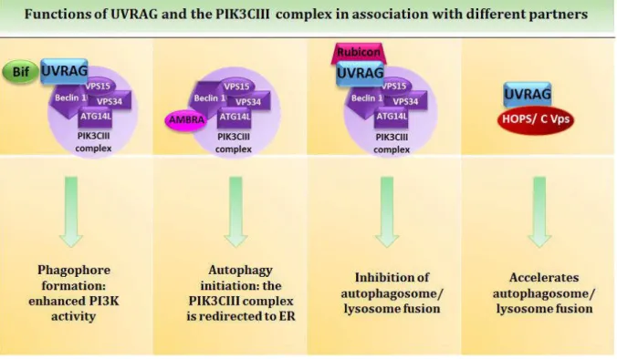
for the phosphorylation of phosphatidylinositol (PI) to produce phosphatidylinositol 3 phosphate (PI3P) (reviewed in Russell et al., 2014). Hence this complex is termed class III phosphatidylinositol 3-kinase (class III PI3K) complex. PI3P seems to be involved in the stabilization of ULK1 at the omegasome but also contributes to the binding of other proteins necessary for the formation of the autophagosome, such as the double FYVE containing protein 1 (DFCP1) (Axe et al., 2008; Karanasios et al., 2013). The VPS34 complex can be associated to three other proteins: ATG14L, the UV resistance associated gene protein (UVRAG) and to Rubicon (RUN domain and cysteine-rich domain containing, Beclin 1-interacting protein) (Fig 7). Even though those proteins bind specifically to Beclin1, it is seemingly never at the same time and they are required for different tasks. ATG14L is essential for the relocalization of the complex to the isolation membrane and more specifically to the omegasome (Fan et al., 2011; Matsunaga et al., 2010). When UVRAG is associated to the complex it has been suggested that it enhances the PI3K activity. Rubicon binding on the other hand has an inhibitory effect, since it decreases the PI3 kinase activity and thus prevents autophagosome formation (Zhong et al., 2009). When in another conformation, Beclin1 is also able to bind the activating molecule in Beclin1-related autophagy 1 (AMBRA1). The latter protein interacts with the dynein motor complex. It induces autophagy upon release from the complex, by directing the ULK1 complex to the ER (Di Bartolomeo et al., 2010). Beclin1-interacting proteins also comprise Bcl-2 (B cell lymphoma 2), which same as Rubicon negatively regulates autophagy when both proteins are bound (Pattingre et al., 2005). Interestingly, Bcl2 also displays anti-apoptotic properties (Bcl-2 and Bcl-xl) when associated to Bax and Bak on mitochondria. This highlights the tight interplays between autophagy and apoptosis, and the homeostatic role of Beclin1 to that respect.
Figure 5: An overview of the autophagic machinery from initiationto elongation.
(1) Detailed here the initiation step controlled by the ULK1 complex (ULK1 ATG13, FIP200 and ATG101). In nutrient rich conditions mTORC1complex interacts with ULK1 and inactivates the complex by phosphorylating ULK1 and ATG13. Under starvation conditions, mTORC1 is inactivated by AMPK leading to the phosphorylation of ULK1 by AMPK and its subsequent activation. (2) Then ULK1 phosphorylates ATG13 and FIP200 and consequently induces Beclin 1 dissociation from Bcl-2 which initiates autophagosome nucleation. ATG9A also requires the phosphorylation by ULK1 to be activated and furnishes the membranes for the formation of the phagophores during the nucleation step which takes place at the omegasome, a structure generated from the ER. This process requires the PIK3CIII complex (Beclin1, VPS34, VPS15 and ATG14L). PI3P produced by VPS34 recruits DFCP1 and WIPI to the isolation membrane. (3) For the elongation process two ubiquitin-like conjugation systems are required. The ATG5-ATG12 and LC3-PE conjugation systems (blue boxes). (4) The ATG12-ATG5 conjugates form a multimeric complex with ATG16L1 through ATG7 and ATG10, E1 and E2-like enzymes respectively. The formed ATG12-ATG5/ATG16L1 conjugate undergoes dimerization and gets recruited to the phagophores by WIPI. (5) In the second system LC3-I is generated from pro-LC3 by the ATG4 protease. Then, through ATG7, ATG3 (E2-like enzyme), and the ATG12-ATG5/ATG16L1 complex (acts as an E3-(E2-like enzyme), LC3 gets conjugated to the phospholipid phosphatidylethanolamine (PE) to form LC3-II. LC3-II localizes to the growing membranes and the ATG12-ATG5/ATG16L1 conjugates are released from the membranes during the formation of the autophagosome. Cytosolic content is captured while membranes are elongating.
ULK1: Unc-51-like kinase 1; mTOR: mammalian target of rapamycin; AMPK: AMP-activated protein kinase; PI3P: Phosphatidylinositol triphosphate; DFCP1: double FYVE containing protein 1, WIPI: WD-repeat PI3P
INTRODUCTION – The Autophagic Pathways in Cellular Homeostasis
The generation of the phagophore membrane per se is mediated by ATG9A. This ATG is ubiquitously expressed and localized to the Golgi apparatus and late endosomes and cycles from one to the other (Feng et al., 2014). Upon autophagy induction by starvation, ATG9A gets phosphorylated by ULK1 which induces its recruitment to the phagophore (Papinski et al., 2014) and contributes to phagophore expansion by feeding lipids to the growing isolation membrane (Yamamoto et al., 2012; Zavodszky et al., 2013). One particular characteristic of this protein is that, with the vacuole membrane protein 1 (VMP1), it is the only known transmembrane ATG protein identified so far. VMP1 has mainly been found associated to the ER membrane and has recently been shown to recruit Beclin1 to the phagophore. VMP1 thus regulates PI3K CIII complex activation (Molejon et al., 2013).
Elongation – Sequestration (Fig 5)
The elongation step following initiation is mainly mediated by two ubiquitin-like conjugation systems. During this step the phagophore grows and at the same time, cytoplasmic material gets engulfed into the forming autophagosome.
The ATG7 ubiquitin-like conjugation system is composed of three key proteins: ATG5, ATG12 and ATG16L1. In order to build this system, ATG12 has to be conjugated to ATG5 and this is achieved through the combined actions of the E1 ubiquitin-like ligase, ATG7 and the E2 ubiquitin-like ligase, ATG10. First ATG12 gets activated by ATG7 at the C-terminal glycine residue in an ATP dependent manner, subsequently inducing the formation of an ATG12-ATG7 thioester intermediate followed by the transfer of ATG12 to ATG10 to also form a thioester intermediate. Then the conjugation to ATG5 gets established through the covalent attachment of the C-te i al gl i e of ATG to ATG ’s l si e at position 130 (Mizushima et al., 2002). The newly formed ATG12-ATG5 conjugates can then bind the ATG16L1 protein in a non-covalent manner, which in turn induces the homodimerization of this complex resulting in an 800kDa conjugate. The WD-repeat PI3P effector protein (WIPI) recuited by PI3P, interacts with ATG16L1 and facilitates the recruitement of the conjugate to the nucleation site (Dooley et al., 2014). The conjugate ATG12-ATG5/ATG16L1 localizes at the growing autophagosome u til it’s o pletel fo ed a d the disso iates f o it (Mizushima et al., 2003).
Figure 6: The essential roles of UVRAG and and the PIK3CIII complex in autophagy.
The second ubiquitin-like conjugation system is involved in the elongation of the autophagosomal membrane. This system requires the lipidation of the microtubule-associated-protein 1 light chain 3 MAP LC o LC a d its pa alogues elo gi g to the GABARAP fa il γ-amino-butyric acid receptor-associated protein), GABARAPL1, and to the GATE family (Golgi-receptor-associated ATPase enhancer), GATE-16. Mammalian LC3 family is composed by different isoforms which are LC3A, LC3B and LC3C in human, the most abundant being LC3B. While the LC3 family proteins are important for the elongation step, the GABARAP/GATE family is rather needed for autophagosome maturation (Weidberg et al., 2010). LC3 is generally localized in the cytosol where it undergoes a number of posttranscriptional modifications, the first being its processing from pro-LC3 to LC3-I mediated by ATG4, a cysteine protease also present through different isoforms in mammalian cells (ATG4A-D also called Autophagin 1-4). The most active isoform, ATG4B (Autophagin 1) catalyzes the cleavage at the C-terminal region of pro-LC3 after a glycine residue. It has recently been reported that upon autophagy induction, ATG4B requires phosphorylation for proper function (Yang et al., 2015). In any case, the glycine residue at the C-terminus of LC3-I constitutes the lipidation site where phosphatidylethanolamine (PE) will be conjugated to LC3-I to form LC3-PE, also called LC3-II. This action is mediated as previously for the ATG5-ATG12/ATG16L1 conjugation, by an E1 and E2 ubiquitin-like catalytic system composed of ATG7, and ATG3 an E2-like conjugation enzyme. ATG12-ATG5/ATG16L1 complex in this second conjugation system allows the binding of the PE to LC3-I through its E3 like ligation enzyme activity. As
INTRODUCTION – The Autophagic Pathways in Cellular Homeostasis
demonstrated by Fujita and colleagues, ATG16L1 acts like a scaffold protein that transfers LC3 from ATG3 to PE (Fujita et al., 2008). Consequently this lipidated form of LC3 can be integrated both in- and outside of the elongating double membrane. While other proteins attached to the autophagosomal membrane end-up dissociating from it in the initial steps of autophagy, LC3-II is the only protein that remains there from initiation on until content degradation. Hence this protein is commonly used as an autophagic marker. One must however remember that LC3 is partially degraded after autophagosome fusion with the lysosomes. Even though the precise function of LC3-II has not been very well determined yet, it has been suggested, with help of liposomes, to be involved in the hemifusion of the autophagosome (Nakatogawa et al., 2007). Nevertheless LC3-II has been shown to be a major player in cargo recognition in collaboration with sequestosome proteins (see sections 1.4.3 and 3.1.1)
Maturation and Degradation (Fig 7)
Maturation begins once the autophagosome is fully formed and the cytoplasmic content is sequestered inside. This step leads to the fusion of the autophagosome with the lysosomes and subsequently to the formation of the autolysosome. Many proteins and complexes are involved in the establishment of this process.
UVRAG, involved in phagophore generation through its interaction with the Beclin1-VPS34-VPS15 complex, also plays an important role in the autophagosome maturation process. This complex actually localizes both in endosomes and autophagosomes. UVRAG interacts with a tethering complex (the homotypic fusion and protein-sorting/class C vacuole protein-sorting (HOPS/class C Vps), composed of VPS16, VPS33 and VPS39 among other proteins) independent of Beclin1 (Fig 6). Liang and colleagues show that UVRAG accelerates endosomal trafficking which leads to an increased autophagic flux. Furthermore knock-down of UVRAG by small interfering ribonucleic acid (siRNA) in HeLa cells substantially reduces the colocalization of VPS16 with the GFP-LC3 fusion protein, suggesting an UVRAG-dependent recruitment of the HOPS/class C Vps to the autophagosome (Liang et al., 2008). Tethering complexes are in fact essential in fusion processes since they mediate the coupling of Rab GTPase activation and soluble N-ethylmaleimide-sensitive factor attachment protein receptors (SNARES) assembly. One element of the HOPS/class C Vps complex, humain VPS39 activates such a GTPase, namely Rab7 (Liang et al., 2008). As a matter of fact, Rab7 is one other major key player in this maturation process. This small GTPase usually involved in late endosomal trafficking has been identified as an important mediator of autophagosome fusion with the lysosomes. In fact it has been shown that Rab7 down-regulation in cardiomyocytes resulted in the accumulation of autophagosomes followed by apoptosis, demonstrating the role of this protein in autophagosome maturation rather than in the initiation. It also highlights the requirement of a complete autophagic flux for the
maintenance of cellular integrity in that context. Rab7 actually plays two roles in this process, first mediating the autophagosome transport along the microtubules from the periphery to the microtubule organization center (MTOC), the concentration site of the lysosomes. Secondly Rab7 favors the autophagosome/lysosome fusion. The HOPS complex discussed earlier, seems also to be able to bind Rab7 and to participate both in trafficking and fusion by tethering the lysosome to the autophagosome (Balderhaar and Ungermann, 2013; Pawelec et al., 2010).
UVRAG can also be associated to Rubicon and, as already mentioned, this Beclin1-VPS15-VPS34-UVRAG-Rubicon complex has an inhibitory function towards autophagy (Fig 6). It has been recently proven that Rubicon binds UVRAG and interacts with VPS34 via its RUN-domain and thus inhibits VPS34 lipid kinase activity by blocking UVRAG mediated VPS34 activation. Rubicon is also able to bind Rab7 and to inactivate its capacity to mediate autophagosome/lysosome fusion (Sun et al., 2011).
The endosomal sorting complexes required for transport (ESCRT complexes 0, I, II and III) are important actors in autophagosomes maturation and are able to interact with Rab7 as well. Their primary role is to participate in endocytosis and in multivesicular bodies (MVBs) biogenesis. MVBs are late endosomes containing intraluminal vesicles that can fuse with autophagosomes in certain circumstances (e.g. starvation). The fusion of late endosomes with autophagosomes forms a structure called amphisome, prior to the fusion with lysosomes. Interestingly the ESCRT complexes have been shown to mediate this fusion. Impairments of their function is associated to autophagy dysregulations in neurodegenerative diseases. It has been suggested that the ESCRT complex II could interact with Rab-interacting lysosomal protein (RLIP), inducing the recruitment of Rab7 and thus priming the fusion process (Wang and Hong, 2006). Of note, the HOPS complex also interacts with ESCRT complexes (Pryor and Luzio, 2009).
Other Rab GTPases have been shown to be involved in fusion mechanisms. The Protein Rab11 for example, is required for the fusion of autophagosomes with multi-vesicular bodies (MVBs) as well. As clearly established, the fusion process is essential for autophagosome maturation and in fact one particular protein family is specialized in catalyzing this process, the SNARE family mentioned previously. Their purpose is to create an opening in the membrane of two adjacent vesicles so that they can fuse. SNARE proteins share a signature sequence of about 60 residues. They often contain a transmembrane domain and tend to be present in opposing membranes destined to fuse (Gerber and Südhof, 2002). The proteins present in each SNARE complex, usually composed of four SNARE proteins forming an alpha helix bundle, depend on the vesicles that need to fuse. The SNAREs Vit1b, VAMP7, VAMP8 and SYNTAXIN17 have been shown to mediate autophagosome/lysosome fusion, in particular in the context of pathogen elimination by xenophagy (see section 3.1.1) (Furuta et al., 2010). It has also been demonstrated that SYNTAXIN17 interacts with tethering complex HOPS (Kim et al., 2001).
INTRODUCTION – The Autophagic Pathways in Cellular Homeostasis
Pryor and Luzio proposed a fusion model involving the interaction of SYNTAXIN17 with the tethering complexes HOPS and Rab7. They suggested that HOPS might block SYNTAXIN17 from mediating membrane pairing. After replacement of Rab5 by Rab7 at the endosomal membrane, it would induce the recruitment of VPS39 and VPS41, two members of the HOPS complex. VPS39 would then activate Rab7 through its guanine-exchange-factor (GEF) activity and lead to the dissociation of SYNTAXIN17 from HOPS complex, thus allowing trans-SNARE pairing and fusion of opposed membranes (Pryor and Luzio, 2009).
One other feature of the maturation process is the vesicular microtubule trafficking which has been shown to be a very important component of the autophagic process. The cytoskeleton is composed of four major constituents: actin filaments, microtubules, intermediary filaments and septin. All of them play a role at different autophagic stages with the microtubules being the ones mainly involved in autophagosome maturation. Autophagosome transport along the microtubules towards the MTOC e ui es the oto p otei d ei . I a ouse odel fo Hu ti gto ’s disease, d ei utatio through deletion of the dynein heavy chain1 gene (Dnch1) actually blocks protein aggregate degradation and thus mediates the development of neurodegenerative diseases (Ravikumar et al., 2005). Studies have shown that dynein is linked to the autophagosomes through its binding to LC3 family proteins as their name (Microtubule-Associated-Protein) already indicates. Another protein that has emerged as an important player in autophagy maturation is the histone deacetylase-6 (HDAC6). Lee and colleagues report that HDAC6 mediates the fusion of autophagosomes with lysosomes through actin remodeling. This protein is known to regulate microtubule-dependent transport. Interestingly the authors observed that HDAC6 recruits an actin-remodeling factor, cortactin, at the site of protein aggregates (Lee et al., 2010b). HDAC6-defecient MEFs accumulate double-membrane structures containing multivesicular bodies, protein aggregates and have furthermore low levels of cortactin indicative of its role in autophagosome/lysosome fusion in order to mediate protein clearance. However inducing starvation in the absence of HDAC6 did not disturb autophagosome maturation suggesting that this protein is mostly required for quality control basal autophagy (see section 1.4.3).
All in all, many proteins and complexes are required for the maturation process which is differentially regulated depending on vesicles that need to fuse even though the order in which the molecular events take pla e does ’t seem to have been clearly identified yet. To sum up the events, it can be said that once the autophagosome is formed it needs to be transported along the microtubules to the MTOC where it comes in contact with the lysosomes or endosomes leading subsequently to their fusion and the formation of the autolysosome (Longatti and Tooze, 2009; Metcalf and Isaacs, 2010).
Figure 7: Schematic representation of endosome, autophagsome maturation and fusion with lysosomes.
(1) Rab5 is attached to the surface of an early endosome. UVRAG interacts with the tethering complex HOPS/C-Vps and mediates endosomal maturation by replacing Rab5 with Rab7 and by activating Rab7. (2) The same complex integrates Rab7 into the membranes of autophagosomes where Rab7 interacts with LC3-II. (3) Starvation promotes the binding of FYCO1 to kinesin and to Rab7 at the membrane of the lysosome while the RILP-dynein complex binds to Rab7 at the autophagosome, a process which requires the intervention of the ESCRT II complex. These interactions provide means to regulate the bidirectional movement of lysosomes and autophagosomes along microtubules towards one another and the MTOC. RILP-Rab7 interaction is further controlled by the UVRAG–HOPS/C-Vps complex which also regulates the fusion process (white square) by tethering the MVB/autophagosome and lysosome/autophagosome to one another. The SNARE complex (VAMP8, VAMP7, Vit1b and SYNTAXIN17) promotes the fusion of the lysosomes with the autophagosome. SYNTAXIN17 (purple) interacts with two proteins of the HOPS complex, Vps39 and Vps41 (not depicted). UVRAG: UV-resistance associated gene protein; FYCO1: FYVE and coiled-coil (CC) domain–containing protein 1; ESCRT: Endosomal sorting complex required for transport;
HOPS/C-Vps: Homotypic fusion and sorting/Class C vacuole protein-sorting; MTOC: Microtubule organization center; RILP: Rab-interacting lysosomal protein.