HAL Id: tel-01837919
https://tel.archives-ouvertes.fr/tel-01837919
Submitted on 13 Jul 2018HAL is a multi-disciplinary open access archive for the deposit and dissemination of sci-entific research documents, whether they are pub-lished or not. The documents may come from teaching and research institutions in France or abroad, or from public or private research centers.
L’archive ouverte pluridisciplinaire HAL, est destinée au dépôt et à la diffusion de documents scientifiques de niveau recherche, publiés ou non, émanant des établissements d’enseignement et de recherche français ou étrangers, des laboratoires publics ou privés.
Host-pathogens interactions during RSV / S.
pneumoniae infection immune response and p53 pathway
Daniela Bandeira Brancante Machado
To cite this version:
Daniela Bandeira Brancante Machado. Host-pathogens interactions during RSV / S. pneumoniae infection immune response and p53 pathway. Virology. Université de Lyon, 2017. English. �NNT : 2017LYSEN083�. �tel-01837919�
Numéro National de Thèse: 2017LYSEN083
THESE de DOCTORAT DE L’UNIVERSITE DE LYON
opérée par
l’Ecole Normale Supérieure de Lyon
Ecole DoctoraleN° 340
Biologie Moléculaire, Intégrative et Cellulaire (BMIC) Discipline: Sciences de la vie
Soutenue publiquement le 12/12/2017, par :
Daniela BANDEIRA BRANCANTE MACHADO
Etude in vitro et in vivo des interactions
hôte/pathogènes dans un modèle de co-infection
VRS/S. pneumoniae
Host-pathogens interactions during RSV / S. pneumoniae infection: immune response and p53 pathway
Devant le jury composé de :
Pr. Renaud MAHIEUX Professeur ENS de Lyon Examinateur
Pr. Jean-Christophe BOURDON Professeur University of Dundee Rapporteur
Dr. Ronan LE GOFFIC Chargé de recherche INRA Rapporteur
Pr. Marilda SIQUEIRA Professeure Fiocruz-Brésil Examinatrice
Dr. Rosita ACCARDI-GHEIT Chargée de recherche IARC Membre invitée
Dr. Glaucia PARANHOS-BACCALA Directrice de recherche FMX Directrice de thèse
ACKNOWLEDGMENT
First, I’d like to thank my advisor Dr. Glaucia Paranhos-Baccala, for supporting and teaching me to grow professionally and personally. Also, I would like to thank Mr. Benoit Miribel, (Executive Director), Dr. Hubert Endtz (Director of the Scientific Department) and all staff of Fondation Mérieux for the fabulous opportunity to work at this Fondation which has the commitment to fight infectious diseases around the world. I would like to thank the Laboratoire des Pathogenes Emergent team for their support throughout my thesis, especially to Dr. Jonathan Hoffmann and Dr. Marie Moroso for their friendship and encouragement.
I express my gratitude to Dr. Olivier Terrier for his commitment, patience, and support in overcoming numerous obstacles I have been facing through my research. I also greet Professor Bruno Lina, Dr. Manuel Rosa-Calatrava and the members of the laboratory of Virology and Human Pathology (VirPath) for their feedback, cooperation, and scientific support.
I am also sincerely grateful to Professor Renaud Mahieux (Professor and Director of the Department of Biology at the Ecole Normale Supérieur, Lyon-France), to Professor Jean-Christophe Bourdon (Professor at University of Dundee), Dr. Ronan Le Goffic (Researcher at the Institut National de la Recherche Agronomique), Dr. Marilda Siqueira (Head of National Influenza Center in Brazil) and Dr. Rosita Accardi-Gheit (researcher at International Agency for Research on Cancer) for having accepted to constitute the jury of my thesis. It is an honor for me to present all results obtained during the last 4 years of research.
A big thanks to all my Brazilian friends in France (Milena, Leandro, Clarice, Paula, Lilian, Bruno, Magda, and Paola) and also my French friends (Laureline, Pierre-Eliot, Gaetan Laetitia, Marie-Amelie, and Guillaume) who shared several funny moments, helping me to enjoy life besides work. I’m very grateful to Jacques for his patience, confidence, care, encouragement and for unforgettable moments together.
And finally, I’d like to thank my family (Roberto, Vera, Pedro, Ana Livia, Julie, Oscar, and Madeleine) who even so far away physically, provides me with unfailing support and continuous encouragement throughout all my years of study and through the process of writing this thesis. Without their love, affection and encouragement this work would not have been possible.
TABLE OF CONTENTS
ACKNOWLEDGMENT ... 2 LIST OF FIGURES ... 6 LIST OF TABLES ... 6 ABREVIATION ... 7 RESUME ... 9 ABSTRACT ... 10 INTRODUCTION ... 111. ACUTE LOWER RESPIRATORY TRACT INFECTIONS ... 12
1.1. Definition and clinical symptoms ... 13
1.2. Etiology ... 14
1.2.1. Respiratory viral infections ... 15
a) Influenza Viruses ... 15
b) Respiratory Syncytial Virus ... 18
c) Others Human Viruses ... 21
1.2.2. Respiratory bacterial infections ... 21
a) Streptococcus pneumoniae ... 22
b) Haemophilus influenzae ... 23
c) Others respiratory bacteria ... 23
1.2.3. Mixed Respiratory infections ... 24
1.3. Diagnostic of lower respiratory tract infections ... 26
1.4. Treatments for lower respiratory tract infections ... 28
1.4.1. Antiviral treatments ... 28
1.4.2. Antibiotics ... 30
1.5. Prevention of lower respiratory tract infection ... 31
1.5.1. Viral prophylaxis ... 31
1.5.2. Pneumococcal vaccine ... 33
2. HOST-PATHOGEN INTERACTIONS ... 35
2.1.1. Innate immune response ... 38 2.1.2. Adaptive immunity ... 41 2.2. Inflammatory response ... 42 2.2.1. NF-kB pathway ... 43 2.2.2. Interferon pathway ... 43 2.3. p53 pathway ... 45 2.3.1. Mechanisms of p53 regulation ... 47 a) Ubiquitinylation ... 47 b) Phosphorylation ... 48 c) Other modifications ... 48 2.3.2. p53 isoforms ... 49
2.3.3. Biological responses induced by p53 target genes ... 50
2.3.4. Functional interplay between pathogens and p53 ... 52
OBJECTIVE ... 56
RESULTS ... 58
CHAPTER 1HOST-VIRAL INTERACTIONS DURING SINGLE OR MIXED INFECTIONS: ROLE OF INNATE IMMUNE RESPONSES ... 61
Article 1. Viral and bacterial co-infection in severe pneumonia ... 65
Article 2. RSV infection in macrophages promotes IP-10 expression during bacterial co-infection ... 79
CHAPTER 2HOST-VIRAL INTERACTIONS DURING SINGLE AND MIXED INFECTIONS: ROLE OF THE CELL GUARDIAN P53 ... 91
Article 1. Role of p53/NF-kB functional balance in RSV-induced inflammation and immune responses ... 94
CHAPTER 3HOST-VIRAL INTERACTIONS DURING SINGLE AND MIXED INFECTIONS: DEVELOPMENT OF SEVERE PNEUMONIA MODEL ... 117
Article 1. Establishing severe pneumonia in non-human primate model during mixed infection ... 119
DISCUSSION AND PERSPECTIVES ... 132
APPENDICES ... 147
Article 1. Influenza A viruses alter the stability and antiviral contribution of host E3-ubiquitin ligase Mdm2 during the time-course of infection ... 148
Article 2. Phylogenetic analyses of influenza A (H1N1)pdm09 hemagglutinin gene during and after the pandemic event in Brazil ... 177
6
LIST OF FIGURES
FIGURE 1 CAUSES OF DEATH WORLDWIDE. ... 12
FIGURE 2 DIFFERENCES BETWEEN PNEUMONIA AND BRONCHIOLITIS. ... 14
FIGURE 3 INFLUENZA VIRUS PARTICLE. ... 16
FIGURE 4 THE REPLICATIVE CYCLE OF IAV. ... 17
FIGURE 5 RESPIRATORY SYNCYTIAL VIRUS PARTICLE AND GENOME. ... 19
FIGURE 6 REPLICATIVE CYCLE OF RSV. ... 20
FIGURE 7 PNEUMOCOCCUS SURFACE STRUCTURE AND MAJOR VIRULENCE FACTORS. ... 23
FIGURE 8 HOST-PATHOGEN INTERACTIONS. ... 36
FIGURE 9 CHRONOLOGICAL COURSE OF INNATE AND ADAPTIVE IMMUNITIES. ... 37
FIGURE 10 INNATE AND ADAPTIVE CELLS. ... 37
FIGURE 11 PRRS SIGNALING PATHWAY. ... 40
FIGURE 12 INFLAMMATORY RESPONSES. ... 42
FIGURE 13 THE P53 NETWORK. ... 46
FIGURE 14 SCHEME OF THE P53 PATHWAY. ... 47
FIGURE 15 P53/MDM2 COMPLEX. ... 48
FIGURE 16 SCHEMATIC OF THE P53 ISOFORMS. ... 49
FIGURE 17 P53 PATHWAY ROLE DURING THE ANTIVIRAL RESPONSE. ... 51
FIGURE 18 P53 AND NF-KB MODULATION. ... 51
FIGURE 19 P53 PATHWAY MODULATION BY IAV AND RSV. ... 55
FIGURE 20 POST-TRANSCRIPTIONALLY P53 MODULATION DURING HPIV-3 INFECTION. ... 138
FIGURE 21 A DIFFERENT PATTERN OF P53 EXPRESSION DURING VIRAL AND MIXED INFECTIONS. ... 139
FIGURE 22 EXPRESSION OF P53 ISOFORMS DURING RSV OR HPIV-3 INFECTION. ... 140
LIST OF TABLES
TABLE 1 PNEUMOCOCCAL VACCINE. ... 33TABLE 2 TLR AND MICROBIAL LIGANDS. ... 39
TABLE 3 ISG MODULATED DURING DIFFERENT VIRUS INFECTION. ... 45
7
LIST OF ABREVIATION
C. pneumoniae Chlamydophila pneumoniae
cRNA complementary RNA
CRP C-reactive protein
DBD DNA-binding domain
F fusion protein
G attachment glycoprotein
H. influenzae Haemophilus influenzae
HA Hemagglutinin protein
hCoV human Coronaviruses
HD hinge domain
hMPV human Metapneumoviruses
hPIV human Parainfluenza viruses
IAV Influenza A viruses
IFN Interferon
IL interleukin
IP-10 Interferon gamma-induced protein 10
IRF interferon regulatory factor
ISGs Interferon-stimulated genes
L subunit of the RSV-polymerase protein
M. pneumoniae Mycoplasma pneumoniae
MAPK mitogen-activated protein kinase
Mdm2 mouse double minute 2
mRNA messenger RNA
MRSA methicillin-resistant Staphylococcus aureus
N nucleoprotein
NA Neuraminidase protein
NF-kB Nuclear factor kappa-light-chain-enhancer of activated B cells
NHP Nonhuman Primates
NLRs NOD-like receptors
NP Nucleoprotein
NS non-structural protein
NTHi nonencapsulated strains of H. influenzae
OD oligomerization domain
P phosphoprotein
PA acid polymerase protein
PAMPs pathogen-associated molecular patterns
PB1 basic polymerase 1 protein
PB2 basic polymerase 2 protein
PCR polymerase chain reaction
PCT procalcitonin
PCV Pneumococcus conjugated vaccine
8
PRD proline-rich domain
PRRs pattern recognition receptors
RLRs RIG-I-like receptors
RNPs Ribonucleoprotein complexes
RSV human Respiratory syncytial viruses
S. aureus Staphylococcus aureus
S. pneumoniae Streptococcus pneumoniae
SH small hydrophobic protein
TA Transactivation domain
Th helper T cells type
TLR Toll-like receptor
TNF Tumor necrosis factor
TRAIL TNF-related apoptosis-inducing ligand
vRNA viral RNA
9
RESUME
Les infections aiguës des voies respiratoires inférieures constituent la troisième cause de décès dans la population mondiale, avec 3,2 millions de décès. Parmi cette mortalité, au tour de 1 million c’était des enfants de moins de 5 ans, représentant la première cause de mortalité dans ce groupe d’âge, selon l'OMS en 2015. Le virus respiratoire syncytial (VRS) est considéré comme un agent étiologique important dans le millieux pediatric et les estimations sont que ce virus cause 3
million d’hospitalization par an. Un aspect important du pronostic des infections
virales est le rôle de co-infection bactérienne. La combinaison d’agents viraux et bactériens a été signalée entre le VRS et les bactéries Streptococcus pneumoniae. En raison de l’importance clinique de cette co-infection et le taux élevé de circulation du VRS, il est important de comprendre comment le système immunitaire est affecté à l'infection de ces deux agents pathogènes. Notre étude identifie la réponse immunitaire dans les macrophages, ainsi comme les interactions entre le VRS et le facteur de transcription p53. Les résultats montrent un profil particulier a cette co-infection mixte dans le macrophages et des modifications dans la réponse immune innée que nous a permis de mieux comprendre les mécanismes de pathogenèse du VRS dans les cellules épithélial pulmonaires en regardant la régulation de p53. Dans la dernière partie, nous avons évalué l'impact direct de l’infection mixte chez les primates non-humain et ce modèle nous a montré les difficultés et complexités des établir une pneumonie sévère.
Mots-clés : VRS ; S. pneumoniae ; co- infection ; réponse immune ; p53 pathway.
10
ABSTRACT
Respiratory viruses play a leading role in the etiology of respiratory infections. Currently, respiratory syncytial virus (RSV) is generally considered to be the etiologic agent of respiratory disease in pediatric importance, as children can develop bronchiolitis and pneumonia when infected with the virus. The first RSV infection occurs in the first two years of life in about 95% of children, with the peak incidence occurring in the first few months of life. An important aspect of the prognosis of viral infections is the role of bacterial co-infection. The combination of viral and bacterial agents has been reported between RSV and Streptococcus pneumoniae bacteria. Because of the clinical importance of this co-infection and the high rate of RSV circulation, it is important to understand how the immune system is affected by the infection of both pathogens. Our study was designed to evaluate the immune response in macrophages, in addition to interactions between RSV and p53 transcription factor. The results show a particular profile of this mixed co-infection in macrophages and p53 regulation that implies several modifications in the innate immune response and that allowed us to better understand the mechanisms of pathogenesis of RSV in pulmonary epithelial cells. In the last part, we evaluated the direct impact of mixed co-infection in non-human primates and this model showed us the difficulties and complexities of establishing severe pneumonia.
Keywords: RSV; S. pneumoniae; mixed-infection; immune response; p53 pathway
11
INTRODUCTION
12 Tous les résultats présentés ici ont été développés dans le cadre du projet de thèse visant à mieux comprendre les infections mixtes au cours de la pneumonie. Ce travail a eu la collaboration ferme et efficace entre les Laboratoire de Pathogènes Emergents et Virpath, permettant l'approfondissement et menant à des conclusions importantes dans la pathogenèse des virus respiratoires lors de la présence ou l'absence de bactéries dans des modèles in vitro et in vivo.
1. ACUTE LOWER RESPIRATORY TRACT
INFECTIONS
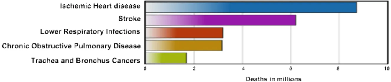
The respiratory tract constitutes a wide and critical frontier at the interface between the body and the environment. This complex organ system is divided into the upper airways and lower airways. The upper airways or upper respiratory tract includes the nose and nasal passages, paranasal sinuses, the pharynx, and the portion of the larynx above the vocal folds (cords). The lower airways or lower respiratory tract includes the portion of the larynx below the vocal folds, trachea, bronchi, and bronchioles. The lungs can be included in the lower respiratory tract or as a separate entity and include the respiratory bronchioles, alveolar ducts, alveolar sacs, and alveoli [1]. Upper respiratory tract infections are less severe whereas lower infections are often associated with high mortality rates [2]. Acute lower respiratory tract infections constitute the third leading cause of human death worldwide with 3.2 million deaths in 2015 (Figure 1), and the first cause of mortality in children under five years of age, according to the World Health Organization (WHO) [3, 4].
Figure 1 Causes of death worldwide.
Lower respiratory infections constitute the third cause of death in the world population (red bar) being responsible for 3.2 million of deaths in 2015. Heart diseases (Ischemic heart disease – blue bar - and strokes – pink bar) were the most cause of deaths. Among the 5 main causes of global death described, lower respiratory infections are the only transmissible infectious disease [3].
13 The diversity of pathological agents makes it difficult to prevent, diagnose and treat these diseases, contributing to high mortality rates [5-8]. These diseases can affect the general population, but severe cases and high mortality rates are found among children up to age 5, immunosuppressed adults and elderly [9-11].
Acute lower respiratory infections constitute a major global health burden due to the emergence of resistance to antimicrobial treatments, the presence of multiple pathogens and the recurrence of infections throughout life [12-14]. In this context, more knowledge about respiratory diseases and their etiological agents are very important to improve or propose novel prophylactic and therapeutic approaches.
1.1. Definition and clinical symptoms
Respiratory tract infections are responsible for a variety of clinical features that range from milder manifestations, such as the common cold to acute lower respiratory tract infections, represented by bronchiolitis and pneumonia [15, 16]
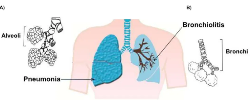
Pneumonia is an inflammatory process that takes place in the alveolar spaces, whereas, in bronchiolitis, inflammation rather occurs in bronchi (Figure 2). The symptoms are relatively similar between pneumonia and bronchiolitis; they begin as a common cold (nasal congestion, high fever, and decreased appetite) and after 2 to 3 days it is possible to observe several complications of the disease. Pneumonia usually accompanies a dry cough, thoracic pain, and extreme tiredness while bronchiolitis accompanies a characteristic wheezing. At this stage of both diseases, cough is persistent and the difficulty of feeding is marked, besides the accelerated breathing. Apnea is a very common symptom in infants less than 2 months during bronchiolitis while confusion occurs in the elderly with pneumonia. Complications such as hypoxia (low oxygen level) and cyanosis (blue-tinged skin) are indicative of both severe diseases [16, 17].
14
Figure 2 Differences between Pneumonia and Bronchiolitis.
Lower respiratory tract diagram showing affected lung regions during respiratory tract infections. A) Pneumonia is characterized by infection in the alveoli airways and normally occurs in the terminal part of the lobular lung. B) Bronchiolitis is restricted in the bronchial region and remains near to principal bronchi. Adapted from Eugenia et al [18].
Bronchiolitis affects children younger than 2 years old with a high mortality rate among preterm infants, due to the characteristics of this age group in which the lung and immune system are functionally immature, forming ideal spots for viral infection [19]. Pneumonia affects all ages but is extremely severe in children up to 5 years of age, immunosuppressed adults and the elderly [20-22]. The most common pneumonia is community-acquired pneumonia, which is acquired in an extra-hospital environmental [6, 23, 24].
The pathogenesis of respiratory infections involves a complex interplay between virulence factors of a number of different pathogens – including bacteria, virus and/or fungi – and host response [25]. An overview of the etiological agents of pneumonia and bronchiolitis is presented in the following chapter and the host/pathogens interactions involved in these respiratory infections will be approached in the second part of this manuscript.
1.2. Etiology
The upper respiratory tract, mainly the nasopharynx, constitutes a rich and diverse niche in microbes. It is believed that most respiratory infections of the lower respiratory tract must have originated from this microbial niche. Thus, commensal microorganisms are also found in cases of severe disease, making it difficult to determine precisely etiological pathogens [2, 26].
15
1.2.1.
Respiratory viral infections
The main viruses associated with lower respiratory tract infections are Influenza Viruses, human respiratory syncytial viruses (RSV), human parainfluenza viruses (hPIV), human metapneumoviruses (hMPV) and human Coronaviruses (CoV) [20, 27, 28].
a) Influenza Viruses
Influenza viruses cause an acute infection popularly known as Flu that has a high rate of recurrence and the ability to infect individuals in all age groups. Influenza viruses epidemics are estimated to result in about 3 to 5 million cases of severe illness, and about 250000 to 500000 deaths [29].
Influenza A, B and C viruses belong to the family Orthomyxoviridae [30]. Among these three types, Influenza A viruses (IAV) is responsible for the major pandemic and seasonal epidemics events being considered more virulent and genetically more variable [31]. IAV have the ability to achieve a large spectrum of animal reservoirs, among mammals and birds. Instead, Influenza B types are composed of two circulating lineages (Victoria and Yamagata) in the human population causing seasonal epidemics but without the capacity to promote pandemic events. Influenza C type is the less common and often only cause a mild infection in children [32-34].
IAV is divided into subtypes according to antigenic properties of the two viral surface glycoproteins, hemagglutinin (HA) and neuraminidase (NA). Historically, three subtypes of HA (H1, H2, and H3) have acquired the ability to be transmitted efficiently among humans and seasonal subtypes H1 and H3 are most co-circulate detected [35]. However, other subtypes, such as H5, H6, H7, and H9 occasionally affect humans and are considered possible threats to a future pandemic [36]. IAV can cause pandemics when a strain which was not previously circulating among humans emerges and transmits among humans. As the majority of the population has no immunity against these viruses, the proportion of persons in a population getting infected may be quite large [37]. Also, zoonotic Influenza viruses can cause
16 sporadic severe infection in humans through direct contact with infected animals, such as swine or birds but rarely spread very far among humans [38].
IAV is spherical and elongated virions with a diameter varying between 95 to 120 nanometers (nm) and morphological virions are shown in Figure 3A [39]. In the viral envelope are inserted the surface glycoproteins HA and NA, and also the M2 channel, which is a protons channel. The viral envelope is constituted from host cell membrane and M1 proteins delimit the inner portion of the viral particle. The negative single strand RNA viral genome is composed of 8 segments and each single RNA strand is coated by nucleoprotein (NP) and associated to an RNA-polymerase-RNA-dependent complex composed by basic polymerase 1 (PB1), basic polymerase 2 (PB2) and acid polymerase (PA) [40]. IAV genome encodes up to 17 structural and nonstructural proteins that play different roles in the architecture and dynamics of viral replication [35, 40-43]. A schematic viral particle is represented in Figure 3B.
Figure 3 Influenza virus particle.
A) Cryo-electron micrographs (Cryo-EM) presenting different strains of Influenza A subtypes. B)
Schematic structure of Influenza viral particle showing surface glycoproteins (HA, NA, and M2). Each segment of the viral genome is also represented and RNP complex is highlighted to show each protein that forms this complex. Also, nonstructural proteins are listed. Adapted from Moulès et al [39].
The HA glycoprotein possesses two primordial functions for the replicative cycle. First, the HA recognizes and binds to sialic acid receptors on the cell surface
17 and then allows the fusion of the viral envelope with the internal membrane of the endosome, allowing the release of vRNPs in the cytoplasm [44]. The other glycoprotein, NA is involved in the liberation of new virions from the surface of the host cell due to its sialidase activity, thus allowing viral dissemination [45]. A schematic representation of the different steps of the replicative cycle Influenza A viruses is presented in Figure 4.
Figure 4 The Replicative cycle of IAV.
IAV binds to the respiratory epithelial cells by residues of sialic acid on the cell surface. Then, the internalization occurs by endocytosis of the virus. The low pH in the endosome triggers the fusion of the viral and endosomal membranes, releasing the genome into the cytoplasm. The genome is imported into the nucleus where they serve as a template for translation and transcription of the viral genome. New proteins are synthesized from messenger RNA (mRNA) viral and the viral genome is replicated by means of a complementary RNA (cRNA) of positive sense. The newly produced vRNA assembles with the viral polymerase and the nucleocapsid protein to form the biologically active ribonucleoprotein complexes (RNPs). After packaging of the RNPs into new virions the virus is released from the cell surface by the action of the NA glycoprotein. Adapted from Dubois et al. [43]
Two important evolutionary mechanisms are involved in the replicative cycle of influenza viruses, the antigenic drift and shift, which allow it to evade the immune system and adapt to new hosts [31]. The antigenic drift mechanism results from point mutations inserted during the replicative process, caused by the low fidelity of the viral RNA-dependent RNA polymerase enzyme, facilitating rapid viral evolution [41]. This process allows viral antigenic evolution by the selection of new variants containing amino acid alterations in HA and NA proteins [41]. More drastic changes in the viral genome may occur, such as the rearrangement of gene segments
18 (antigenic shift), resulting in the emergence of a new variant with pandemic potential, as occurred with the A (H1N1) virus pdm09 [46]. The mechanism of antigenic shift occurs due to the rearrangement between the gene segments of viruses of distinct origins during the simultaneous infection of two viral particles in the same cell.
The seasonality of IAV is complex and influenced by a set of socio-economic (social, behavioral and cultural interactions), viral (the continuous process of generation and selection of new strains) and ecological/environmental factors [47]. Influenza viruses have their peak epidemic from May to September in the temperate regions of the Southern Hemisphere, between December and March in the temperate regions of the Northern Hemisphere, and throughout the year (with a higher incidence in the rainy season) in Tropical and subtropical regions [48].
Due to the evolutionary characteristics of influenza viruses, the annual impact on morbidity and mortality of their epidemics and their pandemic potential, the constant surveillance of these viruses as well as the rapid identification of new emergent strains are extremely important.
b) Respiratory Syncytial Virus
The human Respiratory Syncytial Virus (RSV) is considered as an etiological agent of major pediatric importance in respiratory infectious disease such as pneumonia and bronchiolitis [49, 50].The first RSV infection occurs in the first two years of age and the peak incidence usually occurs in the first months of life [51]. Also, RSV infection during the first year is associated with the development of recurrent wheezing, asthma, and others chronics lung diseases later in life [52, 53]. Cases of reinfection by RSV are common throughout life, but the clinical symptoms in children with older age and adults are milder nature [54]. Some studies associate RSV infection with a relevant morbidity and mortality in children with prematurity, bronchopulmonary dysplasia, and congenital heart disease, in the elderly [22] and in immunocompromised individuals [55, 56].
Human RSV is a member of the new family Pneumoviridae [57] with Bovine Respiratory Syncytial Virus and murine pneumonia virus. RSV particles have irregular spherical morphology, with a diameter around 100-350 nm (Figure 5A).
19 Long filamentous particles having a diameter from 60 to 200 nanometers and more than 10µm in length have also been described in the literature [54].
RSV possesses a negative, non-segmented single-stranded RNA genome [58]. Three surface glycoproteins are inserted at the surface of the viral envelope: the fusion protein (F), the attachment glycoprotein (G) and the small hydrophobic protein (SH). Two proteins compose the viral matrix and are known as M and M2-1. The nucleocapsid - a protein complex associated with the vRNA - is formed by the phosphoprotein (P), the nucleoprotein (N), the largest subunit of the polymerase (L) and the M2-2 transcription factor. RSV genome encodes 11 proteins, with two non-structural proteins, NS1 and NS2 which are expressed only during cell infection and are not packaged into the viral particle [59-65]. A schematic representation of RSV particle is represented in Figure 5B and RSV genome in Figure 5C.
Figure 5 Respiratory syncytial virus particle and genome.
A) Electron transmission micrograph of different stages of the budding process of RSV particles in the
cytoplasm membrane. B) A schematic figure representing RSV viral particle with glycoproteins of the surface (F, G, and SH), matrix protein and viral RNA bound to N, P and L proteins. C) RSV genome showing representative proteins of each genome region. Adapted from Norrby et al. [66].
There are two major groups of the virus, RSV A, and B, based on differences in reaction with monoclonal antibodies against the major structural glycoproteins G and F, and also by genetic comparison analysis [54]. Each group was further subdivided into genotypes based on nucleotide sequence variability [54, 67].
Overall, the role of surface glycoproteins F and G in mediating receptor binding are not completely understood. Some studies have shown that heparin sulfate present at the cell surface is essential for RSV entry into continuous cell lines
20 and interactions with nucleolin, annexin II, ICAMs and Toll-like receptors may also be associated with the process of binding the RSV to cells in vitro [68-76]. The F protein also mediates the fusion of infected cells with adjacent cells that are not infected, contributing to the formation of large multinucleated cells called syncytia [77]. A schematic of RSV replication showing principle steps is represented in Figure 6.
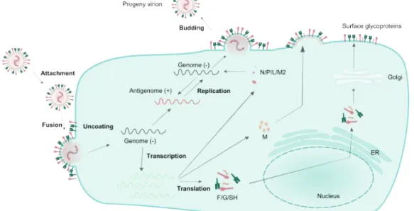
Figure 6 Replicative cycle of RSV.
Replication cycle begins when viral surface proteins interact with a cell receptor. After viral attachment fusion between viral and cell membrane occurs in a pH-independent process. All RSV replication steps occur in the cell cytoplasm and viral maturation occurs with the assembly of the nucleocapsid by combining genomic vRNA with N protein, which is accompanied by the addition of P and L proteins for envelope assembly. Matrix proteins aggregate with the viral surface proteins in the cellular membrane and the complete viral particle is released by budding, taking a portion of the plasma membrane in a reverse process to penetration by fusion. In addition to transcription and translation of proteins, viral genome produces a positively stranded RNA intermediate, which serves as a template to generate copies of the viral genome. Adapted from Collins et al. [54]
Patterns of seasonality and duration of RSV outbreaks vary considerably between geographical regions. In temperate climates, epidemics have been described in the winter months [78-80] while in tropical regions, epidemics appear to occur in rainy seasons [81]. However, it is possible that the seasonality of the virus is not only related to climatic factors but also to socioeconomic factors increasing the risk of RSV contamination [82-84]. In most RSV epidemics reported, the co-circulation of different genotypes of groups A and B were detected [78, 79, 85].
21
c) Others Human Viruses
Others respiratory viruses are capable to cause acute lower respiratory tract infections in humans such as:
Human Parainfluenza viruses (hPIVs) are common respiratory pathogens that induce acute respiratory tract diseases in infants and immunocompromised adults [86, 87]. Serological surveys have indicated that 80% of children are infected with hPIV-3 by 4 years of age and hPIV infections re-occur throughout life. The hPIV belong to a diverse group of enveloped single-stranded RNA viruses within the family
Paramyxoviridae and based on genetic and antigenic analyses, hPIVs have been
divided into four major subtypes (hPIV-1 to hPIV-4), with subtypes 1 and 3 being most frequently found in severe cases [88].
Human metapneumoviruses (hMPV) were first identified in 2001 and constitute a common cause of acute respiratory infection in individuals of all ages worldwide [89, 90]. hMPV is a member of the family Pneumoviridae, that also includes RSV, and two distinct hMPV genotypes, A and B, which can be divided into two subgroups: A1, A2, B1, and B2 circulate worldwide. hMPV and RSV share similar clinic features causing severe disease in the same range of age between children with an incidence around 15% of all respiratory tract infections [91-95].
Human Coronaviruses (HCoV) infections display a wide range of symptoms and their role in pediatric lower respiratory infections is still not clear [96, 97]. There are currently five coronaviruses (family Coronaviridae) known to infect humans and they are associated with both upper and lower respiratory tract infections in all age groups [96, 98-100]. Thus, the role of coronaviruses in pneumonia has not been completely clarified but HCoV 229E and OC43 have been recognized as causes of viral upper respiratory infection and were linked to pneumonia in children and immunocompromised adults [101, 102].
1.2.2.
Respiratory bacterial infections
Etiological studies of acute lower respiratory tract infection identify a high prevalence of different types of bacteria, even more than viral detection. Among the most frequent bacterial causes of pneumonia are Streptococcus pneumoniae,
22
Haemophilus influenzae, and Staphylococcus aureus. All these pathogens are
asymptomatic bacteria which carriage is well described in healthy individuals [103]. Also, Mycoplasma pneumoniae and Chlamydia pneumoniae are opportunistic bacteria considered as important pathogens causing pneumonia and bronchiolitis [104-110].
a) Streptococcus pneumoniae
Streptococcus pneumoniae (S. pneumoniae) can colonize the nasopharynx
asymptomatically but is one of the leading causes of high mortality and morbidity in infants, the elderly and immunocompromised people [111-114]. Prior to the use of antibiotics, more than 75% of pneumonia cases were caused by S. pneumoniae [23]. However, nowadays, studies show that only 5 to 15% of pneumonia cases are caused by S. pneumoniae in developed countries and a higher proportion of cases described in low and income countries [23, 27, 115, 116]. S. pneumoniae induced-pneumonia is commonly named pneumococcal induced-pneumonia.
S. pneumoniae is a gram-positive, encapsulated bacterium classified into 92
serotypes [117, 118] based on the composition of polysaccharide capsule. Despite this diversity, only a limited number of serotypes (around 20%) are responsible for almost 90% of all pneumococcal diseases and serotypes isolated in asymptomatic children generally reflect serotypes that cause disease [119]. The epidemiology of pneumococcal pneumonia exhibits a seasonal fluctuation with a peak incidence during the winter months [120].
S. pneumoniae is detected in the airways of healthy individuals [121] and
carriage rates are around 60% to 80% in children under five years old [122]. Colonization state (the first step to infection) occurs when bacteria promotes
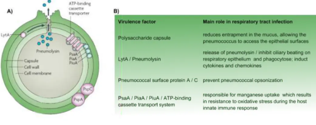
adhesion on the mucosal surface of the nasopharynx. The surface of the bacterium
consists of 3 structures with several virulence factors that could contribute to colonization and development of pneumococcal diseases as described in Figure 7 [26, 123, 124].
23
Figure 7 Pneumococcus surface structure and major virulence factors.
A) Scheme of S. pneumoniae surface structure showing capsule, cell wall, and cell membrane. The
principal's virulence factor PSpA, PspC, LytA, Pneumolysis and ABC complex are identified. B) The virulence factors showed in part A with each main role in respiratory tract infection. Adapted from Kadioglu et al. [123].
b) Haemophilus influenzae
Haemophilus influenza (H. influenzae) is a gram-negative bacterium also
found in the upper and lower respiratory tract as commensal bacteria. H. influenzae can be divided due to differences in the capsular polysaccharide, with six different strains (a-f), and nonencapsulated strains (NTHi) [125]. Polysaccharide capsule is a major virulence factor in protecting the bacterium from phagocytosis and stimulating the inflammatory response [125]. H. influenzae serotype b is considered an important agent causing pneumonia in children under five years of age, elderly and immunosuppressed [126]. Also, viral-bacterial dynamics has been described suggesting that viral infection increases NTHi colonization [127-129].
c) Others respiratory bacteria
Some studies have suggested that other bacteria, such as Staphylococcus
aureus, Mycoplasma pneumoniae, and Clamidophyla pneumoniae, may cause
pneumonia with elevated mortality among HIV positive population [10, 130].
Staphylococcus aureus (S. aureus) is another commensal bacteria of the
human nasopharynx that causes respiratory infections [131]. S. aureus is a gram-positive, facultatively anaerobic bacterium, usually without a capsule. This species of bacteria is widely distributed, being able to live in a wide variety of environments due to its tropism for several tissues. S. aureus is composed of several species and
24 subspecies [132], whereas methicillin-resistant Staphylococcus aureus (MRSA) is the principal responsible for hospital infections, among organisms resistant to antibiotics [133].
Mycoplasma pneumoniae (M. pneumoniae) are distributed globally and are
the smallest prokaryotic microbes present in nature. Mycoplasma is divided into seven species that are pathogenic to humans, including M. pneumoniae [134] that accounts for approximately 20% of all pneumonia and higher rates correlated with the degree of immunosuppression accounts in the HIV-infected population [135].
Chlamydophila pneumoniae (C. pneumoniae), with the two others species C. psittaci and C. trachomatis, are responsible for lung infections and C. pneumoniae
remains a particular problem in the HIV-infected population [8, 130]. C. pneumoniae is an obligate intracellular pathogen that induces an inflammatory reaction which contributes to damages in epithelial respiratory tract [136, 137].
1.2.3.
Mixed Respiratory infections
The upper respiratory tract constitutes a dynamic and equilibrated microbiological niche, notably composed of commensal viruses and bacteria. Perturbation of this equilibrium, by the emergence of a pathogen and/or imbalance of the host immunity, can constitute the starting point of respiratory diseases [2, 26, 138]. Usually, the opportunistic bacteria, such as S. pneumoniae, S. aureus, and H.
influenza are co-detected with respiratory viruses during lower respiratory tract
infections [127, 139-142]. However, determining the contribution of viral/bacterial co-infection to disease severity is highly complex. There is an abundance of distinct viruses and bacterial species carried commensally in the nasopharynx and samples for laboratory diagnosis are generally contaminated with components of upper respiratory tract [143]. The use of the same pathways, cofactors, and the overlap in the inflammatory mediators produced by different pathogens create an opportunity for augmentation of the immune response during dual or sequential infection. The complexity of microbiome interactions in the airways possibly contributes to the susceptibility to exacerbations and the natural course of airway diseases [144, 145].
25 Thus, many aspects of the relationship between co-infection detection and disease severity remain unclear. However, in the literature, some studies have classified viruses/bacteria interactions in two distinct scenarios:
Bacterial superinfection is described when viral infection promotes favorable conditions to commensal bacteria causes an infection in the lower respiratory tract. The association of viruses and bacteria is described by epidemiological studies that show a high prevalence of bacteria in severe disease during seasonal epidemics of the respiratory virus [146]. During IAV pandemics, bacterial superinfection was observed in adults and children associated with increased morbidity and mortality [147-149]. Bacteria superinfection is also demonstrated by quantitative studies that show an increasing of commensal bacteria during a viral infection. For example, IAV and RSV infection increase colonization rates of S. pneumoniae and H. influenzae which can lead to secondary complications contributing to the disease severity [128, 142, 146, 150-155].
Mechanisms associated with viruses predisposing the respiratory tract to bacterial superinfection [156] are poorly understood with two potential explanations:
(I) Viral infection can increase bacterial adherence into epithelial cells, as described for IAV infection which is capable to exposes bacterial receptors on the surface of host cells by cleaving sialic acids residues in the upper respiratory tract [128, 157-161]. RSV, on the other hand, is thought to bind directly to H. influenzae and S. pneumoniae [129, 159, 162], increasing bacterial proximity to the epithelial
monolayer and augmenting attachment to host cell receptors. Also, viral infection can
induce disruption of epithelial cell tract enabling bacteria to access into deep epithelial cells [147].
(II) Viral infection can also predispose bacterial superinfection via the alteration of host’s innate immune response. Viral replication may increase recruitment and activation of pro-inflammatory immune cells and may also directly affect the immune system [163-166]. Additionally, viral presence also affects the production and biological activity of cytokines [167] impairing bacterial clearance in its initial phase.
26 Bidirectional synergism or bacterial predisposition to viral disease is represented by an increased viral susceptibility to bacterial infection. It might be possible that microbial interactions may disturb the equilibrium of the microbiota, creating an opportunity for viral invasion and transmission in the lower respiratory tract.
Epidemiological studies show that pneumococcal conjugate vaccine not only reduced the incidence of pneumonia due to S. pneumoniae but also prevented approximately 33% of pneumonia associated with respiratory viruses [168]. Also, several studies have shown that the presence of a specific bacterial species may promote viral infection in the respiratory tract such as S. pneumoniae was shown to enhanced RSV infection in vitro and in vivo [144, 169, 170]. In addition, pre-exposure of epithelial cells to bacteria alters the response to subsequent viral infection, suggesting that bacterial presence could facilitate viral attachment to host cells [171].
1.3. Diagnostic of lower respiratory tract
infections
The diagnostic of pneumonia or bronchiolitis is performed through clinical examinations taking into account the history and the age of the patient. This clinical diagnosis should be performed according to WHO criteria [120]. In addition, to confirm the diagnosis of pneumonia, a chest X-ray can be performed, being able to show the extent of the disease and to identify the presence of complications that increase the severity of the disease. Usually, clinicians start the treatment without an etiologic detection due to the low sensitivity and/or lack of specificity of current diagnostic tools. In severe cases, laboratory tests capable to identify the pathogen are usually requested and a great effort has been made to improve etiological diagnosis methods [172, 173]. The quality of the collection, packaging, and transport of clinical samples are essential for an optimal diagnosis. In general, nasopharyngeal secretion samples are used for detection of viruses while blood samples are analyzed for bacterial detection [172, 173]. The most common laboratory detection methods are pathogen isolation, molecular detection, immunofluorescence and antibodies detection.
27 Pathogen isolation is a method where samples are incubated in different conditions and the most abundant pathogen is detected and identified. During a long time, this type of identification was considered the preferred method in diagnostics. For bacterial culture, the problems of this method are the false-negative detection due antibiotic pretreatment and nonculturable bacteria [174]. For viral isolation in cell culture, the support and conditions can vary a lot. Influenza virus can be isolated in embryonated chicken eggs or mammalian cell lineages (MDCK) while for RSV isolation, other mammalian cell lineages (HEp-2) are most common. The main disadvantage of these procedures is the relatively long period of time required between 7 to 10 days, depending on the pathogen [173].
Molecular detection is based on nucleic acid amplification and nowadays polymerase chain reaction (PCR) assay is considered a primordial technique for pathogen characterization. This method can be used directly on clinical samples and the rapid nature of the results can greatly facilitate investigation of outbreaks of respiratory illness. In addition, this method allows detecting multiples pathogens together being capable to identify different respiratory pathogens and its subtypes [173]. Also, it is possible to make quantitative analyses correlating pathogen load to the severity of disease [175, 176]. However, although PCR is highly specific, sensitivity has been shown to vary depending on the patient sample tested [177, 178].
Indirect immunofluorescence assay is the most common test in the diagnosis of various respiratory viruses. This technique is based on antibody staining of virus-infected cells in original clinical specimens and is a rapid and sensitive method for diagnosing viral infections [173].
Serological diagnoses are important approaches when clinical specimens are unobtainable or when a laboratory does not have the resources required for pathogen isolation. Serological methods such as the haemagglutination inhibition test are essential for many epidemiological and immunological studies and for evaluation of the antibody response following vaccination for Influenza virus, for example [173].
Biomarkers approaches can help to predict or to recognize potential cases of severity. Biomarkers are biological markers that function as indicators of a pathogen-related disease, or of disease severity [179]. The first biomarker proposed during an
28 infection were white cell count and erythrocyte sedimentary rate but nowadays they have been replaced by C-reactive protein (CRP) and procalcitonin (PCT) which have higher sensitivity and specificity for severity prognostic [180-199]. They seem to have suboptimal sensitivity and specificity for differentiated bacterial to viral pneumonia [187, 200, 201].
Several others biomarkers capable of identifying the etiology and predict complications, outcomes, and mortality of pneumonia have been studied. Tumor necrosis factor (TNF) receptor 2 and interleukin (IL)-10 characterization studies don’t show success but tissue inhibitor of metalloproteinases has shown promise for the identification of bacterial pneumonia in children [195, 202-205]. Also, an association of different biomarkers like CRP, TRAIL, and IP-10 were described and might offer advantages in the differentiation of viral or bacterial pneumonia.
In addition, innovative technologies, including microarray-based whole genome expression arrays, proteomics, and metabolomics, can be a basis for biomarker discovery. For example, specific host responses induced show a microRNA bio-signatures that can be identified using microRNA analyses [199]. However, further studies are necessary before routine use of biomarker assays [199, 206].
1.4. Treatments for lower respiratory tract
infections
Treatments for lower respiratory tract infections depend on the nature of the etiological agent, resulting in antibiotic or antiviral treatment, in the case of bacterial or viral infections, respectively.
1.4.1.
Antiviral treatments
Viral replication is linked to metabolic processes of the host cells and safe antivirals offering benefits by reducing mortality as well as the duration of disease
29 approaches focused on targeting the viral cycle and new antiviral strategies targeting the host instead of the virus [207].
a) Anti-influenza drugs
According to their targets in the viral replicative site, anti-influenza drugs are
classified into three groups: targeting M2 channel, neuraminidase inhibitors, or
polymerase inhibitors.
Inhibition of viral envelope fusion to cell membranes by M2 proton channel blockers is represented by amantadine and rimantadine. These inhibitors were approved by health authorities but transmissible resistance variants rapidly emerged from patients after treatment and since 2009, they are not recommended for clinical use anymore [208].
Viral NA protein is an attractive target for drug action as it is essential for infectivity and has a highly conserved active site across influenza A and B viruses [31]. Inhibitors of NA, which block the sialidase activity of NA and prevent the release of new viral particles, are represented by zanamivir, oseltamivir, peramivir, and laninamivir [209]. This class of antiviral is approved for human treatments and oseltamivir is the most recommended worldwide. Viral resistance to oseltamivir was reported confirming the need for new antiviral therapies [210, 211].
Inhibitors of viral polymerases, which interrupt replication and transcription of the viral genome, include inhibitors of PB2 and NP. Some molecules such as nucleozin, naproxen, RK424 (NP inhibitors) and VX-787 (inhibitors of PB2) are in pre-clinical phases studies with promising results [212].
An alternative strategy less prone to antiviral resistance consists to target the host rather than the viral determinants. Fludase (DAS181) is inhibitory for a range of influenza A and B viruses, altering the ability of the virus to replicate efficiently. Potent antiviral properties during clinical trials with reduced inflammatory responses in mice and ferrets were described. Also, Fludase promotes protection against secondary pneumococcal infection of mice [213, 214]. Another example is the acetylsalicylic acid and its derivate demonstrate antiviral activity against influenza A viruses with some ongoing phase I/II clinical assays [215]. Also, the combination of
30 antiviral agents like oseltamivir with immune modulators like acetylsalicylic acids has been evaluated and shown to increase survival in animal models [212, 215, 216].
In addition, RNA-based screening studies or other similar high-throughput approaches are very helpful to identify new cellular targets. These studies provide a valuable library to select novel cellular drug targets [217, 218]. Targeting cellular rather than viral factors could be an important approach to prevent the problem of resistance to classic antivirals.
b) Anti-RSV drugs
To date, no effective and accessible treatment for RSV is available. The only drug licensed is inhaled ribavirin. Ribavirin is an analog of purine nucleotides which inhibits viral replication. However, its use remains limited because of a lack of results proving its efficacy and suspicion of side effects. Ribavirin is sometimes used in some circumstances but is not recommended in most cases [219-222].
Some anti-RSV treatment targets viral replication cycle by intervention in membrane fusion and RNA synthesis. Membrane fusion inhibition, such as GS-5806, prevents virus-cell fusion and cell-cell syncytium formation and has shown promise results in early-phase clinical trials with efficacy and safety in hospitalized adults [223, 224]. Inhibitors of RNA synthesis during RSV replication are divided into two groups. Nucleoside analogs represented by ALS-8112 and ALS-8176 target the active site of the polymerization domain, and non-nucleoside inhibitors (BI-compoundD) bind to other regions of the polymerase. These molecules are in ongoing studies and show promising results but further evaluation is necessary to determine effectivity and safety [225-228].
Despite the importance of this viral pathogen, there are not adequate treatment options available. Thus, it is important to continue to identify and characterize possible targets for antiviral drugs.
1.4.2.
Antibiotics
Against bacterial infections, antibiotics are widely used worldwide. There are cytotoxic or cytostatic towards the microorganism and often act by inhibiting the
31 synthesis of a bacterial cell wall [229, 230]. For mild to moderate pneumonia suspected to be of bacterial origin, amoxicillin is recommended first-line therapy. Amoxicillin appropriately covers the most prominent invasive bacterial pathogen, S.
pneumoniae [231]. During an atypical bacterial suspicion, a macrolide (azithromycin)
is recommended [232]. In addition, a third-generation cephalosporin is recommended for a specific group of patients [5].
Due to the indiscriminate use of antibiotics, the emergence of antibiotic-resistant strains is considered a serious problem [230]. Thus, preventing lower respiratory tract infections through vaccination and prophylaxis is important.
1.5. Prevention of lower respiratory tract
infection
Prophylaxis is considered one of the best alternatives for combating respiratory tract infections. The prophylaxis method most common are vaccines, but passive immunization has also been overspread worldwide.
1.5.1.
Viral prophylaxis
The development of viral vaccines are based on attenuated, recombinant, inactivated and subunit composition strategies.
A traditional strategy that has worked for several pathogens involves the development of attenuated viral strains. Attenuation can be accomplished by serial passage or cold-adaptation and has the advantage of expressing most of the pathogen’s antigens to improve immune response. Attenuated vaccines for Influenza viruses have been produced for more than 50 years, however, this vaccine has a restricted use in USA and Russia [233, 234]. Using this strategy, attenuated RSV
strains have been developed [235, 236] but failed in some clinical trials [237, 238].
The disadvantage of this strategy is the that, in rare cases, the live attenuated vaccine strain can revert to its virulent wild-type, causing severe disease [239].
Alternatively, recombinant vaccines consisting viral protein expressed in other
32 approach was described for influenza virus which HA protein was expressed in insect cells by baculovirus vectors [233]. For RSV, viral antigens expressed in other viruses, such as Sendai virus, vaccinia virus, adenovirus, and parainfluenza or in bacteria was studied. Despite promising results in murine studies, studies in adults showed relatively low capacity for inducing neutralizing activity [241-243], so they have not been advanced into clinical phases. Another approach of recombinant RSV vaccine carrying host cytokines capable to promote immune responses suggests that these formulations can modulate the immune response being effective alternatives for immunization against RSV [244-249].
Inactivated vaccines are composed of purified virus chemically inactivated and are capable to generate humoral and cellular immunity. The annual Influenza vaccine around the world is an inactivated vaccine [31, 233, 234]. This vaccine, during decades, was composed of 2 IAV strains and one strain of Influenza B virus according to circulated subtypes detected by surveillance. In 2016, WHO decided to add another Influenza B virus subtype to try to increase vaccine coverture [34, 233]. Even if this Influenza vaccine has a great coverture and seroconversion, the constantly evolving of influenza viruses requires continuous global monitoring and annually reformulation of influenza vaccines [250, 251]. For RSV, during 60’s, a clinical trial of a formalin-inactivated RSV vaccine not only failed to prevent RSV infection but caused an increase in severe disease [252]. This experience had a profound negative impact on subsequent RSV vaccine development and the immune mechanisms that led to enhanced disease in this clinical study are not yet fully elucidated, making difficult to advances into clinical evaluation of inactivated RSV vaccines [253].
For RSV vaccine, lability and heterogeneity in particle size represent obstacles for the formulation of a stable vaccine [254] and despite the many approaches developed and tested, there is still no vaccine defined for use in humans.
Thus, the prophylactic palivizumab is indicated in months prior to the seasonality of RSV for premature babies; children with congenital pathology or with chronic lung disease by WHO. Palivizumab is a humanized mouse IgG1 monoclonal antibody directed against a conserved epitope on the surface fusion protein of RSV. This passive prophylaxis shows a potent RSV neutralizing activity and has been
33 clearly demonstrated to protect against RSV. The administration of palivizumab in specific risk groups is limited by its expensive cost in many low and income countries [221]. Consequently, prevention of RSV infection is a public health priority, and global initiatives have advanced numerous efforts to expand the field [255]. Continued research into the pathogenesis of RSV disease and immune responses are important to contribute to the development of RSV vaccines.
1.5.2.
Pneumococcal vaccine
The polysaccharide capsule from encapsulated bacteria is a major virulence factor and can be used as an antigen for vaccine development [256, 257]. However, that does not induce a complete response and cannot provide adequate protection against pneumococcal infection in children [24]. Thus, the polysaccharide was chemically conjugated to different bacterial protein [258] and the first conjugate vaccine used was composed of 7 different serotypes of pneumococcus (PVC 7). In 2009, two new conjugated vaccines were licensed for use with 10 and 13 different serotypes (PCV 10 and PVC 13). Serotypes coverture of each conjugated vaccine is shown in Table 1.
Pneumococcal vaccine Serotypes
PCV 7 4, 6B, 9V, 14, 18C, 19F, 23F
PCV 10 1, 4, 5, 6B, 7F, 9V, 14, 18C, 19F, 23F
PCV 13 1, 3, 4, 5, 6B, 6C, 7F, 9V, 14, 18C, 19C, 19F, 23F
Table 1 Pneumococcal vaccine.
Serotypes included in each pneumococcal vaccines approved to use worldwide [259].
A reduction in pneumococcal disease among vaccinated children have been observed since the introduction of the first PCV vaccine in 2000 [151, 260-264]. In addition, vaccination in children has been shown to reduce pneumococcal disease among the elderly by preventing the transmission due the diminished of the carriage in general population [265-267].
The challenge of the pneumococcal vaccine is the existence of 92 serotypes since vaccinated individuals remain susceptible to serotypes not included in the
34 vaccine [268-270]. Also, changes of serotypes carried out in the nasopharynx of children [271] were detected and the potential risk of other serotypes infections make the pneumococcal disease an important problem of global health, especially in children and must be a priority.
Therefore, new potential vaccines that effectively protect against pneumonia have been investigated and are undergoing clinical trials [24, 272-274].
35
2. Host-pathogen interactions
The human body is constantly exposed to microbes and prevention of opportunistic infections is made by physical or anatomical mechanisms (skin, mucosa), mechanical (cilia in respiratory tract cells, tight junctions) and biochemical (tears, saliva) barriers as well as cellular mechanisms including innate and immune responses [1].
The pathogenesis of respiratory infections involves the complex interplay between virulence factors, environmental conditions, the magnitude and temporal dynamics of the host response, and host susceptibility factors. The severity of disease is associated with an enhanced host immune response which is essential for pathogen control but can cause collateral damage to the tissues, leading to mortality in some cases. In addition, several pathogens evolved strategies to counteract or hijack host responses constitute by multiple elements including diverse cell types (epithelial cells, dendritic cells, macrophages, monocytes, and granulocytes), various pattern recognition receptors, a large array of cytokines and chemokines, cellular stress, and different pathways [25, 275].
Thus, in this chapter, immune response and others cellular mechanisms important during Influenza virus, RSV and S. pneumoniae infection was highlighted as resumed in Figure 8.
36
Figure 8 Host-pathogen interactions.
Important complex cell physiological processes in the control of pathogen infection showing an immune and inflammatory response, in addition to p53 pathway. all these cellular mechanisms are often targeted or modulated by pathogens during infection. Each pathway will be explained in the next part. Adapted from Sun et al. [275].
2.1. Immune response
In the respiratory tract, epithelial cells are constantly in contact with potential pathogens having the important function to activate immune
responses.The immune response is divided into innate and adaptive
immunity. The innate response is the first line of defense induced by a pathogen and aims to control the infection locally and to activate an adaptive response. The adaptive response is specific to the pathogen, more systemic and durable [276]. A chronological course of innate and adaptive immunity is schematically showed in
37
Figure 9 Chronological course of innate and adaptive immunities.
Innate mechanisms confer rapid protection, keeping the invading microorganisms under control until the development of adaptive immunity. It may take several days and even weeks for adaptive immunity to become effective.
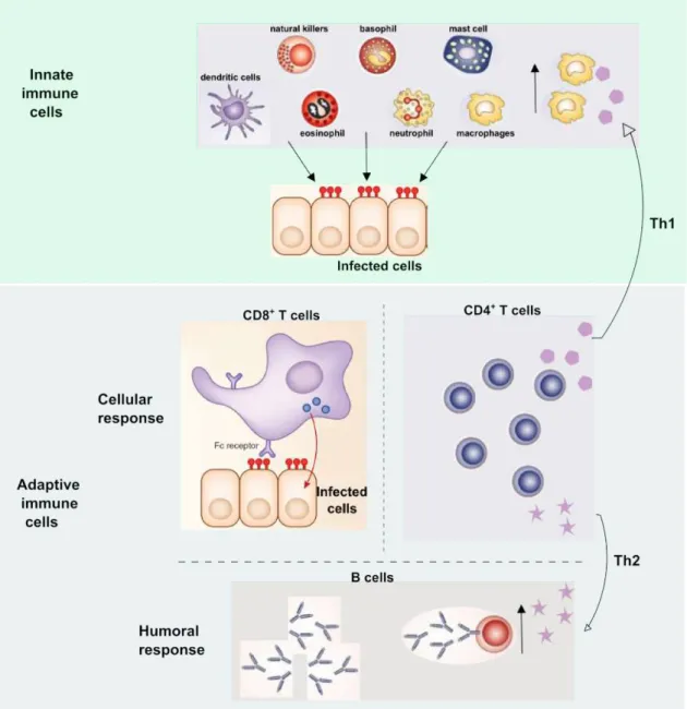
Figure 10 Innate and adaptive cells.
Innate immune cells (macrophages, dendritic cells, natural killers, basophil, neutrophil and mast cells) exists before the invading microorganism, they are effectors cells capable of recognized several molecules of various pathogens. Adaptive cells are developed following exposure to a particular invading agent. It is able to react more quickly and more effectively to subsequent contacts, they specifically identify a molecule with high specificity. Adaptive immunity is divided into a cellular and humoral response. During the cellular response, CD8+ T cells are capable to identify and kill infected cells while CD4+ T cells increase macrophages quantity and/or increases antibodies expression by B cells (humoral response).
38
2.1.1.
Innate immune response
The innate immune response begins as soon as the pathogen enters the target cell and is implicated in recognition and protection of infections. The innate system consists of different cells as shown in Figure 10.
The recognition of the pathogen by receptors is the first step in the host cell membrane during infection. This recognition is based on a limited repertoire of receptors called pattern recognition receptors (PRRs) that detect conserved microbial components known as pathogen-associated molecular patterns (PAMPs) [276]. This initial response triggered by infection is mediated by three major receptor families - PRRs: Toll-like receptors, RIG-I-like receptors (RLRs), and NOD-like receptors (NLRs) that will be detailed above.
Toll-Like Receptors (TLRs) are type 1 transmembrane proteins that are able to recognize PAMPs from bacteria, parasites, fungi, and viruses [277]. TLRs are one of the largest classes of PRRs with 10 receptors (TLRs 1-10). TLRs family is well conserved among organisms and homologous receptors are found in plants, insects and other vertebrates [276, 278]. Normally TLR 1, 2, 4, 5, 6 and 10 are expressed on the cell surface while TLR 3, 7, 8 and 9 are intra-vesicular [276, 279]. The expression profile of TLRs in different cell types may be tissue-specific and highlight the different role between cells of the immune system (macrophages, dendritic cells, neutrophils, B and T cells) and epithelial cells [277].
Viral attachment and fusion proteins, as well as the components of bacterial cell wall, are able to be recognized by these receptors. The summary of TLR receptors and pathogens ligands are described in Table 2.