RESEARCH OUTPUTS / RÉSULTATS DE RECHERCHE
Author(s) - Auteur(s) :
Publication date - Date de publication :
Permanent link - Permalien :
Rights / License - Licence de droit d’auteur :
Bibliothèque Universitaire Moretus Plantin
Institutional Repository - Research Portal
Dépôt Institutionnel - Portail de la Recherche
researchportal.unamur.be
University of Namur
Optimization of label-free nano LC-MS/MS analysis of the placental proteome
Luyten, Leen J.; Dieu, Marc; Demazy, Catherine; Fransolet, Maude; Nawrot, Tim S.; Renard,
Patricia; Debacq-Chainiaux, Florence
Published in:
Placenta
DOI:
10.1016/j.placenta.2020.09.013
Publication date:
2020
Document Version
Publisher's PDF, also known as Version of record
Link to publication
Citation for pulished version (HARVARD):
Luyten, LJ, Dieu, M, Demazy, C, Fransolet, M, Nawrot, TS, Renard, P & Debacq-Chainiaux, F 2020,
'Optimization of label-free nano LC-MS/MS analysis of the placental proteome', Placenta, vol. 101, pp. 159-162.
https://doi.org/10.1016/j.placenta.2020.09.013
General rights
Copyright and moral rights for the publications made accessible in the public portal are retained by the authors and/or other copyright owners and it is a condition of accessing publications that users recognise and abide by the legal requirements associated with these rights. • Users may download and print one copy of any publication from the public portal for the purpose of private study or research. • You may not further distribute the material or use it for any profit-making activity or commercial gain
• You may freely distribute the URL identifying the publication in the public portal ? Take down policy
If you believe that this document breaches copyright please contact us providing details, and we will remove access to the work immediately and investigate your claim.
Placenta 101 (2020) 159–162
Available online 14 September 2020
0143-4004/© 2020 Elsevier Ltd. All rights reserved.
Technical note
Optimization of label-free nano LC-MS/MS analysis of the
placental proteome
Leen J. Luyten
a,b, Marc Dieu
a,c, Catherine Demazy
a,c, Maude Fransolet
a,c, Tim S. Nawrot
b,d,
Patricia Renard
a,c, Florence Debacq-Chainiaux
a,*aUnit´e de Recherche en Biologie Cellulaire (URBC) - Namur Research Institute for Life Sciences (Narilis), University of Namur (UNamur), Namur, Belgium bCentre for Environmental Sciences, Hasselt University (UHasselt), Diepenbeek, Belgium
cMaSUN, Mass Spectrometry Facility, University of Namur (UNamur), Namur, Belgium
dDepartment of Public Health & Primary Care, Occupational and Environmental Medicine, Leuven University (KULeuven), Leuven, Belgium
A R T I C L E I N F O Keywords: Placenta Proteomics Label-free nano LC-MS/MS A B S T R A C T
The placenta can be regarded as a mirror of the events to which the fetus is exposed during development. The placental proteome has been studied with several methodologies differing in sample handling, protein extraction, and processing. We optimized a protocol to analyze the placental proteome by means of label-free nano-LC-MS/ MS mass spectrometry with regard to sample treatment, protein extraction, and protein digestion, in order to obtain a high protein concentration for identification of a specific protein signature according to the conditions studied. We recommend mechanical tissue disruption, blood removal prior to protein extraction, and FASP-based or in-gel digestion.
1. Introduction
The placenta is a complex organ that can be used postnatally to examine the morphological and molecular effects of environmental ex-posures or disease processes during pregnancy. The placental proteome has been studied for numerous adverse pregnancy conditions, such as preeclampsia [1,2] and in the context of environmental stressors, such as maternal smoking during pregnancy [3]. Several techniques are currently used in an attempt to identify the entire placental proteome or a placental sub-proteome [4,5]. However, the placenta is a complex tissue with various cell types and it has proven to be very challenging to study the low molecular weight, low abundance placental proteins [5]. Hence, optimization of protein processing prior to the final analysis, for example with label-free mass spectrometry, is needed to improve iden-tification. Protocols applied in different research groups vary in the size of the placental biopsies and in the methods to process the protein ex-tracts into peptides. Therefore, more attention is needed to optimize and unify how to handle placental tissue samples before proteins are extracted, the technique to separate and concentrate placental proteins, and the digestion of the protein mixture into peptides, prior to peptide
injection in the mass spectrometer.
Two important aspects of proteomics are sensitivity (the ability to detect the protein if it is present) and specificity (the ability to distin-guish a specific target protein from other proteins in the sample) [6], with a high risk of different outcomes for the same research question when different protocols are applied. In addition, since protein material can be lost in each step of the label-free nano LC-MS/MS analysis pro-cess, from protein extraction until the final injection of the peptide mixture, a generalized protocol should entail a high number of proteins identified. In this study we optimized an approach to study the proteome of the placenta through label-free nano LC-MS/MS, since this technique allows the untargeted identification of a substantial part of the protein entity, with high accuracy and precision. We paid specific attention to sample treatment regarding blood removal, protein extraction, and post-extraction sample processing.
Abbreviations: ACN, Acetonitrile; DTT, Dithiothreitol; FASP, Filter-aided sample preparation; IAA, Iodoacetamide; Nano LC-MS/MS, Nano Liquid chromatographic
tandem mass spectrometry; PBS, Phosphate buffered saline; TFA, Trifluoroacetic acid; UA, Urea acetate. * Corresponding author.
E-mail address: [email protected] (F. Debacq-Chainiaux).
Contents lists available at ScienceDirect
Placenta
journal homepage: http://www.elsevier.com/locate/placenta
https://doi.org/10.1016/j.placenta.2020.09.013
Placenta 101 (2020) 159–162
160
2. Methods
2.1. Sample collection
Placental tissue samples were collected from 10 randomly selected participants of the ongoing ENVIRONAGE (ENVIRonmental influence
ON AGEing in early life) birth cohort. The study protocol was approved
by the ethical committees of Hasselt University and East-Limburg Hos-pital (EudraCT B37120107805). Before delivery, an informed consent form was signed by the mothers, and placentas were collected within 10 min after birth [7]. The samples were taken at the fetal side of the placenta, as described by Janssen et al. [7] and were either directly frozen in liquid nitrogen or rinsed shortly in phosphate-buffered saline (PBS) before freezing. The biopsies were kept at − 80 ◦C until protein
extraction.
2.2. Protein extraction
Proteins were extracted from approximately 200 mg placental tissue in 1.5 ml lysis buffer containing 2 M thiourea, 7 M urea, 2% CHAPS, and 2% dithiothreitol (DTT). Four tissue disruption methods were tested: by using a syringe (VWR, Pennsylvanian USA), by three subsequent 10-sec sonication bursts (UP100H, Hielscher, Teltow, Germany), by mechani-cal disruption with an Ultra Turrax T8 mixer (IKA, Staufen, Germany) three times during 10 s, or by crushing the tissue in liquid nitrogen. The lysates were incubated on a shaking plate for 30 min at 1400 rpm and 15 ◦C and subsequently centrifuged for 5 min at 16.000 g. Protein
concentration was determined with the Pierce BCA Protein Assay Kit (Thermo Scientific, Massachusetts, USA).
2.3. Trypsin digestion and detergent removal 2.3.1. Protein precipitation and liquid digestion
Protein extracts were treated with the 2-D Clean-up kit (GE Health-care Life Sciences, Illinois, USA) according to the manufacturers’ in-structions. The resulting pellet was re-suspended in 0.2% Rapigest (Waters, Massachusetts, USA), incubated at 15 ◦C for 30 min,
centri-fuged for 5 min at 13,000 g, heated for 5 min at 100 ◦C and finally kept at
− 80 ◦C. Samples were reduced with 10 mM dithiothreitol (DTT) and
shaken at 500 rpm for 45 min at 37 ◦C, then, alkylated with 40 mM
iodoacetamide (IAA) in the dark under the same conditions. After add-ing 1 mM CaCl2 and trypsin in a ratio of 1/50 w/w, protein digestion
occurred overnight, shaking at 300 rpm and 37 ◦C. The reaction was
stopped by adding 2% trifluoroacetic acid (TFA; Biosolve) and incu-bating for 45 min at 300 rpm and 37 ◦C. After 10 min of centrifugation at
13,000 rpm, the supernatant was used for nano LC-MS/MS analysis.
2.3.2. Liquid digestion with the FASP kit
Samples were digested using filter-aided sample preparation (FASP) digestion, based on modified protocols of the FASP Protein Digestion kit (Expedeon, San Diego, USA) [8,9]. We used 10 μg of protein input, and
protein digestion was performed with either 1/20 or 1/50 w/w trypsin, for either 2 h, 5 h, or overnight at 37 ◦C and 300 rpm. Following final
centrifugation during 10 min at 13,000 g, the digestion was stopped with 2% TFA, after which the peptide mixture was directly analyzed with nano LC-MS/MS.
2.3.3. In-gel digestion
Five μl of loading buffer was added to 20 μg of protein sample. This
mixture was heated at 100 ◦C for 5 min, centrifuged during 2 min at
13,000 rpm and subsequently loaded on a 10% polyacrylamide gel (Biorad, California, USA). Following a run of 5 min at 200 V and 400 mA, the gel containing proteins was cut into pieces of ± 5–6 mm3 and kept in H2O at − 20 ◦C prior to trypsin digestion.
Gel fragments were dehydrated with acetonitrile (ACN), shaking 10 min at 900 rpm. After ACN removal, reduction and alkylation of the in-
gel protein samples were performed similar to that of the liquid diges-tion protocol, by incubadiges-tion with 10 mM DTT and 55 mM IAA. Following two incubation steps with ACN and one with 100 mM NH4HCO3, the in-gel protein mixtures were digested with trypsin at a
final concentration of 6.25 ng/μl, overnight at 37 ◦C and 300 rpm.
2.4. Nano LC-MS/MS
We analyzed the peptide samples with a nano-liquid chromatog-raphy Ultimate 3000 system (Thermo Scientific), connected to a maXis Impact electrospray Ultra-High resolution Q-TOF mass spectrometer (Bruker, Massachusetts, USA). Initial peptide separation occurred by reverse-phase liquid chromatography on a 75 μm by 250 mm C18
col-umn (Acclaim™ PepMap™ 100 C18 LC Colcol-umn, Thermo Scientific). Mobile phase A in this column was composed of 0.1% formic acid and 2% acetonitrile, mobile phase B contained 0.1% formic acid in 80% acetonitrile. For each run, 2 μg of sample was injected, and the organic
concentration of the mobile phase was increased linearly from 4% to 35%. We used a 90 min gradient for in-gel digestion and one of 215 min for other strategies. Subsequently, the resulting flow-through was ionized in the electrospray ionization (ESI) CaptiveSpray (Bruker), which was directly coupled to the C18 column. In survey scan, MS spectra were acquired for 0.5 s in a 50–2200 m/z range.
Peak lists were created using the DataAnalysis 4.0 software (Bruker) and uploaded to the ProteinScape 3.1 software (Bruker) which uses Mascot 2.4 (Matrix Science) as protein identifier search engine, through the UniProt database (version September 2019, Uniprot-HomoIsoform 190904). Finally, we used the Scaffold 4.8 software (Proteome Soft-ware Inc., Portland, Oregon, USA) to visualize the protein identification lists. For the total number of identified proteins, we set the protein and peptide threshold at 1.0% false discovery rate (FDR), and the minimum number of peptides for identification at two. We used the total spectral count method, counting the total number of analyzed spectra associated to a single protein group, including those shared with other proteins.
2.5. Data analyses and publication
We used the built-in statistical package of the Scaffold 4.8 software (Proteome Software Inc.) for initial protein identification data analyses. Relative protein abundance was determined using the spectral counts method. We determined the difference in total protein numbers between liquid digestion and FASP-based digestion, and between one and two PBS washing steps, by a two-sided t-test with SAS 9.4 software (SAS Institute Inc., Cary, NC, USA). The mass spectrometry proteomics data were deposited to the ProteomeXchange Consortium via the PRIDE [10] partner repository with the dataset identifiers PXD020438 and 10.6019/PXD020438.
3. Results
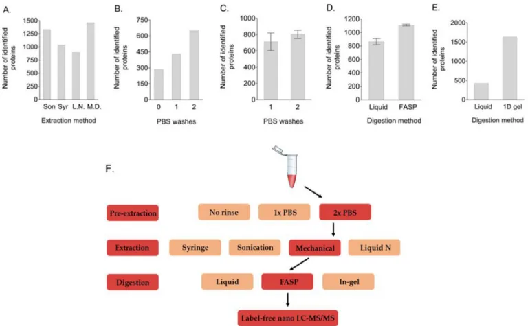
3.1. Mechanical tissue disruption and extra washing improves protein identification
We tested placental tissue disruption with a syringe, sonication, liquid nitrogen, or mechanical disruption. The total number of identified proteins was the highest after mechanical separation (n = 1458) (Fig. 1A), although the number for sonication did not seem to differ greatly (n = 1330). Because of the presence of more uniquely identified proteins and higher unique peptide counts for important functional proteins (such as ATP synthase and superoxide dismutase) in mechani-cally disrupted samples compared to those treated with sonication, for all further optimization tests on pre-extraction blood removal and on protein digestion methods, tissue samples were mechanically disrupted. Using the same biological sample, we performed either no sample treatment, one PBS washing step after tissue collection, or a washing step with PBS both after tissue collection and after thawing to determine
the effect on protein identification. PBS washing increased the number of protein identifications (Fig. 1B). Repeating this experiment, with the conditions of one and two PBS washes in triplicate, showed that after two PBS washes the average total amount of identified proteins increased from 713 to 803, although the difference was not statistically significant (p = 0.27) (Fig. 1C) and that the numbers of several blood- specific proteins decreased (data not shown).
3.2. Increased protein identification with gel- and FASP-based separation and digestion compared to liquid digestion
Firstly, we tested FASP digestion as an alternative for protein pre-cipitation and liquid digestion. We used one biological sample and tested each technique in triplicate. At this optimization stage, all tissue samples were washed both before freezing and after thawing, and were mechanically disrupted for protein extraction. FASP digestion resulted in a higher number of protein identifications (n = 1110) compared to liquid digestion (n = 864) (p = 0.003) (Fig. 1D). Furthermore, we compared the percentage of non-specific cleavages (i.e. peptides were cut at amino acids other than lysine or arginine) and miscleavages (i.e. trypsin failed to cut the carboxyl end of lysine or arginine). The per-centage of miscleavages was approximately the same for liquid- (9.8%)
and FASP-based digestion (10.2%), while the percentage of non-specific cleavages was higher for liquid digestion (9.1% vs 3.7%).
Secondly, we compared liquid-based digestion with in-gel digestion of the same biological replicate. Gel-based digestion increased the number of protein identifications (n = 1640) compared to liquid digestion (n = 462) (Fig. 1E). Although the number of non-specific cleavages was comparable (7.3% for liquid digestion and 5.6% for gel- based digestion), the percentage of miscleavages was higher for gel- based digestion (5.1% vs 17.1%).
In a third test, we compared three digestion periods, i.e. 2 h, 5 h, or overnight, in combination with either 1/20 or 1/50 w/w of trypsin. All samples, derived from one biological replicate, were digested by the FASP method. Although the total number of proteins did not differ substantially, the number of miscleavages was higher for the lower trypsin concentrations during each of the three digestions periods (Table 1). Overnight digestion increased the number of non-specific cleavages, especially for the digestion with 1/20 w/w trypsin. There-fore, a 5-h digestion with 1/20 w/w trypsin appears to be most favorable.
Fig. 1. Increase in the total number of identified proteins after protein extraction through mechanical tissue disruption, after a PBS washing step both before freezing
and after thawing the samples, and either FASP-system-based or gel-based protein digestion. Figure A: Tested methods of sample tissue disruption are depicted in the x-axis. Mechanical disruption of the placental tissue resulted in the highest number of identified proteins. Figures B and C: The x-axis indicates the number of times that the samples had been washed before protein extraction. An extra wash of the placental biopsies after thawing increased the total number of identified proteins. Figure D: Protein digestion with the FASP system increased the total number of identified proteins, compared to the numbers obtained with liquid protein digestion. Figure E: In-gel digestion following 1D gel sample separation increased the total number of identified proteins, compared to the numbers obtained with liquid protein digestion. Figure F: To increase the total number of proteins identified by label-free nano LC-MS/MS, we advise to wash placental tissue samples two times (once directly after taking the biopsies, and a second time before protein extraction) with a neutral substance such as PBS for excess blood removal prior to tissue disruption (line “Pre-extraction”), mechanically disrupt the washed tissue samples (line “Extraction”) for protein extraction, and perform FASP-based protein digestion (line “Digestion”) for 5 h with 1/20 w/w trypsin to minimize the number of miscleavages and non-specific cleavages.
Abbreviations: FASP, filter-aided sample preparation; L. N, crushing tissue that was snap-frozen in liquid nitrogen; M. D., mechanical tissue disruption; PBS, phosphate-buffered saline; Son, sonication bursts; Syr, disruption of placental tissue through a syringe.
Placenta 101 (2020) 159–162
162
4. Discussion
We have evaluated a protocol to analyze the placental proteome through label-free nano-LC-MS/MS. Our key findings are: 1) Blood removal prior to protein extraction and mechanical tissue disruption increase the total amount of identifications; 2) In-gel and FASP-based protein digestion result in a higher number of identified proteins, compared to liquid-based digestion and protein precipitation, and 3) To reduce the number of miscleavages and non-specific cleavages during placental protein digestion, a concentration of 1/50 w/w trypsin and overnight digestion are not advised (Fig. 1F). Depending on the research question and number of samples, research groups can choose between FASP-based digestion or in-gel protein digestion, of which the latter is more labor-intensive and time consuming (due to all gel fragments for one sample being analyzed separately), but results in higher protein identification compared to FASP-based digestion.
One of the challenges of using placental tissue in proteomic research is its complexity and heterogeneity. Concerning cellular compartment distribution, our placental protein extracts contained a higher percent-age of mitochondrial proteins, and a similar number of cytosol- and membrane-related proteins for all extraction techniques in comparison with the human proteome [11–13]. Nuclear proteins could be identified to a lower level compared with the human proteome for each technique, apart from the technique without blood removal before protein extrac-tion, for which no nucleus-related protein was found (Supplemental
Table 1).
Recent research suggests that the placenta contains different sub- proteomes, rather than one proteome entity [14]. Therefore, tissue sampling is an essential initial step in placental proteomic research. Burton and colleagues have optimized a protocol regarding the placental storage and sampling process for various purposes [15]. If these steps proceeding sample treatment and protein digestion would be combined and unified for proteomic research purposes in placental tissue, this would aid reproducibility and reliability in the search for protein bio-markers related to the impact of environmental exposure or disease development during pregnancy.
Declaration of competing interestCOI
All authors declare that this research was conducted in the absence of any commercial or financial relationships that could be considered as a potential conflict of interest.
Acknowledgements
Leen J. Luyten was financed by the Bijzonder Onderzoeksfonds (BOF,
BOF15DOCNA01) partnership between the UHasselt (Diepenbeek, Belgium) and the UNamur (Namur, Belgium). Florence Debacq- Chainiaux is a Research Associate of the FNRS, Belgium.
Appendix A. Supplementary data
Supplementary data to this article can be found online at https://doi. org/10.1016/j.jallcom.2020.156830.
References
[1] W. Zhang, X. Chen, Z. Yan, Y. Chen, Y. Cui, B. Chen, C. Huang, W. Zhang, X. Yin, Q.-Y. He, T. Wang, Detergent-insoluble proteome analysis revealed aberrantly aggregated proteins in human preeclampsia placentas, J. Proteome Res. 16 (2017) 4468–4480, https://doi.org/10.1021/acs.jproteome.7b00352.
[2] F. Wang, Z. Shi, P. Wang, W. You, G. Liang, Comparative proteome profile of human placenta from normal and preeclamptic pregnancies, PLoS One 8 (2013), e78025, https://doi.org/10.1371/journal.pone.0078025.
[3] P. Huuskonen, M.R. Amezaga, M. Bellingham, L.H. Jones, M. Storvik, M. H¨akkinen, L. Keski-Nisula, S. Heinonen, P.J. O’Shaughnessy, P.A. Fowler, M. Pasanen, The human placental proteome is affected by maternal smoking, Reprod, Toxicol 63 (2016) 22–31, https://doi.org/10.1016/j.reprotox.2016.05.009.
[4] D.D. Vandr´e, W.E.I. Ackerman, A. Tewari, D.A. Kniss, J.M. Robinson, A placental sub-proteome: the apical plasma membrane of the syncytiotrophoblast, Placenta 33 (2012) 207–213, https://doi.org/10.1016/j.placenta.2011.12.010.
[5] K. Kedia, C.A. Nichols, C.D. Thulin, S.W. Graves, Novel “omics” approach for study of low-abundance , low-molecular-weight components of a complex biological tissue: regional differences between chorionic and basal plates of the human placenta, Anal. Bioanal. Chem. 407 (2015) 8543–8556, https://doi.org/10.1007/ s00216-015-9009-3.
[6] R. Wilson, Sensitivity and specificity: twin goals of proteomics assays. Can they be combined? Expert Rev. Proteomics 10 (2013) 135–149, https://doi.org/10.1586/ epr.13.7.
[7] B.G. Janssen, N. Madhloum, W. Gyselaers, E. Bijnens, D.B. Clemente, B. Cox, J. Hogervorst, L.J. Luyten, D.S. Martens, M. Peusens, M. Plusquin, E.B. Provost, H. A. Roels, N.D. Saenen, M. Tsamou, A. Vriens, E. Winckelmans, K. Vrijens, T. S. Nawrot, Cohort Profile: the ENVIRonmental influence ON early AGEing (ENVIRONAGE): a birth cohort study, Int. J. Epidemiol. 46 (2017) 1386–1387m,
https://doi.org/10.1093/ije/dyx033.
[8] J.R. Wi´sniewski, A. Zougman, N. Nagaraj, M. Mann, Universal sample preparation method for proteome analysis, Nat. Methods 6 (2009) 359–362, https://doi.org/ 10.1038/nmeth.1322.
[9] U. Distler, J. Kuharev, P. Navarro, S. Tenzer, Label-free quantification in ion mobility–enhanced data-independent acquisition proteomics, Nat. Protoc. 11 (2016) 795–812, https://doi.org/10.1038/nprot.2016.042.
[10] Y. Perez-Riverol, A. Csordas, J. Bai, M. Bernal-Llinares, S. Hewapathirana, D. Kundu, A. Inuganti, J. Griss, G. Mayer, M. Eisenacher, E. P´erez, J. Uszkoreit, J. Pfeuffer, T. Sachsenberg, S. Yilmaz, S. Tiwary, J. Cox, E. Audain, M. Walzer, A. Jarnuczak, T. Ternent, A. Brazma, J. Vizcaíno, The PRIDE database and related tools and resources in 2019: improving support for quantification data, Nucleic Acids Res. 47 (2019) D442–D450, https://doi.org/10.1093/nar/gky1106. [11] A. Pandey, S. Chakraborty, N. Chakraborty, Nuclear proteome: Isolation of Intact
nuclei, extraction of nuclear proteins, and 2-DE analysis, Methods Mol. Biol. 1696 (2018) 41–55, https://doi.org/10.1007/978-1-4939-7411-5_3.
[12] S.M. Smith, Strategies for the purification of membrane proteins, Methods Mol. Biol. 1485 (2017) 389–400, https://doi.org/10.1007/978-1-4939-6412-3_21. [13] P.J. Thul, L. Åkesson, M. Wiking, D. Mahdessian, A. Geladaki, H.A. Blal, T. Alm,
A. Asplund, L. Bj¨ork, L.M. Breckels, A. B¨ackstr¨om, F. Danielsson, L. Fagerberg, J. Fall, L. Gatto, C. Gnann, S. Hober, M. Hjelmare, F. Johansson, S. Lee, C. Lindskog, J. Mulder, C.M. Mulvey, P. Nilsson, P. Oksvold, J. Rockberg, R. Schutten, J.M. Schwenk, Å. Sivertsson, E. Sj¨ostedt, M. Skogs, C. Stadler, D. P. Sullivan, H. Tegel, C. Winsnes, C. Zhang, M. Zwahlen, A. Mardinoglu, F. Pont´en, K. von Feilitzen, K.S. Lilley, M. Uhl´en, E. Lundberg, A subcellular map of the human proteome, Science 356 (2017), https://doi.org/10.1126/science.aal3321
eaal3321.
[14] W.E. Heywood, R. Preece, J. Pryce, J. Hallqvist, R. Clayton, A. Virasami, K. Mills, N.J. Sebire, Proteomic profiling reveals sub proteomes of the human placenta, Placenta 59 (2017) 69–72, https://doi.org/10.1016/j.placenta.2017.09.014. [15] G.J. Burton, N.J. Sebire, L. Myatt, D. Tannetta, Y.-L. Wang, Y. Sadovsky, A.C. Staff,
C.W. Redman, Optimising sample collection for placental research, Placenta 35 (2014) 9–22, https://doi.org/10.1016/j.placenta.2013.11.005.
Table 1
Total number of protein identifications and percentage of miscleavages and non- specific cleavages for each of six conditions combining three different digestion durations and two trypsin concentrations.
Incubation conditions (duration - w/w trypsin)
Total number of
identified proteins Miscleavages (%) Non-specific cleavages (%)
2 h - 1/50 392 23.0 0.3 2 h - 1/20 352 16.0 2.7 5 h - 1/50 382 19.6 1.4 5 h - 1/20 349 11.9 1.5 ON - 1/50 374 11.3 2.2 ON - 1/20 359 7.2 7.3
Abbreviations: ON, overnight.