Review
Wnt signaling:
multiple functions in neural development
F. Ille and L. Sommer*
Institute of Cell Biology, Department of Biology, Swiss Federal Institute of Technology, 8093 Zürich (Switzerland),
Fax : +41 1 633 10 69, e-mail: [email protected]
Received 10 December 2004; received after revision 19 January 2005; accepted 21 January 2005
Available online 09 March 2005
Abstract. Wnt signaling has proven to be essential for
neural development at various stages and across species.
Wnts are involved in morphogenesis and patterning, and
their proliferation-promoting role is a key function in stem
cell maintenance and the expansion of progenitor pools.
Moreover, Wnt signaling is involved in differentiation
DOI 10.1007/s00018-005-4552-2© Birkhäuser Verlag, Basel, 2005
processes and lineage decision events during both central
and peripheral nervous system development. Additionally,
several reports point to a role of Wnt signaling in axon
guidance and neurite outgrowth. This article reviews and
consolidates the existing evidence for the functions of
Wnt signaling in neural development.
Key words. Wnt signaling;
b-catenin; Frizzled receptor; proliferation; apoptosis; stem cell maintenance; differentiation;
lineage decision; axon guidance; neurite outgrowth.
Introduction
During the past few years, technical advances and
im-proved methodology have helped to considerably increase
our understanding of the mechanisms regulating neural
development. However, our knowledge about neural
devel-opment is still peppered with many unanswered questions.
Various signaling molecules and signal transduction
mech-anisms, cell-cell interactions, as well as the extracellular
matrix (ECM) have been implicated in neural
develop-ment. Slowly, a complex scheme is emerging in which a
plethora of factors and signaling cascades are orchestrated
in a spatiotemporal manner. The signals involved include
members of the transforming growth factor-
b (TGF-b)
superfamily [1] such as the bone morphogenic proteins
(BMPs) [2–4] and the growth and differentiation factors
(GDFs) [5, 6]. In addition, members of the Hedgehog
family, fibroblast growth factor (FGF), and many other
cues are crucial for neural development [7–11].
*
Corresponding author.In this review article, we focus on the role of Wnt proteins
in vertebrate neural development. After a short
introduc-tion to the Wnt family, we describe the canonical Wnt
sig-naling pathway and discuss the effects of Wnt sigsig-naling
on key developmental processes like proliferation,
apop-tosis, stem cell maintenance, lineage decision,
differenti-ation, and axon guidance. Much of the data on Wnt
sig-naling are related to embryonic development, such as the
formation of Spemann’s organizer and dorsalization of
the vertebrate central nervous system (CNS), which
in-volve Wnt signaling [12–15]. Moreover, a great deal of
effort has been put into studies on postnatal requirement
and function of Wnt signaling. Various requirements for
Wnt signaling in different cortical cell populations were
recently reported for postnatal mouse brains [16]. Equally
important are the effects of Wnt signaling in the adult
organism in cases of signal deregulation or alteration. In
such situations, aberrant Wnt signaling can act as a
path-omechanism in tumorigenesis [17–20]. Furthermore, Wnt
signaling is thought to play an important role in the onset
of Alzheimer’s disease [21–23].
The Wnt family
The Wnt family consists of a group of proteins encoded by
7 known genes in Drosophila and about 19 genes in
vertebrates. The name Wnt is derived from the Drosophila
gene wingless (wg), which plays a role in segment polarity,
and the mouse gene int-1, which is required for midbrain
and cerebellar formation as well as the generation of
neural crest cells [24–27]. The size of Wnt proteins varies
between 350 and 400 amino acids. They contain around
24 highly conserved cysteine residues most probably
involved in disulfide bond formation, as is typical for
extracellular proteins. The importance of Wnt proteins
and their signaling pathways in development is reflected
in the degree of conservation of protein structure across
species [28–30]. Wnt proteins are mostly glycosylated
prior to secretion. Their only lipid modification is
palmi-toylation, which is not strictly required for Wnt activity
[31], but may be involved in tethering of the protein to the
membrane thereby increasing its activity. Overexpression
of unpalmitoylated Wnt possibly overcomes the lack of
membrane tethering [32]. Wnt proteins act in multiple
disparate signaling pathways and bind to cell surface
receptors to activate signaling cascades. In particular,
three major pathways have been identified, all of which
are thought to signal via Frizzled (Fz) receptors: (i) the
Wnt/
b-catenin pathway also referred to as the canonical
Wnt signaling pathway, in which
b-catenin – a Drosophila
armadillo related protein – is crucially involved (fig. 1);
(ii) the Frizzled/planar cell polarity (Fz/PCP) pathway,
and (iii) the Ca
2+pathway [33, 34].
The canonical Wnt signaling pathway
Extracellular Wnt molecules bind to Fz seven-pass
transmembrane receptors and to low-density lipoprotein
receptor-related proteins (LRP5 or LRP6) to form a
ternary complex [35–38]. This receptor complex induces
the phosphorylation of Dishevelled (Dsh). An alternative
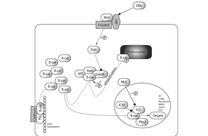
Figure 1. Model of canonical Wnt/b-catenin signaling pathway. Both adherens junctions and the canonical Wnt signaling pathway require b-catenin. By default, b-catenin is phosphorylated in the glycogen synthase kinase-3b(GSK3b), Axin, APC complex and thus directed into the ubiquitin/proteasome degradation pathway. Wnt forms together with Frizzled and LRP 5/6 a trimeric complex and activates intracellu-lar dishevelled (Dsh) by phosphorylation. Dickkopf (DKK) inhibits the formation of the Wnt/receptor complex. Activated Dsh inhibits GSK3band thus leads to stabilization and accumulation of b-catenin in the cytoplasm. Stabilized b-catenin is transported to the nucleus in a concentration-dependent manner. There it activates, together with transcription factors of the TCF/LEF family, the transcription of tar-get genes. Nemo-like kinase (NLK) phosphorylates TCF and regulates its DNA-binding affinity. Pitx2 acts as a transcriptional repressor when bound to b-catenin.
activation of the canonical Wnt signaling pathway via Fz
dimerization only has been proposed from experiments
with Xenopus laevis [39]. Moreover, there is evidence
that Wnt proteins are able to induce phosphorylation of
Dsh via LRP5/6 by pathways other than the canonical,
since phosphorylation of Dsh did not necessarily lead to
b-catenin stabilization [40].
Wnt signaling is regulated at the receptor level by various
regulatory proteins, including LRP5/6-binding factors
(Dickkopfs, DKKs) and secreted Fz-related proteins
(SFRPs) [41–43]. DKKs inhibit the ternary complex
formation between Wnt, Fz, and LRP5/6, and therefore
inhibit canonical Wnt signaling [44]. Interestingly,
DKK1 has been reported to be a direct transcriptional
target of the canonical Wnt signaling pathway [45]. This
might point to a feedback loop modulating canonical Wnt
signaling. Moreover, Golan at al. [46] have shown that
human Fz receptor 6 can act as a negative regulator for
the canonical Wnt signaling pathway downstream of the
b-catenin destruction complex (fig. 1). b-Catenin is not
only a key component of the canonical Wnt signaling
pathway, but also serves as a structural molecule that
anchors the actin cytoskeleton to the intracellular domain
of cadherins [47–49]. As such,
b-catenin is also involved in
cadherin-mediated cell-cell adhesion. Adherens junctions
and canonical Wnt signaling possibly require distinct
molecular forms of beta-catenin. While the cytosolic pool
of
b-catenin can bind to a-catenin and cadherins, Wnt
signaling is thought to promote a specific conformation
of
b-catenin which can bind to certain transcription factors
but not to cadherins [50].
Dsh inhibits the complex formation of Axin, adenoma
poliposis coli protein (APC), glycogen synthase kinase-3
b
(GSK3
b) and b-catenin. This complex is required for
b-catenin phosphorylation, which directs the protein into
the proteasome degradation pathway [51, 52]. If not
degraded,
b-catenin accumulates in the cytoplasm.
Although
b-catenin lacks a nuclear localization signal it
can be translocated into the nucleus in a
concentration-dependent manner. Translocation has been shown to be
independent of the classical nuclear transport pathways
that involve Ran or importin [53, 54]. However,
b-catenin
is too large for passive nuclear transport. A possible
solu-tion for the nuclear transport problem emanates from the
fact that
b-catenin interacts with the androgen receptor
(AR), which might serve as a nuclear transporter. Indeed,
AR agonists affected the nuclear transport kinetics of
b-catenin and the AR in an analogous manner [55].
In the nucleus,
b-catenin interacts with high-mobility
group (HMG) box transcription factors, like the T cell
factor (TCF, also known as lymphoid-enhancer factor
LEF), and forms a transcriptional activator complex [56].
This activator complex targets genes such as cyclin D1,
c-Myc, fibronectin, BMP4, mab-5, and NT-3 [27].
Com-plex formation between the TCF and
b-catenin is inhibited
by a protein called inhibitor of
b-catenin and TCF-4
(ICAT), which can bind to
b-catenin armadillo repeats in
a manner similar to that of the TCFs [57, 58]. In cells with
elevated
b-catenin, ICAT was proposed to sequester a
subpopulation of
b-catenin and thus to buffer increased
b-catenin levels in the cytoplasm [59]. Phosphorylation
of TCF/LEF by activated Nemo-like kinase (NLK) inhibits
the DNA-binding affinity of the complex and thus
indirectly regulates Wnt signaling in the nucleus [60]. Yet
NLK has also been shown to act as a positive regulator of
Wnt signaling in early zebrafish development [61].
Moreover, the factor Pitx2, which is involved in cell
type-specific proliferation, has been shown to be converted
from a transcriptional repressor into a transcriptional
activator when bound to
b-catenin [62]. Overall, the
var-ious levels of regulation of canonical Wnt signaling may
indicate multiple possible interactions with other signaling
cascades, required for differential signal integration.
Proliferation and apoptosis
Precise regulation of proliferation/apoptosis ratios is
essential in neural development [63]. Unbalance results in
severe malformations during embryonic development, and
promotes cancer formation postembryonically. In vitro as
well as in vivo studies have shown that Wnt signaling is
required to expand and maintain neural precursor
popula-tions in the brain and the spinal cord [64–66]. Wnt-1
regu-lates precursor populations in the mid/hindbrain and is
necessary for its development [67, 68]. Wnt-3a signaling
seems to be involved in hippocampal development by
regulating the size of the caudomedial cortex through
progenitor pool regulation and/or stem cell maintenance
[69]. Moreover, Wnt signaling regulates the size of the
cerebral cortex in the mammalian system [70]. In the
spinal cord, progenitors are specified by BMP signaling
that determines domains of Wnt ligand, receptor, and
an-tagonist expression, resulting in spinal cord patterning
[71]. In these BMP-defined progenitor populations, Wnt
signaling is thought to regulate cell cycle exit and thus
progenitor expansion [70, 72]. Similar proliferative
ef-fects of Wnt signaling have been described for stem cells
and progenitors in various tissues like the skin, intestine,
and the hematopoietic system [31, 73, 74].
While most results indicate a proliferative function of
Wnt signaling, there are also reports which claim that
Wnt signaling inhibits proliferation in certain cell types,
such as human endothelial vein cells (HUVECs) [75].
This effect is thought to result from non-canonical
Wnt/Ca
2+pathway signaling that inhibits the proliferative
effects of canonical Wnt signaling [76–78].
Wnt signaling not only affects proliferation but has also
been implicated in apoptosis. In cancer research, for
example, Wnt signaling has been related to drug
resis-tance in cancer therapy where vinblastine and vincristine
are used as apoptosis-inducing drugs. The effect of these
drugs has been overruled by Wnt signaling and cell
sur-vival maintained, while inhibition of Wnt signaling by
expression of dominant-negative TCF4 rendered the cells
responsive to the drugs [79]. Another study on 3T3-L1
cells revealed a TCF4-independent mechanism by which
Wnt signaling inhibits apoptosis. While overexpression
of dominant-negative TCF4 triggered the expression of
apoptotic genes, Wnt signaling upregulated the expression
of insulin-like growth factors (IGF I/II) which mediate
antiapoptotic effects [80]. Moreover, conditional ablation
of
b-catenin in the dorsal spinal cord led to increased
apoptosis, although whether this was due to impaired
canonical Wnt signaling or to disturbed cell-cell
interac-tions was difficult to assess [F. Ille, R. Kemler and L.
Sommer, unpublished data]. Canonical Wnt signaling has
been proposed to suppress apoptosis by inhibiting
c-Myc-induced release of cytochrome c and caspase
activa-tion [81, 82]. In addiactiva-tion to the antiapoptotic effects of
canonical Wnt signaling, apoptotic inhibition has been
attributed to
b-catenin-independent Wnt signaling via
Janus kinase (Jnk) [83]. Despite the evidence for
anti-apoptotic effects of Wnt signaling, Wnt may also induce
apoptosis. In particular, Hasegawa and colleagues have
shown that stabilizing
b-catenin by conditional APC
ablation leads to massive induction of apoptosis in neural
crest cells in the mouse model [84].
Stem cell maintenance, differentiation, and lineage
decision
Stem cell maintenance and self-renewal are cellular
processes closely associated with proliferation. Given the
role of Wnt signaling in cell cycle regulation, Wnt
sig-naling has not surprisingly also been implicated in the
control of stem cell development [85]. Activation of
Wnt/
b-catenin signaling in human and mouse embryonic
stem cells (ESCs) by administration of pharmacological
GSK3
b inhibitor maintains their self-renewal capacity as
well as their pluripotency [86]. Exposure of hematopoietic
stem cells to Wnt molecules and sustained expression of
b-catenin in long-term cultures maintains self-renewal as
well as the capacity of these cells to reconstitute the
hematopoietic lineages in vivo [87]. Nonetheless, ablation
of
b-catenin seems to impair neither hematopoiesis nor
lymphopoiesis under physiological conditions [88]. For
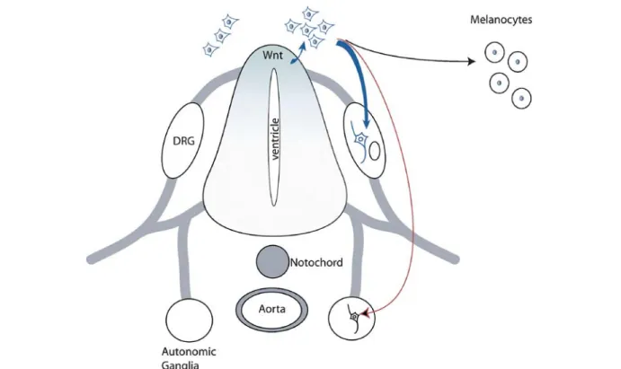
Figure 2. Wnt signaling in neural crest stem cell development. A gradient of Wnt molecules is established in the dorsal neural tube, which is involved in neural tube patterning and the generation of the neural crest cell population. Some of the early neural crest stem cells (eNCSC; blue-colored cells) are thought to become committed to the sensory lineage (blue arrow) in response to canonical Wnt/b-catenin signaling in the dorsal neural tube. Delaminated eNCSCs migrate toward their homing positions. At these positions, NCSCs respond to local signals and are involved in the generation of structures such as the dorsal root ganglia (DRG), the autonomic ganglia (red arrow), and structures of the skin (black arrow), and others.
these processes, the data suggest either an alternative
rescue pathway or the involvement of a factor upstream of
b-catenin in the Wnt signaling cascade.
The effects of Wnt signaling on stem cells seem to be
diverse. While it inhibits neural differentiation and
main-tains pluripotency in ESCs [86, 89], it can also promote
differentiation in other stem and progenitor cells [85].
Marretto et al. [90] identified a TCF-dependent reporter
gene in differentiating cortical neurons during
develop-ment, suggesting a potential role of Wnt/
b-catenin
signal-ing in the differentiation process. In agreement with this,
Wnt-7a signaling induces differentiation in neural
precur-sor cells (NPCs) of the neocortex [91]. This process
reduces the NPC pool at late developmental stages (E13.5)
whereas NPCs at earlier developmental stages (E10.5) do
not seem to differentiate in the presence of Wnt. Recently,
Wnt-3a has been reported to promote differentiation into
the neural and astrocyte lineage by inhibiting neural stem
cell maintenance [92]. Moreover Wnt/
b-catenin signaling
is required for neural differentiation in ESCs [93].
In neural crest stem cells (NCSCs), Wnt signaling has
been linked to cell fate decision (fig. 2). Neural crest cells
generate various cell types of the peripheral nervous
system (PNS) as well as craniofacial, skin, and heart
struc-tures [94]. These cells are derived from the border of the
neural plate adjacent to the ectoderm, and are strictly
dependent on Wnt signaling [95, 96]. Wnt/
b-catenin
signaling regulates cell fate decisions in early neural crest
stem cells (eNCSCs) by driving these cells into the sensory
lineage, rather than affecting the population size. Hari at
al. [97] have shown by cell type-specific gene ablation
that loss of
b-catenin-function in NCSCs prevents sensory
ganglia and melanocyte formation in vivo [97].
Intrigu-ingly,
b-catenin-deficient NCSCs emigrate and proliferate
normally in culture, but they fail to acquire a sensory
neuronal fate [97]. Conversely, a
b-catenin
gain-of-function study shows that, in vivo, continuous Wnt/
b-catenin signaling in NCSCs leads to sensory neurogenesis
at the expense of all other neural crest derivatives [98].
However, stabilized
b-catenin does not affect NCSC
migration and proliferation. Consistent with the in vivo
data, practically all mutant cells lose NCSC features and
adopt a sensory fate in cell culture. Although all eNCSCs
are Wnt responsive, only a subpopulation of the cells
generate sensory cells in vivo, indicating the presence of
factors counteracting Wnt activity [85]. The development
of the hippocampus, and in particular the generation of
dentate gyrus granule cells, also appears to be regulated
by LEF1/TCF transcription factors [99]. Similar effects
have been reported for skin stem cells, in which
b-catenin
signaling determines differentiation into follicular and
epidermal lineages [100, 101]. Taken together, these
results underline the role of Wnt signaling in lineage
decision, but the molecular mechanisms remain to be
resolved.
Axon guidance and neurite outgrowth
One essential aspect in CNS development is ‘wiring,’ a
process which involves patterning, migration, axon
guidance, and synapse formation. As mentioned before,
Wnt molecules have been implicated in morphogenesis,
where they establish tissue patterning together with other
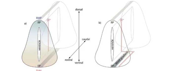
Figure 3. Guidance of commissural axons in the developing spinal cord. (a) Commissural axons of certain neurons are repelled by the dorsal bone morphogenetic protein (BMP) gradient (blue) that originates from the roofplate (RP). At the same time, the axons are attracted by the ventral sonic hedgehog (SHH) gradient in the floorplate (FP). Therefore, commissural axons extend ventrally toward the floorplate. (b) Once the axons reach the floorplate, they cross it and turn rostrally. After turning toward the head, the axons extend along a Wnt-4 gradient until they reach their target area and form synapses.
proteins like sonic hedgehog (SHH) and BMPs. In the
early neural tube, for example, BMP4 expressed in the
roofplate and SHH expressed in the floorplate generate
two dorsoventral countergradients which influence
proneural gene expression. Wnt-1 and Wnt-3a are
expressed in the dorsal neural tube at later stages, and are
thought to determine proneural domains of the dorsal
neural tube by mechanisms which still remain to be
elucidated [102, 103]. Wnt-4, however, is expressed in the
ventral neural tube and has been related to axon guidance
in commissural axons [104]. Commissural axon migration
is repelled from the roofplate by BMP signaling, and at
the same time, these axons are attracted toward the
floor-plate by SHH. Once these axons reach the floorfloor-plate, they
cross it, turn rostrally, and extend toward the brain
(fig. 3). Lyuksyutova et al. [104] have found Wnt-4 to be
expressed in a rostrocaudal gradient in the floorplate,
which attracts postcrossing commissural axons. Likewise,
commissural axons which lack the Wnt receptor Fz3
exhibit rostrocaudal guidance defects in postcrossing
commissural axons [104]. Since these results have not
been tested in Wnt-4 knockout animals, the direct
involvement of Wnt-4 in axon guidance remains to be
proven. Nevertheless, evidence from invertebrate systems
favors the hypothesis that Wnt molecules can act as
guid-ance molecules. In Drosophila, Wnt-5 seems to fulfill a
guidance function. By signaling through the Derailed
receptor expressed in the growth cones, Wnt-5 acts as a
chemorepellent [105–107].
In vertebrates, ablation of Ryk, the mammalian
homo-logue of Drosophila Derailed, leads to axon guidance
defects in vivo [108]. Unlike Derailed, Ryk acts as a Fz
coreceptor and forms a ternary complex with Fz and
Wnt-1. It also mediates TCF activation induced by Wnt-1
and is able to bind to Wnt-3a. Moreover, Ryk is required
for neurite outgrowth in dorsal root ganglion neurons,
induced by Wnt-3a [108]. Similarly, Wnt-7a has been
described to remodel axon spreading and branching
in developing cerebellar granule cells [109]. Axonal
remodeling involves MAP-1B, a microtubule-associated
protein that has been identified as substrate for GSK3
b
and implicated in axonal outgrowth [110]. Upon GSK3
b
inhibition, the amount of phosphorylated MAP-1B
decreases in the cells, a process that naturally occurs prior
to axonal remodeling and leads to changes in microtubule
dynamics. Recently, Ciani et al. [111] have shown that the
Wnt/
b-catenin signaling component Dsh also induces
axonal remodeling and stabilizes microtubules in
devel-oping neurons. Intriguingly, this process seems to involve
neither
b-catenin nor TCF factors, suggesting an
alterna-tive regulatory pathway for canonical Wnt signaling,
downstream of GSK3
b. Similar evidence comes from
experiments in neuroblastoma [112]. Here, GSK3
b and
Axin have been reported to promote neurite outgrowth in
a
b-catenin-independent manner.
Conclusion
The roles of Wnt signaling in neural development are
manifold, and seem at times contradictory. While one of
its most important functions is promotion of
prolifera-tion, in some contexts, Wnt signaling is able to inhibit it.
Similarly, apoptosis is abolished by Wnt signaling in
cer-tain cases whereas in others it is induced. As much as Wnt
signaling is responsible for stem cell maintenance, it is to
a similar extent essential for differentiation and lineage
decisions, axon guidance, and neurite outgrowth. With
every newly described aspect of Wnt signaling, more
questions arise. Some answers to these questions may lie
in understanding the intrinsic status of a cell that reflects
its ‘history’ and that is modified depending on the
posi-tion of the cell in the organism at a given timepoint. In
other words, the effects of Wnt signaling are tissue and
cell type specific and represent the results of
combinato-rial signal integration in an environment of dynamic
signaling networks.
1 Derynck R. and Zhang Y. E. (2003) Smad-dependent and Smad-independent pathways in TGF-beta family signalling. Nature 425: 577–584
2 Takahashi, Y., Tonegawa, A., Matsumoto, K., Ueno, N., Kuroiwa, A., Noda, M. et al. (1996) BMP-4 mediates inter-acting signals between the neural tube and skin along the dor-sal midline. Genes Cells 1: 775–783
3 Mehler M. F., Mabie P. C., Zhang D. and Kessler J. A. (1997) Bone morphogenetic proteins in the nervous system. Trends Neurosci. 20: 309–317
4 Timmer J. R., Wang C. and Niswander L. (2002) BMP signal-ing patterns the dorsal and intermediate neural tube via regu-lation of homeobox and helix-loop-helix transcription factors. Development 129: 2459–2472
5 Lee S. J. (1990) Identification of a novel member (GDF-1) of the transforming growth factor-beta superfamily. Mol. En-docrinol. 4: 1034–1040
6 Rankin C. T., Bunton T., Lawler A. M. and Lee S. J. (2000) Regulation of left-right patterning in mice by growth/differen-tiation factor-1. Nat. Genet. 24: 262–265
7 Guerrero I. and Ruiz i Altaba A. (2003) Development. Long-ing for ligand: hedgehog, patched, and cell death. Science
301: 774–776
8 Thibert C., Teillet M. A., Lapointe F., Mazelin L., Le Douarin, N. M. and Mehlen, P. (2003) Inhibition of neuroepithelial patched-induced apoptosis by sonic hedgehog. Science 301: 843–846
9 Gritli-Linde A., Lewis P., McMahon A. P. and Linde A. (2001) The whereabouts of a morphogen: direct evidence for short-and graded long-range activity of hedgehog signaling pep-tides. Dev. Biol. 236: 364–386
10 Kuschel S., Ruther U. and Theil T. (2003) A disrupted balance between Bmp/Wnt and Fgf signaling underlies the ventraliza-tion of the Gli3 mutant telencephalon. Dev. Biol. 260: 484–495 11 Lobjois V., Benazeraf B., Bertrand N., Medevielle F. and Pituello F. (2004) Specific regulation of cyclins D1 and D2 by FGF and Shh signaling coordinates cell cycle progression, patterning, and differentiation during early steps of spinal cord development. Dev. Biol. 273: 195–209
12 Sokol S., Christian J. L., Moon R. T. and Melton D. A. (1991) Injected Wnt RNA induces a complete body axis in Xenopus embryos. Cell 67: 741–752
13 Crease D. J., Dyson S. and Gurdon J. B. (1998) Cooperation between the activin and Wnt pathways in the spatial control of organizer gene expression. Proc. Natl. Acad. Sci. USA 95: 4398–4403
14 Niehrs C. (1999) Head in the WNT: the molecular nature of Spemann’s head organizer. Trends Genet. 15: 314–319 15 Xanthos J. B., Kofron M., Tao Q., Schaible K., Wylie C. and
Heasman J. (2002) The roles of three signaling pathways in the formation and function of the Spemann Organizer. Develop-ment 129: 4027–4043
16 Shimogori T., VanSant J., Paik E. and Grove E. A. (2004) Members of the Wnt, Fz, and Frp gene families expressed in postnatal mouse cerebral cortex. J. Comp. Neurol. 473: 496–510
17 Brown A. M. (2001) Wnt signaling in breast cancer: have we come full circle? Breast Cancer Res. 3:: 351–355
18 Giles R. H., Es J. H. van and Clevers H. (2003) Caught up in a Wnt storm: Wnt signaling in cancer. Biochim. Biophys. Acta
1653: 1–24
19 Uren A., Wolf V., Sun Y. F., Azari A., Rubin J. S. and Toretsky J. A. (2004) Wnt/Frizzled signaling in Ewing sarcoma. Pediatr. Blood Cancer 43: 243–249
20 Worm J., Christensen C., Gronbaek K., Tulchinsky E. and Guldberg P. (2004) Genetic and epigenetic alterations of the APC gene in malignant melanoma. Oncogene 23: 5215–5226 21 De Ferrari G. V. and Inestrosa N. C. (2000) Wnt signaling function in Alzheimer’s disease. Brain Res. Brain Res. Rev.
33: 1–12
22 Inestrosa N., De Ferrari G. V., Garrido J. L., Alvarez A., Olivares G. H., Barria M. I. et al. (2002) Wnt signaling involvement in beta-amyloid-dependent neurodegeneration. Neurochem. Int.
41: 341–344
23 Caricasole A., Copani A., Caruso A., Caraci F., Iacovelli L., Sortino M. A. et al. (2003) The Wnt pathway, cell-cycle activation and beta-amyloid: novel therapeutic strategies in Alzheimer’s disease? Trends Pharmacol. Sci. 24: 233–238 24 McMahon A. P. and Bradley A. (1990) The Wnt-1 (int-1)
proto-oncogene is required for development of a large region of the mouse brain. Cell 62: 1073–1085
25 McMahon A. P., Joyner A. L., Bradley A. and McMahon J. A. (1992) The midbrain-hindbrain phenotype of Wnt-1–/Wnt-1– mice results from stepwise deletion of engrailed-expressing cells by 9.5 days postcoitum. Cell 69: 581–595
26 Ikeya M., Lee S. M., Johnson J. E., McMahon A. P. and Takada S. (1997) Wnt signalling required for expansion of neural crest and CNS progenitors. Nature 389: 966–970
27 Nusse. R. (2004) http://www.stanford.edu/~rnusse/wntwin-dow.html
28 Nusse R. and Varmus H. E. (1992) Wnt genes. Cell 69: 1073–1087
29 Cadigan K. M. and Nusse R. (1997) Wnt signaling: a common theme in animal development. Genes Dev. 11:: 3286–3305
30 Miller J. R. (2001) The Wnts. Genome Biol. 3: REVIEWS3001.1–3001.15
31 Willert, K., Brown, J. D., Danenberg, E., Duncan, A. W., Weissman, I. L., Reya, T. et al. (2003) Wnt proteins are lipid-modified and can act as stem cell growth factors. Nature
423: 448–452
32 Nusse R. (2003) Wnts and Hedgehogs: lipid-modified proteins and similarities in signaling mechanisms at the cell surface. Development 130: 5297–5305
33 Strutt D. (2003) Frizzled signalling and cell polarisation in
Drosophila and vertebrates. Development 130: 4501–4513
34 Kuhl M., Sheldahl L. C., Park M., Miller J. R. and Moon R. T. (2000) The Wnt/Ca2+ pathway: a new vertebrate Wnt signal-ing pathway takes shape. Trends Genet. 16: 279–283 35 Adler P. N., Krasnow R. E. and Liu J. (1997) Tissue polarity
points from cells that have higher Frizzled levels towards cells that have lower Frizzled levels. Curr. Biol. 7: 940–949
36 Bhano P., Brink M., Samos C. H., Hsieh J. C., Wang Y., Macke J. P. et al. (1996) A new member of the frizzled family from
Drosophila functions as a Wingless receptor. Nature 382:
225–230
37 Hey P. J., Twells R. C., Phillips M. S., Yusuke N., Brown S. D., Kawaguchi Y. et al. (1998) Cloning of a novel member of the low-density lipoprotein receptor family. Gene 216: 103–111 38 Brown S. D., Twells R. C., Hey P. J., Cox R. D., Levy E. R.,
Soderman A. R. et al. (1998) Isolation and characterization of LRP6, a novel member of the low density lipoprotein receptor gene family. Biochem. Biophys. Res. Commun. 248: 879–888
39 Carron C., Pascal A., Djiane A., Boucaut J. C., Shi D. L. and Umbhauer M. (2003) Frizzled receptor dimerization is suffi-cient to activate the Wnt/beta-catenin pathway. J. Cell Sci.
116: 2541–2550
40 Gonzalez-Sancho J. M., Brennan K. R., Castelo-Soccio L. A. and Brown A. M. (2004) Wnt proteins induce dishevelled phosphorylation via an LRP5/6-independent mechanism, irrespective of their ability to stabilize beta-catenin. Mol. Cell. Biol. 24: 4757–4768
41 Glinka A., Wu W., Delius H., Monaghan A. P., Blumenstock C. and Niehrs C. (1998) Dickkopf-1 is a member of a new family of secreted proteins and functions in head induction. Nature 391: 357–362
42 Hoang B., Moos M. Jr, Vukicevic S. and Luyten F. P. (1996) Primary structure and tissue distribution of FRZB, a novel protein related to Drosophila frizzled, suggest a role in skeletal morphogenesis. J. Biol. Chem. 271: 26131–26137
43 Rattner A., Hsieh J. C., Smallwood P. M., Gilbert D. J., Copeland N. G., Jenkins, N. A. et al. (1997) A family of secreted proteins contains homology to the cysteine-rich ligand-binding domain of frizzled receptors. Proc. Natl. Acad. Sci. USA 94: 2859–2863
44 Zorn A. M. (2001) Wnt signalling: antagonistic Dickkopfs. Curr. Biol. 11: R592–R595
45 Niida A., Hiroko T., Kasai M., Furukawa Y., Nakamura Y., Suzuki Y. et al. (2004) DKK1, a negative regulator of Wnt signaling, is a target of the beta-catenin/TCF pathway. Oncogene 23: 8520–8526
46 Golan T., Yaniv A., Bafico A., Liu G. and Gazit A. (2004) The human Frizzled 6 (HFz6) acts as a negative regulator of the canonical Wnt beta-catenin signaling cascade. J. Biol. Chem.
279: 14879–14888
47 Ozawa M., Baribault H. and Kemler R. (1989) The cytoplasmic domain of the cell adhesion molecule uvomorulin associates with three independent proteins structurally related in different species. EMBO J. 8: 1711–1717
48 Ozawa M., Ringwald M. and Kemler R. (1990) Uvomorulin-catenin complex formation is regulated by a specific domain in the cytoplasmic region of the cell adhesion molecule. Proc. Natl. Acad. Sci. USA 87: 4246–4250
49 Kintner C. (1992) Regulation of embryonic cell adhesion by the cadherin cytoplasmic domain. Cell 69: 225–236 50 Gottardi C. J. and Gumbiner B. M. (2004) Distinct molecular
forms of beta-catenin are targeted to adhesive or transcrip-tional complexes. J. Cell. Biol. 167: 339–349
51 Aberle H., Bauer A., Stappert J., Kispert A. and Kemler R. (1997) Beta-catenin is a target for the ubiquitin-proteasome pathway. EMBO J. 16: 3797–3804
52 Ikeda S., Kishida S., Yamamoto H., Murai H., Koyama S. and Kikuchi A. (1998) Axin, a negative regulator of the Wnt signaling pathway, forms a complex with GSK-3beta and beta-catenin and promotes GSK-3beta-dependent phosphory-lation of beta-catenin. EMBO J 17: 1371–1384
53 Fagotto F., Gluck U. and Gumbiner B. M. (1998) Nuclear localization signal-independent and importin/karyopherin-independent nuclear import of beta-catenin. Curr. Biol. 8: 181–190
54 Yokoya F., Imamoto N., Tachibana T. and Yoneda Y. (1999) Beta-catenin can be transported into the nucleus in a Ran-unassisted manner. Mol. Biol. Cell 10: 1119–1131 55 Pawlowski J. E., Ertel J. R., Allen M. P., Xu M., Butler C.,
Wilson E. M. et al. (2002) Liganded androgen receptor interaction with beta-catenin: nuclear co-localization and modulation of transcriptional activity in neuronal cells. J. Biol. Chem. 277: 20702–20710
56 Nusse R. (1999) WNT targets: repression and activation. Trends Genet. 15: 1–3
57 Tago K., Nakamura T., Nishita M., Hyodo J., Nagai S., Murata Y. et al. (2000) Inhibition of Wnt signaling by ICAT, a novel beta-catenin-interacting protein. Genes Dev. 14: 1741–1749 58 Daniels D. L. and Weis W. I. (2002) ICAT inhibits beta-catenin
binding to Tcf/Lef-family transcription factors and the general coactivator p300 using independent structural modules. Mol. Cell 10: 573–584
59 Gottardi C. J. and Gumbiner B. M. (2004) Role for ICAT in beta-catenin-dependent nuclear signaling and cadherin functions. Am. J. Physiol. Cell Physiol. 286: C747–C756 60 Ishitani T., Ninomiya-Tsuji J. and Matsumoto K. (2003)
Regulation of lymphoid enhancer factor 1/T-cell factor by mitogen-activated protein related Nemo-like kinase-dependent phosphorylation in Wnt/beta-catenin signaling. Mol. Cell. Biol. 23: 1379–1389
61 Thorpe C. J. and Moon R. T. (2004) Nemo-like kinase is an essential co-activator of Wnt signaling during early zebrafish development. Development 131: 2899–2909
62 Kioussi C., Briata P., Baek S. H., Rose D. W., Hamblet N. S., Herman T. et al. (2002) Identification of a Wnt/Dvl/beta-catenin
Æ
Pitx2 pathway mediating cell-type-specific prolif-eration during development. Cell 111: 673–68563 Sommer L. and Rao M. (2002) Neural stem cells and regulation of cell number. Prog. Neurobiol. 66: 1–18
64 Dickinson M. E., Krumlauf R. and McMahon A. P. (1994) Evidence for a mitogenic effect of Wnt-1 in the developing mammalian central nervous system. Development 120: 1453–1471
65 Megason S. G. and McMahon A. P. (2002) A mitogen gradient of dorsal midline Wnts organizes growth in the CNS. Development 129: 2087–2098
66 Zechner D., Fujita Y., Hulsken J., Muller T., Walther I., Taketo M. M. et al. (2003) Beta-catenin signals regulate cell growth and the balance between progenitor cell expansion and differentiation in the nervous system. Dev. Biol. 258: 406–418 67 Lee S. M., Danielian P. S., Fritzsch B. and McMahon A. P. (1997) Evidence that FGF8 signalling from the midbrain-hindbrain junction regulates growth and polarity in the devel-oping midbrain. Development 124: 959–969
68 Panhuysen M., Vogt Weisenhorn D. M., Blanquet V., Brodski C., Heinzmann U., Beisker W. et al. (2004) Effects of Wnt-1 signaling on proliferation in the developing mid-/hindbrain region. Mol. Cell. Neurosci. 26: 101–111
69 Lee S. M., Tole S., Grove E. and McMahon A. P. (2000) A local Wnt-3a signal is required for development of the mam-malian hippocampus. Development 127: 457–467
70 Chenn A. and Walsh C. A. (2002) Regulation of cerebral cortical size by control of cell cycle exit in neural precursors. Science 297: 365–369
71 Panchision D. M., Pickel J. M., Studer L., Lee S. H., Turner P. A., Hazel T. G. et al. (2001) Sequential actions of BMP recep-tors control neural precursor cell production and fate. Genes Dev. 15: 2094–2110
72 Chesnutt C., Burrus L. W., Brown A. M. and Niswander L. (2004) Coordinate regulation of neural tube patterning and proliferation by TGFbeta and WNT activity. Dev. Biol. 274: 334–347
73 Alonso L. and Fuchs E. (2003) Stem cells of the skin epithe-lium. Proc. Natl. Acad. Sci. USA 100 (suppl 1): 11830–11835
74 Pinto D., Gregorieff A., Begthel H. and Clevers H. (2003) Canonical Wnt signals are essential for homeostasis of the intestinal epithelium. Genes Dev. 17: 1709–1713
75 Cheng C. W., Smith S. K. and Charnock-Jones D. S. (2003) Wnt-1 signaling inhibits human umbilical vein endothelial cell proliferation and alters cell morphology. Exp. Cell Res.
291: 415–425
76 Torres M. A., Yang-Snyder J. A., Purcell S. M., DeMarais A. A., McGrew L. L. and Moon R. T. (1996) Activities of the Wnt-1 class of secreted signaling factors are antagonized by the Wnt-5A class and by a dominant negative cadherin in early
Xenopus development. J. Cell Biol. 133: 1123–1137
77 Olson D. J. and Gibo D. M. (1998) Antisense wnt-5a mimics wnt-1-mediated C57MG mammary epithelial cell transforma-tion. Exp. Cell Res. 241: 134–141
78 Topol L., Jiang X., Choi H., Garrett-Beal L., Carolan P. J. and Yang Y. (2003) Wnt-5a inhibits the canonical Wnt pathway by promoting GSK-3-independent beta-catenin degradation. J. Cell Biol. 162: 899–908
79 Chen S., Guttridge D. C., You Z., Zhang Z., Fribley A., Mayo M. W. et al. (2001) Wnt-1 signaling inhibits apoptosis by activating beta-catenin/T cell factor-mediated transcription. J. Cell Biol. 152: 87–96
80 Longo K. A., Kennell J. A., Ochocinska M. J., Ross S. E., Wright W. S. and MacDougald O. A. (2002) Wnt signaling protects 3T3-L1 preadipocytes from apoptosis through induction of insulin-like growth factors. J. Biol. Chem. 277: 38239–38244
81 You Z., Saims D., Chen S., Zhang Z., Guttridge D. C., Guan K. L. et al. (2002) Wnt signaling promotes oncogenic trans-formation by inhibiting c-Myc-induced apoptosis. J. Cell Biol.
157: 429–440
82 Kanei-Ishii C., Ninomiya-Tsuji J., Tanikawa J., Nomura T., Ishitani T., Kishida S. et al. (2004) Wnt-1 signal induces phosphorylation and degradation of c-Myb protein via TAK1, HIPK2, and NLK. Genes Dev. 18: 816–829
83 You L., He B., Uematsu K., Xu Z., Mazieres J., Lee A. et al. (2004) Inhibition of Wnt-1 signaling induces apoptosis in beta-catenin-deficient mesothelioma cells. Cancer Res 64: 3474–3478
84 Hasegawa S., Sato T., Akazawa H., Okada H., Maeno A., Ito M. et al. (2002) Apoptosis in neural crest cells by functional loss of APC tumor suppressor gene. Proc. Natl. Acad. Sci. USA 99: 297–302
85 Kleber M. and Sommer L. (2004) Wnt signaling and the regulation of stem cell function. Curr. Opin. Cell Biol. 16: 681–687
86 Sato N., Meijer L., Skaltsounis L., Greengard P. and Brivanlou A. H. (2004) Maintenance of pluripotency in human and mouse embryonic stem cells through activation of Wnt signaling by a pharmacological GSK-3-specific inhibitor. Nat. Med. 10: 55–63
87 Reya T., Duncan A. W., Ailles L., Domen J., Scherer D. C., Willert K. et al. (2003) A role for Wnt signalling in self-renewal of haematopoietic stem cells. Nature 423: 409–414 88 Cobas M., Wilson A., Ernst B., Mancini S. J., MacDonald H.
R., Kemler R. et al. (2004) Beta-catenin is dispensable for hematopoiesis and lymphopoiesis. J. Exp. Med. 199: 221–229 89 Haegele L., Ingold B., Naumann H., Tabatabai G., Ledermann B. and Brandner S. (2003) Wnt signalling inhibits neural differentiation of embryonic stem cells by controlling bone morphogenetic protein expression. Mol. Cell. Neurosci. 24: 696–708
90 Maretto S., Cordenonsi M., Dupont S., Braghetta P., Broccoli V., Hassan A. B. et al. (2003) Mapping Wnt/beta-catenin signaling during mouse development and in colorectal tumors. Proc. Natl. Acad. Sci. USA 100: 3299–3304
91 Hirabayashi Y., Itoh Y., Tabata H., Nakajima K., Akiyama T., Masuyama N. et al. (2004) The Wnt/beta-catenin pathway
directs neuronal differentiation of cortical neural precursor cells. Development 131: 2791–2801
92 Muroyama Y., Kondoh H. and Takada S. (2004) Wnt proteins promote neuronal differentiation in neural stem cell culture. Biochem. Biophys. Res. Commun. 313: 915–921
93 Otero J. J., Fu W., Kan L., Cuadra A. E. and Kessler J. A. (2004) Beta-catenin signaling is required for neural differen-tiation of embryonic stem cells. Development 131: 3545–3557
94 Le Douarin N. M. and Dupin E. (2003) Multipotentiality of the neural crest. Curr. Opin. Genet. Dev. 13: 529–536 95 Chang C. and Hemmati-Brivanlou A. (1998) Neural crest
induction by Xwnt7B in Xenopus. Dev. Biol. 194: 129–134 96 Garcia-Castro M. I., Marcelle C. and Bronner-Fraser M.
(2002) Ectodermal Wnt function as a neural crest inducer. Science 297: 848–851
97 Hari L., Brault V., Kleber M., Lee H. Y., Ille F., Leimeroth R. et al. (2002) Lineage-specific requirements of beta-catenin in neural crest development. J. Cell Biol. 159: 867–880 98 Lee H. Y., Kleber M., Hari L., Brault V., Suter U., Taketo M.
M. et al. (2004) Instructive role of Wnt/beta-catenin in sensory fate specification in neural crest stem cells. Science
303: 1020–1023
99 Galceran J., Miyashita-Lin E. M., Devaney E., Rubenstein J. L. and Grosschedl R. (2000) Hippocampus development and generation of dentate gyrus granule cells is regulated by LEF1. Development 127: 469–482
100 Huelsken J., Vogel R., Erdmann B., Cotsarelis G. and Birchmeier W. (2001) Beta-catenin controls hair follicle morphogenesis and stem cell differentiation in the skin. Cell
105: 533–545
101 Merrill B. J., Gat U., DasGupta R. and Fuchs E. (2001) Tcf3 and Lef1 regulate lineage differentiation of multipotent stem cells in skin. Genes Dev. 15: 1688–1705
102 Cauthen C. A., Berdougo E., Sandler J. and Burrus L. W. (2001) Comparative analysis of the expression patterns of Wnts and Frizzleds during early myogenesis in chick embryos. Mech Dev 104: 133–138
103 Muroyama Y., Fujihara M., Ikeya M., Kondoh H. and Takada S. (2002) Wnt signaling plays an essential role in neuronal specification of the dorsal spinal cord. Genes Dev. 16: 548–553
104 Lyuksyutova A. I., Lu C. C., Milanesio N., King L. A., Guo N., Wang Y. et al. (2003) Anterior-posterior guidance of commissural axons by Wnt-frizzled signaling. Science 302: 1984–1988
105. Bonkowsky J. L., Yoshikawa S., O’Keefe D. D., Scully A. L. and Thomas J. B. (1999) Axon routing across the midline controlled by the Drosophila Derailed receptor. Nature 402: 540–544
106 Yoshikawa S., McKinnon R. D., Kokel M. and Thomas J. B. (2003) Wnt-mediated axon guidance via the Drosophila Derailed receptor. Nature 422: 583–588
107 Yoshikawa S. and Thomas J. B. (2004) Secreted cell signaling molecules in axon guidance. Curr. Opin. Neurobiol. 14: 45–50
108 Lu W., Yamamoto V., Ortega B. and Baltimore D. (2004) Mammalian Ryk is a Wnt coreceptor required for stimulation of neurite outgrowth. Cell 119: 97–108
109 Lucas F. R. and Salinas P. C. (1997) WNT-7a induces axonal remodeling and increases synapsin I levels in cerebellar neurons. Dev. Biol. 192: 31–44
110 Lucas F. R., Goold R. G., Gordon-Weeks P. R. and Salinas P. C. (1998) Inhibition of GSK-3beta leading to the loss of phosphorylated MAP-1B is an early event in axonal remodel-ling induced by WNT-7a or lithium. J. Cell. Sci. 111: 1351–1361
111 Ciani L., Krylova O., Smalley M. J., Dale T. C. and Salinas P. C. (2004) A divergent canonical WNT-signaling pathway regulates microtubule dynamics: dishevelled signals locally to stabilize microtubules. J. Cell. Biol. 164: 243–253
112 Orme M. H., Giannini A. L., Vivanco M. D. and Kypta R. M. (2003) Glycogen synthase kinase-3 and Axin function in a beta-catenin-independent pathway that regulates neurite outgrowth in neuroblastoma cells. Mol. Cell. Neurosci. 24: 673–686