ORIGINAL ARTICLE
18
F-Fluoride PET/CT for detection of sacroiliitis
in ankylosing spondylitis
Klaus Strobel&Dorothee R. Fischer&Giorgio Tamborrini&Diego Kyburz&
Katrin D. M. Stumpe&Rolf G. X. Hesselmann&A. Johayem&
Gustav K. von Schulthess&Beat A. Michel&Adrian Ciurea
Received: 3 October 2009 / Accepted: 29 March 2010 / Published online: 27 May 2010 # Springer-Verlag 2010
Abstract
Purpose The aim of this study was to evaluate the performance of 18F-fluoride-PET/CT (PET/CT) for the diagnosis of sacroiliac joint (SIJ) arthritis in patients with active ankylosing spondylitis (AS).
Methods Included in the study were 15 patients with AS according to the modified New York criteria (AS group) and with active disease and 13 patients with mechanical low back pain (MLBP; control group) who were investi-gated with whole-body18F-fluoride PET/CT. The ratio of the uptake in the SIJ and that in the sacrum (SIJ/S) was calculated for every joint.
Results The mean SIJ/S ratio of 30 quantified joints in the AS group was 1.66 (range 1.10–3.07) with PET/CT, and the mean SIJ/S ratio of 26 quantified joints in the MLBP group was 1.12 (range 0.71–1.52). The area under the receiver operating characteristic curve for SIJ arthritis was 0.84. With plain radiography as a the gold standard and taking an SIJ/S ratio of >1.3 as the threshold, the
sensitivity, specificity and accuracy on a per patient basis were 80%, 77% and 79%, respectively. On a per SIJ basis, the greatest sensitivity (94%) was found in grade 3 sacroiliitis (n=16).
Conclusion Our results suggest that quantitative 18 F-fluo-ride PET/CT may play a role in the diagnosis of sacroiliitis in active AS and is an alternative to conventional bone scintigraphy in times of molybdenum shortage.
Keywords Fluoride PET/CT . Ankylosing spondylitis . Sacroiliac joint arthritis . Quantification
Introduction
Ankylosing spondylitis (AS) is the prototype of a group of chronic rheumatic diseases called spondyloarthritides (SpA) that generally affect young people with an estimated prevalence of 0.3–1.4% [1,2]. The main clinical feature is inflammatory back pain caused by sacroiliitis. Inflamma-tion may extend to other locaInflamma-tions of the spine as spondylitis, spondylodiscitis or spondylarthritis, and ulti-mately result in axial fusion, causing substantial functional disability [3–5]. Peripheral arthritis and enthesitis, as well as anterior uveitis, may also occur. Unequivocal radio-graphic sacroiliitis of at least grade 2 bilaterally or grade 3 unilaterally, in addition to at least one clinical criterion, is needed for the diagnosis of AS according to the modified New York criteria [6]. Therefore, plain radiography is still regarded as the imaging gold standard in the evaluation of AS. By contrast, magnetic resonance imaging (MRI) is considered to be the most sensitive imaging technique for the detection of acute inflammation of the sacroiliac joints (SIJ) [7–9]. It allows early diagnosis of SpA long before postinflammatory chronic changes can be documented on
K. Strobel
:
D. R. Fischer:
K. D. M. Stumpe:
G. K. von SchulthessDepartment of Medical Radiology, Division of Nuclear Medicine, University Hospital,
Zurich, Switzerland
G. Tamborrini
:
D. Kyburz:
B. A. Michel:
A. Ciurea Department of Rheumatology, University Hospital Zurich, Zurich, SwitzerlandR. G. X. Hesselmann
:
A. Johayem Radiopharmacy, University Hospital Zurich, Zurich, SwitzerlandK. Strobel (*)
Nuclear Medicine, Cantonal Hospital Lucerne, Lucerne, Switzerland
e-mail: klaus.strobel@ksl.ch DOI 10.1007/s00259-010-1464-7
plain radiographs [10, 11]. Quantitative analysis of sacro-iliac biopsies has demonstrated a good correlation between cellularity and MRI enhancement [12]. However, new bone formation and fusion (ankylosis) in AS may be uncoupled from inflammation, as exemplified by the failure so far of immunosuppression via TNF-alpha inhibition to inhibit radiographic disease progression [13, 14]. In this regard, nuclear imaging techniques may detect increased bone turnover not only during inflammation, but probably also paralleling postinflammatory reparative changes at the starting point of new bone formation [15].
Conventional scintigraphy with bone-seeking tracers such as99mTc-labelled diphosphonates have been used for decades in patients with AS [16]. Recently, a systematic literature review including 25 articles showed that scintigraphy reaches sensitivities of 52% in patients with established AS and 49.4% in patients with probable sacroiliitis [17]. With the worldwide shortage of molybdenum [18, 19], the increasing availability of positron emission tomography (PET) scanners and the superior image quality,18F-fluoride PET/CT imaging has attracted increasing interest [20]. For example, 18F-fluoride PET seems to be superior to planar bone scintigraphy for the evaluation of metastatic disease [21]. The aim of this study was to evaluate the performance of 18F-fluoride PET/CT for the diagnosis of sacroiliitis in patients with AS and to answer the question as to whether this tracer can be used as an alternative in times of molybdenum shortage. Patients with mechanical low back pain (MLBP) served as a control group.
Materials and methods Patient selection
The AS group comprised 15 patients with active AS who fulfilled the modified New York criteria and were recruited from a single rheumatology outpatient clinic. The minimal disease activity required for inclusion was a Bath ankylosing spondylitis disease activity index (BAS-DAI) of at least 4 and/or spine pain of at least 4 (BASDAI item 2: second question of the BASDAI instrument dealing with back pain) on a numerical rating scale from 0 to 10 [22]. A BASDAI of 4 or higher is considered to reflect a high disease activity [23]. C-reactive protein (CRP) and erythrocyte sedimentation rate (ESR) were measured in all patients at the time of imaging.
The control group comprised 13 patients with non-traumatic chronic MLBP for at least 3 months and a pain intensity of at least 4 on a numerical rating scale ranging from 0 to 10. MLBP was defined as back pain exacerbating with exercise and improving with rest, as
well as with a duration of morning stiffness, if present, of less than 15 minutes. The diagnoses in the MLBP patients were obtained from the treating rheumatologist. Patients with concomitant neoplastic or other inflamma-tory rheumatic diseases were excluded. A plain radio-graph of the pelvis or lumbar spine that ruled out radiographic SIJ arthritis was available.
Patients were recruited for this study nonconsecutively (“convenience sample”), provided that they met the inclusion criteria, had been diagnosed as having AS or MLBP, and were willing to participate. The local ethics committee approved the protocol and all patients gave written informed consent. Grading of SIJ on conventional radiographs
Conventional radiographs of the SIJ were available in all AS patients and were classified by two experienced rheumatolo-gists independently for sacroiliitis according to the radio-graphic modified New York criteria [6] in consensus (0 normal; 1 changes suggestive of SIJ arthritis; 2 minimal abnormality with small localized areas of erosions, sclerosis, joint space widening or narrowing; 3 definitive abnormalities with erosions, sclerosis, joint space widening or narrowing, or partial ankylosis; 4 complete ankylosis).
18
F-Fluoride PET/CT protocol
All the data were acquired on a combined PET/CT in-line system (Discovery STE or Discovery Rx; GE Health Systems, Milwaukee, WI). In these dedicated systems a PET scanner (GE Advance Nxi; GE Health Systems) is integrated with a multislice helical CT scanner (Lightspeed 16, lightspeed VCT; GE Health Systems) permitting the acquisition of coregistered CT and PET images in one session. No special patient preparation was needed before radiotracer administration. Scanning was performed 30–45 min after intravenous
Fig. 1 Quantification of the SI joints with fluoride PET/CT showing regions of interest over both SIJ and the sacrum
injection of 100–150 MBq18
F-fluoride. Initially, a low-dose CT scan was acquired covering the entire skeleton using the following parameters: 40 mAs, 140 kV, 0.5 s per tube rotation, slice thickness 4.25 mm, scan length 867 mm, data acquisition time 22.5 s. The low-dose CT data were used for correction of attenuation and localization of the lesions. Immediately following the CT acquisition, a PET emission scan was acquired with an acquisition time of 1.5 min (scanner 1) or 2 min (scanner 2) per cradle position with a one-slice overlap in 3-D mode. Thus, 12 or 13 cradle positions starting from the head to the toes resulted in an acquisition time of approximately 18–26 min. PET images were reconstructed using a standard iterative algorithm (OSEM) and sent to a AW workstation (GE Health Systems, Milwaukee, WI) for further evaluation.
Quantification of SIJ with fluoride PET/CT
Quantification was performed in analogy to the method established by conventional scintigraphy by drawing a rectangular region of interest box over each SIJ and the sacrum. The CT part of the PET/CT study helped to place the box exactly and to exclude uptakes not related to the joint. The maximal standard uptake value (SUV) in every box was measured and the ratio of the SUV over the SIJ to the SUV over the sacrum (SIJ/S ratio) was calculated for every joint as shown in Fig.1.
Statistical evaluation
Data were analysed using SPSS 15 for Windows (SPSS, Chicago, IL). Receiver operating characteristic (ROC) curves were used to evaluate the diagnostic performance of PET [24].
Results
Patient characteristics
The 15 patients with AS (11 men, 4 women) had a median age of 46.6 years (range 31–64 years). The patients had active disease with a mean (±SD) BASDAI of 5.8±1.5 and a Bath ankylosing spondylitis functional index of 5.6±2.2. The mean (±SD) CRP was 13.7±13.8 mg/l and the ESR (first hour) was 22±18.7 mm.
The 13 MLBP patients (5 men and 8 women) had a median age of 51.5 years (range 32–74 years). These patients were diagnosed as having osteoarthritis of the spine (n=8), lumbar strain/sprain (n=2), fibromyalgia (n=2) and herniated
lum-Fig. 2 Box plots showing the performance of 18F-fluoride PET for the diagnosis of SIJ arthritis using SIJ/S ratios for quantification
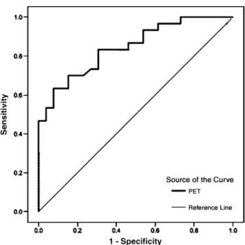
Fig. 3 ROC curve analysing the performance of18F-fluoride PET/CT for the diagnosis of sacroiliitis
Table 1 Sensitivity, specificity and accuracy of 18F-fluoride PET quantification for the diagnosis of sacroiliitis in 15 patients with AS and 13 patients with MLBP
Per patient analysis Per SIJ analysis
Sensitivity 80% (12/15) 70% (21/30)
Specificity 77% (10/13) 85% (22/26)
bar disc (n=1). The mean (±SD) CRP in the control group was 2.4±1.5 mg/l and the ESR (first hour) was 7.8±3.2 mm. Radiographic SIJ grading in the AS group
In patients with AS SIJ grading on plan radiographs showed grade 2 sacroiliitis in 10 joints (33%), grade 3 in 16 joints (53%) and grade 4 in 4 joints (13%). No joints with grade 1 sacroiliitis were seen.
Quantitative18F-fluoride PET/CT using the SIJ/S ratio 18
F-Fluoride PET/CT quantification was feasible in all patients. The mean SIJ/S ratio of 30 quantified joints was 1.66 (range 1.10–3.07) in the AS group. In the MLBP group, the mean SIJ/S ratio of 26 quantified joints was 1.12 (range 0.71–1.52) (Fig.2).
The area under the ROC curve for sacroiliitis was 0.84 (Fig. 3). Taking a SIJ/S ratio of >1.3 as a cut-off, the sensitivity, specificity and accuracy on a per patient basis were 80%, 77% and 79%, respectively, with conventional radiology as the gold standard. In a per joint basis equivalent values were 70%, 85% and 77%,
Fig. 4 A 49-year-old female patient with ankylosing spondy-litis.18F-Fluoride PET/CT with increased uptake (arrows) in both SIJs with more uptake in the left joint. MIP image (a), axial PET (b), CT (c) and fused PET/CT images (d) demonstrate the morphologically severely al-tered joints with sclerosis and multiple erosions on both sides (arrows)
Fig. 5 Box plots of SIJ/S ratios of18F-fluoride PET/CT in relation to conventional SIJ grading using plain radiographs in patients with AS
respectively (Table 1). The sensitivity of PET imaging was 40% in grade 2 sacroiliitis (n=10), 94% in grade 3 sacroiliitis (n=16) (Fig.4) and 50% in grade 4 sacroiliitis (n=4) (Fig.5).
Discussion
To our knowledge, this is the first study evaluating the performance of 18F-fluoride PET/CT for the diagnosis of sacroiliitis in AS patients with active disease. Quantitative PET/CT using the SIJ/S ratio with a cut-off of 1.3, as established for conventional bone scintigraphy, is feasible. The finding of an overall sensitivity of 80% for the diagnosis of sacroiliitis may indicate the superiority of 18
F-fluoride PET/CT in comparison to scintigraphy. For the latter imaging method a sensitivity of 50–55% and a specificity of about 80% was calculated following a recent systematic literature review. The following characteristics of the PET tracer might explain its superiority: rapid blood clearance with high and quick bone uptake, resulting in a better lesion-to-background ratio compared to 99mTc la-belled phosphonates [20]. Additionally, PET devices are superior to conventional gamma cameras with regard to spatial resolution and sensitivity.
The lower sensitivity of PET in patients with grade 2 than in those with grade 3 sacroiliitis is intriguing, as we would not expect the intensity of acute bone inflammation to considerably differ between these two subgroups. We assume that increased fluoride uptake might not only be a consequence of inflammation but also of postinflammatory repair osteoproliferation (greater areas of erosions and sclerosis in joints with grade 3 sacroiliitis) [15]. The low sensitivity of PET in patients with radiographic grade 4 sacroiliitis, as already demonstrated for scintigraphy [17], might be explained by the fact that inflammation can no longer be expected and osteoproliferation is already complete in these fused joints (complete ankylosis). The direct correlation between 18F-fluoride PET/CT, MRI and histopathological examinations, which is the subject of current research, might unravel this issue.
The CT part of the PET/CT imaging technique allows the exact localization of the active lesion, as well as the visualization of chronic changes including erosions, scle-rosis and ankylosis. We intentionally ignored this informa-tion in the present study and used the low-dose CT part only for attenuation correction. We expect that combined reading of PET and CT data would further increase the sensitivity of this technique for the detection of sacroiliitis not only in AS, but most importantly in patients with early SpA. However, with regard to radiation exposure and costs, PET/CT might only play a role in the routine diagnosis of sacroiliitis if it is shown to provide a better
correlation with disease progression than the currently used MRI [25].
We have tried to minimize exposure to radiation from 18
F-fluoride PET/CT by lowering the applied18F activity to 100–150 MBq per patient in comparison to 300–550 MBq used in other institutions for the detection of bone metastases. A low-dose protocol (40 mAs) was performed for CT acquisition [20, 21, 26]. The estimated radiation dose for the combined technique is similar to that of bone scintigraphy (3–5 mSv).
We focused on evaluation of the SIJ in this pilot study. In our experience 18F-fluoride PET/CT is also promising for the evaluation of spine involvement as well as peripheral arthritis and enthesitis during the same exami-nation.
In conclusion, quantitative 18F-fluoride PET/CT may play a role in the diagnosis of sacroiliitis in AS, and is a possible alternative to conventional bone scintigraphy in times of molybdenum shortage.
Acknowledgment We thank our technicians, especially Sabine Knöfel, Thomas Berthold and Ennio Müller for scanning the patients.
References
1. Braun J, Sieper J. Ankylosing spondylitis. Lancet 2007;369:1379– 90.
2. Sieper J. Developments in the scientific and clinical understanding of the spondyloarthritides. Arthritis Res Ther 2009;11:208. 3. Baraliakos X, Landewe R, Hermann KG, et al. Inflammation in
ankylosing spondylitis: a systematic description of the extent and frequency of acute spinal changes using magnetic resonance imaging. Ann Rheum Dis 2005;64:730–4.
4. Baraliakos X, Listing J, von der Recke A, Braun J. The natural course of radiographic progression in ankylosing spondylitis – evidence for major individual variations in a large proportion of patients. J Rheumatol 2009;36:997–1002.
5. Boonen A, van der Linden SM. The burden of ankylosing spondylitis. J Rheumatol Suppl 2006;78:4–11.
6. van der Linden S, Valkenburg HA, Cats A. Evaluation of diagnostic criteria for ankylosing spondylitis. A proposal for modification of the New York criteria. Arthritis Rheum 1984;27:361–8.
7. Battafarano DF, West SG, Rak KM, Fortenbery EJ, Chantelois AE. Comparison of bone scan, computed tomography, and magnetic resonance imaging in the diagnosis of active sacroiliitis. Semin Arthritis Rheum 1993;23:161–76.
8. Blum U, Buitrago-Tellez C, Mundiger A, Krause T, Laubenberger J, Vaith P, et al. Magnetic resonance imaging (MRI) for detection of active sacroiliitis– a prospective study comparing conventional radiography, scintigraphy, and contrast enhanced MRI. J Rheu-matol 1996;23:2107–15.
9. Yu W, Feng F, Dion E, Yang H, Jiang M, Genant HK. Comparison of radiography, computed tomography and magnetic resonance imaging in the detection of sacroiliitis accompanying ankylosing spondylitis. Skeletal Radiol 1998;27:311–20.
10. Oostveen J, Prevo R, den Boer J, van de Laar M. Early detection of sacroiliitis on magnetic resonance imaging and subsequent
development of sacroiliitis on plain radiography. A prospective, longitudinal study. J Rheumatol 1999;26:1953–8.
11. Rudwaleit M, Jurik AG, Hermann KG, et al. Defining active sacroiliitis on magnetic resonance imaging (MRI) for classifica-tion of axial spondyloarthritis: a consensual approach by the ASAS/OMERACT MRI group. Ann Rheum Dis 2009;68:1520–7. 12. Bollow M, Fischer T, Reisshauser H, et al. Quantitative analyses of sacroiliac biopsies in spondyloarthropathies: T cells and macrophages predominate in early and active sacroiliitis – cellularity correlates with the degree of enhancement detected by magnetic resonance imaging. Ann Rheum Dis 2000;59:135–40. 13. Baraliakos X, Listing J, Brandt J, et al. Radiographic progression
in patients with ankylosing spondylitis after 4 years of treatment with the anti-TNF-alpha antibody infliximab. Rheumatology 2007;46:1450–3.
14. van der Heijde D, Landewe R, Einstein S, et al. Radiographic progression of ankylosing spondylitis after up to two years of treatment with etanercept. Arthritis Rheum 2008;58:1324–31. 15. Sieper J, Appel H, Braun J, Rudwaleit M. Critical appraisal of
assessment of structural damage in ankylosing spondylitis: implications for treatment outcomes. Arthritis Rheum 2008;58:649–56.
16. Van Laere M, Veys EM, Mielants H. Strontium 87m scanning of the sacroiliac joints in ankylosing spondylitis. Ann Rheum Dis 1972;31:201–6.
17. Song IH, Carrasco-Fernandez J, Rudwaleit M, Sieper J. The diagnostic value of scintigraphy in assessing sacroiliitis in ankylosing spondylitis: a systematic literature research. Ann Rheum Dis 2008;67:1535–40.
18. Perkins A, Hilson A, Hall J. Global shortage of medical isotopes threatens nuclear medicine services. BMJ 2008;337:a1577. 19. Gould P. Medical isotope shortage reaches crisis level. Nature
2009;460:312–3.
20. Grant FD, Fahey FH, Packard AB, Davis RT, Alavi A, Treves ST. Skeletal PET with 18F-fluoride: applying new technology to an old tracer. J Nucl Med 2008;49:68–78.
21. Even-Sapir E, Metser U, Mishani E, Lievshitz G, Lerman H, Leibovitch I. The detection of bone metastases in patients with high-risk prostate cancer: 99mTc-MDP planar bone scintigraphy, single- and multi-field-of-view SPECT, 18F-fluoride PET, and 18F-fluoride PET/CT. J Nucl Med 2006;47:287–9.
22. Garrett S, Jenkinson T, Kennedy LG, et al. A new approach to defining disease status in ankylosing spondylitis: the Bath Ankylosing Spondylitis Disease Activity Index. J Rheumatol 1994;21:2268–91.
23. Braun J, Davis J, Dougados M, Sieper J, et al. First update of the international ASAS consensus statement for the use of anti-TNF agents in patients with ankylosing spondylitis. Ann Rheum Dis 2006;65:316–20.
24. Hanley JA, McNeil BJ. The meaning and use of the area under a receiver operating characteristic (ROC) curve. Radiology 1982;143:29–36.
25. Maksymowych WP. Disease modification in ankylosing spondy-litis. Nat Rev Rheumatol 2010;6:75–81.
26. Beheshti M, Vali R, Waldenberger P, Fitz F, Nader M, Loidl W, et al. Detection of bone metastases in patients with prostate cancer by 18F-fluorocholine and 18F-fluoride PET-CT: a comparative study. Eur J Nucl Med Mol Imaging 2008;35:1766–74.