Abstract The testis-expressed human TPTE is a putative
transmembrane tyrosine phosphatase, probably involved
in signal transduction pathways of the endocrine and/or
the spermatogenetic function of the testis. TPTE was
mapped to the pericentromeric region of human
chromo-somes 21 and 13, and to chromochromo-somes 15, 22, and Y. It is
unknown which of the TPTE copies are transcribed,
con-tain intronic sequences, and/or have open reading frames.
Here, in silico analysis of the genomic sequence of human
chromosome 21 allowed the determination of the genomic
structure of a copy of the TPTE gene. This copy consists
of 24 exons and spans approximately 87 kb. The mapping
position of this copy of TPTE on the short arm of
chro-mosome 21 was confirmed by FISH using the BAC
15L0C0 clone as a probe that contains almost the entire
TPTE gene. This is the first description of the genomic
se-quence of a non-RNR gene on the short arm of human
acrocentric chromosomes.
Introduction
The short arms of the five human acrocentric
chromo-somes, 13p, 14p, 15p, 21p, and 22p are mainly composed
of different types of repetitive elements and RNR gene
cluster (Choo et al. 1989; Greig et al. 1993; Henderson et
al. 1973; Vissel et al. 1992). These short arms share
ex-tensive homology among the different acrocentric
chro-mosomes (Van Camp et al. 1992). It is believed that 21p
and the p-arms of all other acrocentrics do not contain
sin-gle copy expressed genes other than RNR.
We have recently identified a cDNA, named TPTE
(Transmembrane Phosphatase with Tensin homology),
encoding a predicted protein of 551 amino acids with
po-tential transmembrane domains, a tyrosine phosphatase
motif and sequence homology to several phosphatases
in-cluding the tumor suppresser PTEN (Chen et al. 1999).
The gene is strongly expressed exclusively in testis. There
are copies of TPTE sequences on the pericentromeric
re-gions of chromosomes 21, 13, 15, 22, and the Y
chromo-some (Chen et al. 1999). The total copy number of the
TPTE genes and potential pseudogenes is polymorphic
but unknown. Furthermore, it is unknown how many of
TPTE gene copies are transcribed and if there is
chromo-some-specific expression. Finally, it is not clear whether
all TPTE copies contain introns, and their reading frames
are all open. One copy of TPTE has been assigned by
phys-ical mapping to chromosome 21p in a YAC contig
be-tween markers D21S237 and D21Z4 (Wang et al. 1999).
The advances of the sequencing of human chromosomes
will provide a better understanding of the different TPTE
copies and help to distinguish expressed genes from the
pseudogenes.
We report here the genomic structure, the sequence,
and the confirmation of the mapping of a copy of the
hu-man TPTE gene on the short arm of chromosome 21 that
is likely to be expressed.
Materials and methods
In silico analysis of human chromosome 21 genomic DNA sequence
The chromosome 21 sequencing group in the Department of Genome Analysis of the GBF Institute in Braunschweig Germany (http://genome.gbf.de/seqproj.html#Human21) determined the nu-cleotide sequences of BACs B7L1C4 (106.7 kb; Genbank No. AL078476) and B15L0C0 (80.6 kb; Genbank No. AL078471), and a contig (Genbank No. NT_002217) of approximately 144.3 kb
Michel Guipponi · Marie-Laure Yaspo ·
Lisa Riesselman · Haiming Chen · Albertina De Sario ·
Gérard Roizès · Stylianos E. Antonarakis
Genomic structure of a copy of the human TPTE gene
which encompasses 87 kb on the short arm of chromosome 21
Hum Genet (2000) 107 : 127–131
Digital Object Identifier (DOI) 10.1007/s004390000343
Received: 4 May 2000 / Accepted: 9 June 2000 / Published online: 1 August 2000
O R I G I N A L I N V E S T I G AT I O N
M. Guipponi · H. Chen · S. E. Antonarakis (✉) Division of Medical Genetics,
University of Geneva Medical School, Geneva, Switzerland e-mail: [email protected], Tel.: +41-22-7025708, Fax: +41-22-7025706 S. E. Antonarakis
University and Cantonal Hospital,
1 rue Michel Servet 1211 Geneva 4, Switzerland M.-L. Yaspo · L. Riesselman
Max-Planck-Institut fur Molekulare Genetik, D-14195 Berlin-Dahlem, Germany
A. De Sario · G. Roizès
Séquences Répétées et Centromères Humains,
CNRS UPR 1142, Institut de Biologie, Montpellier, France © Springer-Verlag 2000
was constructed. These BACs were cloned from the WAV17 mouse human somatic cell hybrid that contains only human chro-mosome 21 (Kozak et al. 1977). Detailed analysis of the nucleotide sequences of the contig was performed using numerous computer programs (Williams et al. 1998) interfaced by NIX. This program integrates most gene-identification software (GRAIL, Fex, Hexon, MZEF, Genemark, Genefinder, FGene, Polyah, RepeatMasker, tRNAscan), Blast searches (against many databases), and also al-lows personal annotations with a graphical interface.
The genomic structure of the TPTE gene was determined using the “EST_genome” molecular biology software available at HGMP (http://www.hgmp.mrc.ac.uk/Registered/Option/est_genome.html). This software predicts the genomic organization of genes by com-paring the genomic to the cDNA sequence. The TPTE cDNA (Genbank Nos. NM_013315 or AF007118) was used for sequence comparisons.
FISH analysis
Metaphase chromosomes were prepared from human peripheral blood lymphocytes. Standard FISH protocols were followed (Ward et al. 1995). BAC 15L0C0 and the alpha satellite were la-beled by nick translation. The alpha satellite probe was Z13/21 (Vissel and Choo 1991). Labeled BAC DNA was coprecipitated with human Cot-1 competitor DNA (GIBCO-BRL) and herring sperm carrier DNA to block repetitive elements. Biotinylated probes were detected by fluorescein isothiocyanate (FITC)-conju-gated avidin (Vector) and digoxygenated probes by Cy3-conju-gated anti-digoxygenin antibody (Dianova). Chromosomes were counterstained with 4,6-diamidino-2-phenylindole (DAPI). Images
Fig. 1A–D Schematic representation of the TPTE locus on
mosome 21p. A Ideogram of human chromosome 21. B The chro-mosome 21p YAC contig that defines the position of the TPTE lo-cus (Wang et al. 1999). The positions of the markers and the hmc01a06 trapped exon (exon 10 of TPTE) are shown. C BAC clones that contain the TPTE gene. D The structure and some fea-tures of the TPTE gene are shown. TPTE exons are in black and numbered underneath. The presence of repeats (Alus, L1s, MERs, MIRs, etc.) and CpG islands are also indicated. Numbers within the thick black line are nucleotide numbers. The orientation of the transcription is from the centromere to the telomere
were taken with a Zeiss epifluorescence microscope equipped with a thermoelectronically cooled CCD camera (Photometrics CH250).
Results and Discussion
Figure 1 schematically represents the mapping and
ge-nomic organization of the chromosome 21 copy of TPTE
(we designate this copy as TPTE-21). The gene spans
87 kb and is divided into 24 exons. There is 100% identity
between the cDNA sequence determined by Chen et al.
(1999) and the genomic sequence of contig NT_002217.
Only two nucleotide substitutions were found, a c.1495G>A
and a c.1748C>T (nucleotide numbers are from the
Genbank entry with accession number NM_013315 or
AF007118). Only the second change results in an amino
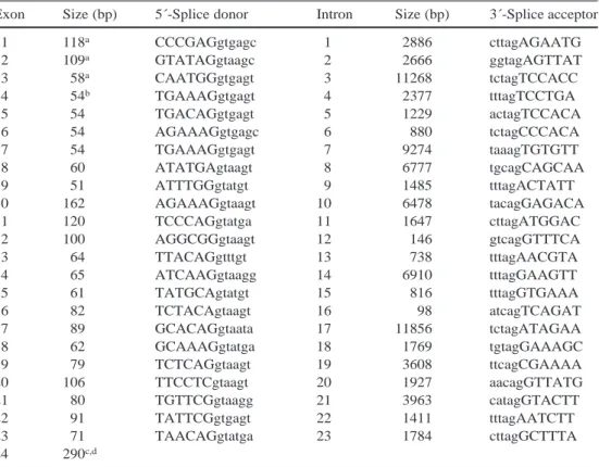
acid substitution, P470L. Table 1 lists the size of each
exon and intron and the sequences of intron-exon
junc-tions. Exons range from 51 to 162 nucleotides; intron
sizes range from 98 (intron 16) to 11,856 nucleotides
(in-tron 17). All splice junction sequences conform to the
GT-AG rule. The codon ATG for the initiator methionine
is on exon 4; there are therefore at least three untranslated
5´-exons. There is no experimental evidence for
alterna-tive splicing; however, this possibility needs to be
investi-gated in testicular RNA once the genomic organization of
the TPTE gene family is elucidated. There is a predicted
CpG island in exon 1 and 5´-UTR, implying that it may
represent part of the promoter sequences in this copy of
TPTE.
FISH analysis using a TPTE containing BAC clone
from chromosome 21 clearly showed a hybridization
sig-nal on 21p (Fig. 2). This confirms the previous mapping of
TPTE in a YAC contig of 21p (Wang et al. 1999). A
hy-bridization signal on 13p was also observed, as expected.
Database searches revealed the existence of unordered
pieces of sequences from the human chromosome 13 clone
RP11–95E16 (Genbank No. AL139386, first draft
se-quence from the Sanger Center) containing all except the
last TPTE exon. Analysis of the sequences indicates that
chromosome 13 also contains at least one copy of TPTE.
There was complete identity among the exons of the
chro-mosome 21 and 13 TPTEs. The introns were also highly
conserved (98–100% identity). The coding regions of the
chromosome 13 TPTE sequence only show one
nucleo-tide difference from the TPTE cDNA. This is c.1784A>C,
resulting in a Y482S amino acid substitution. However,
the chromosomal mapping of this clone by FISH showed
signal on both 21p and 13p
(http://www.sanger.ac.uk/cgi-bin/humace/searcher.cgi). It is not clear that this clone
originates from chromosome 13 and not 21.
Blast searches identified additional genomic sequences
with high homology to TPTE. These sequences are
“work-ing draft” quality, unordered and not precisely mapped to
specific human chromosomes. For example, a homologous
draft sequence with mapping position to chromosome Y
(BAC clone RP11–428D10; Genbank No. AC019099)
has also been mapped by the Sanger Center to
chromo-some 13 by FISH (http://www.sanger.ac.uk/cgi-bin/
humace/searcher.cgi). Because of the preliminary nature
129Table 1 Intron-exon
bound-aries and exon and intron sizes of the human chromosome 21 copy of TPTE
aUntranslated, contains longest cDNA sequence known bForty-three nucleotides to start codon
cOne hundred and thirty-six nucleotides to stop codon dTwo hundred and seventy-two nucleotides to the polyadenyla-tion signal
Exon Size (bp) 5´-Splice donor Intron Size (bp) 3´-Splice acceptor
1 118a CCCGAGgtgagc 1 2886 cttagAGAATG 2 109a GTATAGgtaagc 2 2666 ggtagAGTTAT 3 58a CAATGGgtgagt 3 11268 tctagTCCACC 4 54b TGAAAGgtgagt 4 2377 tttagTCCTGA 5 54 TGACAGgtgagt 5 1229 actagTCCACA 6 54 AGAAAGgtgagc 6 880 tctagCCCACA 7 54 TGAAAGgtgagt 7 9274 taaagTGTGTT 8 60 ATATGAgtaagt 8 6777 tgcagCAGCAA 9 51 ATTTGGgtatgt 9 1485 tttagACTATT 10 162 AGAAAGgtaagt 10 6478 tacagGAGACA 11 120 TCCCAGgtatga 11 1647 cttagATGGAC 12 100 AGGCGGgtaagt 12 146 gtcagGTTTCA 13 64 TTACAGgtttgt 13 738 tttagAACGTA 14 65 ATCAAGgtaagg 14 6910 tttagGAAGTT 15 61 TATGCAgtatgt 15 816 tttagGTGAAA 16 82 TCTACAgtaagt 16 98 atcagTCAGAT 17 89 GCACAGgtaata 17 11856 tctagATAGAA 18 62 GCAAAGgtatga 18 1769 tgtagGAAAGC 19 79 TCTCAGgtaagt 19 3608 ttcagCGAAAA 20 106 TTCCTCgtaagt 20 1927 aacagGTTATG 21 80 TGTTCGgtaagg 21 3963 catagGTACTT 22 91 TATTCGgtgagt 22 1411 tttagAATCTT 23 71 TAACAGgtatga 23 1784 cttagGCTTTA 24 290c,d
of these draft sequences, we did not extend our analysis to
TPTE copies in other chromosomes.
To our knowledge, the TPTE described here is the first
gene besides the RNR gene family that maps the short
arm of an acrocentric human chromosome. The
determi-nation and analysis of the genomic sequence of TPTE
provide tools for mutation analyses in selected
pheno-types and the elucidation of the evolutionary history of
this family of genes and pseudogenes. The genomic
orga-nization of the additional members of the TPTE gene
fam-ily is likely to be determined in the next couple of years.
It is currently unknown which members of the TPTE gene
family are expressed in the testis. The TPTE-21 has the
potential of being expressed; however, its mapping
posi-tion near the centromere may prevent its expression. It is
well known that genes placed in the heterochromatin
gion of Drosophila are silenced (see Karpen 1994 for
re-view). It is possible that similar mechanisms may operate
in human chromosomes and prevent the expression of
genes located near the centromeres.
Acknowledgements This study was supported by grants from the
Swiss FNRS 31-57149.99, the European Union/OFES BMH4-CT98-3039, and funds from the University and Cantonal Hospital of Geneva.
References
Chen H, Rossier C, Morris MA, Scott HS, Gos A, Bairoch A, An-tonarakis SE (1999) A testis-specific gene, TPTE, encodes a putative transmembrane tyrosine phosphatase and maps to the pericentromeric region of human chromosomes 21 and 13, and to chromosomes 15, 22, and Y. Hum Genet 105:399–409
Fig. 2 FISH mapping of BAC 15L0C0 containing part of the
hu-man TPTE gene on the short arm of huhu-man chromosome 21. Chro-mosomes were stained with DAPI. The BAC 15L0C0 signal is shown in red and the alpha satellite signal is shown in green. On both chromosomes 21 the BAC hybridization is clearly on the short arms; labeling on chromosome 13 is also observed because of the known sequence sharing between chromosome 21 and 13 in the centromeres and short arms
Choo KH, Vissel B, Earle E (1989) Evolution of alpha-satellite DNA on human acrocentric chromosomes. Genomics 5:332– 344
Greig GM, Warburton PE, Willard HF (1993) Organization and evolution of an alpha satellite DNA subset shared by human chromosomes 13 and 21. J Mol Evol 37:464–475
Henderson AS, Warburton D, Atwood KC (1973) Ribosomal DNA connectives between human acrocentric chromosomes. Nature 245:95–97
Karpen GH (1994) Position-effect variegation and the new biology of heterochromatin. Curr Opin Genet Dev 4:281–291
Kozak CA, Lawrence JB, Ruddle FH (1977) A sequential staining technique for the chromosomal analysis of the interspecific mouse/hamster and mouse/human somatic cell hybrids. Exp Cell Res 105:109–117
Van Camp G, Cruts M, Backhovens H, Wehnert A, Van Broeck-hoven C (1992) Unique sequence homology in the pericen-tromeric regions of the long arms of chromosomes 13 and 21. Genomics 12:158–160
Vissel B, Choo KH (1991) Four distinct alpha satellite subfamilies shared by human chromosomes 13, 14 and 21. Nucleic Acids Res 25:271–277
Vissel B, Nagy A, Choo KH (1992) A satellite III sequence shared by human chromosomes 13, 14, and 21 that is contiguous with alpha satellite DNA. Cytogenet Cell Genet 61:81–86
Wang SY, Cruts M, Del-Favero J, Zhang Y, Tissir F, Potier MC, Patterson D, Nizetic D, Bosch A, Chen H, Bennett L, Estivill X, Kessling A, Antonarakis SE, Broeckhoven C van (1999) A high-resolution physical map of human chromosome 21p using yeast artificial chromosomes. Genome Res 9:1059–1073 Ward DC, Boyle A, Haaf T (1995) Fluorescence in situ
hybridiza-tion techniques. Metaphase chromosomes, interphase nuclei, and extended chromatin fibers. In: Verma RS, Babu A (eds) Human chromosomes. Principles and techniques. MacGraw-Hill, New York, pp 184–192
Williams GW, Woollard PM, Hingamp P (1998) NIX: a nucleotide identification system at the HGMP-RC. http://www.hgmp.mrc. ac.uk/NIX/