DOI 10.1007/s00402-010-1143-y
T R A U M A S U R G E R Y
Anatomical considerations of the internal iliac artery
in association with the ilioinguinal approach for anterior
acetabular fracture
Wxation
Nakul Karkare · Richard A. Yeasting · Nabil A. Ebraheim · Norman Espinosa · Max J. Scheyerer · Clément M. L. Werner
Received: 12 March 2008 / Published online: 29 June 2010 © Springer-Verlag 2010
Abstract
Introduction Vascular injury may be encountered during an anterior approach to the pelvis or acetabulum—be it due to hematoma decompression, clot dislodgement during fracture manipulation, or iatrogenic. This can be associated with signiWcant bleeding, hemodynamic instability, and subsequent morbidity. If the exact source of bleeding can-not be easily identiWed, compression of the internal iliac artery may be a lifesaving procedure.
Materials and methods We describe an extension of the lateral window of the ilioinguinal (or Olerud) approach elaborated on cadavers.
Results The approach allows emergent access the internal iliac artery and intraoperative cross-clamping of the inter-nal iliac vessels to control bleeding.
Conclusion The approach allows rapid access to the inter-nal iliac artery. The surgeon should be familiar, however, with the surgical anatomy of this region to avoid potential injury to the ureter, peritoneum, lymphatics, and sympathetic nerves overlying the vessels when using the approach described.
Keywords Ilioinguinal approach · Acetabulum · Pelvic ring · Bleeding · Complication
Introduction
Pelvic hemorrhage might be encountered during either an anterior approach to the ilium or SI-joint (Olerud), or dur-ing an iliodur-inguinal [13, 15] approach to the acetabulum. This might be due to injuries to the internal iliac artery associated with pelvic fractures, clot dislodgement during fracture manipulation, or iatrogenic (e.g. injury to the corona mortis) [1, 6–10, 12, 13, 17, 18, 22, 24–28, 30, 31,
33, 34]. These major sources of bleeding are addressed by open exploration and repair, ligation, intraoperative embolization, packing, or percutaneous selective arterio-graphic embolization [2, 3, 14, 16, 18, 32]. If the exact source of bleeding cannot be immediately identiWed intra-operatively, approach the proximal vasculature to control bleeding might be warranted. For such a scenario, opera-tive strategy remains controversial in literature [33]. A method of access to the internal iliac artery using the lat-eral window of the ilioinguinal approach—to the best our knowledge—has yet not been described. An extension of the given ilioinguinal approach allowing rapid access to the internal iliac artery has been carried out on cadavers and is described.
Technique
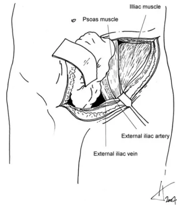
The approach described is an extension of the lateral win-dow of the ilioinguinal approach [13] (similar to Olerud [23] approach to the SI-joint, Fig.1). It gives rapid access to the bifurcation of the iliac vessels. The psoas muscle is identiWed and serves as a major landmark for further surgi-cal dissection. Staying extraperitoneal, the plane of dissec-tion is between the medially retracted pelvic content/ peritoneum, and the psoas muscle (Fig.2).
N. Karkare · N. A. Ebraheim Department of Orthopaedic Surgery, Medical College of Ohio, Toledo, OH, USA
R. A. Yeasting
Department of Anatomy,
Medical College of Ohio, Toledo, OH, USA
N. Espinosa · M. J. Scheyerer · C. M. L. Werner (&) Department of Orthopaedics, University of Zurich,
Uniklinik Balgrist, Forchstrasse 340, 8008 Zurich, Switzerland e-mail: clement.werner@balgrist.ch
236 Arch Orthop Trauma Surg (2011) 131:235–239
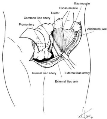
The common iliac and the external iliac arteries are par-allel and in close vicinity of the medial border of the psoas (Fig.3). The external iliac artery can be palpated in the operative Weld infero-laterally and is a pointer to the bifur-cation of the iliac vessels. If necessary (obese patients), the skin incision can be extended parallel to the operating table along the course of the psoas muscle to get a better access to the deep retroperitoneal structures (Fig.1 dotted line). The dissection is carried out bluntly along this ves-sel leaving the space between its surrounding connective tissue and the antero-medial aspect of the psoas muscle undisturbed. This is important, since the sympathetic nerves and the lymphatics overlying the external iliac artery—as well as the common iliac vessels and the inter-nal iliac artery—are to be protected. As the dissection is carried medially beyond the external iliac artery, the ure-ter and the inure-ternal iliac arure-tery can be identiWed (Fig.4). If in doubt, light touching of the ureter leads to peristaltic movements in vivo. The internal iliac artery is the vessel that runs oV the common iliac and the medial border of the psoas to disappear in the inner pelvis (Fig.1). It can be distinguished from the middle sacral artery by its closer
relationship with the psoas muscle and its more lateral position. Both, the ureter and the internal iliac artery remain unseparated from the posterolateral structures they lie on. The bifurcation of the common iliac vessels (into internal and external iliac) can be expected at the level of the anterior superior iliac spine. Once it is reached, no fur-ther dissection is necessary.
In the midline of the body, the lumbosacral prominence is an important landmark which can easily be palpated. At its lateral aspect, the internal iliac artery is in close vicinity and can be compressed against the bone to control bleed-ing. Also, the artery can be cross-clamped from antero-medially, should ligation be required. Compared to simple compression to the bone for temporary control of bleeding, cross-clamping and ligation needs a more extensive dissec-tion of the vessel.
Another possible way of compressing the internal iliac artery is to apply pressure laterally and compressing it between the psoas and the pelvic brim. Attempts to expose the internal iliac from the lateral aspect (underneath the psoas major) is likely to be more time consuming and may also damage the sympathetic Wbers lying over the common iliac and the external iliac vessels. After compression or clamping of the internal iliac artery, pulses should be checked in the lower limb to ensure that the external iliac artery is intact.
Fig. 1 Proximal extension of the incision parallel to the operating table (dotted line)
Fig. 2 The psoas muscle serves as a landmark for further surgical dis-section. Staying extraperitoneal, the plane of dissection is between the medially retracted pelvic content/peritoneum, and the psoas muscle
Cadaver series
Five embalmed cadavers (two males and three females) were used in this study. The age of cadavers ranged from 40 to 85 years. They were placed in a supine position and bilateral ilioinguinal approaches [13] were performed. In all cases, we were able to approach the bifurcation of the iliac vessels on both sides using this approach. The bifurcation of the common iliac artery was consistently found to be at the level of the anterior superior iliac spine. Three speci-mens had calciWcation at the level of the common iliac bifurcation. Compression of the internal iliac artery against bone was possible in all cases. It was felt to be more diY-cult, however, in subjects with calciWed vessels.
One specimen had a partial sigmoid resection that made medial retraction more diYcult. Numerous nerves (sympa-thetic) and lymph nodes overlying the iliac vessels were encountered in all the dissections. If the dissection was carried out too close to the external iliac vessels, these were likely to be damaged. The ureter was in close relationship with the iliac bifurcation and had to be identiWed and protected.
We also attempted to approach the internal iliac artery (1) by dissecting posterior to the iliacus muscle in the retro-peritoneal space, and (2) dissecting between the iliacus and the psoas muscles. Both of these alternate approaches, how-ever, were not only found to be more diYcult due to limited visualization, but also to be associated with possible dam-age to the soft tissue/sympathetic Wbers surrounding the vessels.
Discussion
Fractures of both the pelvis and acetabulum may be associated with injury to the major pelvic vessels. This can be associated with the primary injury in more severe fracture patterns [9, 10, 12, 17, 18, 24, 27, 34], or later due to clot dislodgement or iatrogenic trauma during fracture manipulation [1, 6–10, 12, 13, 17, 18, 22, 24–
28, 30, 31, 33, 34].
The sources of bleeding have been reported to be more posterior (internal iliac vessels or their posterior branches) in patients with unstable posterior pelvic fractures, while in patients with lateral compression injuries they are located more anteriorly (pudendal or obturator vessels) [7, 19]. Treatment of these vascular injuries can be open exploration Fig. 3 The common iliac and the external iliac arteries are parallel
and in close vicinity of the medial border of the psoas. The external il-iac artery can be palpated in the operative Weld infero-laterally and is a pointer to the bifurcation of the iliac vessels
Fig. 4 Sympathetic nerves and the lymphatics overlying the external iliac artery are to be protected. As the dissection is carried medially beyond the external iliac artery, the ureter and the internal iliac artery can be identiWed. The promontory, against which the artery can be pressed, is marked with dotted lines
238 Arch Orthop Trauma Surg (2011) 131:235–239
and repair, exploration and ligation, intraoperative emboli-zation, or percutaneous selective arteriographic embolization [2, 3, 7, 14, 16, 32].
The common iliac divides into the internal and external iliac arteries at the level of the anterior superior iliac spine. The internal iliac artery divides into anterior and posterior trunks. The anterior trunk gives rise to the vesical branches, the middle rectal, the obturator, internal pudendal, inferior gluteal, and the uterine arteries. The iliolumbar, lateral sac-ral, and superior gluteal vessels arise from the posterior trunk.
Waiting for a vascular surgeon to arrive and access the artery can be time consuming even at a level 1 trauma cen-ter. Compression of the internal iliac artery by the orthope-dic surgeon maybe a lifesaving procedure allowing to minimize the pooling of blood and to identify the source of bleeding even when the exact source of bleeding is not immediately apparent.
The approach used by vascular surgeons to get access to the pelvic vessel usually is a midline retro- or transperito-neal approach. An additional midline incision might, how-ever, put the skin at risk where the ilioinguinal and midline approach meet. In a given situation, where the ilioinguinal (or Olerud) approach is already made at the time serious bleeding is encountered, the method described in this article may allow more rapid access to the vessels. Also, even in case the approach has to be extended, it does not burn any bridges for the vascular surgeon in case a midline incision is later needed.
Although signiWcant variations of the pelvic vasculature have been described [1, 4, 6, 8, 11, 20–22, 25, 26, 29, 30], the anatomical relationship at the bifurcation of the common iliac has been found to be fairly constant.
The corona mortis [1, 22, 26, 30] is an anastomotic branch between the inferior epigastric and obturator vessels in the obturator canal. Compression of the internal iliac will not completely stop bleeding from this anomalous artery since it has a contribution from the external iliac artery. Nevertheless, the maneuver described could also be utilized to control any contribution of the internal iliac artery to a corona mortis.
A possible drawback of accessing the internal pelvic vessels with the method described might be the limited exposure of the contralateral side. Although not common, the surgeon might be in doubt as to which side of the body a source of bleeding is to be expected. In these cases, the classic midline approach used by vascular surgeons to control bleeding [5] might be the better way to go.
In conclusion, we have to record that this is a cadaveric study and that in a clinical setting the access might be more diYcult due to bleeding. Further investigations are needed to give evidence about the failure rate or the learning curve for an average orthopedic trauma surgeon.
We still think, however, that it represents a valuable alternative to the classic midline transperitoneal approach to the vessels.
References
1. Berberoglu M, Uz A, Ozmen MM, Bozkurt MC, Erkuran C, Taner S, Tekin A, Tekdemir I (2001) Corona mortis: an anatomic study in seven cadavers and an endoscopic study in 28 patients. Surg Endosc 15(1):72–75
2. Brown JJ, Greene FL, McMillin RD (1984) Vascular injuries asso-ciated with pelvic fractures. Am Surg 50:150–154
3. Downs AR (1988) Hemorrhage and pelvic fractures. Can J Surg 31:89–90
4. Ebraheim NA, Xu R, Farooq A, Yeasting RA (1996) The quanti-tative anatomy of the iliac vessels and their relation to anterior lumbosacral approach. J Spinal Disord 9(5):414–417
5. Ertel W, Keel M, Eid K, Platz A, Trentz O (2001) Control of severe hemorrhage using C-clamp and pelvic packing in multiply injured patients with pelvic ring disruption. J Orthop Trauma 15(7):468–474
6. Feugier P, Fessy MH, Bejui J, Bouchet A (1997) Acetabular anat-omy and the relationship with pelvic vascular structures. Implica-tions in hip surgery. Surg Radiol Anat 19(2):85–90
7. Hamill J, Holden A, Paice R, Civil I (2000) Pelvic fracture pattern predicts pelvic arterial haemorrhage. Aust N Z J Surg 70(5):338–343 8. Hong HX, Pan ZJ, Chen X, Huang ZJ (2004) An anatomical study of corona mortis and its clinical signiWcance. Chin J Traumatol 7(3):165–169
9. Huijbregts JE, Luitse JS, Goslings JC, Eijer H (2004) Entrapment of the external iliac vein in a both-column acetabular fracture. J Orthop Trauma 18(9):630–633
10. Hureau J, Franco JL (1971) Rupture of the left common and exter-nal iliac veins during a dislocation-fracture of the pubic symphy-sis. Surgical repair and healing. Chirurgie 97(4):287–291 11. Karakurt L, Karaca I, Yilmaz E, Burma O, Serin E (2002) Corona
mortis: incidence and location. Arch Orthop Trauma Surg 122(3):163–164
12. Kataoka Y, Maekawa K, Nishimaki H, Yamamoto S, Soma K (2005) Iliac vein injuries in hemodynamically unstable patients with pelvic fracture caused by blunt trauma. J Trauma 58-4:704– 708 (discussion 8–10)
13. Letournel E (1993) The treatment of acetabular fractures through the ilioinguinal approach. Clin Orthop Relat Res 292:62–76 14. Mansour MA, Moore FA, Moore EE (1990) Hypogastric arterial
embolization in pelvic fracture hemorrhage: case report. J Trauma 30(11):1417–1418
15. Matta JM (2006) Operative treatment of acetabular fractures through the ilioinguinal approach: a 10-year perspective. J Orthop Trauma 20(1)Suppl:S20–S29
16. Maull KI, Sachatello CR (1976) Current management of pelvic fractures: a combined surgical-angiographic approach to hemor-rhage. South Med J 69(10):1285–1289
17. McMurtry R, Walton D, Dickinson D, Kellam J, Tile M (1980) Pelvic disruption in the polytraumatized patient: a management protocol. Clin Orthop Relat Res 151:22–30
18. Moreno C, Moore EE, Rosenberger A, Cleveland HC (1986) Hemorrhage associated with major pelvic fracture: a multispecial-ty challenge. J Trauma 26(11):987–994
19. O’Neill PA, Riina J, Sclafani S, Tornetta P 3rd (1996) Angiographic Wndings in pelvic fractures. Clin Orthop Relat Res 329:60–67 20. Okamoto K, Wakebe T, Saiki K, Nagashima S (2005)
Consider-ation of the potential courses of the common iliac artery. Anat Sci Int 80(2):116–119
21. Okcu G, Erkan S, Yercan HS, Ozic U (2004) The incidence and location of corona mortis: a study on 75 cadavers. Acta Orthop Scand 75(1):53–55
22. Pungpapong SU, Thum-umnauysuk S (2005) Incidence of corona mortis; preperitoneal anatomy for laparoscopic hernia repair. J Med Assoc Thai 88(Suppl 4):51–53
23. Ragnarsson B, Olerud C, Olerud S (1993) Anterior square-plate Wxation of sacroiliac disruption. 2–8 years follow-up of 23 consec-utive cases. Acta Orthop Scand 64–2:138–142
24. Rothenberger DA, Fischer RP, Strate RG, Velasco R, Perry JF Jr (1978) The mortality associated with pelvic fractures. Surgery 84(3):356–361
25. Sarikcioglu L, Sindel M (2002) Multiple vessel variations in the retropubic region. Folia Morphol (Warsz) 61(1):43–45
26. Sarikcioglu L, Sindel M, Akyildiz F, Gur S (2003) Anastomotic vessels in the retropubic region: corona mortis. Folia Morphol (Warsz) 62(3):179–182
27. Sriussadaporn S (2000) Abdominopelvic vascular injuries. J Med Assoc Thai 83(1):13–20
28. Stephen DJ, Kreder HJ, Day AC, McKee MD, Schemitsch EH, El-Maraghy A, Hamilton P, McLellan B (1999) Early detection of arterial bleeding in acute pelvic trauma. J Trauma 47(4):638–642 29. Teague DC, Graney DO, Routt ML Jr (1996) Retropubic vascular
hazards of the ilioinguinal exposure: a cadaveric and clinical study. J Orthop Trauma 10–3:156–159
30. Tornetta P 3rd, Hochwald N, Levine R (1996) Corona mortis. Inci-dence and location. Clin Orthop Relat Res 329:97–101
31. Totterman A, Madsen JE, Roise O (2006) Multifocal arterial haemorrhage in a partially stable pelvic fracture after a crush inju-ry: a case report. Arch Orthop Trauma Surg 126(2):113–117 32. van Urk H, Perlberger RR, Muller H (1978) Selective arterial
embolization for control of traumatic pelvic hemorrhage. Surgery 83(2):133–137
33. Vicq P, Hajji A, Le Reveille R, Darrieus H, Chaussard JF (1989) Vascular complications of fractures of the pelvis. J Chir (Paris) 126(10):507–513
34. Wali MA (2003) Internal iliac artery injury in a fractured pelvis. Ann Thorac Cardiovasc Surg 9(5):337–339