HAL Id: tel-01726946
https://tel.archives-ouvertes.fr/tel-01726946
Submitted on 8 Mar 2018HAL is a multi-disciplinary open access archive for the deposit and dissemination of sci-entific research documents, whether they are pub-lished or not. The documents may come from teaching and research institutions in France or abroad, or from public or private research centers.
L’archive ouverte pluridisciplinaire HAL, est destinée au dépôt et à la diffusion de documents scientifiques de niveau recherche, publiés ou non, émanant des établissements d’enseignement et de recherche français ou étrangers, des laboratoires publics ou privés.
Regulation of DNA methylation by DNA glycosylases
MBD4 and TDG
Abdulkhaleg Ibrahim
To cite this version:
Abdulkhaleg Ibrahim. Regulation of DNA methylation by DNA glycosylases MBD4 and TDG. Ge-nomics [q-bio.GN]. Université de Strasbourg, 2015. English. �NNT : 2015STRAJ019�. �tel-01726946�
!!!!!!!!!!!!!!!!!!!!! !!!!!!!!!!!!! !!!!!!!!!! ! ! ! ! !
Regulation of DNA methylation by DNA glycosylases
MBD4 and TDG
by
Abdulkhaleg IBRAHIM
Thesis submitted to the University of Strasbourg for the degree of
DOCTOR OF PHILOSOPHY
Discipline:
Life and Health Sciences
Specialization:
Molecular and Cellular Biology
Public PhD defence MAY 19th, 2015
Thesis Jury
Thesis Director:...
Dr Christian BRONNER
Rapporteur:...
Pr Gilles SALBERT
Rapporteur:...
Dr Pierre-Antoine DEFOSSEZ
Examiner:...
Pr Philippe BOUCHER
!
!
"#$%&:...''''''''...
Dr Ali HAMICHE
! ! ! ! ! ! ! ! ! ! ! ! ! ! ! ! ! ! ! ! ! ! ! ! ! ! ! ! ! ! ! ! ! ! ! ! ! ! ! ! ! ! ! ! ! ! !
! "
Acknowledgements
I am deeply grateful to my supervisor, Dr Christian Bronner, for his guidance with kindness and patience in helping me during my thesis. I appreciated very much the opportunity he gave me to discover the epigenetic field and the time he spent to have highly interesting scientific discussion with me.
I would like to express my sincere gratitude to our team leader Dr Ali Hamiche for giving me the opportunity to join his valuable laboratory. He helped me how to think scientifically, and provided me many helpful suggestions, important expert advice and constant encouragement over the years of my thesis. I deeply appreciate what you have done for me Ali.
My special thanks are going to Dr Pierre-Antoine Defossez, Pr Gilles Salbert and Pr Philippe
Boucher for their acceptance to be members in my thesis jury and for their valuable time spent to
critically read and comment on my thesis work.
I am especially grateful to Dr Christophe Papin for his enthusiasm, guidance and stimulating discussions throughout all the period we worked together. He worked with me to figure out reasons whenever I had problems in experiments and provided kindly recommended solutions.
In no particular order, I thank my colleagues; Khalid, Catherine, Philippe, Isabelle, Hatem,
Chrysa, Maria and Liam. Thank you all, for useful instruction, interesting scientific discussions,
and hearty laughs.
I am grateful for my financial support from the National Authority for Scientific Research and the Biotechnology Reseach Center (BTRC), Libya.
Thanks for the Affaires Culturelles Bureau in the libyan embassy in Paris for their efforts to facilitate my staying in France during my study and for their kind collaboration.
! ""
Summary
In mammals, methylation is an epigenetic mark targeting the 5th carbon of cytosine mainly in a CpG context producing a methylcytosine (5mC). The majority (70 to 80%) of CpGs are methylated but non-methylated in CpG-rich regions, referred as CpG islands (CGI). Repeated sequences cover almost half the genome in mammals and are highly methylated in healthy cells. In cancer cells, they can be specifically targeted by a demethylation process. However, the interest of repeated sequences has long been ignored in favor of the more functionally important regulatory regions such as CpG islands.
DNA methylation is not stable regarding that 5mC is highly sensitive to a spontaneous or enzymatic deamination leading to thymine and thus forming G/T mismatch. 5mC can also be successively oxidized to hydroxymethylcytosine (5hmC), formylcytosine (5FC) and 5-carboxylcytosine (5caC) by TET protein family (TET1, 2 and 3). These modifications (deamination and oxidation) of 5mC are considered as participating in active demethylation processes. But how the methylation state of these CGIs at silenced promoters is preserved, remains, however unclear. In mammals, the thymine in G/T mismatch (deamination product of 5mC) is recognized and cleaved by TDG and MBD4 glycosylase proteins. TDG is able also to specifically recognize and excise the 5FC and 5caC modifications. Therefore, TDG and MBD4 could play an essential role in the active demethylation process.
MBD4 is especially intriguing since it is the sole mammalian protein having a methylated DNA binding domain MBD (Methyl Binding Domain) associated with a glycosylase domain. In vitro, MBD4 possesses a monofunctional glycosylase activity that specifically cleaves the N-glycosidic bond of thymidine mismatched to a guanine (G/T), leading to the formation of an abasic site.
This thesis was to clarify the function of TDG and MBD4 in the dynamics of 5mC. We started by identifying proteins associated with MBD4 in vivo trying to understand its mechanism of action. To this end, we purified MBD4 complex from HeLa cells. Analysis by mass spectrometry of this complex shows that MBD4 is associated with PMS2, MLH1, MSH2 and MSH6 proteins, four proteins involved in DNA mismatch repair (MMR, mismatch repair). The in
vitro enzymatic tests show that MBD4/MMR complex has a bifunctional glycosylase/ lyase
activity specific for G/T and directed by methylation. Biochemical analysis of point mutants of MBD4 reveals that the integrity of MBD and glycosylase domains is required for this function.
! """
Our data suggest an activator and/or enzymatic role of MMR proteins in the bifunctional activity of MBD4 complex. To study this hypothesis, we purified recombinant MBD4, PMS2/MLH1 dimer and MSH2/MSH6 dimer proteins, and verified that MBD4 protein physically interacts with the MMR proteins. In vitro, low nuclease activity was detected with the MBD4 protein suggesting that the enzymatic activity of the native complex MBD4/MMR is catalyzed by MBD4 and regulated by MMR proteins.
Considering that the MLH1 protein is the most abundant subunit of MBD4 complex in our analysis by mass spectrometry, we wondered whether MLH1 could act as activator of MBD4 within the MBD4/MMR complex. Consistent with this hypothesis, we reconstituted a MBD4/MLH1 complex (Fig. 8A). Interestingly, while the nuclease activity of MBD4 is low and is not sensitive to methylation, the complex MBD4/MLH1 shows intense nuclease activity strongly induced by the methylation. The lack of enzymatic activity detected with the purified complex from a catalytic mutant MBD4 (MBD4 D554A/MLH1), demonstrates that the cleavage reaction is catalyzed by MBD4 and is dependent on its binding to MLH1.
We then sought to verify the role of TDG in the genome-wide DNA methylation dynamic. To this goal, we targeted this enzyme by shRNA technique in MEF cells and characterized the distribution of modified cytosine (5mC, 5hmC, 5FC and 5caC). Strikingly, our results show an enrichment of modified cytosines specifically at repeat sequences level. While 5mC target all the repeated sequences, with a preference for SINEs, oxidized forms (5hmC, and 5caC 5FC) are found preferentially in microsatellite (simple repeats). The distribution of 5mC, 5hmC, 5FC and 5caC at SINEs level in MEFs reveals a dynamic of these changes, regulated by TDG, only at the SINEs B1m and B2m family.
The genomic distribution analysis of SINEs B1m and B2m showed that the most preserved mouse specific SINEs are those the most close to a TSS (Transcription Start Site), suggesting an important function in the regulation of gene expression.
The dynamic changes was also observed at LINEs and LTRs. Similar to the obtained results at SINEs, the TDG-dependent regulation is observed only at the evolutionary newer retro-elements (mouse specific lineage), the L1Md family for LINEs and IAP family for the LTRs. We concluded that TDG regulates the dynamics of methylation/demethylation at repeated sequences during differentiation. Conversely, MBD4 targets promoters repressed by methylation and protects them against deamination mechanisms.
! "#!
Table of contents
Acknowledgements... i
Summary ... ii
Table of contents ...iv
List of figures ...vii
Abbreviations ...viii Chapter 1 Introduction 1.1 Overview of Epigenetics...1 1.2 DNA methylation...2 1.3 DNA methyltransferases...3
1.4 Genomic distribution of methylated cytosines...5
1.4.1 CpG islands methylation...6
1.4.2 Non-CpG methylation...8
1.5 CpG islands and Establishment of a permissive chromatin...8
1.6 DNA methylation and regulation of transcription...9
1.7 Methyl-CpG binding proteins as intermediates in transcriptional repression...11
1.7.1 Methyl-CpG binding proteins...11
1.7.1.a MeCP2...11
1.7.1.b MBD1 ...12
1.7.1.c MBD2/MBD3...12
1.7.1.d MBD4...13
1.7.2 Kaiso and Kaiso-like proteins...13
1.7.3 SRA domain proteins...14
1.8 DNA demethylation...14
1.8.1 Passive DNA demethylation...15
1.8.2 Active DNA demethylation...15
1.8.3 Mechanisms of active DNA demethylation...17
! "!
1.8.3.2 Radical SAM mechanism...18
1.8.3.3 Nucleotide Excision Repair (NER) to erase 5mC...18
1.8.3.4 Direct excision of 5mC followed by Base excision repair (BER)...19
1.8.3.5 Hydrolytic deamination of 5mC followed by BER...20
1.8.3.6 Oxidative modification of 5mC...22
1.8.3.6.a Passive dilution of oxidized 5mC...24
1.8.3.6.b ctive removal of oxidized methyl group...25
1.9 DNA glycosylases...26
1.9.1 Glycosylase activity of MBD4...27
1.9.2 MBD4/Substrate interaction...28
1.9.3 Glycosylase activity of TDG...29
1.9.4 TDG/Substrate interaction...30
1.9.5 TDG/AP site dissociation...32
1.10 Aims...35
Chapter 2 Results 2.1 The methyl-directed nuclease activity of MBD4-MLH1 complex is required to protect silenced promoters from demethylation. 2.2 Combinatorial DNA methylation code at repetitive elements. Chapter 3 Discussion 3.1. Function of MBD4/MMR complex in the protection of methylated cytocine...36
3.1.1 MBD4 interacts with mismatch repair proteins...36
3.1.2 MBD4 is a bifunctional glycosylase/lyase enzyme when associated to MLH1...37
3.1.3 Function of MBD4/MMR complex in the protection of methylcytosine...38
3.2. TDG-dependent Methylation/oxidation dynamic at repetitive elements...39
3.2.1 Methylation dynamics at IAP LTRs...40
3.2.2 Methylation dynamics at mouse-specific SINEs...40
! "#!
3.2.4 Methylation dynamics at CA repeats...41
Chapter 4
Conclusion and perspectives...43
Chapter 5
! "##!
List of figures
Figure 1. Cytosine methylation
Figure 2. Structure of DNA methyltransferases
Figure 3. Mechanisms of RE influence on gene transcription Figure 4. Passive DNA demethylation
Figure 5. DNA methylation changes during developmental epigenetic reprogramming Figure 6. 5meC modifying pathways
Figure 7. Schematic and Catalytic reaction of Tet enzymes. Figure 8. TET-induced 5meC oxidation
Figure 9. TET-induced DNA demethylation Figure 10. Mono- and Bi-functional glycosylases.
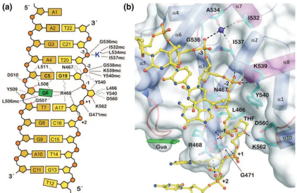
Figure 11. Interactions between MBD4 glycosylase domain and DNA sbstrate. Figure 12. Overview of TDG/ sbstrate structure.
! "###!
List of abbreviations
ADD ATRX-DNMT3-DNMT3L-type zinc finger domain AID Activation-induced Cytidine Deaminase
AP Apurinic/Apyrimidinic
APE1 APurinic/apyrimidinic Endonuclease 1
APOBEC Apolipoprotein B mRNA Editing enzyme, Catalytic polypeptide ATRX Alpha Thalassemia/Mental Retardation Syndrome X-Linked BAH Bromo-Adjacent Homology domain
BDNF Brain-Derived Neurotrophic Factor BER Base Excision Repair
C Cytosine
CFP1 CXXC Finger Protein 1 CGI CpG Island
CMT3 ChromoMethylase 3
CpG Cytosine-phosphate-Guanine
CREB C-AMP Response Element-Binding protein CtBP C-terminal Binding Protein
CXXC Cysteine rich region CYP27B1 CYtochrome P450 27B1
DIP-seq DNA-ImmunoPrecipitation-sequencing DNMT DNA methyltransferase
DRM2 Domains Rearranged Methyltransferase 2 DSBH Double-Stranded B-Helix
ElP3 Elongator complex Protein 3 ER! Oestrogen Receptor-! ERVs Endogenous RetroViruses ESC Embryonic Stem Cells EXO1 Exonuclease 1
! "# G Guanine
Gadd45 Growth arrest and DNA-damage-inducible protein 45 GFP Green Fluorescent Protein
HA HemAgglutinin
H3K27ac Histone H3 Lys27 acetylation
H3K36me2 Histone H3 di-methylated on lysine 36 H3K4me3 Histone H3 tri-methylated on lysine 4 H3K9ac Histone H3 Lys27 acetylation
HAT Histone AcetylTransferase HDAC2 Histone DeACetylase 2
HEK293 Human Embryonic Kidney 293 cells HP1 Heterochromatin Protein1
IAP Intracisternal A Particle ING4 INhibitor of Growth protein 4 JBP1 J-Binding Protein1
KDM2A Lysine-specific DeMethylase 2A LINEs Long INterspersed Elements LTRs Long Terminal Repeats MBD Methyl-CpG Binding Domain MBPs Methyl-CpG-Binding Proteins MeCP2 Methyl-CpG-binding Protein 2 MED Methyl-CpG-binding Endonuclease MLH1 MutL homolog 1
MMR Misatch Repair
MSH2 MutS protein homolog 2 MTA2 Metastasis-associated protein Mtase Methyltransferase domain. NCoR Nuclear receptor Co-Repressor NER Nucleotide Excision Repair NLS Nuclear localization signal
! "
NURF NUcleosome Remodeling Factor ORF Open Reading Frame
PARP1 Poly-ADP-Ribose Polymerase 1 PHD Plant Homeo Domain
PGC Primordial Germ Cells Pol III RNA polymerase III
PWWP Proline-tryptophan-tryptophan-Proline REs Repetitive DNA Elements
RMSK UCSC Repeat-Masker ROS1 Repressor Of Silencing 1
RRBS Reduced Representation Bisulfite Sequencing SAM S-Adenosyl Methionine
SETDB1 SET Domain, Bifurcated 1 shRNA short hairpin RNA
SINEs Short INterspersed Elements siRNA Small interfering RNA
SRA Set and Ring Associated domain SUMOs Small Ubiquitin Like Modifiers SWI/SNF SWItch/Sucrose NonFermentable T Thymine
TDG Thymine DNA Glycosylases TET Ten-Eleven Translocation TFF1 TreFoil Factor 1
TFIID Transcription Factor IID TFIIIC Transcription Factor IIIC TFs Transcription Factors Tg Thymine glycol THF TetraHydroFuran
TpG Thymine-phosphate-Guanine
TRD Transcriptional Repression Domain TSA TrichoStatin A
! "# TSGs Tumor Suppressor Genes TSS Transcription Start Site U Uracil
UHRF1 Ubiquitin-like containing plant Homeo-domain and RING Finger domain 1
UTR UnTranslated Region
ZBTB38 Zinc finger and BTB domain containing 38 ZF-CXXC Zinc Finger domain
5caC 5-carboxylCytosine 5fC 5-formylCytosine 5-FU 5-Fluorouracil 5hmU 5-hydroxymethylUracil 5hmC 5-hydroxymethylCytosine 5-hm 5-hydroxymethyl 5mC 5-methyl-Cytosine 8-oxoG 8-oxo-7,8-dihydroguanine 3-meA 3-methyl-Adenine 7-meG 7-methyl-Guanine
CHAPTER 1
INTRODUCTION
! "!
1.1 Overview of Epigenetics
All cells of a multicellular mammalian organism, except germinal cells, contain the same DNA in terms of nucleotide sequence. Considering that DNA is the layer of heredity and cell identity, how can arise cell diversity and differentiation from the same DNA sequence? The question is important and is one of the major challenges for the scientific community. Epigenetics is the research field that tries to answer this question by deciphering a tremendous number of cellular mechanisms of gene regulation embedded in the chromatin but not related to changes in DNA sequences.
The term “Epigenetic” comes from the Greek prefix “Epi” which means “above” or “over” and the term “Genetic” which is related to the study of genes. Therefore, “Epigenetic” literally means the study of gene expression variation that is not due to alterations of DNA sequences. In other words, it refers to external modifications to DNA that turn genes "on" or "off.!
Since it was originally introduced by Conrad Waddington in 1942, epigenetics showed a large progression in its understanding. Waddington used the term epigenetic to describe heritable changes in a cellular phenotype not related to alterations in the DNA sequence. He hypothesized that environmental stimulus could be converted into an internal genetic factor. Since then, different definitions of the term epigenetics have been proposed. In 2001, Rakyan has redefined the epigenetic as the mechanism of physically marking the DNA or its associated proteins to maintain stably gene expression which allows genotypically identical cells to be phenotypically distinct (Rakyan et al. 2001). Other definition is that “epigenetic” is the stably inherited phenotype resulting from changes in a chromosome without alterations in the DNA sequence (Berger et al. 2009). These tissue-specific patterns are essential for normal cellular function, and control of different stages of development by affecting gene expression (Eckhardt et al. 2006, Jones P. L. et al. 1998, Razin and Riggs 1980). Disturbance in these modifications or failure to maintain them can induce irregular gene transcription which lead to various forms of disease (Robertson 2005).
Three categories of signals have been proposed to be accompanied in the establishment of a stably heritable epigenetic state: an ‘‘Epigenator’’ which is a signal acquired from the environment and triggers an intracellular pathway; the second signal is
! #! the ‘‘Epigenetic Initiator’’ and this responds to the Epigenator and defines the precise location of the modification on chromatin. The Initiator could be a DNA-binding protein or a noncoding RNA. The third type of signal is the ‘‘Epigenetic Maintainer’’ to sustain the chromatin environment in the first and subsequent cell generations. Epigenetic
Maintainers involve many different epigenetic marks. These various maintainer marks
act together as an epigenetic machinery complex (Illingworth and Bird 2009).
At the molecular level, an epigenetic mark is by definition a chemical signal outside DNA or above DNA that regulates gene expression and that is transmitted to its descent. There are many different epigenetic marks, including DNA methylation, histone post-translational modifications, histone variants, micro-RNAs (Berger et al. 2009, Bronner et al. 2010) and we might imagine that more new epigenetic marks can be uncovered in a near future.
As my thesis work is dealing with DNA methylation, my manuscript will mainly focus on this epigenetic mark. However, we have to keep in mind that DNA methylation is intemely connected to other epigenetic marks such as for instance methylation of lysine 9 of histone H3, mediated by UHRF1 (Liu X. et al. 2013).
1.2 DNA methylation
DNA methylation occurs on the 5th carbon of cytosine in eukaryotes leading to form 5’-methylcytosine (5mC), whereas in prokaryotes, it happens on various bases giving rise to 5-methylcytosine, N6-methyladenine and N4-methylcytosine, and contributes to host restriction systems and protects the cells from foreign genetic material such as viral DNA and destruction by proper restriction enzymes. DNA methylation is a stable epigenetic mark that plays a key role in several biological functions including gene expression regulation, transposon silencing, X chromosome inactivation, imprinting, cell differentiation, development and other diverse processes (Bird A. 2002, Cedar and Bergman 2012). DNA methylation is a post-replicative mechanism that refers to transfer of a methyl group from the methyl donor S-adenosyl methionine (SAM) to cytosine (Figure 1).
! $! DNMT SAM-CH3 SAM Cytosine 5-Methylcytosine H3C Methylation DNMT SAM-CH3 SAM Cytosine Cytosine 5-Methylcytosine H3C 5-Methylcytosine H3C Methylation
Figure 1 – Cytosine methylation on
the 5-position in a reaction
catalysed by DNMT enzymes using
S-adenosyl methionine as a
substrate.
The methylated cytosine was first reported in 1948 by using paper chromatography (Hotchkiss 1948). Later, it was suggested that this modification of DNA can serve as a heritable epigenetic modification that conveys the epigenetic memory to progeny cells (Holliday and Pugh 1975, Riggs 1975). The fraction of methylated cytosine in humans is 0.76-1% depending on tissues, corresponding to 4-5% of all cytosine bases (Ehrlich et al. 1982). The human sperm DNA is highly methylated as that of eggs. However, after fertilization the paternal becomes rapidly demethylated, whereas that of the maternal undergoes slower demethylation (Figure 5). Paternal and maternal genomes become remethylated at the right place and at the right moment, through as a yet mysterious mechanism, which might also affect non-CpG regions.
DNA methylation is established and maintained by a family of DNA methyltransferases (DNMTs), which will be now reviewed.
1.3 DNA methyltransferases
The DNA methyltransferases (DNMTs) family fits into two general classes based on their preferred DNA substrate, and the way to achieve this process. First, DNMT3A and B establish de novo methylation patterns at previously unmethylated cytosine after embryos implantation during early stages of mammalian development, that leads to a wide spread of global DNA methylation, and second, the established global methylation patterns are faithfully maintained through the action of DNA methyltransferase1 (DNMT1) enzyme. This enzyme is able, with the help of its obligate functional partner UHRF1 (Ubiquitin-like containing plant homeo-domain and RING finger domain 1), to recognize the methylated DNA resulted from DNA replication. The hemi-methylated double strand DNA is specifically recognized by the SRA (Set and Ring
! %! Associated) domain of UHRF1. The SRA-DNA interaction may serve as an anchor to keep UHRF1 at a hemi-methylated CpG site, where it recruits DNMT1 for DNA methylation maintenance, i.e., methylation of the newly synthesized DNA strand (Arita et al. 2008, Avvakumov et al. 2008, Bostick et al. 2007, Hashimoto et al. 2008, Sharif et al. 2007). An attractive model has been proposed, which suggests that UHRF1 is sliding along the DNA in order to detect the hemimethylated CpG (m1/2CpG) and once it had, a “message” would be transmitted to DNMT1 through a yet unidentified mechanism (Bronner et al. 2010) and then UHRF1 would slide to the next m1/2CpG. This will ensure the duplication of symmetric DNA methylation patterns.
It is evident that this model cannot be applied for asymmetric DNA methylation inheritance. Accordingly, it has been proposed that DNMT3A is involved in DNA methylation maintenance in non-CpG regions (Ramsahoye et al. 2000).
A fourth DNA methyltransferase is called DNMT2. This DNMT family member shows a weak DNA methyl-transferase activity in vitro (Hermann et al. 2003). But it seems not to have an essential role in setting DNA methylation patterns because targeted deletion of the DNMT2 gene in embryonic stem cells causes no detectable effect on global DNA methylation (Okano et al. 1998). An other DNMT-related protein, called DNMT3L, has not DNA methyltransferase activity, but associates physically with DNMT3A and DNMT3B and modulates their catalytic activities (Okano et al. 1999). Most of DNMT proteins share common C-termini that are responsible for the methyltransferase activity, while the N-terminal part of DNMT3A and DNMT3B consists of a cysteine-rich domain and a tryptophan-rich region (PWWP) domain (Figure 3). This tryptophan-rich domain is required for directing enzymes to the major satellite repeats at pericentric heterochromatin (Chen T. et al. 2004).
DNA methyltransferases are essential for viability as mice with disrupted alleles of DNA-methyltransferase1 (Dnmt1) or double-null for Dnmt3a and Dnmt3b die early in embryogenesis and show inappropriate expression of a large number of genes (Li E. et al. 1992, Okano et al. 1999).
! &!
Figure 2; Structure of DNA methyltransferases. The N-terminal contains motifs of interaction with proteins or DNA. The C-terminal contains the conserved methyltransferase domains. NLS stands for nuclear localization signal; CXXC for cysteine rich region; BAH for bromo-adjacent homology domain; PWWP for proline-tryptophan-tryptophan-proline domain; ADD for ATRX-DNMT3-DNMT3L-type zinc finger domain; and Mtase stands for methyltransferase domain.
1.4
Genomic distribution of DNA methylationIn adult mammals, cytosine methylation occurs almost exclusively at CpG dinucleotides (a cytosine and a guanine separated by a phosphate). An interesting study performed on two cell lines, i.e., IMR90 (fetal lung fibroblasts) and H1 (human embryonic stem cells) showed that in fetal cells 99,98% of cytosine methylation is located within CpG dinucleotides whereas in embryonic stem cells cytosine methylation was found in different context: in CpG dinucleotides (75,5%), in CHG (17,3%) and in CHH (7,2%), where H accounts for A, T or C (Lister et al. 2009). These results have been confirmed later by several studies supporting that cytosine methylation can be found outside of CpG, providing that cells arise from embryonic stem cells (Laurent et al. 2010, Lister et al. 2009, Ziller et al. 2011). A comparative study among different ESC lines showed that the highly methylated non-CpG sites were conserved at TACAG (Chen P. Y. et al. 2011).
Altogether, these studies support the notion that gametic genomes, which are heavily methylated, undergo demethylation followed by remethylation through CpG dinucleotides but also through CHH and CHG. The requirement through these later is not
! '! clear but may involve DNA methyltransferases other than DNMT1. In Arabidopsis
thaliana, DNA methylation frequently occurs at CpG, CHG and CHH, which is
maintained by CMT3 and DRM2, respectively (Law and Jacobsen 2010). It appears that in mammals DNMT3A and DNMT3B are involved in the maintainance of non-CpG methylation (Arand et al. 2012). However, so far the mechanisms of non-CpG methylation maintenance, establishment and functions are still elusive.
1.4.1 CpG islands methylation
As seen before, the methylation happens mostly on cytosine within CpG dinucleotides. The majority of CpG sites (approximately 70-80%) throughout the genome are methylated (Bird A. 2002, Ehrlich et al. 1982). These methylated CpGs are not randomly distributed across the genome, they are instead mainly distributed within repetitive elements such as SINEs (Short INterspersed Elements), LINEs (Long Interspersed Elements) or LTRs (Long Terminal Repeats) and coding regions of functional genes, while unmethylated CpGs are often found clustered in CpG rich DNA regions called CpG islands (CGI). CGIs are defined as DNA regions of around 500 bp in length with a G-C content greater than 55% (Takai and Jones 2002). Most of CGIs co-localize with promoters in mammalian genome, and approximately 70% of mammalian gene promoters contain CGIs. These CGIs remain unmethylated in the majority of promoters, especially those located at the promoters of housekeeping genes and tumor suppressor genes (TSGs) (Illingworth and Bird 2009). However, in pathological situations, such as cancer, promoters can be abnormally methylated, particularly those of TSGs (Alhosin et al. 2011).
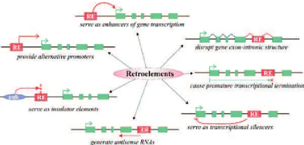
Repetitive DNA elements, also known as retroelements (REs), have been reported to exhibit various deleterious effects on genome structure and functioning ( Figure 2). Given that the most of the methylated cytosines in mammalian genome reside in repetitive elements, it has been proposed that DNA methylation evolved primarily to suppress the activity of transposable elements and to protect the host cell (Yoder et al. 1997). Supporting to this, hypomethylation of REs was demonstrated to be associated with
! (!
Figure 3; Different mechanisms of RE influence on gene transcription. Red boxes retroelements, green boxes gene exons, green arrow gene transcriptional start site, purple oval enhancer element. (Gogvadze and Buzdin 2009).
Currently, more than 175 families of SINEs have been described in a wide variety of eurkaryotes, whereas in mammals around 70 families are supposed to exist (Vassetzky and Kramerov 2013). SINEs are DNA sequences of typically 100 to 500 base pairs in length (Singer 1982). In contrast, LINEs are 5000 to 8000 base pairs length (Treangen and Salzberg 2011). They can occupy more than 10% of the genome under the existence of more than 1 million of copies (Ichiyanagi T. et al. 2013). SINEs are nonautonomous retrotransposons transcribed by the cellular RNA polymerase III (pol III) from an internal promoter, and their reverse transcription depends on the reverse transcriptase of partner LINEs (Gogvadze and Buzdin 2009). In contrast, LINEs can be autonomous, i.e., they encode the proteins necessary for their proliferation and transposition. They are transcribed by the cellular RNA polymerase II (Vassetzky and Kramerov 2013). Regions enriched in SINEs and LINEs are found in different parts of the genome, i.e., in gene-rich and gene-poor respectively (Lander et al. 2001).
Human Alu, and mouse B1 & B2 are among the most represented retrotransposons in mammalian genomes. These elements can amplify their copy numbers by transposition via transcription. The human Alu of approximately 290 bp in length carries up to 25 CpG sites both inside and outside of its Pol III promoter. These CpG sites are highly methylated in somatic tissues. This methylation results in inhibition of Pol III binding and therefore in prevention of Alu and tRNA transcription (Besser et al. 1990, Englander et al. 1993, Liu W. M. and Schmid 1993). The underlying mechanism is a hindering of the
! )! TFIIIC (a co-factor of PolIII) to the A- and B- boxes, and thus, Pol III cannot load to the promoter.
The mouse B1 (145bp) contains up to 8 CpG sites in its sequence and is highly methylated in somatic cells but weakly in germ cells and in preimplantation embryos. The level of methylation in B1 is negatively correlated with RNA abundance (Ichiyanagi K. et al. 2011). Considering that SINEs are closely located to transcription start sites (TSS) of PolII genes, it has been suggested that methylation of mouse B1 SINEs can harbor gene expression regulatory mechanism (Ichiyanagi et al. 2013*+!
!
1.4.2 Non-CpG methylation
Non-CpG methylation, i.e., methylation that occurs at cytosine in CHH or CHG trinucleotides, is known to be enriched in germ cells and ES cells (Lister et al. 2009, Tomizawa et al. 2011). Non-CpG methylation, are mainly found in SINEs, LINEs and LTRs. These retrotransposable elements constitute about half of mammalian genomes (Deininger and Batzer 2002) and have long been considered as selfish or junk DNA (Orgel and Crick 1980). Recent findings, however, suggest that some of these elements may exert gene regulation as well as chromatin structure organization functions.
Non-CpG methylation, in contrast to CpG methylation, is asymmetric, hence it is of interest to study the methylation patterns of DNA on both strands. This was recently done in H1 cells, (embryonic stem cells) by Guo et al., (2014), who observed that in introns, non-CpG sites are more heavily methylated on the antisense strand than those on the sense strand. Such skew of non-CpG methylation was not observed in exons but was more pronounced at intron boundaries (Guo et al. 2014).
1.5 CpG islands and Establishment of a permissive chromatin
Non-methylated CGIs are recognized by a family of zing finger (ZF-CXXC) proteins which contributes to the establishment of a permissive chromatin. The CXXC finger protein 1 (CFP1) binds to non-methylated CGIs and recruits the SET1 methyltransferase activity complex to these regions (Lee and Skalnik 2005). SET1
! ,! complex catalyzes the histone H3 lysine 4 tri-methylation (H3K4me3) (Thomson et al. 2010). H3K4me3 serves as a platform recruitment of PHD domain proteins (Plant Homeo Domain) such as TFIID, ING4 containing histone acetyltransferase (HAT) and NURF, which are known to be involved in the initiation of transcription (Blackledge and Klose 2011). Similar to CFP1, the lysine-specific demethylase 2A (KDM2A) specifically binds non-methylated CGI, but acts in a different way. KDM2A catalyzes the demethylation of H3K36me2 (Blackledge et al. 2010), a modification recognized by the histone deacetylase HDAC activity complex, known to exert gene transcription inhibitory activity. Thus, ZF-CXXC domain proteins cooperate at a single CGI to form a unique chromatin architecture (without H3K36me2 and enriched with H3K4me3), which provide ideal conditions to promote the initiation of transcription.
Non-methylated CGIs are also recognized by the TET1 protein, which unlike CFP1 and KDM2A, is involved in the recruitment of polycomb repressor complex family PRC1 and PRC2 (Wu H. et al. 2011). TET1 recruits PRC2, which catalyzes the histone H3 lysine 27 tri-methylation (H3K27me3). This modification is then recognized by PRC1, which in turn inhibits transcription elongation by promoting histone H2A ubiquitination and chromatin compaction (Eskeland et al. 2010, Stock et al. 2007). This mechanism of transcription repression is independent of CGI methylation.
The analysis of methylation distribution across the whole genome (or methylome) in somatic cells showed that, a fraction of CGI becomes methylated in a tissue-specific manner during developpement (Mohn et al. 2008, Weber et al. 2005). The majority of the CGI-promoters, that acquires methylation during cell differentiation, is already suppressed by the polycomb proteins within ESCs (Mohn et al. 2008), demonstrating that DNA methylation is not an initiator event in transcriptional repression, but rather acts as a heritable epigenetic marker which maintains repression in time
1.6 DNA methylation and regulation of transcription
DNA methylation is now well-known to play a role in regulating gene expression and chromatin structure beside its role in imprinting, X-chromosome inactivation and a possible role in silencing of repetitive DNA elements (Li E. 2002).
! "-! state (Bird A. P. and Wolffe 1999). In 2011, by associating RNA-sequencing approach with Whole-Genome-Bisulfite-Sequencing (WGBS), Bell and colleagues showed a genome-wide significant negative correlation between DNA methylation and gene expression level, and that the hypomethylated regions colocalize with the CGI-containing TSS of highly expressed genes (Bell et al. 2011). The effect of DNA methylation on gene control depends on the distribution of methylation (position of 5mC) on the transcriptional unit. For example, methylation at TSS blocks transcriptional initiation, whereas the methylation in gene body could stimulate transcription elongation, and may also have an impact on splicing (Jones P. A. 2012). At repeat regions such as centromeres, methylation has been suggested to suppress the expression of transposable elements, while at the same time allowing transcription of the host gene to run through them, and thus to have a role in genome stability (Li E. 2002, Yoder et al. 1997). Enhancers have low CpG content, however, methylation at these regions has been suggested to regulate their activity (Lister et al. 2009), whereas oxidation of 5mC to 5hmC leads to enhancer activation (Serandour et al. 2012).
There are two known mechanisms by which DNA methylation affects gene expression: First, modification of cytosine bases can interrupt the association of some DNA binding transcription factors (TFs) with their cognate DNA recognition sequences. Most of transcription factors in mammals have GC-rich binding sites, and binding of several of these factors is impeded or abolished by methylation of CpG (Bird A. P. and Wolffe 1999, Watt and Molloy 1988). Consistent with these observations, it has been found that CGIs that remain unmethylated in normal and in malignant cells contain specific sequence motifs that are identical to the consensus sequence for general TFs (Gebhard et al. 2010). The absence of methylation, at these transcription factor binding sites, is important for facilitating the binding of these factors to their target genes (Bell et al. 2011). Some examples validating this model include E2F1, CREB and c-myc (Campanero et al. 2000, Iguchi-Ariga and Schaffner 1989).
Second, proteins that recognize and bind methylated cytosine at CpG (methyl-CpG-binding proteins MBPs) recruit transcriptional co-repressor molecules to silence the transcription of genes (Nan et al. 1993). These recruited co-repressors modify the surrounding chromatin to be in a repressive state providing a link between DNA methylation and chromatin remodelling modification during the regulation of gene-expression (Cedar and Bergman 2009, Thomson et al. 2010, Zhang Y. et al. 1999). It has
! ""! also been shown that DNA methylation levels correlate negatively with the presence of histone marks that target active genes such as H3K27ac, H3K4me3 and H3K9ac which are positively correlated with transcription levels (Heintzman et al. 2009, Lister et al. 2009).
1.7 Methyl-CpG binding proteins as intermediates in transcriptional
repression
So far, three families of proteins are known to bind methylated DNA, methyl-CpG binding domain (MBD) protein family, Kaiso and Kaiso-like proteins and SET and Ring finger Associated (SRA) protein family.
At present, it is established that methyl-CpG binding proteins interact with histone deacetylases and histone methylase activities, that modify chromatin leading to the prevention of transcription initiation (Bird A. P. and Wolffe 1999). Each of these MBPs has a role in repressing the transcription in a DNA methylation-dependent manner. These MBPs will now be discussed.
1.7.1 Methyl-CpG binding domain proteins (MBDs)
The MBD family consists of the methyl-CpG-binding protein 2 (MeCP2), the methyl-CpG-binding-domain proteins MBD1, MBD2, MBD3 and MBD4 (Buck-Koehntop and Defossez 2013, Defossez and Stancheva 2011). These family members have been shown to play an intermediate role in gene repression.
1.7.1.a
MeCP2MeCP2 was the first discovered methyl-CpG binding protein member by Adrian Bird in 1992 (Lewis et al. 1992). This protein contain two fonctional domains, a N-terminal methyl-CpG binding domain (MBD) and a C-N-terminal transcriptional repression domain (TRD) (Nan et al. 1993). MeCP2 was co-purified with Sin3A/HDAC2 complex in mammalian cells, which interaction is essential for MeCP2-mediated transcription
! "#! repression (Jones P. L. et al. 1998). Nan and colleagues showed by co-immunoprecipitation that the interaction of MeCP2 with Sin3A/HDAC2 complex is occurring via the TRD domain, and they went to confirm the transcriptional repression fonction of this interaction by transfection, using a Gal4-targeted TRD. Furthermore, they could show that the HDAC inhibitor, trichostatin A (TSA) can partially modulate the induced repression of a reporter gene that contain GAL4 DNA-binding sites (Nan et al. 1998). MeCP2 interacts also with ATRX (Alpha Thalassemia/Mental Retardation Syndrome X-Linked) and Brahma (Brm1), which belong to SWI/SNF chromatin remodelling complex, this interaction results in transcription repression of the associated genes (Kokura et al. 2001). In spite of its role as a transcriptional repressor, MeCP2 was found to associate with the transcription activator CREB1 at the promoters of active genes. Moreover lack or overexpressed MeCP2 in the hypothalamus of mice leads to changes either positively or negatively of expression levels of thousands of genes, and most of these genes seemed to be activated by MeCP2 (Chahrour et al. 2008).
1.7.1.b
MBD1MBD1 protein acts as a histone deacetylation-independent transcriptional repressor (Ng et al. 2000). In contrast, MBD1 seems to act on histone H3 lysine 9 (H3K9) mrthylation as it is found to interact with two (H3K9) methylase activities, SETDB1 and SUV39H, as well as the heterochromatin protein HP1 (Sarraf and Stancheva 2004). It has been described that H3K9me2/3 recruits HP1!/", which then recruits the H3K9 methyltransferase SUV39H1, which in turn methylates more heavily H3K9. This propagation of the H3K9me2/3 mark and HP1, itself, then serve to bind additional proteins leading to heterochromatin formation conducting to gene silencing (Lachner et al. 2001). In addition the C-terminus of MBD1 binds AM/MCAF the co-factor of SETDB1 (SET domain, bifurcated 1). This binding leads to stimulate SETDB1 activity in order to allow more efficient di- and trimethylation of H3K9 (Fujita et al. 2003, Wang et al. 2003).
1.7.1.c
MBD2/MBD3MBD2 and MBD3 have almost similar structure and outside the MBD domain they have 77 % identity to each other. But in spite of this similarity, they do not have the same affinity toward the methylated DNA. In contrast to MBD2, MBD3 cannot specifically
! "$! bind methylated DNA because it has a phenylalanine instead of the conserved tyrosine at position 34 (Fraga et al. 2003). These two proteins co-purified with the protein complex NuRD (nucleosome remodelling and histone deacetylation), which contains chromatin remodelling factors such as ATPase Mi-2, HDAC1 and HDAC2 histone deacetylases (Zhang Y. et al. 1999).
1.7.1.d
MBD4MBD4 has a special structure within the MBD protein family. Beside its MBD domain, it exhibits a DNA glycosylase domain. The MBD of this protein has a high affenity to bind to symmetrically methylated DNA. It has also been shown, by GFP fusion MBD4, that this protein localised in a methylation-dependent manner to the peri-centromeric heterochromatin (Hendrich and Bird 1998). MBD4 has been described as an HDAC-dependant transcriptional repressor as it is able to recruit HDAC complexes to the hypermethylated promoters of the p16INK4a and hMLH1 genes, and the knockdown of
Mbd4 leads to an upregulation of these genes (Kondo et al. 2005).
Unlike to the other MBDs, MBD4 can bind to T:G mismatches (deamination product of methyl-Cytosine). Via its glycosylase domain, MBD4 can efficiently remove thymine from T:G mismatch to restore 5mC at the CpG sites (Hendrich et al. 1999). The ability of MBD4 to excise the T (deaminated m5C) from TpG to be replaced by a new C, which will be subsequently remethylated, could be a key mechanism in maintaining and/or repairing the methylation state of these regions. By this action, MBD4 participates to maintain the CGIs in a methylated state at the promoters of the methylation-dependent repressed genes. This role could reflect another indirect way by which MBD4 ensures the role as a methyl-CpG-dependent transcritional repressor.
1.7.2 Kaiso and Kaiso-like proteins
Similar to MBD family, this group of proteins specifically bind to methylated CpGs. Kaiso and Kaiso-like proteins have no structure similarity with MBDs and consist of an N-terminal POZ domain and C-terminal zinc finger domain. They use the zinc finger domain to bind the methylated CpGs. Via the zinc finger domain, Kaiso binds also to DNA regions that lack CpG dinucleotides with a mild affinity (Daniel et al. 2002).
Kaiso family proteins repress transcription in HDAC-dependent pathway. Indeed, Kaiso was shown to co-purify with NCoR (nuclear receptor co-repressor), from HeLa
! "%! cell nuclear extracts, containing histone deacetylase HDAC3. Furthermore, it has been reported that the association of Kaiso with NCoR is required for silencing of methylated
MTA2 (Metastasis-associated protein) promoter (Yoon et al. 2003). Moreover, it has been
shown that the depletion of Kaiso protein leads to derepression of methylated genes in Xenopus embryos (Ruzov et al. 2004). Another member of Kaiso-like proteins, ZBTB38 is able to interact with histone deacetylases1, 3 (HDAC1, HDAC3) as well as the co-repressor CtBP (C-terminal binding protein) (Sasai et al. 2005).
1.7.3 SRA domain proteins
The SRA (SET- and RING-associated domains) family of proteins bind to methylated DNA via SRA domain. The founder member of this family is UHRF1, also known as ICBP90 in human and Np95 in mouse (Bronner et al. 2007). In human another UHRF1 paralog UHRF2 is defined to have similar domains to UHRF1. UHRF1 and 2 can bind to methylated DNA. UHRF1 has higher affinity towards to hemi-methylated than binding to fully-hemi-methylated DNA (Avvakumov et al. 2008). This protein interacts with and recruit DNMT1 to methylate the newly synthesized DNA strand during the replication (Bronner et al. 2010). UHRF1 recruits G9a (Histone H3 lysine 9 methyltransferase) and thanks to this function is considered as a transcriptional repressor (Kim J. K. et al. 2009a).
1.8 DNA demethylation
Although it is considered that 5mC is a stable epigenetic mark and strongly maintained, DNA methylation patterns are dynamic during development and during pathological situations such as cancer. Changes in the methylation patterns happens both at the global and local levels. We will now review the proposed different pathways of DNA demethylation, which can result from either passively at the new DNA strand after replication or actively, by a replication-independent process.
! "&!
1.8.1 Passive DNA demethylation
During DNA replication, the symmetrical CpG methylation pattern is restored by the maintenance proteins UHRF1 through the methylation of the unmodified cytosine in the nascent DNA strand. Therefore, the passive loss or dilution of 5mC can be achieved in the absence of functional DNMT1/UHRF1 through successive cycles of DNA replication (Figure 4). One example of such a mechanism of demethylation is the global erasure of 5mC in the maternal genome during mouse preimplantation development. The first S phase in the newly formed zygote is initiated 8 to10 hours after fertilization, this give rise to the first opportunity for passive demethylation to act as a demethylation mechanism (Rougier et al. 1998). Supporting to this mechanism, it has been shown that in this stage of development, the abundance of DNMT1 is largely reduced by the active exclusion of DNMT1 protein from the nucleus to the subcortical region (Carlson et al. 1992, Howell et al. 2001), where it resides throughout most of the period of preimplantation, thus supporting a step-wise loss of DNA methylation from the maternal genome (Ratnam et al. 2002). Furthermore, alteration in UHRF1/DNMT1 tandem, via UHRF1 over-expression or through disrupted interaction between UHRF1 and DNMT1, induces global genome-wide demethylation, which is a hallmark of cancer cells (Mudbhary et al. 2014, Pacaud et al. 2014).
Figure 4; Passive DNA demethylation. Lack of methylation maintenance during DNA replication leads to 5mC dilution, eventually leading to fully unmethylated DNA. (Hill et al., 2014, Genomics).
1.8.2 Active DNA demethylation
Active DNA demethylation refers to an enzymatic process that results in the changing or removal of the methyl group from 5mC. After fertilization, the paternal and
! "'! maternal genomes show a global loss of the methylation pattern in mammalian fertilized oocytes (Mayer et al. 2000, Oswald et al. 2000). The excessive wave of demethylation observed on the paternal genetic material starts 4-8 hours after fertilization (Mayer et al. 2000) (Figure 5) gave rise to the concept of active DNA demethylation after it has long been controversial. This global fast reduction of DNA methylation levels takes place before the first round of DNA replication begins (Oswald et al. 2000). Thus, it is unlikely to be achieved by the passive dilution pathway. Moreover, when zygotes were treated with aphidicolin, a DNA replication inhibitor, paternal genome demethylation was still detected (Kishigami et al. 2006). Active DNA demethylation has also been reported in somatic cells, which happens at specific genomic loci in response to certain signals. One example, within 20 minutes of stimulation, activated T lymphocytes undergo active demethylation at the interleukin-2 promoter-enhancer region in the absence of DNA replication (Bruniquel and Schwartz 2003). The locus-specific demethylation has also been observed at the promoter of brain-derived neurotrophic factor (BDNF), which is, when methylated, recognized and bound by the MeCP2. Following the depolarization with KCl, BDNF is upregulated, coinciding with the release of MeCP2 and demethylation of the promoter (Martinowich et al. 2003).
The local active DNA demethylation has also been reported to take place during nuclear hormone-regulated gene activation. For example, the pS2 (also known as TFF1) promoter exhibits periodic methylation and demethylation that coincides with cyclical binding of oestrogen receptor-! (ER!) and expression of pS2 (Metivier et al. 2008).
Figure 5. DNA methylation changes during developmental
epigenetic reprogramming.
Following sex-determination,
new DNA-methylation
landscapes are established in germ-cell precursors in an asymmetrical fashion in male
and female embryos. In the
male embryo (blue line), de novo methylation takes place before meiosis in mitotically arrested cells. In the female embryo (red line), primary oocytes enter meiosis and arrest in prophase-I (diplotene stage); DNA methylation is established after birth during the follicular/oocyte growth phase. Following fertilisation, a new wave of DNA demethylation takes place that is distinct on the parental genomes. In the zygote, DNA methylation of the paternal genome is rapidly erased by an active mechanism (blue line). Demethylation of the maternal genome is slower (red line) and is dependent on DNA replication (passive demethylation). Concomitant with blastocyst implantation and cell-lineage determination, new methylation landscapes become established, associated with cellular differentiation. (Smallwood and Kelsey, Trends in Genetics, 2012).
! "(!
1.8.3 Mechanisms of active DNA demethylation
Regarding the importance of DNA methylation in diverse biological processes, beside the observations of active DNA demethylation in embryonic development and somatic cells, extensive efforts have been made to identify a DNA demethylase, and/or to understand the mechanisms by which this process is achieved.
So far, various mechanisms have been proposed as possible pathways by which active DNA demethylation can occur, including enzymatic removal of the methyl group of 5mC, radical SAM mechanism, base excision repair (BER) through direct excision of 5mC, deamination of 5mC to T, followed by BER of the T:G mismatch, nucleotide excision repair (NER) and the oxidation of 5mC to hydroxymethylcytosine (5hmC), 5-formylcytosine (5fC) and 5-carboxylcytosine (5caC) (Franchini et al. 2012, Li C. J. 2013, Wu H. and Zhang 2014, Wu S. C. and Zhang 2010). We will now review hereafter the different putative mechanisms underlying “DNA demethylation”.
1.8.3.1
Enzymatic removal of the methyl group of 5mCInitially, the search for DNA demethylation mechanisms focused on the identification of an enzymatic activity that directly removes the methyl group from 5!methylcytosine, which would be, if occurs, the most straightforward way to achieve DNA demethylation. However, this direct pathway needs a thermodynamically unfavorable reaction to break the carbon!carbon bond, which makes this pathway unlikely to occur in living cells. Nevertheless, a study proposed earlier, that the direct removal of methyl group could be catalyzed by the methyl-CpG-binding domain protein 2 (MBD2) leading to the release of methanol (Bhattacharya et al. 1999). Since then, no other laboratory could reproduce this enzymatic activity of MBD2, leaving this mechanism largely controversial. Moreover, other studies have shown that MBD2 can stably bind methylated DNA (Hendrich and Bird 1998, Ng et al. 1999) which give rise to the question of how MBD2 can bind to 5mC if it can efficiently remove the methyl group. Moreover, further study has shown that MBD2 null mice are viable and also exhibited normal methylation patterns (Hendrich et al. 2001). Importantly, the paternal pronucleus of MBD2-null zygotes still exhibits normal demethylation activity (Santos et al. 2002). All these findings have raised serious doubts on the capacity of MBD2 to serve as a DNA demethylase and is now completely given up.
! ")!
1.8.3.2
Radical SAM mechanismEfforts to define a protein(s) responsible for the observed paternal genome demethylation in zygotes led to propose a possible role of elongator complex protein 3 (ElP3) in DNA demethylation of embryonic development, and that Fe–S radical SAM domain of this protein is required for the demethylation process (Okada et al. 2010). ElP3 is a member of the core elongator complex (ElP1–ElP3), which associates with an other subcomplex (ElP4–ElP6) to form the holo-elongator complex (Hawkes et al. 2002). Knockdown of the ElP1 and ElP4 components impaired paternal genome demethylation, suggesting that it is likely that the entire elongator complex may be involved in the demethylation process (Okada et al. 2010).
Although the proposed role of SAM domain could provide a clue for an enzymatic mechanism of ElP3, other studies suggest that the Cys-rich domain of ElP3 is required for the integrity of the elongator complex (Greenwood et al. 2009) raising the possibility that the Fe–S radical SAM motif may has a structural rather than an enzymatic role. In spite of this proposed role, direct biochemical evidence of the demethylation activity of ELP or the elongator complex and genetic evidence using ElP3-null oocytes are still lacking.
1.8.3.3
Nucleotide Excision Repair (NER) to erase 5mCTheoretically, the erasure of 5mC is possible by the excision repair of short genomic regions that contain methylated cytosine nucleotides. Via the NER pathway, cells can repair bulky DNA lesions formed by exposure to radiation or chemicals. Once the bulky DNA lesion is recognized, specific enzymatic activities introduce dual incisions flanking the damage region resulting in a single-stranded gap (usually 24–32 nucleotides). The resulted gap is then filled in by DNA repair polymerases and ligases. Supporting to this way, it has been reported that the Gadd45 (growth arrest and DNA-damage-inducible protein 45), stimulates active DNA demethylation via NER (Barreto et al. 2007). However, the role of this protein family in DNA demethylation remains uncertain as Gadd45a- and Gadd45b null mice are quite normal and have no global alteration in DNA methylation levels (Engel et al. 2009, Ma et al. 2009).
! ",!
1.8.3.4
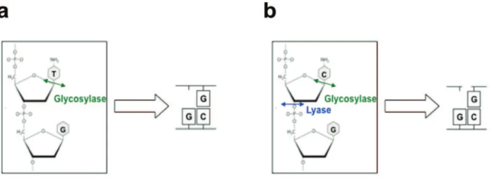
Direct excision of 5mC followed by Base excision repair (BER)One hypothesis, proposed for some time, is that DNA demethylation can be achieved through the base excision repair (BER) pathway. This mechanism of repair involves a DNA glycosylase that cleaves the N-glycosidic bond between the 5-mC base and the deoxyribose to remove the target base resulting in an abasic (apurinic and apyrimidinic: AP) site. The DNA backbone is subsequently nicked by an AP lyase activity to generate a 5# phosphomonoester and a 3# sugar phosphate residue. An AP endonuclease then removes the 3# sugar group leaving a single nucleotide gap that is ultimately filled in by DNA repair polymerases and ligases (Sancar et al. 2004). These steps lead to remove 5mC without previous modification of this base. This way of active demethylation has been reported in flowering plants (e.g., Arabidopsis thaliana), where the reaction is mediated by the Demeter (DME)/repressor of silencing 1 (ROS1) family of DNA glycosylases and base excision repair (BER) machinery (Gehring et al. 2006, Gong et al. 2002). These findings led to think that such a mechanism could also occur in mammalian cells. However, mammalian orthologs of DME/ROS1 enzymes have not been identified. Therefore, mammalian cells may achieve active DNA demethylation using different mechanisms rather than directly removing of 5mC.
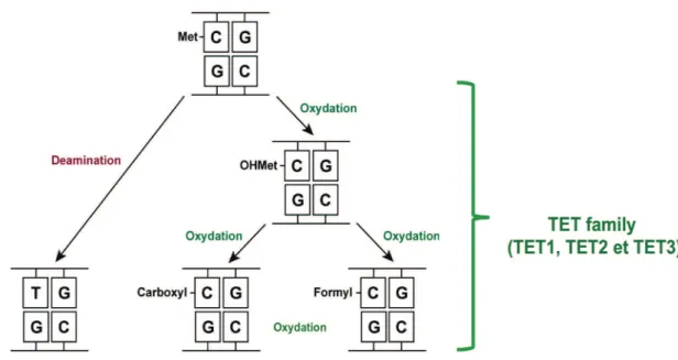
Evidences demonstrating that mammalian cells first tend to modify the 5mC followed by the repair of this modified base. Indeed, two pathways has been reported to modify 5mC in mammalian cells, first; hydrolytic deamination of 5mC to thymine (T) leading to form a G:T mismatch, and second; oxidation of 5mC to 5hmC, 5fC and 5caC by the Tet family (Figure 6). Here we review these two pathways;
! #-!
Figure 6. 5meC modifying pathways. Deamination of 5mC to thymine (T) leading to form a G:T mismatch (Left part), and oxidation of 5mC by the Tet family leads to 5hmC, 5fC and 5caC (Right part).
1.8.3.5
Hydrolytic deamination of 5mC followed by BERThe 5mC undergoes spontaneous hydrolytic deamination, which subsequently generates thymine (Gehring et al. 2006). Unmethylated cytosine could also be deaminated but with a lower rate resulting in the formation of uracil. Both deaminase activities towards C or mC are considered mutagenic, because if left unrepaired, the resulting G:U and G:T mispairs will give rise to a C to T transitions upon replication.
It has been suggested that some members of the vertebrate-specific Activation-induced Cytidine Deaminase (AID)/Apolipoprotein B mRNA Editing enzyme, Catalytic polypeptide (APOBEC) family such as AID (Morgan H. D. et al. 2004, Popp et al. 2010), APOBEC1, APOBEC2 (Guo et al. 2011, Morgan H. D. et al. 2004), could play a key role in active DNA demethylation. AID and APOBEC proteins are zinc-dependent cytidine deaminases acting on single-stranded polynucleotides and deaminate cytosines in different contexts (Chelico et al. 2006). Among the AID/APOBEC family, Aid and APOBEC1 are expressed in mammalian oocytes and embryos, which points to a possible role in global DNA demethylation occurs in these stages (Morgan H. D. et al. 2004). Moreover, it has been reported that locus-specific and global demethylation in zebrafish embryos is mediated by the AID/Apobec family of deaminases and the DNA glycosylase MBD4 (Rai et al. 2008). Overexpression of AID and MBD4 together in zebrafish
! #"! embryos, causes demethylation of the bulk genome and injected methylated DNA fragments. Importantly, overexpression of the glycosylase alone, does not lead to such changes demonstrating a deaminase activity of AID towards the 5mC (Rai et al. 2008). Moreover, a bisulphite sequencing study indicated that DNA methylation levels of male and female PGCs derived from AID-null embryos increased about 4% (from 18% to 22%) and 13% (from 7% to 20%), respectively, when compared to their wildtype counterpart (Popp et al. 2010), suggesting that AID may contribute to primordial germ cells (PGC) demethylation. However, DNA methylation levels in AID-null PGCs (~20%) are still relatively low compared with ES or somatic cells (70–80%) and significant demethylation still occurs in the absence of AID, indicating that other factors could be responsible for PGC demethylation.
In addition to AID and APOBEC, other studies showed that DNMTs are implicated in 5mC deamination, even though they are commonly known for their ability to catalyse DNA methylation. The starting evidence, indicating their involvement in the deamination process, initially came from studies in bacteria where the methyltransferases M. EcoRII (Wyszynski et al. 1994) and M. HpaII (Zingg et al. 1996) were shown to possess deaminase activities. Consistent with studies on bacteria, the mammalian counterparts, DNMT3A and DNMT3B, have been shown to possess deaminase activity toward 5mC in
vitro, but this proposed deaminase activity of DNMT1 can only occur under conditions
where SAM concentrations are very low or absent (Metivier et al. 2008). However, given that SAM is present at relatively high levels in all cell types, the physiological relevance of this reaction remains uncertain.
In support of 5mC deamination/BER mechanism, inhibitors of poly-ADP-ribose polymerase 1 (PARP1), a member of the BER pathway proteins, and of apurinic/apyrimidinic endonuclease 1 (APE1), an essential downstream enzyme required to generate an abasic site, were found to alter paternal-specific DNA methylation (Hajkova et al. 2010).
Altogether, these observations lead to accept that DNA demethylation can be achieved by deamination of 5mC followed by BER to replace the mismatched T with unmethylated C.
! ##!
1.8.3.6
Oxidative modification of 5mCThe searching for enzymatic activity modifying 5mC through oxidation was prompted by the discovery of the biosynthesis of ‘‘base J’’ (b-D-glucosyl-hydroxymethyluracil), a modified base present in the genome of the parasite Trypanosome brucei. Biosynthesis of Base J is achieved by a two-step process. First, thymine (T) is oxidized to form 5-hydroxymethyluracil (5hmU) by J-binding protein (JBP) 1 and 2. These two proteins are members of the Fe (II)/ ! ketoglutarate (!-KG)-dependent dioxygenase family (Loenarz and Schofield 2011). Then, a glycosyltransferase completes the synthesis of base J by adding glucose groups to 5hmU (Borst and Sabatini 2008). It has also been shown that further oxidation of 5hmU leads to form 5-formyluracil and 5-carboxyluracil. This later base then undergoes a decarboxylation process by isoorotate decarboxylase completing a cycle from T to U, with a putative similar mechanistic to 5mC demethylation (Wu S. C. and Zhang 2010).
Efforts to find mammalian homologues with similarity to the dioxygenase domains of JBP proteins led to the identification of the ten-eleven translocation family of proteins (Tahiliani et al. 2009). TET1 was first identified in Purkinje cells of the brain, and later in mouse embryonic stem cells (Kriaucionis and Heintz 2009, Tahiliani et al. 2009). Since then, two other TET family members, TET2 and TET3, harbouring the dioxygenase motif, have been identified (Ito et al. 2010).
All TET proteins contain a C-terminal catalytic domain that includes a Cys-rich insert and a large double-stranded b-helix (DSBH) domain. TET1 and TET3 proteins also contain a N-terminal CXXC domain (Kohli and Zhang 2013) (Figure 7 a). Concerning the oxidation activity toward 5mC, overexpression of TET1 in cultured cells results in reduction of genomic 5mC level, and recombinant TET1 proteins can oxidize 5mC in
vitro resulting in 5hmC. Similar to that achieved by JBP1/2, this reaction, requires Fe(II)
and alpha-ketoglutarate binding to complete the oxidation of 5mC to 5hmC (Tahiliani et al. 2009) (Figure 7 b). The same activity was later demonstrated with all TET (TET1-3) proteins in mouse (Ito et al. 2010).
! #$!
Figure 7; Schematic and Catalytic reaction of Tet enzymes. a, Schematic of mouse Tet
enzymes, showing the double-stranded !-helix (DS!H) fold core oxygenase domain, a
preceding cysteine(Cys)-rich domain and a CXXC domain in Tet1 and Tet3. b, Catalytic mechanism for generation of 5hmC by Tet enzymes. An active site Fe(II) (left) is bound by conserved His-His-Asp residues in Tet and coordinates water and "-ketoglutarate ("-KG). A two-electron oxidation of "-KG by molecular oxygen yields CO2 and enzyme-bound succinate, and results in a high-valent Fe(IV)-oxo intermediate (Costello et al.). The intermediate reacts with 5mC to yield 5hmC, with a net oxidative transfer of the single oxygen atom to the substrate, resulting in regeneration of the Fe(II) species. (Kohli and Zhang., 2013).
Ito and colleagues have later reported that TET proteins are capable of further oxidizing 5hmC to 5fC and 5caC (Ito et al. 2011) (Figure 8). Indeed, the important observation that the loss of paternal DNA methylation coincides with a highly increase in 5hmC levels (Inoue and Zhang 2011, Iqbal et al. 2011, Wossidlo et al. 2011) and 5fC/5caC (Inoue and Zhang 2011) strengthens the proposed oxidative demethylation mechanism as a main pathway in the paternal genome demethylation after fertilization. Within the TET family, TET3 is highly enriched (30-fold) in the zygote comparing to TET1 and TET2, which makes it as the most likely candidate responsible for the
! #%! oxidation of 5mC in the paternal genome during this development period (Iqbal et al. 2011). This role was later confirmed by small interfering RNA (siRNA) approach against TET3. Targeting TET3 lead to abrogate 5hmC and to an important increase of 5mC levels (Wossidlo et al. 2011). The oxidative demethylation pathway could further include a passive dilution of the oxidation products of 5mC (5hmC, 5fC or 5caC) (in a replication-dependent manner), or another enzymatic activity to remove the final oxidation products (Shen et al. 2013).
Figure 8; TET-induced 5meC oxidation. TET proteins are able to oxidize 5mC to 5hmC and further to 5fC and 5caC.
1.8.3.6.a
Passive dilution of oxidized 5mCSimilar to 5mC, the oxidized products (5hmC, 5fC and 5caC) could be diluted by a replication-dependent manner (Figure 9a). The dilution of the oxidized 5mC may be effective even in the presence of a functional methylation maintenance machinery (DNMT1/UHRF1) because the affinity of this machinery towards the oxidized bases is less than towards 5mC. It has been established that DNMT1 is significantly less efficient (10-60 fold) in methylating hemihydroxi-methylated (hCG:GC) than hemimethylated (mCG:GC) sites in vitro (Hashimoto et al. 2012). The decreased efficiency of DNMT1 to methylate the newly strand could also occur, but it is not yet proven, in the presence of hemiformylcytosine (fCG:GC) or hemicarboxylcytosine (caCG:GC).
In summary, TET proteins may initiate a two-step demethylation process in proliferating cells that involves initial active modification of 5mC through oxidation and subsequent replication-dependent (passive) dilution of 5hmC and potentially 5fC/ 5caC (Wu H. and Zhang 2014).