HAL Id: tel-01531928
https://tel.archives-ouvertes.fr/tel-01531928
Submitted on 2 Jun 2017HAL is a multi-disciplinary open access archive for the deposit and dissemination of sci-entific research documents, whether they are pub-lished or not. The documents may come from teaching and research institutions in France or abroad, or from public or private research centers.
L’archive ouverte pluridisciplinaire HAL, est destinée au dépôt et à la diffusion de documents scientifiques de niveau recherche, publiés ou non, émanant des établissements d’enseignement et de recherche français ou étrangers, des laboratoires publics ou privés.
Functional characterisation of the mechano-electrical
transduction complex of the auditory hair cells
Ménélik Labbe
To cite this version:
Ménélik Labbe. Functional characterisation of the mechano-electrical transduction complex of the auditory hair cells. Neurons and Cognition [q-bio.NC]. Université Pierre et Marie Curie - Paris VI, 2016. English. �NNT : 2016PA066543�. �tel-01531928�
Université Pierre et Marie Curie
Ecole doctorale Complexité du Vivant
Unité de Génétique et Physiologie de l’Audition
Caractérisation fonctionnelle du complexe de transduction
mécano-électrique des cellules ciliées du système auditif
Functional characterisation of the mechano-electrical transduction
complex of the auditory hair cells
Par Ménélik LABBÉ
Thèse de doctorat de Neurosciences
Dirigée par le Professeur Christine PETIT
Présentée et soutenue publiquement le 12 avril 2016
Devant un jury composé de :
Serge PICAUD Professeur Président
Fabrice GIRAUDET Maître de conférences Rapporteur
Christophe VINCENT Professeur Rapporteur
Acknowledgements
Firstly, I would like to thank Christine Petit for giving me the opportunity to undertake my PhD in her lab, and by the same token, allowing me to meet and discuss with a number of passionate scientists on campus at the Institut Pasteur in Paris. Secondly, I would like to express my sincere gratitude and appreciation to my supervisor, Nicolas Michalski, on whom I have been able to count for any matter, whether it be scientific or personal.
I would also like to thank Serge Picaud for accepting to be the president of the jury of my thesis committee, as well as Fabrice Giraudet and Christophe Vincent for accepting to be the appo teu s of my thesis committee.
Contents
INTRODUCTION
I. The human ear ………. 1
I.1. Anatomy and function of the mammalian inner ear ………. 1
I.2. Anatomy and physiology of the vestibule ………. 2
I.3. Anatomy and physiology of the cochlea………. 3
I.3.A. The basilar membrane ………. 5
I.3.B. The organ of Corti and the hair cells ……….. 6
I.3.C. The hair bundle and mechano-electrical transduction (MET) ……….. 8
I.3.C.1. Morphological aspects of the hair bundle ……… 8
I.3.C.2. The MET channel ………15
I.3.C.3. Biophysical properties of the MET channel ……….21
I.3.C.4. Adaptation ………..24
II. Usher syndrome ………..29
II.1. Clinical aspects of Usher syndrome ……….29
II.2. Molecular aspects of Usher syndrome ………..30
II.2.A. Usher syndrome type 1 ………..30
II.2.B. Usher syndrome type 2 ………..35
II.2.C. Usher syndrome type 3 ………..37
II.3. Usher syndrome type 2 and the ankle link (AL) complex ……….38
II.3.A. Whirlin ………38
II.3.B. Usherin ………..43
II.3.C. Adgrv1 ………46
II.3.D. Pdzd7 ………50
MATERIALS & METHODS
Immunofluorescence studies ………..57
RT-PCR and transcripts analysis ……….58
Scanning electron microscopy……….58
Electrophysiological recordings ……….59
In vivo recordings and data analysis………...60
Statistical tests ………60
RESULTS Usherin project ……….67
Sans rescue project ………...85
Cib2 project ……….93
Protocadherin-15 project ………..101
DISCUSSION Sound wave propagation along the tectorial membrane in Ush2aΔTM/ΔTM mice? ………..108
What causes the delay in onset of auditory defects in Ush2aΔTM/ΔTM mice? ……….111
Does usherin form a new type of basal lateral links, distinct from the ankle links ………112
Gene therapy as a cure for Usher syndrome? ……….113
Determining a robust genotype/phenotype correlation is essential for better diagnosis of USH patients ………114
Perspectives ……….117
Conclusion ……….118
List of figures
Figure 1. The human ear ... 2
Figure 2. Cross-section of mammalian cochlea ... 4
Figure 3. Ionic concentrations in the perilymph and endolymph ... 5
Figure 4. Scanning electron micrograph illustrating the 3 rows of OHCs, and the single row of IHCs in the organ of Corti ... 7
Figure 5. Schematic representation of sequential stages of hair bundle development ... 8
Figure 6. Schema and immunofluorescent image illustrating the crescent-shape disposition of PCP proteins in wild-type mice ... 9
Figure 7. Scanning electron micrograph of a normal vestibular hair bundle ... 10
Figure 8. The different interstereociliary links during hair bundle development ... 13
Figure 9. Schematic representation and scanning electron micrograph of the tip link ... 14
Figure 10. Scanning electron micrograph of the ankle links ... 15
Figure 11. Localisation of mechano-electrical transduction channel by extracellular potential measurements ... 16
Figure 12. Schematic representation of the gating spring model ... 18
Figure 13. Calcium entry through the mechano-electrical transduction channel after hair bundle deflection ... 19
Figure 14. Calcium entry into the small- and middle-row stereocilia of the hair bundle... 20
Figure 15. Localisation of mechano-electrical transduction channel by high-speed calcium imaging ... 20
Figure 16. Examples of single-channel current recordings in turtle hair cells ... 23
Figure 17. Mechano-electrical transduction current amplitude and characteristic parameters of adaptation ... 25
Figure 18. The current-displacement (I(X)) curve ... 25
Figure 19. Illustration of the calcium-dependent adaptative shift ... 26
Figure 20. USH1 proteins ... 34
Figure 21. USH1 proteins interacting and forming the MET complex ... 34
Figure 22. Schematic representation of USH2 proteins ... 37
Figure 23. Schematic represention of the USH3 protein clarin ... 37
Figure 24. Scanning electron micrographs of P35 +/wi and wi/wi IHCs ... 39
Figure 25. IHC stereocilia length along the tonotopic axis ... 40
Figure 26. Whirlin distribution in wild-type mouse IHCs and OHCs at P4 ... 42
Figure 27. Usherin distribution in murine cochlear and vestibular hair cells ... 45
Figure 28. Domain architecture and disease-related mutations of Vlgr1 protein ... 48
Figure 29. USH2 protein distribution in Adgrv1+/+ and Adgrv1-/- mice ... 49
Figure 30. Scanning electron micrographs of Adgrv1+/- and Adgrv1-/- cochlear hair bundles . 49 Figure 31. Longest alternatively spliced isoform of PDZD7 ... 50
Figure 32. Pdzd7 distribution in P2 rat cochlear and vestibular hair cells ... 51
Figure 34. Mechano-electrical transduction current recordings, by mechanical stimulation of
the hair bundle in the excitatory direction ... 73
Figure 35. Hearing impairment at low and mid-high sound frequencies in Ush2aΔTM/ΔTM mice ... 75
Figure 36. Abnormally efficient masking of mid-high frequency sounds by lower frequency
sounds in a subset of Ush2aΔTM/ΔTM mice ... 77
Figure 37. Scanning electron micrographs (SEM) of cochlear hair bundles in Ush2aΔTM/ΔTM
and Ush2aΔTM/ΔTM P5 mice ... 80
Figure 38. Hair bundle distribution of adgrv1, pdzd7 and whirlin in the cochlear hair bundles
of Ush2a+/ΔTM and Ush2aΔTM/ΔTM P6 mice ... 81
Figure 39. Mechano-electrical transduction currents and I(X) curves in injected and
non-injected Ush1g+/- and Ush1g-/- IHCs... 88
Figure 40. Mechano-electrical transduction currents and I(X) curves in injected and
non-injected Ush1g+/- and Ush1g-/- OHCs ... 89
Figure 41. Auditory brainstem response recordings in 1-month old injected Ush1g-/- and
Ush1g+/- mice ... 90
Figure 42. Mechano-electrical transduction current recordings, by mechanical stimulation of
OHC hair bundles in the excitatory direction ... 95
Figure 43. Mechano-electrical transduction current recordings, by mechanical stimulation of
UHC hair bundles in the excitatory direction ... 95
Figure 44. Scanning electron micrographs of P2, P7, and P18 Cib2-/- cochlear hair bundles .. 97
Figure 45. Schematic representation of pcdh15 isoforms ... 104 Figure 46. Averaged peak amplitudes of mechano-electrical transduction currents and I(X)
curves in Pcdh15+/Δe and Pcdh15Δe /Δe OHCs ... 104
Figure 47. OHC stereocilia imprints on the lower surface of the tectorial membrane in
List of abbreviations
ABR auditory brainstem response
Adgrv1 adhesion G protein-coupled receptor v1
BAPTA 1,2-bis(o-aminophenoxy)ethane-N,N,N’,N’-tetraacetic acid
Cdh23 cadherin-23
CIB2 calcium and integrin binding family member-2
DFNB autosomal-recessive for of deafness
DPOAE distorsion product otoacoustic emission
IHC inner hair cell
MET mechano-electrical transduction
OHC outer hair cell
PBM PDZ binding motif
Pcdh15 protocadherin-15
PCP planar cell polarity
PDZ postsynaptic density 95/disc large/zonula occludens-1 domain
PDZD7 PDZ domain containing 7
SEM scanning electron microscopy
SPL sound pressure level
TM transmembrane domain
USH Usher syndrome
INTRODUCTION
1
I.
The human ear
I.1
Anatomy and function of the mammalian inner ear
The mammalian ear is composed of three distinct parts: the outer ear, the middle ear and the inner ear (Figure 1. The e te al ea ’s fu tio is to aptu e a d ha el sou d waves along the external auditory meatus. The waves then propagate through the middle ear, an air-filled cavity delimited by two membranes: the tympanum (or eardrum) and the oval window. Upon sound stimulation, the tympanum vibrates, transforming the airborne acoustic waves into solid mechanical waves, capable of propagating through the three ossicles located in the middle ear: the malleus (or hammer), the incus (or anvil) and the stapes (or stirrup). Through the lever action of the ossicles and a reduction in the area of force distribution, the sound wave is amplified. Amplification at this level allows the sound wave to cross the oval window and into inner ear. Because the inner ear is a fluid-filled cavity, if the sound wave were to arrive at the oval window, an air/liquid interface, without amplification, it would almost entirely be reflected back towards where it came from. The role of the middle ear is thus to act as an impedance adaptor, by compensating the loss of energy resulting from the changes in medium that occur between the external and inner ear.
While the external and middle ears are solely dedicated to auditory functions, the inner ear however is involved in auditory as well as equilibration functions. It is composed of the vestibule, the organ responsible for equilibration, and the cochlea, the organ responsible for hearing (Figure 1).
2
Figure 1. The human ear (adapted from Kandel et al., 2000)
I.2.
Anatomy and physiology of the vestibule
The vestibule is the organ responsible for equilibration. Just like the cochlea, the vestibule uses highly specialised sensory cells, called hair cells, to detect the linear accelerations and rotational movements of the head. Hair cells are equipped at their apex with a mechanical antenna, called the hair bundle, where the transformation from mechanical to chemical signal is initiated. The information is sent to the central nervous system which coordinates head and eye movements, as well as body posture, to maintain balance.
In the vestibule, hair cells are grouped into 5 distinct neuroepitheliums: the saccule and the utricule, that detect linear accelerations, and the three ampullæ, that detect the angular accelerations of the head. The utricule is responsible for the detection of linear
3
accelerations on the horizontal plane, while the saccule is responsible for detecting accelerations on the vertical plane. The ampullæ detect angular movements of the head in the three possible axes: from left to right, up and down and the tilting of the head. They are situated in the three semi-circular canals and are orthogonal to one another.
Each one of these neuroepitheliums contains a sensory epithelium, called the macula, which is composed of sensory hair cells surrounded by supporting cells. Sensory hair cells are capable of producing electrical signals, although they are bereft of any axon or dendrites. The hair bundles of the hair cells are covered by the otoconial membrane, a gelatinous structure, formed by intertwined filaments organised in a mesh-like pattern, and the otoconial membrane is covered by calcite crystals, called otoconies, which play an important role in the sensitivity of the maculæ. When acceleration occurs, a force is exerted on the otoconies, and because of their inertia, this leads to a synchronised movement of the hair bundles. Depending on their orientation, certain hair cells are depolarised or hyperpolarised and the resulting information is then transmitted to the brain through the vestibular nerves.
I.3.
Anatomy and physiology of the cochlea
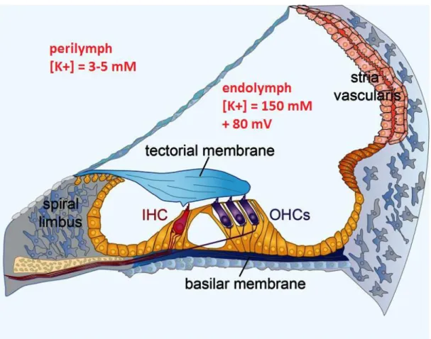
The cochlea is a bony tube coiled around a conical bony core called the modiolus. A human cochlea consists of slightly less than three coils whereas in mice it is 1.75 coils long. The cochlea contains three fluid-filled compartments: the scala vestibuli, the scala media (or cochlear duct) and the scala tympani (Figure 2). The scala vestibuli and scala tympani are filled with a classic extracellular medium, rich in Na+ ions (140 mM) and poor in K+ ions (3- 5
4
mM), called the perilymph. The scala media, however, is filled with a very specific liquid called the endolymph. This liquid is abnormally rich in K+ ions (150 mM) and poor in Na+ ions (1mM). These unexpected extracellular concentrations are possible thanks to the stria
vascularis which lines the external wall of the scala media and reabsorbs the Na+ and secretes K+ against their gradients (Figure 3). Because of the differences in ionic concentrations and the electrical isolation of the scala media, the endolymph has an electric potential of approximately +80 mV with respect to the perilymph. This electric potential is called the endocochlear potential and is crucial to the mechano-electrical transduction process which I will describe a little further in the manuscript.
Figure 2. Cross-section of mammalian cochlea (adapted from Kandel et al., 2000)
5
Figure 3. Ionic concentrations in the perilymph and endolymph (figure adapted from Michalski & Petit, 2015)
I.3.A. The basilar membrane
The scala vestibuli and the scala media are separated by a thin membrane, called the Reissner membrane, whereas the scala media and the scala tympani are separated by the basilar membrane, upon which the auditory neurosensory epithelium, the organ of Corti, rests. The basilar membrane possesses very particular mechanical properties which are responsible for the frequency discrimination capabilities characteristic of the cochlea. The work of von Bekesy (Von Bekesy, 1954) has shown that a pure tone does not provoke the oscillation of the basilar membrane in a unique position, but rather that there is a wave that
6
reaches a maximum amplitude at a specific characteristic frequency position, before rapidly petering out in direction of the cochlea apex. In humans, and in other species including mice, the basilar membrane is larger and more flexible at the apex than at base. High-frequency sounds reach their maximum amplitude in the basal region of the basilar membrane, whereas low frequency sounds reach their maximum amplitude in the apical region. This frequency gradient is known as the tonotopic organisation of the cochlea, and it determines the audible frequency range for a given species.
I.3.B. The organ of Corti and the hair cells
The organ of Corti, the hearing sensory neuro-epithelium, rests upon the basilar membrane, and is covered by an acellular gel called the tectorial membrane (Figure 3). It is composed of two families of cells: the auditory sensory hair cells and the supporting cells.
Supporting cells are non-sensory cells that form tight junctions with the auditory sensory hair cells. These tight junctions are located at the apex of the cells and act as a barrier between the two extracellular media, the endolymph and the perilymph. In this way, hair bundles of the sensory hair cells bathe in endolymph whereas the cell bodies bathe in perilymph. This compartmentalisation of the organ of Corti is essential to the mechano-electrical transduction (MET) process, even though MET solely occurs in sensory hair cells.
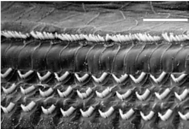
In the cochlea, hair cells subdivide into two categories: the inner hair cells (IHCs) and the outer hair cells (OHCs). There are approximately 3000 to 3500 IHCs, and 9000 to 12000 OHCs in humans which, compared to other sensory epitheliums in the body, represents a very small number. For example, the sensory organ of vision, the retina, contains several
7
million photoreceptor cells. Furthermore, following a gradient from the base to the apex of the cochlea, the cell body diameter of the hair cells varies from 10 to 20 m and their length, from 10 to 50 m. Hair cells of the cochlea are organised in such a way that IHCs form a single row of cells on the neural side (modiolus) of the organ of Corti, whereas OHCS form 3 rows of cells, situated toward the abneural side of the organ (Figure 4). IHCs and OHCs are separated by the pilar cells, which form the tunnel of Corti, and contribute to the overall stiffness of the organ. IHCs e ei e % of the o ga of Co ti’s affe e t e e fi es hile OHCs on the other hand, mainly receive efferent nerve fibres. IHCs are the genuine auditory sensory cells, whereas OHCs play a major role in sound amplification. This is possible thanks to a protein located in the membrane of the OHC cell body, called prestin. Prestin possesses piezo-electric properties which allow OHCs to contract or extend their cell body length in response to a modification of their membrane potential. OHC contraction/extension movements are transmitted to the basilar membrane through the Deiter cells, thus contributing to cochlear amplification.
Figure 4. Scanning electron micrograph illustrating the 3 rows of OHCs, and the single row of IHCs in the organ of Corti
8
I.3.C. The hair bundle and mechano-electrical transduction (MET)
I.3.C.1.
Morphological aspects of the hair bundle
The first studies on the development of the hair bundle were conducted on the chicken (Tilney et al., 1992), but with the introduction of genetics to the study of hearing deficits, mice then became the preferred animal model, and results obtained in mice mirrored those first found in the chicken.
There are three main phases in hair bundle development. During the first phase, several small microvilli appear at the apex of the hair cells and aggregate around a central cilium, called the kinocilium. In a second phase, the kinocilium and the growing microvilli then migrate to the periphery of the apical surface and the microvilli situated closest to the kinocilium begin to increase in length. Subsequently, the microvilli situated a little further from the kinocilium also increase in length. In the third phase, the kinocilium and the growing microvilli settle in their final position (Figure 5).
Figure 5. Schematic representation of sequential stages of hair bundle development (adapted from Schwander et al., 2010)
9
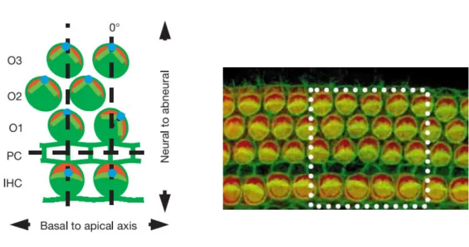
The final position of the kinocilium, and thus the proper orientation of the hair bundle, is of the utmost importance, and there are several planar cell polarity genes implicated in this crucial process. These genes include Vangl1, Vangl2, Frizzled-3, Frizzled-6,
Disheveled-1, Disheveled-2, Disheveled-3, as well as genes responsible for Usher syndrome
type 1, a syndrome I will detail later in the manuscript, as it is an important part of my PhD work. Furthermore, GTP- i di g p otei αi su u its Gαi o t ol itoti spi dle o ie tatio (Walsh et al., 2010; Doherty et al., 2012) and were recently found to be involved in kinocilium migration, and in hair bundle shape and orientation (Ezan et al., 2013; Tarchini et
al., 2013). These proteins are located in the apical region of the hair cell, on its abneural
side, between the cell junction and the hair bundle, forming a crescent-shaped domain (Figure 6 . The ole of Gαi i hai u dle shape as o fi ed i Gαi mutant mice that display flattened hair bundle shapes and mislocalised kinocilia (Lefevre et al., 2008; Ezan et
al., 2013).
Figure 6. Schema and immunofluorescent image illustrating the crescent-shape disposition of PCP proteins in wild-type mice
10
The formation of the staircase pattern, typical of the hair bundle, is initiated by the elongation of the microvilli closest to the kinocilium. During a 2- to 3-day period, depending on the species, the number of actin filaments inside each microvilli (or stereocilium) increases simultaneously across the whole cochlea. During the final phase of stereocilia growth, all the rows grow at the same rate but the stereocilia from the shortest row stop growing first and the ones from the tallest row stop last. The difference in elongation time is also the reason behind the difference in stereocilia length between the apex (long stereocilia) and the base of the cochlea (short stereocilia). The shorter the stereocilia are, the higher their resonance frequency will be, and in this way their sensitivity to high-frequency sound stimuli is increased. Hair bundles come in all shapes and sizes, with variations observed from one species to another, from one organ to another and even differences within the same neuroepithelium. Hair bundles can be of different lengths, have stereocilia of different widths or have an altogether different shape. The explanation for these differences lies within the fact that different hair bundles process different types of information. For example, vestibular hair bundles in mice form a very compact hexagonal structure and have a kinocilium located next to the tallest stereocilia, which persists at adult stages (Figure 7).
Figure 7. Scanning electron micrograph of a normal vestibular hair bundle (image courtesy of Vincent Michel)
11
This is not the case in the mouse cochlea, where the kinocilium disappears approximately a week-to-10 days after birth. In the cochlea, IHC and OHC hair bundle structures are very distinct. Both types of hair bundles are composed of three rows of stereocilia but IHC hair bundles are U-shaped and progressively become linear along the tonotopic axis. OHC hair bundles on the other hand are a V-shaped structure, pointing towards the abneural side of the cochlea (Figure 4). The tallest row of OHC stereocilia is directly coupled to the tectorial membrane (Verpy et al., 2011), and during acoustic stimulation, when the basilar membrane vibrates, the movement leads to tension at the stereocilia/tectorial membrane junction along the apex-to-base axis of the cochlea as well as along the radial (neural-to-abneural) axis. The V-shape of the OHC hair bundles plays a critical role, as it allows the stereocilia to take into account both components of the movement simultaneously.
The formation of the hair bundle and the maintenance of its cohesiveness are orchestrated by several types of links that come into play at different developmental stages. In 1984, using the guinea pig as a model, and by using both scanning and transmission electron microscopy, Pickles and collaborators showed the existence of an array of cross-links between stereocilia. Most notably, they observed thatthe tip of each shorter stereocilium on the hair cell gave rise to a single, upwards-pointing link, which connected with the stereocilium of the adjacent taller row. This link was called the tip link and it was suggested that its distortion lead to sensory transduction (Pickles et al., 1984). It was only in 1991 however, through biochemical experiments, that Assad J.A. and collaborators were able to show that the tip link was indeed physiologically implicated in the MET process. The different types of cochlear links were first biochemically characterised by testing their relative resistance to chemical treatments. The two main chemical products used are
1,2-12
bis(o-aminophenoxy)ethane-N,N,N',N'-tetraacetic acid (BAPTA), which is a high-affinity calcium chelator, and a protease called subtilisin. It was shown that tip links were disrupted by BAPTA but not by subtilisin. In the presence of BAPTA, MET currents could no longer be measured, but if sufficient time was given after BAPTA treatment, tip links and MET currents reappeared (Assad et al., 1991; Crawford et al., 1991; Zhao et al., 1996).
In mice, fibrous links connecting the stereocilia appear before birth and undergo a rapid evolution within a few developmental days (Figure 8). At E17.5, towards the base of the cochlea, where hair bundles already have a certain degree of organisation, a fairly important number of links are present, called lateral links. At this age, kinocilial and slanted apical links are clearly visible but the rest of the links situated below are more difficult to make out. As of postnatal day 2 (P2) however, from top to bottom of the hair bundle, three types of links can be described: the tip links (previously the slanted apical links), the shaft connectors and the ankle links (Figure 8) (Goodyear et al., 2005). Tip links play a direct role in the MET process, as it has previously been mentioned, and in hair bundle development, while shaft connectors and ankle links only play a role in hair bundle development. Despite them only being a transient structure in the developing hair bundle in the mouse cochlea, i.e. they exist from P2 to P9, ankle links play an essential role in hair bundle cohesion and shaping, as I will further discuss in the Results chapter of my manuscript.
13
Figure 8. The different interstereociliary links during hair bundle development (adapted from Michalski and Petit, 2015)
In P2 OHCs and IHCs, tip links and ankle links are clearly present, while shaft connectors, distinct from tip links and ankle links, can be seen sporadically. From P2 to P9, although it increases in length, and stereocilia move slightly apart from one another, the hair u dle does ’t d a ati all ha ge i appea a e. As of P ho e e , a kle li ks sta t to disappear, and another type of link, the horizontal top connectors, that join the apical regions of adjacent stereocilia within OHC hair bundles, start to appear. By the onset of hearing at P12, ankle links are no longer present, and horizontal top connectors are continuing to develop and are seen more frequently than at P9. It is only at P14 that horizontal top connectors finally reach their mature appearance. Furthermore, from P19 onwards, OHCs have highly organised horizontal top connectors that exist both between the stereocilia within the same row and between the stereocilia of adjacent rows (Goodyear et
14
Figure 9. (A) Schematic representation of the tip link (B) Scanning electron micrograph of an IHC and a tip link
(adapted from Pepermans and Petit, 2015)
B
A
15
Top connectors are the most resilient type of lateral link in the hair bundle, as they are the only links to persist in presence of both BAPTA and subtilisin, and ankle links are the weakest links because in presence of either BAPTA or subtilisin, they are disrupted (Jacobs
and Hudspeth, 1990; Assad et al., 1991; Goodyear and Richardson, 1999; Goodyear et al.,
2003)
Figure 10. Scanning electron micrograph of the ankle links (image courtesy of Vincent Michel)
I.3.C.2.
The MET channel
The first step of the MET process takes place in the hair bundle, and the first important biophysical properties of this process were characterised by A.J. Hudspeth, D.P. Co e a d R. Ja o s i the late ’s (Hudspeth and Corey, 1977; Hudspeth and Jacobs,
1979). Using the Rana Catesbeiana bullfrog as a working model, Hudspeth and his
collaborators were able to simultaneously control hair bundle movement as well as measure hair cell receptor potential. Through their experiments, they showed that the hair cell is depolarised following a displacement of its hair bundle in the excitatory direction, i.e.
16
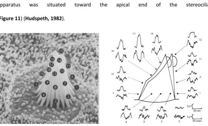
towards the tallest stereocilia, and is hyperpolarised when the hair bundle is displaced in the i hi ito di e tio . The also sho ed that the hai ell’s espo se to s all displa e e ts (up to 0.4 m) is linear, but then starts to saturate as the displacement gets bigger. Finally, by isolating the kinocilium, they showed that the stereocilia, and not the kinocilium, were well and truly the structure where mechano-electrical transduction occured.
Several hypotheses were put forward to explain hair cell depolarisation but the most plausible one was that an ionic channel was located somewhere along the stereocilia or at the junction between stereocilia and the hair cell apex. To prove the veracity of this hypothesis, extracellular potentials were measured at different positions of mechanically stimulated hair bundles. These potentials were consistently larger near the top of the hair bundle than anywhere else around the hair bundle, suggesting that the transduction
apparatus was situated toward the apical end of the stereocilia
(Figure 11) (Hudspeth, 1982).
Figure 11. Localisation of mechano-electrical transduction channel by extracellular potential measurements
17
On the back of these initial results, which type of mechanism activated the transduction apparatus was investigated. Three types of mechanisms were considered possible: (i) an enzymatic mechanism, (ii) a mechanism involving secondary messengers and (iii) a direct mechanical coupling of the MET channel to stereocilia. The first two mechanisms were rapidly discarded for kinetic reasons, i.e. the MET channel opening time is too fast to be compatible with processes involving enzymatic regulation or secondary messengers (Corey and Hudspeth, 1983). Mechanical coupling of the MET channel to stereocilia was thus considered to be the most consistent way of viewing the MET process. This hypothesis consisted in assuming that through a direct elastic link, the gating spring (which would later turn out to be the tip link), displacement of the stereocilia would directly influence the energy difference between open and closed states of the MET channel, as does the change of potential for voltage-activated channels (Figure 12) (Corey and Hudspeth, 1983; Howard
and Hudspeth, 1987).
Further evide e suppo ti g the gati g sp i g theo as gi e i the late ’s Howard and Hudspeth. By measuring hair bundle stiffness, they observed an instantaneous relaxing of the hair bundle upon mechanical stimulation. This mechanism is called the gating
compliance (Howard and Hudspeth, 1988) and can be described in the following way: a
positive deflection of the hair bundle increases tension in the gating spring, and while the channel remains closed, only the length of the spring increases. However, because the spring is directly coupled with the MET channel, once the channel opens, the length of the spring instantly decreases leading to a general relaxing of the hair bundle. In addition, Howard and Hudspeth showed that in presence of gentamycine, a MET channel blocker, there is no relaxing of the hair bundle, providing further evidence in favour of the gating spring and gating compliance theories (Howard and Hudspeth, 1988).
18
Figure 12. Schematic representation of the gating spring model (adapted from Peng et al., 2011)
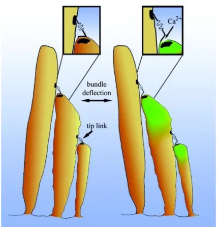
Because MET channels are highly permeable to calcium, calcium-entry monitoring systems were used to locate the MET channel to the tip of the stereocilia (Lumpkin and
Hudspeth, 1995), and determine the number of channels per stereocilium. A study led by
Denk and colleagues in 1995, using two-photon imaging of calcium influx into single stereocilia of bullfrog hair bundles showed that calcium signals occurred in all three rows of stereocilia, during bundle stimulation. The conclusion of the study was to suggest that the MET channels were located both at the upper and lower end of the tip link (Denk et al.,
1995). There were, however, certain limitations in this work. In order to visualise individual
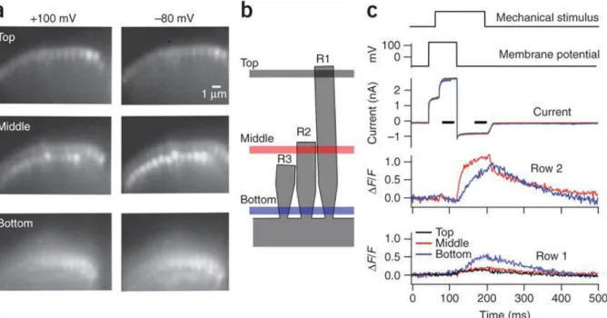
stereocilia, the localisation experiments were done at the base of the hair bundle, but without the adequate temporal resolution to determine the origin of calcium entry, the results of the study were later proven to be inaccurate. It was only in 2009, that Beurg and colleagues managed to show that, in fact, the MET channels are only located at the tip of the smallest and middle, and not the tallest, row of stereocilia (Figure 14) (Beurg et al., 2009). In their study, they overcame the problem related to spatial resolution, by using rat cochlear
19
inner hair cells, where the step in height between rows of large-diameter stereocilia allowed individual stereocilia to be imaged at the apical end of the bundle. To circumvent the temporal resolution problem, they used a high-speed camera coupled to a swept-field confocal system. In this way, they were able to visualise calcium signals predominantly in the small- and middle-row stereocilia. This meant that the MET channels could only be located at the lower end of the tip link (Figure 15). Furthermore, because the number of functional stereocilia was proportional to the amplitude of MET currents, it was suggested that there were about two channels per stereocilium (Beurg et al., 2009).
Figure 13. Calcium entry through the mechano-electrical transduction channel after hair bundle deflection
20
Figure 14. Calcium entry into the small- and middle-row stereocilia of the hair bundle (adapted from Beurg et al., 2009)
Figure 15. Localisation of mechano-electrical transduction channel by high-speed calcium imaging
(adapted from Beurg et al., 2009)
small row tall row middle row
21
I.3.C.3.
Biophysical properties of the MET channel
The MET channel is a non-selective, cationic and voltage-independent channel, with a pore diameter of approximately 0.7 nm (Corey and Hudspeth, 1979; Hudspeth and Jacobs,
1979; Ohmori, 1985; Crawford et al., 1991). The relative permeability to certain cations,
with respect to Na+, are as follows: Li+, 1.14 ; Na+, 1 ; K+, 0.96 ; Rb+, 0.92 ; Cs+, 0.82 ; Ca2+, 3.8 ; Sr2+, 2.3 ; Ba2+, 2.2 ; Mg2+, 2 (Ohmori, 1985) These initial findings, illustrated a preference of the MET channel for Ca2+ ions, although the MET current is carried predominantly by K+ ions. However, these results were based on the assumption that the atio s did ot i te a t ith the ha el’s po e. Yet, i te a tio at the po e le el a i fa t happen, as is seen with the fact that Ca2+ is a MET channel blocker. The channel can also be blocked by several other chemical components. Amilorides specifically and reversibly block the MET channel, in a dose- and voltage-dependent manner, with a half-blocking concentration of 50 mM (Jorgensen and Ohmori, 1988). Aminoglycosides, such as gentamicin and dihydrostreptomycin, are also capable of reversibly blocking the MET channel in a voltage-dependent manner, with a half-blocking concentration in the range of 2 to 95 M (Kroese et al., 1989).
The conductance of the MET channel is also sensitive to extracellular concentration of Ca2+ ions (Crawford et al., 1991). When Ca2+ extracellular concentration varies from 2.8 mM to 0.05 mM, MET currents increase. When Ca2+ concentration is brought back to the 2.8 mM concentration, MET currents return to their original value as well. However, if the extracellular concentration of Ca2+ is brought down to the 1 M range, MET currents totally disappear, and this effect is irreversible. Crawford and colleagues suggested that the decrease in the concentration of extracellular Ca2+ down to 1 M caused the disruption of
22
the tip links, which in turn prevented MET currents from occurring (Crawford et al., 1991). This hypothesis was later confirmed by a study done on the hair bundles from the saccule of adult bullfrogs (Assad et al., 1991). When hair bundles were treated with a calcium chelator, BAPTA, MET currents were abolished, tip links were disrupted, voltage-dependent movement was eliminated and a positive displacement of the hair bundle could be observed. These results strongly pointed to the tip link conveying tension to the MET channels (Assad et al., 1991).
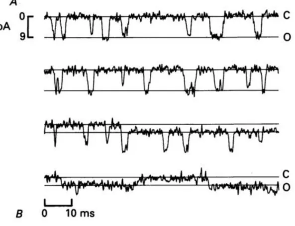
Furthermore, by locally perfusing hair bundles with a solution of 1M Ca2+
, and thus destroying almost all the MET channels in a hair bundle, Crawford and colleagues were able to determine single-channel currents (Figure 16), as well as estimate the conductance of the MET channel to approximately 100 pS (Crawford et al., 1991). Although MET channel conductance estimates can vary significantly from one species to another (single-channel conductance in mammals can reach 300 pS for example), it is important to note that these values are far greater than those of other more classical ionic channels, such as Ca2+ channels, K+ channels, and Na+ channels, which are in the 10-20 pS range.
The sensitivity of the MET channel can be determined by studying its current/displacement curve (also called I(X) curve). Through a series of experiments in the ’s a d ’s, the relationship between inward transducer currents and bundle displacement, i.e. the I(X) curve, was shown to be an asymmetrical sigmoid (Hudspeth and
Corey, 1977; Crawford et al., 1989). This led to the first biophysical model representing the
function of the MET channel. The channel was considered to have one open state and two closed states:
23
Figure 16. Examples of single-channel current recordings in turtle hair cells (adapted from Crawford et al., 1991)
Furthermore, the effects of extracellular calcium concentration on the I(X) curve were also investigated. When the extracellular concentration of Ca2+ is lowered, the I(X) curve is shifted leftward (towards the smallest displacements). This observed shift corresponds to an increase in the number of MET channels that are open at the resting state of the hair bundle (Corey and Hudspeth, 1983). This can easily be explained by the fact that Ca2+ is a blocker of the MET channel, so when its extracellular concentration is decreased, there are less blocked channels, and hence a bigger number of open channels. In return, when extracellular calcium concentration is increased, there is a decrease in MET current amplitude (Corey and Hudspeth, 1983; Crawford et al., 1991; Lumpkin et al., 1997).
24
I.3.C.4.
Adaptation
In an inner ear hair cell, a deflection of the hair bundle in the positive direction elicits a MET current. A decline in the aforementioned current can then be observed despite the sustained stimulus, and this decline is referred to as adaptation (Figure 17). In other words, adaptation can be defined as the decay of a sensory response to a sustained stimulus, and it has been observed in all vertebrate species studied to date: bullfrog (Eatock et al., 1987), turtle (Crawford et al., 1989), mouse (Russell et al., 1989; Kros et al., 1992) and rat (Kennedy et al., 2003). This has allowed for an extensive description of the phenomenon.
Adaptation occurs over several distinct time frames from microseconds to tens of seconds, and it is commonly accepted that there are 2 major processes involved: the fast adaptation process (which operates on a millisecond or submillisecond time scale) and the slow adaptation process (acting over tens of milliseconds) (Wu et al., 1999; Vollrath and
Eatock, 2003). Despite the extensive description, and several studies tackling the question,
the underlying mechanisms of adaptation remain, to date, unclear and debated. There is however one generally accepted view, which is that adaptive mechanisms are calcium-dependent (although it should be mentioned that a recent study has challenged this view in mammalian cochlear hair cells: (Peng et al., 2013)). In 1989, Assad and colleagues showed that when the cell membrane was depolarised to +80 mV (a membrane potential close to the Ca2+ equilibrium), Ca2+ influx into the cell body was drastically reduced, and adaptation was abolished (Assad et al., 1989). Furthermore, it was shown that adaptation necessarily involves a change in the I(X) curve, since the MET current, at a given displacement, declines.
25
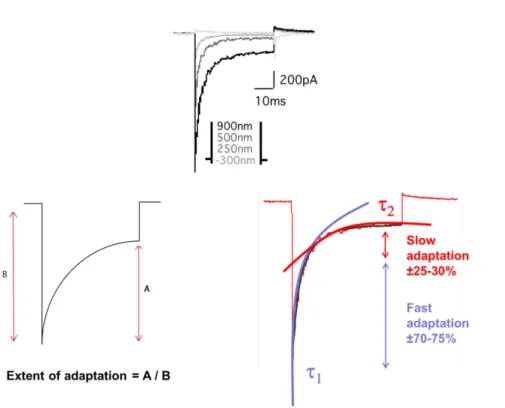
Figure 17. Mechano-electrical transduction current amplitude and characteristic parameters of adaptation
26
The I(X) curve shifts towards the larger displacements, without a change in shape or decrease in maximum current. This is called the adaptive shift (Figure 19) (Eatock et al.,
1987). In actual fact, the position of the I(X) curve on the displacement axis is sensitive to
the extracellular Ca2+ concentration and intracellular Ca2+ buffering (Hacohen et al., 1989;
Ricci and Fettiplace, 1998; Ricci et al., 1998). Changes in stereociliary Ca2+ concentration reset the range of bundle displacements over which the MET channel is activated. Elevating the Ca2+ influx, as it occurs during sustained channel opening, shifts the I(X) curve towards larger displacements, causing desensitisation. Reducing Ca2+ influx however, as is the case when channels are closed for a prolonged period or when extracellular Ca2+ concentration is lowered, shifts the I(X) curve towards smaller displacements. The site(s) at which binding of Ca2+ triggers adaptation are specific for this ion, and other alkaline earth cations do not substitute for Ca2+ (Hacohen et al., 1989), even though some, such as Sr2+, permeate the channel with comparable ease (Ohmori, 1985).
Figure 19. Illustration of the calcium-dependent adaptative shift (adapted from Fettiplace & Kim, 2014)
27
When it comes to molecular mechanisms, a change in the upper attachment point of the tip link has been used to explain slow adaptation. During a positive bundle deflection, the upper end of the tip link slips down the side of the stereocilium to reduce the tension in the elastic element (Howard and Hudspeth, 1987). Furthermore, it has been suggested that the resting tension in the tip link is maintained by an isoform of myosin. Previous studies have implicated myosin 1c in the process (Holt et al., 2002; Gillespie and Cyr, 2004), but it has also been shown that myosin1c is localised to both upper and lower ends of the tip links in frog vestibular hair cells (Garcia et al., 1998), and all along the stereocilia of cochlear hair cells (Schneider et al., 2006). This diffuse labeling contrasts sharply with that for three other myosins examined: myosin 3a and myosin 15a are both confined to the stereociliary tips (Schneider et al., 2006), whereas myosin 7a clusters in a complex with sans and harmonin-b at the upper end of the tip link (Grati and Kachar, 2011). Functional evidence for the role of myosin 7a in cochlear hair cells comes from the observation that several mutations of this motor protein affect adaptation of the MET current (Kros et al., 2002). Myosin 7a climbing up the actin filaments in the stereocilium may also be responsible for maintaining the resting tip link tension in mammalian OHCs (Grati and Kachar, 2011).Furthermore, how do myosins, located at the upper end of the tip link, get access to calcium entering through the MET channel, now believed to be located at the lower end of the tip link (Beurg et al., 2009)? How then are the myosins regulated and what determines the sliding of the attachment point with an increase in tension? Which specific myosin isoform(s) are implicated in adaptation, as well as their mode of action, is still up for debate.
Fast adaptation has a time constant inferior to 5 ms in bullfrogs and mouse vestibular hair cells (Vollrath and Eatock, 2003), in auditory hair cells of turtle (Crawford et al., 1989;
28
Ricci and Fettiplace, 1997) and rat (Kennedy et al., 2003; Ricci, 2005). Therefore,
ATPase-mediated mechanisms, which are behind myosin activity, are incompatible with the speed of the fast component of adaptation. The millisecond time constant of fast adaptation is more consistent with adaptation occurring at a site very close to the MET channel (Crawford et al.,
1989; Ricci and Fettiplace, 1998; Wu et al., 1999). One hypothesis along those lines is that,
by binding to the MET channel pore, Ca2+ induces a change in the force sensitivity of the pore, making it harder to open (Cheung and Corey, 2006).
29
II.
Usher syndrome
II.1. Clinical aspects of Usher syndrome
Initially the small number of hair cells in the inner ear (a few thousands) hampered the progression of understanding the molecular components and mechanisms underlying hearing. In the early 1990s, human genetics, the efficiency of which is independent from the number of hair cells, emerged as the best approach to identify molecules involved in MET. By using genetic approaches, scientists were able to, first, identify several causative genes responsible for hearing loss in patients, and second, engineer mouse models that mimicked the human pathologies. Through this approach, approximately 90 deafness genes were identified and more than 120 deafness loci characterised (Hereditary Hearing Loss Homepage, www.hereditaryhearingloss.org).
Syndromic deafness is defined as deafness accompanied by one or several other symptoms. Among the 400+ syndromes involving deafness (Toriello et al. 2004), Usher syndrome is one the most incapacitating, as it combines congenital hearing loss with progressive loss of vision. The latest data reveals a prevalence of 1/16 000 to 1/50 000, depending on the geographic region, among patients who are both deaf and blind. In the late ’s, Da e po t a d olla orators (Davenport et al., 1978) classified the syndrome into three subtypes: Usher type 1 (USH1), Usher type 2 (USH2), Usher type 3 (USH3). USH1 is the most severe form of the syndrome, and is characterised by severe to profound congenital sensorineural deafness, constant vestibular dysfunction and a pre-pubertal onset of visual impairment by retinitis pigmentosa. Retinitis pigmentosa is characterised by the death of cells in the retina called cones and rods. Rods, which are responsible for peripheral
30
and night vision, die first, and then cones, which are responsible for color perception and central vision, degenerate. The result of retinitis pigmentosa is hence, tunnel vision, night blindness and eventually total loss of vision. USH2 is defined by a moderate to severe congenital hearing loss, an absence of vestibular dysfunction and a post-pubertal onset of retinitis pigmentosa. USH3 is the least severe form of Usher syndrome, and is characterised by progressive hearing loss, retinitis pigmentosa diagnosed between the second and forth decade of life, and only the occasional presence of vestibular dysfunction (Petit, 2001). Up to now, 10 causative genes of Usher syndrome have been identified: 6 causing USH1, 3 causing USH2 and 1 causing USH3.
II.2. Molecular aspects of Usher syndrome
II.2.A.
Usher syndrome type 1
The 6 identified causative genes for USH1 have been shown to interact with one another and play an essential role in one or several of the following aspects: hair bundle morphogenesis, MET transduction and stereocilia length regulation. Here we briefly describe the 6 USH1 genes:
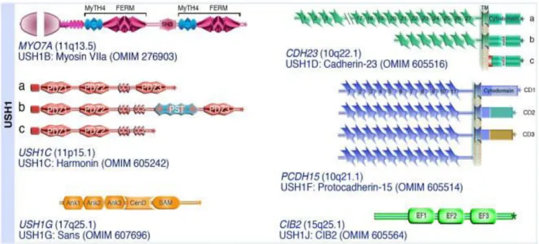
o USH1B (MYO7A): this was the first deafness gene to be identified (Weil et al., 1995).
In humans MYO7A is responsible for USH1B, and the murine orthologue (Myo7a) was found to be the causative gene in shaker-1 mice (Gibson et al., 1995). Myo7a codes for myosin VIIa, an unconventional myosin consisting of a motor domain, a neck region and a cargo-binding tail. The motor domain, which has an unusually high affinity for ADP, is responsible for actin- and ATP-binding. The neck consists of five
31
IQ-motifs (isoleucine and glutamine), that are expected to bind calmodulin, a calcium-binding protein. The tail region is responsible for binding to interaction partners and could also be responsible for inducing dimerization (Sakai et al., 2011). In the inner ear, myo7a plays several roles, as it is implicated in intracellular transport, endocytosis, cell-cell adhesion, and in hair cell adaptation. Furthermore, myo7a is essential for stereocilia differentiation and organisation (Self et al., 1998;
Boëda et al., 2002; Kros et al., 2002; Wolfrum et al. 2003)
o USH1C (HARMONIN): this gene encodes the scaffold protein harmonin (Hmn), a
component of the USH interactome. In the inner ear, there are three different isoforms of harmonin: Hmn-a, -b and –c. Through their PDZ-domains (Post-synaptic density protein - Drosophila disc large tumor suppressor – Zonula occludens-1), these proteins interact with the C-terminal PDZ-binding motif (PBM) of most of the other USH proteins. Furthermore, Hmn-b was shown to be located at the upper tip link density in mature auditory hair cells, further confirming harmonin as an essential element for stereocilia formation and hair bundle function (Bitner-Glindzicz et al.,
2000; Verpy et al., 2000; Boëda et al., 2002; Grillet et al., 2009; Michalski et al.,
2009).
o USH1D (CDH23): this gene codes for cadherin-23, an unconventional member of the
cadherin superfamily. The murine orthologue (Cdh23) was found to underlie the abnormal phenotype in Waltzer (v) mice (Bolz et al., 2001a; Bork et al., 2001; Di
Palma et al., 2001). Cdh23 comprises an extracellular domain of 27 extracellular
32
with a type I C-terminal PBM. In the immature hair bundle it is found all along the stereocilia, where it forms part of the transient lateral links during development. It also forms the stereocilia-associated part of the kinocilial links (Michel et al., 2005;
Goodyear et al., 2010), and is the upper-most component of the two that make up
the tip link (Siemens et al., 2004).
o USH1F (PCDH15): this gene codes for protocadherin-15, also an unconventional
member of the cadherin superfamily, and the murine orthologue of the gene (Pcdh15) has been identified as the causative gene in Ames Waltzer (av) mice. Pcdh15 consists of an extracellular domain of 11 EC repeats, a single transmembrane domain and a cytoplasmic domain with a C-terminal PBM. Three transmembrane splice isoforms can be distinguished based on the C-terminal exon: CD1 (exon 35), CD2 (exon 38) and CD3 (exon 39). A forth secreted isoform (SI) lacking the transmembrane domain also exists. Pcdh15 makes up the lower part of the tip link while cdh23 makes up the upper part. Furthermore, it has recently been shown that the CD2 isoform of pcdh15 is the only pcdh15 isoform to persist in mature auditory hair cells, at the tip link (Ahmed et al., 2001; Alagramam et al., 2001a, 2001b;
Pepermans et al., 2014).
o USH1G (SANS): this gene codes for sans (Scaffold protein containing ANkyrin-repeats
and a SAM domain), a small scaffold protein which consists of 4 ankyrin repeats, a central domain, a SAM domain and a C-terminal type I PBM. In the stereocilia, sans has been localised both at the upper and at the lower part of the MET machinery at different time points in maturation (Caberlotto et al., 2011b; Grati and Kachar, 2011)
33
but it has also been shown in the synaptic region, at the cuticular plate and in the adhesion region. Sans is essential to stereocilia formation and in the absence of this protein there is a loss of short- and middle-row stereocilia (Caberlotto et al., 2011b). The murine orthologue Sans is responsible for the phenotype in Jackson shaker mice (js).
o
USH1J (CIB2): this gene codes for a calcium and integrin binding protein. The USH1Jlocus was first shown to be responsible for DFNB48, which is a genetic form of isolated deafness (Ahmad et al., 2005). Later on, however, mutations in this gene, resulting in USH1J, were identified (Riazuddin et al., 2012). Cib2 contains three EF-hand domains that change conformation upon binding of Ca2+ and presumably mediate calcium signalling (Gentry et al., 2005; Blazejczyk et al., 2009). It is present all along the hair bundle, in the cuticular plate and is also present in supporting cells. Furthermore, cib2 interacts with whirlin and myosin 7a at the tips of the stereocilia in cochlear hair cells of mice.
34
Figure 20. USH1 proteins (adapted from El Amraoui & Petit, 2014)
Although the role of cib2 in cochlear hair cells remains unclear, as I will show in the Results chapter, all the other known USH1 proteins interact together directly or indirectly, and form the MET complex (Figure 21).
Figure 21. USH1 proteins interacting and forming the MET complex (adapted from Michalski & Petit, 2015)
35
II.2.B.
Usher syndrome type 2
As it has been mentioned previously, 3 causative genes for USH2 have, to date, been identified. Together with the protein encoded by the modifier gene Pdzd7, the three proteins encoded by the USH2 genes, whirlin, usherin and adgrv1, form the ankle link complex (Adato et al., 2005; Michalski et al., 2007; Chen et al., 2014; Zou et al., 2014). The ankle links are a subset of filamentous lateral links connecting stereocilia at the base, and the ankle link complex has been reported as being essential to hair bundle shaping, rigidity as well as IHC thickness (Zou et al., 2015). In the next section, I will go further into detail about the ankle link complex and its components but here I give a brief description of the three genes responsible for USH2:
o USH2A (USHERIN): this gene encodes usherin, which was first described as a
basement membrane protein, before being described as a transmembrane protein of the inner ear, implicated in the formation of the ankle link complex. In the human ear and retina, there exists two isoforms of usherin: usherin a-isoform and usherin b-isoform. The a-isoform is an extracellular matrix protein (Pearsall et al., 2002) whereas the b-isoform is a transmembrane protein with a relatively short cytoplasmic domain and a C-terminal type I PBM. The extracellular domain of the b-isoform consists of one laminin G domain, one laminin N-terminal domain, 10 laminin EGF-like domains, 2 laminin G-like domains and 36 fibronectin type III domains, all of which are common to adhesion proteins (Weston et al., 2000) . Usherin has been localised at the ankle link region of the hair bundle and in the synaptic region (Adato
36
o USH2C (ADGRV1): this gene encodes adgrv1 (adhesion G protein-coupled receptor
v1), a protein responsible for reflex seizures prior to its identification as an USH protein. It was formally referred to as mass1, vlgr1 or gpr98. Several isoforms have been identified, but the b-isoform is responsible for USH. This isoform consists of an N-terminal extracellular domain, a 7-transmembrane receptor domain formed by 3 intracellular and 3 extracellular loops, a relatively short cytoplasmic domain with a C-terminal class I PBM. The extracellular domain consists of 35 Calx-beta domains (calcium binding domains), 6 EAR (epilepsy associated repeats) and a laminin globular domain. Just before the 7-transmembrane domain, a GPCR proteolytic site (GPS) can be found. This is a signature domain of family B G-protein coupled receptors (McMillan et al., 2002). The b-isoform of adgrv1 is located at the ankle link region of the hair bundle as well as at the synaptic region (McGee et al., 2006).
o
USH2D (WHIRLIN): this gene codes for whirlin, a scaffold protein that interacts withusherin, adgrv1 through their PBMs. Two isoforms of the protein exist and mutations in WHIRLIN can cause either USH2 or non-syndromic deafness (DFNB). Only the long isoform, containing an N-terminal harmonin homology domain, 2 type I PDZ domains followed by a second harmonin homology domain (Florent Delhommel, unpublished
results), a proline-rich region, a third type I PDZ domain and a C-terminal type II PBM,
is expressed in the retina. In the inner ear however, both long and short isoforms (the short isoform only contains the C-terminal proline-rich region, the third PDZ domain and the PBM) are expressed.
37
Figure 22. Schematic representation of USH2 proteins
PDZ: (PSD95/Dlg1/ZO-1) domain, PR: proline-rich regions, HNL: harmonin-N like domain (adapted from Chen et al., 2014)
II.2.C.
Usher syndrome type 3
Up to date, there is only one gene that has been identified as responsible for USH3. This gene is CLRN1 and leads to USH3A. The protein encoded by this gene is a four-transmembrane domain protein that is found in hair cell and photoreceptor cell synapses.
38
II.3. Usher syndrome type 2 and the ankle link (AL) complex
As mentioned previously, to ensure normal mechano-electrical transduction, an essential step in sound perception, auditory hair cell hair bundle cohesion is essential. During development, the stereocilia of hair bundles differentiate from microvilli and grow differentially to reach their final lengths, thicknesses and rigidity. Throughout this process, several types of lateral links between the stereocilia develop, helping to maintain hair bundle cohesion (Goodyear et al., 2005). One set of links, which has been reported as essential to proper hair bundle cohesion and rigidity, is the ankle links, located at the base of the hair bundle. Up until now, it has been generally accepted that two transmembrane proteins coded by USH2 genes, adgrv1 and usherin, make up the ankle links, per se (Adato et
al., 2005; McGee et al., 2006; Michalski et al., 2007). Through their interaction with a third
protein, also coded by an USH2 gene, whirlin, and pdzd7, a protein coded by modifier gene
Pdzd7, these proteins form what is called, the ankle link complex (Chen et al., 2014; Zou et al., 2014). In this section I will give detailed information about the function of each of the
four proteins which make up the ankle link complex, as well as detailed explanations of how they interact together.
II.3.A Whirlin
The first gene to be discovered that formed part of, what was to be later called, the USH2 complex, was the gene encoding whirlin (WHRN). The murine ortholog of WHRN, was first mapped to chromosome 4 in mice in 1963 (Lane, 1963). In that study, mice presenting with an autosomal recessive mutation (whirler (wi)) in the Whrn gene were reported to display deafness, rapid circling and head-tossing. It was only in 2002, however, that the
39
human orthologue of Whrn, DFNB31, first reported to be responsible for an autosomal recessive form of hearing loss, was identified in a Palestinian consanguineous family from Jordan (Mustapha et al., 2002). Later that year, a study showed that whirlin was implicated in the process of IHC and OHC stereocilia elongation and maintenance in mice (Holme et al.,
2002). Stereocilia of cochlear hair cells in whirler mutant mice were reported to have
abnormal lengths and shapes. In 35-day old (P35) wi/wi mutant IHCs, stereocilia were approximately half the length of the equivalent stereocilia in +/wi control mice (Figure 24), all along the tonotopic axis of the cochlea (Figure 25). The difference in stereocilia length could be observed as early as embryonic day 18.5 (E18.5). While mutant mice stereocilia were reported to elongate at a normal rate until P1, between P1 and P4 they prematurely stopped elongating. Their length then decreased, before disappearing completely. Furthermore, wi/wi mutant OHCs were closer to normal in length but were arranged in a rounded, "U" shape rather than the normal "W" or "V" shape.
Figure 24. Scanning electron micrographs of P35 +/wi and wi/wi IHCs (adapted from Holme et al., 2002)
40
Figure 25. IHC stereocilia length along the tonotopic axis (adapted from Holme et al., 2002)
Whirlin has two reported isoforms: a short C-terminal isoform, and a long isoform, and it is a PDZ domain-containing protein (Mburu et al., 2003). As previously mentioned, the long isoform contains three PDZ domains while the short isoform only contains one PDZ domain. In mouse inner ear hair cells, PDZ domains have been shown to mediate the assembly of proteins into functional complexes (Sheng and Sala, 2001). These complexes, in at least some cases, are thought to provide a link between transmembrane proteins and the actin cytoskeleton (Fanning and Anderson, 1999). Because mutations in whrn lead to shortened stereocilia in whirler mutant mice, it was hypothesised that whirlin mediated the organisation of other proteins to form a complex required for stereocilia elongation, such as espin and myosin 15a. Using an antibody raised against its short isoform, whirlin was initially reported to be located at the tip of cochlear hair bundle stereocilia (Kikkawa et al., 2005). In the same study, myosin 15a was shown to play a major role in the stabilisation of whirlin expression in the stereocilia. The interaction between these two proteins was shown to occur via the PDZ domain of whirlin and the carboxy-terminal PDZ-ligand of myosin 15a (Belyantseva et al., 2005). It was only in 2006 that the interaction of whirlin with the USH2 proteins usherin and vlgr1 was reported. Whirlin was shown to colocalise with usherin and
41
adgrv1 in photoreceptor cells (synaptic regions, connecting cilium and outer limiting membrane) and auditory hair cells (synapse, stereocilia) (van Wijk et al., 2006; Michalski et
al., 2007). In 2007, a study, on human patients, revealed that mutations in exons 1 to 6,
which are specific for the long isoform of whirlin, were responsible for a new form of USH2, named USH2D (Ebermann et al., 2007). This was the first time a clear causal relation between whrn and USH2 was established. Indeed, mutations in the C-terminal half of whirlin (common to both long and short isoforms) were only previously reported in non-syndromic deafness (DFNB31). These observations were later confirmed in mice. Indeed, it was shown that mutations near the N-terminus of whirlin lead to retinal and inner ear defects, reproducing the clinical features of the human USH2 disease (Yang et al., 2010). In the same
study, it was also shown that only the long, and not the short, isoform of whirlin was expressed in the retina, while both long and short isoforms were expressed in the inner ear. The N-terminal PDZ domains of the long isoform of whirlin mediate the formation of the ankle link complex in the inner ear, and a multi-protein complex located in the periciliary membrane complex, in the retina.
The precise distribution, as well as the functional role, of the different whirlin isoforms was only very recently confirmed (Mathur et al., 2015). While the long isoform of whirlin is important for the USH2 protein complexes in hair cell stereociliary bases and photoreceptors, both the long and short isoforms participate in stereociliary elongation in hair cell stereociliary tips. Mutations in the whirlin N-terminal region led to retinal degeneration and moderate to severe hearing loss, while a mutation in the C-terminal region eliminated all normal whirlin isoforms and generated a truncated N-terminal whirlin protein fragment, which was partially functional in the retina, thus preventing retinal degeneration (Mathur et al., 2015).
42
Figure 26. (A) Whirlin distribution in wild-type mouse IHCs and OHCs at P4
(B) Schematic representation of the distribution of whirlin in photoreceptors and inner ear hair cells
(adapted from Mathur et al., 2015)