HAL Id: hal-02378714
https://hal.archives-ouvertes.fr/hal-02378714
Submitted on 24 Nov 2020
HAL is a multi-disciplinary open access
archive for the deposit and dissemination of
sci-entific research documents, whether they are
pub-lished or not. The documents may come from
teaching and research institutions in France or
abroad, or from public or private research centers.
L’archive ouverte pluridisciplinaire HAL, est
destinée au dépôt et à la diffusion de documents
scientifiques de niveau recherche, publiés ou non,
émanant des établissements d’enseignement et de
recherche français ou étrangers, des laboratoires
publics ou privés.
Tripartite Split-GFP Association
Stéphanie Cabantous, Hau Nguyen, Jean-Denis Pedelacq, Faten Koraïchi, Anu
Chaudhary, Kumkum Ganguly, Meghan Lockard, Gilles Favre, Thomas
Terwilliger, Geoffrey Waldo
To cite this version:
Stéphanie Cabantous, Hau Nguyen, Jean-Denis Pedelacq, Faten Koraïchi, Anu Chaudhary, et al.. A
New Protein-Protein Interaction Sensor Based on Tripartite Split-GFP Association. Scientific Reports,
Nature Publishing Group, 2013, 3 (1), �10.1038/srep02854�. �hal-02378714�
A New Protein-Protein Interaction Sensor
Based on Tripartite Split-GFP Association
Ste´phanie Cabantous1, Hau B. Nguyen2, Jean-Denis Pedelacq3, Faten Koraı¨chi1, Anu Chaudhary4,
Kumkum Ganguly2, Meghan A. Lockard5, Gilles Favre1, Thomas C. Terwilliger2& Geoffrey S. Waldo2
1INSERM UMR1037, Cancer Research Center of Toulouse, Universite´ de Toulouse, Institut Claudius Regaud, F-31052 Toulouse,
France,2Bioscience Division, MS-M888, Los Alamos National Laboratory, Los Alamos, NM 87545, USA,3CNRS, IPBS (Institut de
Pharmacologie et de Biologie Structurale), F-31077 Toulouse, France; Universite´ de Toulouse; UPS, IPBS, F-31077 Toulouse, France,
4Department of Microbiology, University of Washington, Seattle, WA 98195, USA,5Rockefeller University, New York, NY 10065,
USA.
Monitoring protein-protein interactions in living cells is key to unraveling their roles in numerous cellular processes and various diseases. Previously described split-GFP based sensors suffer from poor folding and/ or self-assembly background fluorescence. Here, we have engineered a micro-tagging system to monitor protein-protein interactionsin vivo and in vitro. The assay is based on tripartite association between two twenty amino-acids long GFP tags, GFP10 and GFP11, fused to interacting protein partners, and the complementary GFP1-9 detector. When proteins interact, GFP10 and GFP11 self-associate with GFP1-9 to reconstitute a functional GFP. Using coiled-coils and FRB/FKBP12 model systems we characterize the sensorin vitro and in Escherichia coli. We extend the studies to mammalian cells and examine the FK-506 inhibition of the rapamycin-induced association of FRB/FKBP12. The small size of these tags and their minimal effect on fusion protein behavior and solubility should enable new experiments for monitoring protein-protein association by fluorescence.
S
ignal transduction and gene expression often involves protein-protein interactions. Optical tracking of these interactions can help to illuminate regulatory mechanisms and identify aberrant processes in diseases. Fluorescent protein biosensors have been developed to measure protein complexes in living cells1. Forexample, bioluminescence resonance energy transfer (FRET and BRET) enable dynamic observations of protein-protein interactions2. More recent developments include protein-fragment complementation assays (PCA) that
monitor protein-protein interactions by reconstitution of fragments of various enzymes split into two pieces (as fused tags on passenger proteins) including fragments of dihydrofolate reductase3, b-galactosidase4, b-lactamase5,
and the firefly and Gaussia luciferases6. Though sensitive, these have the limitation that the observed products
diffuse away from the protein interaction site. PCA based on split green fluorescent protein (GFP) and color variants7is termed bimolecular fluorescence complementation (BiFC). BiFC relies on (i) interactions between
bait and prey proteins that bring together two non-fluorescent split protein domains and (ii) subsequent co-folding into the b-barrel structure to form the chromophore8. Because assembly of the GFP fragments is
irre-versible, the stability of the BiFC enables integration, accumulation, and subsequent detection even of transient interactions and low affinity complexes9,10. These remain attached to the interacting proteins, enabling tracking of
the complex. However, improving existing BiFCs based on large and bulky fragments is challenging as increasing BiFC fragment solubility and folding can increase background signals from spontaneous assembly of the fluor-escent protein fragments11. For example, BiFC fragments12obtained by fragmentation of folding-reporter GFP
(FR-GFP)13and superfolder GFP (sfGFP)14at permissive sites 156 and 173 were aggregation-prone and had high
backgrounds from self-assembly (Supplementary Fig. S1). Using a smaller tag can reduce aggregation and folding interference. For example, one split GFP uses a very small 15 amino acid tag for quantification of soluble protein and tracking proteins in vivo15–17. However the large size of the remaining piece and self-assembly of the fragments
makes this system less suited for protein-protein interaction studies. We had previously developed a sponta-neously assembling split GFP based on GFP 10–11 (residues 194–233, the tag) and GFP1-9 (residues 1–193, the detector) (Waldo lab, unpublished results). Here we describe the further engineering of this split GFP to yield an entirely new protein interaction assay based on the association of three fragments of the GFP: two short peptides GFP10 (residues 194–212) and GFP11 (residues 213–233) each tagged to one of the interacting partners, and a third large GFP1-9 (residues 1–193) detector fragment (Fig. 1). We present the characterization of the assay using attractive and repulsive pairs of charged coiled-coils peptides18and the rapamycin mediated heterodimerization
SUBJECT AREAS:
MOLECULAR ENGINEERING SENSORS AND PROBES PROTEIN DESIGN FLUORESCENT PROTEINS Received 5 June 2013 Accepted 16 September 2013 Published 4 October 2013 Correspondence and requests for materials should be addressed to S.C. (cabantous. stephanie@ claudiusregaud.fr) or G.S.W. (waldo@lanl. gov)
of FK506 binding protein (FKBP) and FKBP12-rapamycin binding domain (FRB)19. The assay correctly reports the localization of
pro-tein-protein complexes in mammalian cells with very low back-ground fluorescence levels. The chief advantage over BiFC is the small size of the tags (ca. 20 amino acids), and the concomitant reduced interference with passenger protein folding. The tripartite split GFP assay may find broad utility in the visualization of protein complexes specially where bulkier BiFC fragments might impede localization or interfere with folding.
Results
Engineering a three-body split-GFP system for improved solubi-lity and complementation.We previously identified self-assembling split-GFP fragments corresponding to the C-terminal b-hairpin GFP10–11 of sfGFP14(residues 194–238) and the large fragment
GFP1–9 (residues 1–193) (Fig. 2). In order to improve GFP 1–9 folding efficiency, we performed two rounds of directed evolution and obtained a new variant of GFP1–9 named GFP1–9 M1 that contained five additional mutations relative to sfGFP1–9 (Fig. 2, Supplementary Note). We converted this bimolecular pair into a three-body split-GFP in a stepwise manner. First, in attempt to destabilize the GFP10–11 b-hairpin self-assembly, we inserted a long flexible linker that included a cloning site between GFP10 and GFP11 and evolved the whole cassette for improved solubility and complementation efficiency with GFP1–9 M1 (See Methods). Next, we spaced out GFP10 and GFP11 further by inserting a partially soluble bait protein, hexulose phosphate synthase or HPS15in the
cloning site of the linker. HPS alone was ,60% soluble and was used in our earlier study as a bait protein to drive the evolution of more soluble split-GFP tags15(Supplementary table 1 and Supplementary
Note). In the final step, we evolved GFP1–9 M1 to recognize this ‘‘sandwich’’ bait topology. When complemented with the optimized GFP10 and GFP11 tags attached to the N and C termini of the HPS15,
the resulting variant named GFP1–9 OPT, exhibited a 40-fold improvement in the rate of formation of fluorescence (Fig. 3a, black line) relative to GFP 1–9 M1 (Fig. 3a, gray line). Differences between the GFP1–9 M1 were less obvious when complemented with the GFP10–11 hairpin (displayed on permissive loop of a cherry fluorescent protein, Waldo lab, manuscript under review) (Fig. 3a, right panel), presumably due to the reduced entropy of the GFP 10– 11 hairpin relative to the GFP10-HPS-GFP11 ‘‘sandwich’’ format. GFP1–9 OPT amino-acid sequence revealed the presence of four additional mutations relative to GFP1–9 M1 (Fig. 2). We then Figure 1|Principle of the tripartite split-GFP complementation assay.
b-strand 10 (GFP10) and b-b-strand 11 (GFP11) are fused to bait (A) and prey (B) proteins, respectively and the detector fragment GFP1–9 (b-strand 1– 9) is added separately. When protein interaction occurs, GFP10 and GFP11 are tethered and then spontaneously associate with GFP1–9 fragment to form a full-length GFP. If proteins A and B do not interact, GFP10 and GFP11 are not tethered and entropy is too high to allow complementation with GFP1–9.
Figure 2|Secondary structure diagram of GFP 1–9 variants with corresponding mutations. Superfolder GFP1–9 (green dots), GFP1–9 M1 (yellow), and GFP1–9 OPT (orange).
examined whether optimized GFP10 and GFP11 would affect the solubility of fused proteins. We cloned set of eighteen P. aero-philum test proteins into a modified pTET vector as GFP10-POI-GFP11 ‘‘sandwiches’’ (POI 5 protein of interest) to assay solubility levels as previously described16. We had previously measured the
solubility of these eighteen controls without tags and shown that the GFP11 M3 tag engineered in the earlier study did not perturb passenger solubility16. We first assayed the sandwich fusions using
GFP1–10 to detect only the GFP11 tag (Fig. 3b). Co-expressing GFP1–10 and the sandwich resulted in bright colonies reflecting total protein expression (Fig. 3b, top). On the other hand, expressing the sandwich first, shutting off expression to allow the proteins to remain soluble or aggregate, then expressing the GFP1– 10 detector (sequential induction) gave weak fluorescence for the protein expressed in insoluble form (proteins #6,8,15,16) as expected. This pattern was very similar to that previously seen for the GFP11 M3 tag study16 indicating that the GFP10-POI-GFP11
sandwich did not strongly perturb the solubility of the test proteins. Note that the version of GFPS11 developed in this manuscript differs slightly from the GFP11 M3 tag described earlier16. Next we studied
the complementation of the sandwich with GFP1–9 OPT (subse-quently referred to as GFP1-9) (Fig. 3b, bottom). Interestingly, whe-ther the sandwich and GFP1–9 were co-expressed or sequentially expressed, only the soluble proteins gave fluorescent colonies (Fig. 3b, bottom). When GFP1–10 is co-expressed, insoluble protein can be labeled (Fig. 3b, top), ostensibly because the available GFP1–10 can capture the GFP11 M3 tag early on, committing the fluorophore to
form regardless of the subsequent fate of the fusion. In contrast, even when co-expressed with GFP1–9, one or both tags of insoluble proteins must become inaccessible prior to productive binding with GFP1–9. This suggests the GFP 1–9 could be used to strin-gently assess protein solubility, without the concern of false-positives caused by capturing transiently soluble proteins as with GFP1–10.
Validating a protein-protein interaction sensor using coiled-coil heterodimerization.To test whether GFP1–9 is capable of detecting GFP10 and GFP11 fused to separate interacting protein molecules, we selected two pairs of coiled-coils developed by Hodges and coworkers18based on their ability to direct preferential interaction
between oppositely charged K1/E1 coiled-coils or repulsion for the negatively charged E1/E1 pair (Supplementary Fig. S2). This model system features rapid association of the K/E heterodimer and a lack of E coil homodimerization18. Pair-wise combinations of E1-GFP11
and GFP10-E1, or GFP10-K1 and E1-GFP11 were expressed from an anhydrotetracycline (Antet) inducible bicistronic pTET vector (Supplementary Fig. S3), and transformed into E. coli cells ex-pressing the GFP1–9 from an isopropyl b-D-1-thiogalactopyra-noside (IPTG) inducible pET vector (Fig. 4a). After 3 h E. coli GFP1–9 cells co-expressing GFP10-K1 and E1-GFP11 turned brightly fluorescent, thus demonstrating efficient K/E coiled-coil heterodimerization. In contrast, co-expression of GFP10-E1 and E1-GFP11 produced residual background fluorescence, com-parable with mock induction after either Antet or IPTG induction Figure 3|Complementation of the tripartite split-GFPin vitro and in vivo. (a) Complementation curves of GFP1–9 M1 (gray line) and GFP1–9 OPT (black line) with equimolar GFP10-sulfite reductase (SR)-GFP11 fusion protein (left) or GFP10–11 hairpin domain displayed on a permissive loop of a superfolder red fluorescent protein (right) (Waldo lab, manuscript under review). (b) In vivo solubility screen of 18 Pyrobaculum test proteins15expressed in a ‘‘sandwich’’ configuration with N-terminal GFP10 and C-terminal GFP11 (GFP10-A-GFP11, A 5 protein of interest) from pTET-GFP10/11 plasmid assayed with either GFP1–10 (top) or GFP1–9 (bottom) expressed from a pET vector in BL21(DE3) E. coli.15Fluorescence pictures of E. coli colonies on plates after 1–1/2 h Antet induction followed by IPTG induction (sequential induction) (SEQ) or 3 h co-induction (CO). Legend indicates of the tagged proteins determined by SDS-PAGE (soluble (green), partially soluble (yellow), insoluble (red)).
in E. coli (Fig. 4b). These results suggest minimal GFP10 and GFP11 self-association in the absence of interacting fused proteins. Using the K1/E1 model, we also chose the GFP10 construct (residue 194– 212) as the optimal ‘break point’ for detecting protein-protein interactions (Supplementary Fig. S4). To assess the sensitivity of the assay, GFP10-K1, GFP10-E1, and E1-GFP11 fusion proteins were individually expressed from pET vectors in E. coli and puri-fied using immobilized metal affinity chromatography (Supplemen-tary Fig. S5a). Quantified GFP10-K1 or GFP10-E1 were mixed with E1-GFP11 at varying concentrations, and supplemented with a molar excess of the GFP1–9 fragment (8 mM) to initiate rapid complementation and ensure that kinetic rate is indepen-dent of GFP1–9 concentration. Fluorescence of the GFP10-K1/E1-GFP11 interaction assay was linearly related to the concentration of E1-GFP11 substrate, as previously observed with self-assembling split-GFP assays15 (Figure 4c). The assay could detect K1/E1 coil
interaction down to 1 nM which corresponds roughly to the dissociation constant of the heterodimer18. As expected, the E1/E1
pair interaction signal remained at a basal level of fluorescence even at very high concentrations (1 mM) (Fig. 4c). These results are completely consistent with a model in which tripartite split-GFP complementation occurs only when GFP10 and GFP11 are brought into close proximity by interacting protein partners, presumably by reduction in entropy. To study how different concentrations of the detecting reagent (GFP1–9) affected the
assay, two-fold successive dilutions of GFP1–9 were added to the protein mixtures containing GFP10-K1 and E1-GFP11 at the same final concentration (1 mM each). We observed a slower kinetic complementation rate as the reagent concentration decreased, com-pletely consistent with simple bimolecular reaction kinetics (Fig. 4d). For example, the initial rate for appearance of fluorescence drops by a factor of two each time the concentration of GFP1–9 is halved. Although further studies are needed to understand the precise mechanism of assembly, these observations are consistent with a model in which the coils bring the smaller fragments into proxi-mity prior to detection by GFP1–9, i.e., approximating a two-body system with the tethered GFP10 and GFP11 acting as a module interacting with GFP1–9.
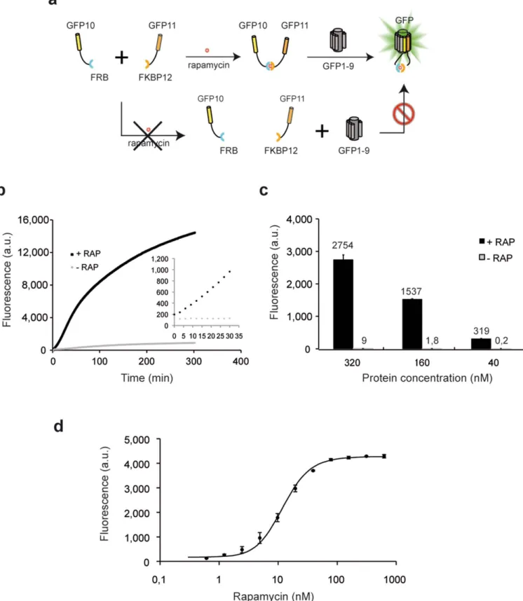
Inducible FRB/FKBP interactions.To further test the specificity and dynamic range of the method, we used the well-studied FKBP12-FRB rapamycin inducible protein interaction19. We fused GFP10 to the
rapamycin-binding domain of the mammalian target of rapamycin (mTOR) kinase (FRB) and GFP11 to the FK506-binding protein 12 (FKBP) and tested their association in vitro upon addition of recom-binant GFP 1–9 protein (Fig. 5a). GFP10-FRB and FKBP-GFP11 fusion proteins were co-expressed from bicistronic pTET GFP10-IRBS-GFP11 vector (Supplementary Fig. S3). In earlier studies of interacting proteins in cell extracts using BiFC, misfolding and aggregation issues interfered with complementation unless the Figure 4|Characterization of protein-protein interactions using coiled-coil heterodimerization. (a) Expression vectors for tripartite split-GFP interaction assays in E. coli: bicistronic pTET tetracycline inducible vector harboring GFP10 and GFP11 tags with cloning sites for test proteins; IPTG inducible pET vector (T7 promoter) for stable expression of GFP1–9 OPT. (b) Colony fluorescence on plates after Antet and IPTG co-induction driving expression of GFP10-K1/E1-GFP11 pair (K/E) (top) and GFP10-E1/E1-GFP11 (E1/E1) (bottom) with GFP1–9 detection fragment. (c) In vitro quantification of K1/E1 or E1/E1 interaction with increasing amount of E-GFP11 (0.4 nM up to 800 nM). Fluorescence values 1 h after initiating complementation with GFP1–9. (d) Initial velocity rates of complementation reactions with various concentrations of GFP1–9 detection reagent (0.5 mM up to 8 mM) in GFP10-K1/E1-GFP11 assay (1 mM of each tagged species).
Figure 5|Application of the tripartite split-GFP to study the ramapycin inducible FRB/FKBP interaction. (a) GFP10 and GFP11 tags were fused to FRB and FKBP proteins. Rapamycin ligand binding brings both protein fusions into proximity, permitting GFP fluorescence reconstitution upon addition of GFP1–9. (b) Raw fluorescence progress curves for GFP1–9 complementation with soluble extracts of GFP10-FRB and FKBP-GFP11 fusions in presence (1RAP) or absence (2RAP) of rapamycin (starting time marked by addition of rapamycin to initiate complementation) (c) Fluorescence levels of GFP10-FRB and FKBP-GFP11 assayed at various concentrations (320, 160, and 40 nM in each) initiated by addition of rapamycin to 150 nM final concentration (black bar) and no rapamycin (gray bar). (d) Rapamycin dose curve (0.6 to 300 nM) for FRB/FKPB binding in vitro measured as final fluorescence after 1 h.
proteins had been co-expressed or were refolded after denaturing8. In
contrast, our soluble crude E. coli cell extracts could be mixed with a 4 mM solution of purified GFP1–9 and incubated for several minutes prior to addition of 150 nM rapamycin, after which the rapid increase in fluorescence indicated soluble interacting GFP10-FRB and FKBP-GFP11 protein. A two-fold increase in fluorescence was easily detectable after only 10 minutes incubation (Fig. 5b, inset), with a half-life of 60 minutes. Without added rapamycin fluorescence remained near blank levels (Fig. 5b). To further examine the behavior of the protein interaction reporter, we studied purified 6-His-GFP10-FRB and 6-His-FKBP-GFP11 fusion proteins (Supple-mentary Fig. S5b). In the presence of 150 nM rapamycin (much greater than the reported Kd of ca. 12 nM19), endpoint fluorescence
was directly proportional to the amount of added protein complex as expected (Fig. 5c). To further evaluate reaction kinetics and equilibria of rapamycin-FKBP/FRB protein association, we tested the effect of increasing concentrations of rapamycin (0.05 to 300 nM). The rapamycin dose response curve presents a half maximal value of 13.5 nM, in accordance with previously published data19(Fig. 5d).
Monitoring the association of protein complexes in E. coli.Protein signaling modules often involve more than two interacting partners. To examine the ability of our tagging system to detect multimeric complexes, we selected two polycistrons from the E. coli genome encoding the heterotrimer Tus BCD complex (YheNML)20 and a
putative allophanate hydrolase constituted by the dimeric assembly YBGK/YBGJ21. We also studied an alternate version of the Tus
complex where YheN subunit was omitted, thus preventing stable association of the complex as previously reported20. These
polycistrons were PCR amplified from genomic DNA including their internal ribosome binding sites. Translation of individual complex subunits was dependent on natural IRBS present in the polycistron, so the first subunit had an N-terminal GFP10 and the last subunit had a C-terminal GFP11 furnished by the pTET SpecR vector (Fig. 6a). To ensure sufficient flexibility between complex subunits, especially for the YheNML whose subunits have nearly buried N-termini in the assembled complex20, 30 and 25-mer
linkers were inserted between the polycistron and the split-GFP tags (Fig. 6b and Supplementary Fig. S3). These constructs were transformed into E. coli cells expressing either the GFP1–9 fragment for complex detection (requiring both the GFP10 and GFP11 tag), or the GFP1–10 fragment16 to assess the expression
level and solubility by monitoring the GFP11 tag (Fig. 6b and 6c). All the complexes were well expressed and highly soluble as shown in Fig. 6c, column A, B. Cells expressing the trimeric YheNML complex became fluorescent after 4h complementation with GFP1–9, as expected (Fig. 6c, 1D). In contrast, weak fluorescence was seen for colonies expressing only YheM and YheL (missing the YheN subunit) with GFP1–9 (Fig. 6c, 2D). Colonies expressing the YBGJ/YBGK control complex were brightly fluorescent after
Figure 6|Monitoring complex formation and stability inE. coli. (a) Analysis of YheNML heterotrimeric complex formation using split-GFP. GFP1–9 complementation provides information on interaction of GFP10 and GFP11 tagged domains, while GFP1–10 binds to GFP11 tagged protein, thus providing expression and solubility levels of the GFP11 tagged protein or complex. (b) E. coli polycistrons YheNML, YheML and YBJG/K were subcloned between GFP10 and GFP11 strands with extended linkers (l25 and l30) into pTET GFP10/11 tet inducible plasmids. (c) Split-GFP assay in E. coli cells expressing either GFP1–9 or GFP1–10. Sequential or co-induction was performed as previously described. Corresponding semi-quantitative measure of colony fluorescence using NIH Image (right).
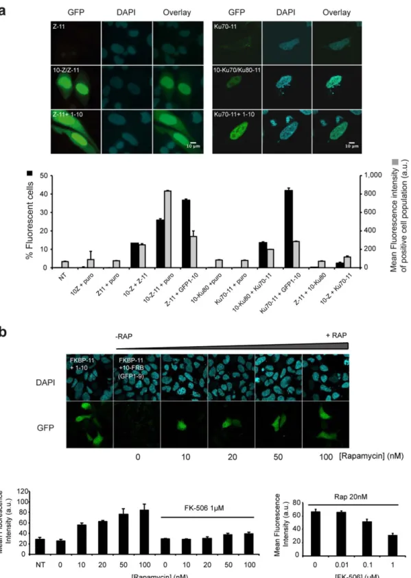
Figure 7|Visualization of complex formation in mammalian cells. (a) Leucine zipper and Ku70/80 heterodimerization. Cells transiently expressing GFP1–9 with GCN4 zipper (Z-11) (left panel) or Ku70-GFP11 (Ku70-11) (right panel) display no background fluorescence. Heterodimerization is visualized as fluorescence with interacting leucine zippers GFP10-Z and Z-GFP11 in CHO cells expressing GFP1–9. Complementation of GFP11-Z with GFP1–10 confirms localization of the zipper alone (bottom). Ku70/80 complex formation is visualized in cell nuclei (right panel). Expression of one Ku component tagged with GFP11 is monitored with GFP1–10. FACS analysis of HEK 293_GFP 1–9 cell lines transfected with corresponding constructs (24 h after transfection). Percentage of fluorescent cells (black bars); mean fluorescence intensity of the positive cell population (gray bars). Self-associating GFP10-Z-GFP11 domain is used as positive control of transfection and complementation with GFP1–9 (mean 6 SD; N 5 3). (b) Rapamycin induced FRB/FKBP interaction in mammalian cells. Stable HEK 293 cells expressing GFP1–9 were co-transfected with GFP10-FRB and FKBP-GFP11 constructs and stimulated with increasing concentrations of rapamycin (RAP) (0, 10, 20, 50 and 100 nM). Bipartite complementation of FKBP-11 and GFP1–10 is shown in the most left image; Green fluorescence at 488 nm excitation (GFP), DAPI nuclear staining (cyan). Scale bars 5 10 mm. Bottom panel: FACS quantification of rapamycin induced association with or without addition of competitive inhibitor FK-506 (1 mM). Right graph: addition of increasing concentrations of FK-506 (20 nM rapamycin; 0.01, 0.1 and 1 mM FK-506) (mean 6 SD; N 5 3).
complementation with GFP1–9 as expected (Fig. 6c, 3D). These results are in good agreement with those obtained in our previous bead-based assays using GFP1–10 complementation21.
Visualization of protein-protein interactions in mammalian cells. We adapted the split-GFP fragments for optimized expression in mammalian cells and tested the formation of several eukaryotic complexes by fluorescence microscopy. As proof of principle, we first fused the yeast GCN4 leucine zipper22 to each of the GFP10
and GFP11 tags and co-transfected both constructs along with the GFP1–9 detector fragment into CHO cells to study the formation of GCN4 heterodimer (Fig. 7a, left panel). Nuclear fluorescence corresponding to leucine zipper heterodimerization was observed in cells expressing GFP1–9 after 24 h of transfection (Fig. 7a, lane 2). As expected, co-expression of GFP1–9 and one of the leucine zipper domain C-terminally fused to GFP11 did not produce fluore-scence (Fig. 7a, lane 1). In a separate experiment we observed the fluorescence complementation of the GFP11 tagged proteins with GFP1–10, which measures soluble protein levels in cells15. As
expected, the leucine zipper domain alone was strongly localized in the nucleus and only weakly in the cytoplasm (Fig. 7a, lane 3). We were also able to detect much larger protein complexes, such as the Ku70–Ku80 complex23that is required in the non-homologous
end-joining (NHEJ) pathway in DNA repair. We successfully detected heterodimerization between GFP10–Ku80 and Ku70–GFP11 in HEK 293 cells expressing stably the GFP1–9 fragment (Fig. 7a, right panel). To shed additional light on possible back-ground from the tripartite assay in living cells, we co-expressed leucine-zipper and Ku proteins that localize in the same sub cellular compartment. Quantification of the fluorescence levels by flow activated cell sorting (FACS) indicated basal levels of fluore-scence from individual split-GFP controls and between non-interacting proteins tested. Pairwise leucine zipper and Ku subunit heterodimerization led to fluorescence levels comparable to the bipartite split-GFP assay, which titrates soluble protein levels in cells15 (Fig. 7a). In parallel, we co-expressed GFP10-FRB and
FKBP-GFP11 protein fusions in HEK 293_GFP1–9 cells, stimu-lated with increasing concentrations of rapamycin. Fluorescence levels for the unstimulated cells remain basal, whereas bright fluorescent cells could be visualized with as little as 10 nM rapamy-cin, in the range of the EC50value measured for the ternary complex
in vitro24 (Fig. 7b). Moreover, pre-addition of an excess of
competitive inhibitor FK-506 totally prevented FRB/FKBP associa-tion by rapamycin in an FK-506 dose dependent manner (Fig. 7b, bottom panel), thus demonstrating that tripartite split-GFP complementation can be used to monitor inhibitors of protein-protein interactions by small molecule compounds prior to association of interacting partners.
Discussion
Here we describe the first protein-protein interaction reporter based on tripartite split-GFP association. Instead of the bulky and poorly folded BiFC fragments of GFP12,25, our tags based on small
engi-neered b-strands of GFP (,20 amino acids long) minimize protein interference and aggregation. Interaction assays using the E-coil/ K-coil model18 have a sensitivity limit in the picomole range.
Chemically induced interactions of the FRB/FKBP complex are detectable using the split-GFP within a few minutes after addition of rapamycin in vitro. Bulky BiFC fragments from various fluor-escent proteins are expressed largely in E. coli in inclusion bodies8,
and the effect of the fragments from other enzyme-based PCA assays on the folding of fused proteins is poorly understood26. Here we show
that our tagging system is highly soluble, permitting production of high yield of fusion proteins at 37uC in E. coli. The assay is therefore not temperature dependent, unlike other BiFC for which co-express-ion and decreasing growth temperature, or refolding denatured
extracts are the only ways to improve assembly of split-GFP frag-ments8,27. We have shown that the method is sensitive enough to
detect association of proteins expressed from E. coli polycistronic mRNAs, and fluorescence is correlated with independently measured protein complex formation and stability. As shown for the TusABC (YheNML) studies above, we could monitor the forma-tion of larger trimeric complexes by extending the length of the linkers between the GFP tag and the protein of interest. This suggests that precise geometry of the GFP10 and GFP11 is not critical for interaction with GFP1–9, but rather that reduction of entropy is important for triggering assembly of the GFP10, GFP 11, and GFP1–9.
The newly described tripartite split-GFP assay is a promising tool to study protein-protein interactions in vitro and in living cells. Its chief advantage over the bulky aggregation-prone fragments of exist-ing BiFC is the small sizes of the GFP10 and GFP11 taggexist-ing peptides. It is therefore well suited to study interactions of unstable protein complexes that are difficult to detect with larger GFP tags. Our sys-tem greatly expands the prospect for protein-interaction screening and the design of new biosensors for protein complex assembly and association28. Although association of the 3-body GFP fragments is
currently irreversible, this system can be exploited to turn on the detection of protein complex formation by simple addition of GFP1–9 reagent. The method is particularly well adapted to high-throughput interaction screens of libraries of protein-protein inter-faces and domains29, the study of complex formation stability, and
the robust detection of soluble proteins and protein complexes by flow cytometry. The technology should prove useful for screening small molecule compounds, for example inhibitors for the interfaces of macromolecular protein complexes. Further work might yield split tripartite GFPs whose assembly can be regulated with light30.
Methods
Cloning.K1-coil and E1-coil DNA, FRB (NM_004958) and FKBP (NM_000801) sequences were amplified by PCR using synthetic oligonucleotides (SI note) and cloned into a pTET ColE1 SpecR GFP10/11 via NdeI:KpnI (GFP10 fusion) or SpeI:BamHI (GFP11 fusion). Linker extension of pTET GFP10/11 was performed using inverse PCR with synthetic oligonucleotides (Supplementary Fig. S3). GFP1–9 was cloned into a pET28a p15 Kan vector via NdeI:BamHI sites. Proteins expressed with GFP10 (E1 and K1 coils, FRB) and proteins expressed with GFP11 (E1 coil, FKBP) were subcloned into a pET vector bearing a C-terminal or N-terminal 6-His tag respectively, prior to purification and characterization. For mammalian expression, all the constructs used in the study were derived from pcDNA 3.1 Zeo vector backbone encoding GCN4-hGLuc1 and hLuc2-GCN4 kindly provided by Stephen Michnick. Split-luciferase domains were replaced with GFP10 and GFP11 fragments. Ku80, FRB were cloned into BspeI:XbaI sites of pcDNA_GFP10; Ku70 and FKBP were amplified from cDNA and inserted into NotI:ClaI cloning sites of pcDNA_GFP11 vector.
Directed evolution of split-GFP fragments for tripartite complementation.The DNA construct encoding (GFP10)-l1(DVGSGGGS)-NdeI::GGGSGSGG::BamHI-l2 (GGGSGGGS)-(GFP11), where GFP10 and GFP11 sequences were derived from superfolder GFP (Supplementary Note), was evolved by DNA shuffling. Libraries of GFP10–11 variants expressed from pTET SpecR vector were screened for improved solubility using a sequential induction protocol16in E. coli cells containing GFP1–9
M1 on a pET p15 vector. At each round, protein solubility of selected optima was verified by complementation with GFP1–10 in vitro. From the six brightest clones sequenced after three rounds of evolution, one best mutant (#5) was identified, termed GFP11 M4 (Supplementary Note). Upstream GFP10 fragment (GFP10 M1) was further evolved by DNA shuffling and primer doping mutagenesis with a pool of fourteen synthetic oligonucleotide primers. Each primer was centered at one of the fourteen amino acids of the GFP10 M1 domain, containing an NNN coding degeneracy at the central target amino acid and flanking homology to the GFP10 M1 in the context of the cloning vector. A partially soluble protein HPS was then inserted in the NdeI:BamHI cloning site to obtain (GFP10 mutant M1)-l1-HPS-l2-(GFP11 mutant M1) in pTET SpecR vector. After three rounds of selection using the sequential induction format from the pTET and pET plasmids16, one best-performing
clone, termed (GFP10 M2)-L1-Nde1::HPS::BamH1-L2-(GFP11 M4) was isolated. GFP10 M2-HPS-GFP11 M4 fusion and GFP1–9 M1 inserts were swapped between pTET and pET plasmids to perform directed evolution of GFP1–9 M1. Briefly, cDNAs libraries of GFP1–9 were expressed from pTET SpecR plasmids and tested for in vivo complementation assays in an E. coli strain expressing GFP10 M2-HPS-GFP11 M4 fusion in pET p15. At the second round, GFP1–9 OPT was isolated and sequenced.
GFP1–9 in vitro assays.Production of recombinant GFP1–9 OPT protein fragment was performed according to previous protocol described for split-GFP 1–1016. GFP 1–
9 was expressed from a pET p15 vector without a 6His tag and produced after induction 1 mM IPTG for 4 hours at 37uC. The protein was refolded from washed inclusions bodies (see GFP1–10 protocol16) and solubilized in TNG buffer. To
perform kinetic characterization of the newly engineered GFP1–9 OPT and GFP 1–9 M1, 180 ml of equal amounts of GFP1–9 refolded pellet fractions (ca. 0.5 mg/ml) were mixed with 20 ml of a soluble protein control sulfite reductase (SR) in fusion with sandwich GFP10 and GFP11 (GFP10-SR-GFP11) or GFP10–11 peptide (3.5 mM each). For K1/E1 and E1/E1 kinetic saturation studies, 25 ml of E1-GFP11 (6.25 mM) were mixed with 50 ml of GFP10-K1 or GFP10-E1 at various concentrations (800 nM down to 0.4 nM). For ligand induced interactions, rapamycin (LC laboratories) was diluted in DMSO and added in 10 ml aliquot to the final FRB/FKBP protein assay mix 20 ml of GFP10-FRB 1 20 ml FKBP-GFP11 (0.5 mg/ml). Assay was repeated by diluting FRB/FKBP samples in TNG buffer. Complementation was induced by addition of a large excess of GFP1–9 OPT (150 ml, 0.25 mg/ml). Fluorescence kinetic (lexc5488 nm/lem5530 nm) was monitored with a FL600 Microplate
Fluorescence Reader (Bio-Tek), at 3 min intervals, for 15 h. The background fluorescence of a blank sample (150 ml of GFP1–9 OPT, 0.25 mg/ml and 50 ml 0.5% (w/v) BSA in TNG buffer) was subtracted from the final fluorescence values. Mammalian cell interactions assays.CHO cells were grown in Ham’s F-12 medium (Gibco, Invitrogen Co) and 10% (v/v) fetal bovine serum (FBS); HEK 293 cells were grown in Dulbecco’s Modified Eagle Medium (DMEM) and 10% (v/v) FBS. CHO cells were co-transfected with Lipofectamine 2000 (Gibco, Invitrogen Co.) with plasmids encoding for GFP1–9 OPT, GCN4-GFP11, GFP10-GCN4 or GCN4-GFP11 M41 GFP1–10. Stable GFP1–9 cell lines were produced by lentiviral transduction of HEK 293. HEK 293_GFP1–9 cells were co-transfected with jetPRIME reagent (Polyplus transfection) with 0.5 mg of each plasmid (GFP10 and GFP11 fusions); Twenty-four hours after transfection, cells were washed with PBS and mounted in DAPI-containing ProLong Gold antifading reagent (Molecular Probes). Imaging was performed using a LEICA DM-RB fluorescence microscope, with a 403 and 1003 oil immersion objective to visualize the stained cells. Images from CHO cells were acquired with a Photometric CoolSNAP HQ camera and analysed with Metamorph or ImageJ softwares. Imaging of HEK cells was performed using a Zeiss (Carl Zeiss) confocal laser scanning inverted microscope (LSM 710 NLO with Quazar spectral detector array). Flow cytometry measurements were performed using a BD Biosciences FACSCaliburTMcytometer. Data analysis was performed using
CellQwestH software (BD Biosciences).
1. Day, R. N. & Davidson, M. W. The fluorescent protein palette: tools for cellular imaging. Chem Soc Rev 38, 2887–2921 (2009).
2. Pfleger, K. D. & Eidne, K. A. Illuminating insights into protein-protein interactions using bioluminescence resonance energy transfer (BRET). Nat Methods 3, 165–174 (2006).
3. Pelletier, J. N., Arndt, K. M., Pluckthun, A. & Michnick, S. W. An in vivo library-versus-library selection of optimized protein-protein interactions. Nat Biotechnol 17, 683–690 (1999).
4. Rossi, F., Charlton, C. A. & Blau, H. M. Monitoring protein-protein interactions in intact eukaryotic cells by beta-galactosidase complementation. Proc Natl Acad Sci U S A 94, 8405–8410 (1997).
5. Galarneau, A., Primeau, M., Trudeau, L. E. & Michnick, S. W. Beta-lactamase protein fragment complementation assays as in vivo and in vitro sensors of protein protein interactions. Nat Biotechnol 20, 619–622 (2002).
6. Shekhawat, S. S. & Ghosh, I. Split-protein systems: beyond binary protein-protein interactions. Curr Opin Chem Biol 15, 789–797 (2011).
7. Hu, C. D. & Kerppola, T. K. Simultaneous visualization of multiple protein interactions in living cells using multicolor fluorescence complementation analysis. Nat Biotechnol 21, 539–545 (2003).
8. Magliery, T. J. et al. Detecting protein-protein interactions with a green fluorescent protein fragment reassembly trap: scope and mechanism. J Am Chem Soc 127, 146–157 (2005).
9. Morell, M., Espargaro, A., Aviles, F. X. & Ventura, S. Detection of transient protein-protein interactions by bimolecular fluorescence complementation: the Abl-SH3 case. Proteomics 7, 1023–1036 (2007).
10. MacDonald, M. L. et al. Identifying off-target effects and hidden phenotypes of drugs in human cells. Nat Chem Biol 2, 329–337 (2006).
11. Kodama, Y. & Hu, C. D. An improved bimolecular fluorescence complementation assay with a high signal-to-noise ratio. Biotechniques 49, 793–805 (2010). 12. Hu, C. D., Chinenov, Y. & Kerppola, T. K. Visualization of interactions among
bZIP and Rel family proteins in living cells using bimolecular fluorescence complementation. Mol Cell 9, 789–798 (2002).
13. Waldo, G. S., Standish, B. M., Berendzen, J. & Terwilliger, T. C. Rapid protein-folding assay using green fluorescent protein. Nat Biotechnol 17, 691–695 (1999).
14. Pedelacq, J. D., Cabantous, S., Tran, T., Terwilliger, T. C. & Waldo, G. S. Engineering and characterization of a superfolder green fluorescent protein. Nat Biotechnol 24, 79–88 (2006).
15. Cabantous, S., Terwilliger, T. C. & Waldo, G. S. Protein tagging and detection with engineered self-assembling fragments of green fluorescent protein. Nat Biotechnol 23, 102–107 (2005).
16. Cabantous, S. & Waldo, G. S. In vivo and in vitro protein solubility assays using split GFP. Nat Methods 3, 845–854 (2006).
17. Kaddoum, L., Magdeleine, E., Waldo, G. S., Joly, E. & Cabantous, S. One-step split GFP staining for sensitive protein detection and localization in mammalian cells. Biotechniques 49, 727–728, 730, 732 passim (2010).
18. Tripet, B. et al. Engineering a de novo designed coiled-coil heterodimerization domain for the rapid detection, purification and characterization of recombinantly expressed peptides and proteins. Protein Eng 10, 299 (1997). 19. Banaszynski, L. A., Liu, C. W. & Wandless, T. J. Characterization of the FKBP.
rapamycin. FRB ternary complex. J Am Chem Soc 127, 4715–4721 (2005). 20. Numata, T., Fukai, S., Ikeuchi, Y., Suzuki, T. & Nureki, O. Structural basis for
sulfur relay to RNA mediated by heterohexameric TusBCD complex. Structure 14, 357–366 (2006).
21. Lockard, M. A. et al. A high-throughput immobilized bead screen for stable proteins and multi-protein complexes. Protein Eng Des Sel 24, 565–578 (2011). 22. Remy, I. & Michnick, S. W. A highly sensitive protein-protein interaction assay
based on Gaussia luciferase. Nat Methods 3, 977–979 (2006).
23. Cary, R. B., Chen, F., Shen, Z. & Chen, D. J. A central region of Ku80 mediates interaction with Ku70 in vivo. Nucleic Acids Res 26, 974–979 (1998). 24. Marz, A. M., Fabian, A. K., Kozany, C., Bracher, A. & Hausch, F. Large
FK506-binding proteins shape the pharmacology of rapamycin. Mol Cell Biol 33, 1357–1367 (2013).
25. Ghosh, I. Antiparallel Leucine Zipper-Directed Protein Reassembly: Application to the Green Fluorescent Protein. J. Am. Chem. Soc. 122, 5658–5659 (2000). 26. Ozawa, T. Designing split reporter proteins for analytical tools. Anal Chim Acta
556, 58–68 (2006).
27. Robida, A. M. & Kerppola, T. K. Bimolecular fluorescence complementation analysis of inducible protein interactions: effects of factors affecting protein folding on fluorescent protein fragment association. J Mol Biol 394, 391–409 (2009).
28. Kellermann, S. J., Rath, A. K. & Rentmeister, A. Tetramolecular fluorescence complementation for detection of specific RNAs in vitro. Chembiochem 14, 200–204 (2013).
29. Pedelacq, J. D. et al. Experimental mapping of soluble protein domains using a hierarchical approach. Nucleic Acids Res (2011).
30. Do, K. & Boxer, S. G. Thermodynamics, kinetics, and photochemistry of beta-strand association and dissociation in a split-GFP system. J Am Chem Soc 133, 18078–18081 (2011).
Acknowledgements
We gratefully acknowledge Dr. P. Calsou and Dr. P. Frit for providing Ku80 and Ku70 cDNAs, S. Michnick for the GCN4-hGLuc1 and hLuc2-GCN4 vectors. We thank Sophie Allart and Astrid Canivet for technical assistance at the Cellular Imaging Facility of CPTP, Toulouse. This work was supported by the National Institutes of Health NIH 1 U54 GM074946-01 to SC, GSW and TCT (LANL), and LDRD/DOE ER projects 20110443ER and 20100520ER (LANL). the French ‘Re´gion Midi-Pyre´ne´es’, INSERM and University of Toulouse to FK, SC, JDP, GF, and a DTRA-TMTI grant to AC.
Author contributions
G.W. and S.C. designed the study. G.W., S.C., H.N., F.K., K.G. and M.L. performed experiments. S.C., H.N., J.D.P. and G.W. wrote the main manuscript text. All authors revised the manuscript. A.C., G.F. provided reagents. G.W. and T.T. supervised the project.
Additional information
Supplementary informationaccompanies this paper at http://www.nature.com/ scientificreports
Competing financial interests:The authors declare competing financial interests. The split-GFP technologies are the subject of domestic and foreign patent applications by Los Alamos National Laboratories on behalf of the Department of Energy and LANS, L.L.C. How to cite this article:Cabantous, S. et al. A New Protein-Protein Interaction Sensor Based on Tripartite Split-GFP Association. Sci. Rep. 3, 2854; DOI:10.1038/srep02854 (2013).
This work is licensed under a Creative Commons
Attribution-NonCommercial-NoDerivs 3.0 Unported license. To view a copy of this license, visit http://creativecommons.org/licenses/by-nc-nd/3.0
1
A New Protein-‐Protein Interaction Sensor Based on Tripartite
Split-‐GFP Association
Supplementary information
Stéphanie Cabantous1,*, Hau B. Nguyen2, Jean-Denis Pedelacq3, Faten Koraïchi1, Anu
Chaudhary4, Kumkum Ganguly2, Meghan A. Lockard5, Gilles Favre1, Thomas C.
Terwilliger2, & Geoffrey S. Waldo2,*
Supplementary Fig.S1: Solubility and in vitro complementation of folding reporter (FR)
and superfolder (SF) GFP BIFC fragments.
Supplementary Fig. S2: Helical coiled-coils model sequences.
Supplementary Fig. S3: Vector map of pTET Bicistronic GFP10-IRBS-GFP11 vector.
Supplementary Fig. S4: Screen of the junction split-point between GFP9 and GFP10
strands.
Supplementary Fig. S5: SDS-PAGE of soluble and Talon® IMAC purified fractions
from 6-His-tagged GFP10 and GFP11 fusion proteins.
Supplementary Table S1: Amino-acid sequence of GFP10 and GFP11 tags
Supplementary Note: Methods and sequences.
2
Fr
ac
%o
n(
so
lu
bl
e(
0(
0,2(
0,4(
0,6(
0,8(
1(
16156(
1576238(
16173(
1746238(
FR(
SF(
b)#
Fluor
esc
enc
e#
ki
ne/
c#
rate
#/
fr
ac
/o
n#
so
lu
bl
e#
0,00E+00#
5,00E+05#
1,00E+06#
1,50E+06#
2,00E+06#
2,50E+06#
3,00E+06#
1=156S#
1=156R#
1=173S#
1=173R#
short#FR# short#SF#Supplementary Figure S1: Solubility and in vitro complementation of folding
reporter (FR) and superfolder (SF) GFP BiFC fragments expressed individually in
E. coli.
We previously developed a bipartite split-GFP system to quantify protein solubility and
follow protein localization in vivo
1,2. Considering that self-association between GFP11 and
GFP1-10 makes this system unsuitable for protein-protein interaction studies, we then studied the
behavior of BiFC fragments
3obtained by fragmentation of folding-reporter GFP
4(FR-GFP) and
Superfolder GFP
5(SF-GFP) at permissive sites 156 and 173
6. a) FR and SF Split-GFP fragments
were produced by splitting GFP at permissive 156 and 173 positions, as described in BiFC
assays
3. Fragments (1-157) and (158-238), (1-173) and (174-238) were expressed from pET
vectors in E. coli at 37°C for 3h. Soluble and insoluble fractions were quantified by SDS-PAGE.
Small GFP fragments are mostly soluble whereas large fragments are largely insoluble. b) In
vitro complementation assay combining soluble fractions from small split-GFP fragments with
the soluble (S) or refolded (R) fractions of the corresponding large fragments 156) and
(1-173). As observed with other fluorescent protein fragments used in refolding-based BiFC
3,
split-GFP fragments from refolded FR-split-GFP self-associate detectably (gray bars). Improved folding in
SF-GFP fragments resulted in improved solubility, but also increased spontaneous fluorescence
for all conditions (whether refolded or soluble extracts) (black bar).
3
KVSALKENVSALKEKVSALTEKVSALKEKVSALKE!
K1#coil#
KVSALENEVSALEKEVSVLEKEVSALEKEVRALEK!
E1#coil#
A"rac&ve)coiled)coil)pair)
Repulsive)coiled)coil)pair))
KVSALENEVSALEKEVSVLEKEVSALEKEVRALEK!
E1#coil#
KVSALENEVSALEKEVSVLEKEVSALEKEVRALEK!
E1#coil#
Supplementary Figure S2: Helical coiled-coils as a model to study protein-protein
interactions.
Developed by Hodges and coworkers
7, E and K coils are constituted by five
repeated heptads of the EVSALEK or KVSALKE motifs (gray boxes). Electrostatic interactions
introduced in the first and sixth positions of the heptads direct preferential interaction, allowing
interchain ionic attraction (K/E) or repulsion (E/E) (black arrows). Valine (V) and Leucine (L)
residues maintain hydrophobic interactions between coils (bold). Directed evolution of wild-type
sequences (G. S. Waldo, unpublished data) resulted in additional mutations that greatly increased
solubility of K coil mutant 1 (K1) and E coil mutant 1 (E1) (underlined) while maintaining the
specificity and tight binding of the wild type. E1 and K1 coils were used in our interaction
studies.
4
a)
!"#$%&' ()*+' ,-' /0123'!"#$%&' 4' *' /0122'5
.-' ,6%(' ;:#(' +:%(' *789(' !"#<2'=&"' $%$)' +:%>)':?<?'
/0123@()*+@/0122'
!
"#"$ /0123' /0122'5
b)
long linker (30-mer) GFP10 tag
D V G G G G S E G G G S G G P G S G G E G S A G G G S A G G G S GACGTTGGTGGTGGCGGATCAGAAGGAGGCGGTAGCGGGGGCCCTGGTTCGGGAGGGGAAGGTTCTGCTGGGGGAGGGAGCGCTGGCGGGGGGTCT CTGCAACCACCACCGCCTAGTCTTCCTCCGCCATCGCCCCCGGGACCAAGCCCTCCCCTTCCAAGACGACCCCCTCCCTCGCGACCGCCCCCCAGA
linker (25-mer) GFP11 tag
G S G A G G S P G G G S G G S G S S A S G G S T S GGATCCGGCGCTGGCGGAAGCCCTGGGGGCGGGAGCGGTGGCTCTGGTTCTTCTGCTAGTGGCGGCTCAACATCT CCTAGGCCGCGACCGCCTTCGGGACCCCCGCCCTCGCCACCGAGACCAAGAAGACGATCACCGCCGAGTTGTAGA
Supplementary Figure S3: Vector map of pTET Bicistronic GFP10-IRBS-GFP11
vector.
a) The pTET GFP10-IRBS-GFP11 vector is based on our previously designed
expression vector
8. It includes a bicistronic expression cassette under the control of the tet
promoter. In one tagging module, N-terminal GFP10 is separated from protein A by a 12-mer
linker (cloning sites NdeI/KpnI). An internal ribosome-binding site separates it from a second
module comprising SpeI/BamHI cloning sites for protein B), a 12-mer linker, and the C-terminal
GFP11 tag. b) Longer linkers of 30-mer (GFP10 tag) and 25-mer (GFP11 tag) were used for
interaction assays of larger proteins (>50kD). The pTET ColE ori GFP10-IRBS-GFP11 vector
was used alone for in vitro assays or in combination with compatible pET p15 ori GFP1-9 OPT
plasmid for studying interactions in E. coli.
5
Supplementary Figure S4: Effect of split-point at the linker junction between GFP9
and GFP10 ß-strands.
a) To investigate the optimal split position between ß-strands GFP9 and
GFP10, we designed GFP1-9 with extended C-termini (red arrows) and corresponding GFP10
with truncated N-terminus (black arrows). b) Several truncated versions of GFP1-9 were
expressed from pET vectors and tested for complementation in vivo with the non-interacting E/E
or interacting K1/E1 coiled-coil pair expressed from pTET bicistron (GFP10-E1/E1-GFP11) or
(GFP10-K1/E1-GFP11). c) Comparison of the output signal of the same bicistronic constructs as
a function of N-terminal GFP10 truncations, GFP1-9 being kept fixed at amino acid position 193.
Colony fluorescence after 2h induction at 37°C was compared for K1/E1 and E1/E1 coils pairs to
evaluate the optimal split-point. Last permissive complementation was given for position 194,
designating the final version of GFP10 used throughout the study. However, extension of
C-termini of the GFP1-9 fragment (incrementally from amino-acid 193 to 197) had no effect on the
fluorescence levels observed for interacting K1/E1 versus non-interacting E1/E1 coils.
!"!#$%
!"!#&%
!"!#'%
!"!#(%
!"!#)%
*+*%
,+*%
-.%
/.%
0%
'0%
!00%
!'0%
100%
!#$%
!#&%
!#'%
!#(%
!#)%
*+*%
,+*%
2"3456789:%;<=7><8%?@A%!0%
B<:<8
C%DE<5
4=/4
8/4
%%
?@A!0%
?@A%!"#%
170 180 190 200 210 220 230 |....|....|....|....|....|....|....|....|....|....|....|....|... !!!!!!!!!SVQLADHYQQNTPIGDGPVLLPDNGSDLPDDHYLSTQTILSKDLNEKRDHMVLLEYVTAAGITDAS % % %%%%%%%%%!" ! ! ! !!!!!!!#$ ! !!! ! !!!##%9.%
6
Mw# Mw# 1# 2# 3# 4# SOL# FT# EL1# 1# 2# 3# 4# 1# 2# 3# 4# 1# N6his3E13GFP11# 2# GFP103l303K13C6his# 3# GFP103l303K1# 4# GFP103l303E13C6his# 116# 97# 66# 55# 36# 31# 21# 14# 6#a)#
5" 6" 7" 8" SOL" 5" 6" 7" 8" EL" SR"control" "5"""2"""1μl" Mw" Mw" 116" 97" 66" 55" 36" 31" 21" 14" 6" 5" N6his<FKBP<l25<GFP11" 6" GFP10<l30<FRB<C6his" 7" 8" N6his<FKBP<GFP11" GFP10<FRB<C6his"b)"
Supplementary Figure S5: SDS-PAGE of soluble and Talon® purified fractions
from 6HIS-tagged GFP10 and GFP11 fusion proteins
with various linker sizes: 12mer,
25mer (indicated as l25) or 30mer (indicated as l30). To further characterize protein-protein
interaction in vitro, K1 coil, E1 coil, or FRB were purified to 95% homogeneity. A 200 ml culture
of BL21(DE3) cells expressing expressing each construct was grown to OD
600nm~ 0.5 in LB
medium supplemented with 1 mM kanamycin, induced with 1 mM IPTG for 3 h at 37ºC. Soluble
extracts were purified on a 50% v/v slurry of metal affinity resin beads (Talon® resin, Clontech)
in TNG buffer (100mM Tris pH 8.0, 150mM NaCl, 10% glycerol) according to standard
procedures. Samples corresponding to the soluble and eluted fractions were resolved on a 4-20%
gradient Criterion SDS-PAGE gel (Bio-Rad Hercules). Protein samples were stained using Gel
Code Blue stain reagent (Pierce) and imaged using a GS-800 Calibrated Densitometer and
quantified by Bio-Rad Protein Assay (Bio-Rad). a) Coiled-coils. Both C-6His (#2) and untagged
(#3) versions of GFP10-K1 fusion expressed mostly in the soluble fraction. Flow-through
fractions (FT) contained cellular proteins and untagged proteins. 90% pure proteins were
recovered after one-step purification in the eluted fraction (EL) b) FKBP-GFP11 and
GFP10-FRB fusions with different linker lengths. A sulfite reductase control protein (SR) sandwich with
GFP10 and GFP11 tags was used as positive control of protein expression and complementation
with GFP1-9.
7
Fragment
a,b,cAmino acid sequence
GFP10 SF
GFP10 M1 (evolved 10-linker-11) (G194D,
S205T, A206I, S208L, P211L)
GFP10 M2 (evolved 10-HPS-11)
G194D, N198D, S205T, A206I, S208, P211L)
195 200 205 210
|....|....|....|..
MGLPDNHYLSTQSVLSKDPN
MDLPDNHYLSTQTILLKDLN
MDLPDDHYLSTQTILSKDLN
GFP11 SF
GFP11 M4 (F223Y)
215 220 225 230
|....|....|....|..
EKRDHMVLLEFVTAAGITGAS
dEKRDHMVLLEYVTAAGITDAS
aUnderlined residues correspond to structural ß-strand sequence from Superfolder GFP.
bBold residues correspond to the additional point mutations introduced by mutagenesis.
cNumbering corresponds to position in full-length SF GFP.
8
1. Amino-acid sequences of evolved split-GFP fragments
Amino-acid sequence of GFP 10-11 hairpin
GFP10 and GFP11 are separated by a long flexible linker (wt, gray shade) that includes two
cloning sites NdeI and BamHI. The entire cassette was shuffled and selected for optimized
complementation with GFP1-9. Six optima were isolated and sequenced. SM5 corresponds to the
best variant, with mutations identified both in GFP10 (GFP10 M1) and only one mutation in
GFP11 (GFP11 M4), thus differing from the previous GFP11M3 split-GFP domain identified
with GFP1-10 complementation
1. Amino-acids corresponding to structural ß-strands GFP10 and
GFP11 are underlined.
GFP10 NdeI BamHI GFP11 WT YTMGLPDNHYLSTQSVLSKDPNGTGGGSGGGSHMGGGSGSGGGSGGGSTSEKRDHMVLLEFVTAAGITGAS* SM1 YTMDLPDNHYLSTQTILLKDLNGTGVGSGGGSHMGGGSGSGGGSGGGSTSEKRDHMVLLEYVTAAGITDAS* SM2 YTMDLPDNHYLSTQTILLKDLNGTGVGSGGGSHMGGGSGSGGESGGGSTGEKRDHMVLLEYVTAAGITGAS* SM3 YTMDLPDNHYLSTQTILLKDLNGTGVGSGGGSHMGGGSGSGGGSGGGSTSEKRDHMVLLEYVTAAGITDAS* SM4 YTMDLPDNHYLSTQTILLKDLNGTDVGSGGGSHMGGGSGSDGGSGGGSTGEKRDHMVLLEYVTAAGITGAS* SM5 YTMDLPDNHYLSTQTILLKDLNGTGVGSGGGSHMGGGSGSGGGSGGGSTSEKRDHMVLLEYVTAAGITDAS* SM6 YTMDLPDNHYLSTQTILLKDLNGTGGGSGDGCHMDGGSGSGGGSGGGSTGEKRDHMVLLEYVTAAGITGAS*Amino-acid sequence of GFP10 optima in HPS fusion
A partially soluble protein hexulose phosphate synthase (HPS) was cloned into the NdeI and
BamHI cloning sites and the cassette was evolved, excluding HPS. Additional rounds of doped
mutagenesis with oligonucleotides targeting GFP10 M1 were realized, and resulted in best mutant
GFP10 M2. GFP11 sequence was unchanged, as solubility of this fragment was optimized.
GFP10 M1 YTMDLPDNHYLSTQTILLKDLNGTGVGSGGGSHM-(HPS)-GSGGGSGGGSTSEKRDHMVLLEYVTAAGITDAS* GFP10 M2 YTMDLPDDHYLSTQTILSKDLNGTDVGSGGGSHM-(HPS)-GSGGGSGGGSTSEKRDHMVLLEYVTAAGITDAS*
Alignment of GFP 1-9 variants obtained by directed evolution (aa 1-196)
10 20 30 40 50 60 ....|....|....|....|....|....|....|....|....|....|....|....|
GFP1-9 SF MSKGEELFTGVVPILVELDGDVNGHKFSVRGEGEGDATNGKLTLKFICTTGKLPVPWPTL GFP1=9 M1 .R.............I...........F...........S................. GFP1=9 OPT .R.............I...........F.......I...S................. 70 80 90 100 110 120 ....|....|....|....|....|....|....|....|....|....|....|....| GFP1-9 SF VTTLTYGVQCFSRYPDHMKRHDFFKSAMPEGYVQERTISFKDDGTYKTRAEVKFEGDTLV GFP1=9 M1 ............................................................ GFP1=9 OPT ......................................Y..................... 130 140 150 160 170 180 ....|....|....|....|....|....|....|....|....|....|....|....| GFP1-9 SF NRIELKGIDFKEDGNILGHKLEYNFNSHNVYITADKQKNGIKANFKIRHNVEDGSVQLAD GFP1=9 M1 .............................................T.............. GFP1=9 OPT ............................K........N.......T.............. 190 ....|....|....|. GFP1-9 SF HYQQNTPIGDGPVLLP* GFP1=9 M1 ................* GFP1=9 OPT ................*
9
binding to sandwich 10-HPS-11: N39I, S99Y, K149N and K158N.
2. Oligonucleotides used to amplify E. coli polycistrons
Forward NdeI primer to YBGJ of YBGJ/YBGK E.coli
AGATATACATATGCAACGAGCGCGTTGTTATCTGATAGG
Bottom BamHI non-stop to YBGK of YBGJ/YBGK E.Coli
AATTCGGATCCATTTTCATTGTGCAGCCGCCACGCTA
Forward NdeI primer to YheN E.coli
AGATATACATATGCGTTTTGCCATCGTGGTGACCGGGCC
Forward NdeI primer to YheM E.coli
AGATATACATATGAAACGAATTGCGTTTGTTTTTTCTAC
Reverse BamHI primer to YheL E coli
AATTCGGATCCCCAGGCCATCTGGCTGGAGTGCTTAA