HAL Id: inserm-00123527
https://www.hal.inserm.fr/inserm-00123527
Submitted on 11 Jan 2007
HAL is a multi-disciplinary open access
archive for the deposit and dissemination of
sci-entific research documents, whether they are
pub-lished or not. The documents may come from
teaching and research institutions in France or
abroad, or from public or private research centers.
L’archive ouverte pluridisciplinaire HAL, est
destinée au dépôt et à la diffusion de documents
scientifiques de niveau recherche, publiés ou non,
émanant des établissements d’enseignement et de
recherche français ou étrangers, des laboratoires
publics ou privés.
Astroglial expression of the P-glycoprotein is controlled
by intracellular CNTF.
Christelle Monville, Christiane Fages, Anne-Marie Feyens, Veronique d’Hondt,
Catherine Guillet, Ann Vernallis, Hugues Gascan, Marc Peschanski
To cite this version:
Christelle Monville, Christiane Fages, Anne-Marie Feyens, Veronique d’Hondt, Catherine Guillet, et
al.. Astroglial expression of the P-glycoprotein is controlled by intracellular CNTF.. BMC Cell
Biology, BioMed Central, 2002, 3, pp.20. �inserm-00123527�
BMC Cell Biology 2002,
3 x
Research article
Astroglial expression of the P-glycoprotein is controlled by
intracellular CNTF
Christelle Monville
1
, Christiane Fages
1
, Anne-Marie Feyens
2
,
Véronique d'Hondt
2
, Catherine Guillet
3
, Ann Vernallis
4
, Hugues Gascan
3
and Marc Peschanski*
1
Address: 1INSERM U421/IM3, Créteil, France, 2Université Catholique de Louvain, Bruxelles, Belgium, 3INSERM E9928, CHU Angers, France and 4Aston University, Birmingham, United Kingdom
E-mail: Christelle Monville - monvillec@cf.ac.uk; Christiane Fages - fages@im3.inserm.fr; Anne-Marie Feyens - feyens@onco.ucl.ac.be; Véronique d'Hondt - dhondt@onco.ucl.ac.be; Catherine Guillet - catherine.guillet@univ-angers.fr; Ann Vernallis - a.b.vernallis@aston.ac.uk; Hugues Gascan - hugues.gascan@univ-angers.fr; Marc Peschanski* - peschanski@im3.inserm.fr
*Corresponding author
Keywords: differentiation, IL-6-type cytokines, astrogliosis, multi-drug resistance
Abstract
Background: The P-glycoprotein (P-gp), an ATP binding cassette transmembrane transporter, is expressed by astrocytes in the adult brain, and is positively modulated during astrogliosis. In a search for factors involved in this modulation, P-gp overexpression was studied in long-term in vitro astroglial cultures.
Results: Surprisingly, most factors that are known to induce astroglial activation in astroglial cultures failed to increase P-gp expression. The only effective proteins were IFNγ and those belonging to the IL-6 family of cytokines (IL-6, LIF, CT-1 and CNTF). As well as P-gp expression, the IL-6 type cytokines - but not IFNγ - stimulated the expression of endogenous CNTF in astrocytes. In order to see whether an increased intracellular level of CNTF was necessary for induction of P-gp overexpression by IL-6 type cytokines, by the same cytokines analysis was carried out on astrocytes obtained from CNTF knockout mice. In these conditions, IFNγ produced increased P-gp expression, but no overexpression of P-gp was observed with either IL-6, LIF or CT-1, pointing to a role of CNTF in the intracellular signalling pathway leading to P-gp overexpression. In agreement with this suggestion, application of exogenous CNTF -which is internalised with its receptor - produced an overexpression of P-gp in CNTF-deficient astrocytes. Conclusions: These results reveal two different pathways regulating P-gp expression and activity in reactive astrocytes, one of which depends upon the intracellular concentration of CNTF. This regulation of P-gp may be one of the long searched for physiological roles of CNTF.
Background
Recent studies have reported expression of the
transmem-brane transporter P-glycoprotein (P-gp) in astrocytes, in addition to the previously determined endothelial
locali-Published: 31 July 2002 BMC Cell Biology 2002, 3:20
Received: 10 May 2002 Accepted: 31 July 2002 This article is available from: http://www.biomedcentral.com/1471-2121/3/20
© 2002 Monville et al; licensee BioMed Central Ltd. This article is published in Open Access: verbatim copying and redistribution of this article are permitted in all media for any non-commercial purpose, provided this notice is preserved along with the article's original URL.
BMC Cell Biology 2002, 3 http://www.biomedcentral.com/1471-2121/3/20
sation. This second cellular location raises new questions as to the function of P-gp in the brain because, whereas endothelial P-gp clearly participates directly in the trans-port of substrates across the blood-brain barrier for a number of identified substrates [1–4], the physiological roles and substrates of its astroglial counterpart are un-known. One potential clue to these issues is the fact that P-gp expression is stimulated in astrocytes activated by various brain insults [5].
P-gp consists of a group of closely related, intrinsic mem-brane proteins encoded by the multidrug-resistance (mdr) genes [6]. It acts as an ATP-driven efflux pump [7] that ac-cepts a wide range of structurally different substrates, among which are drugs, hormones, corticosteroids, cy-tokines or phospoholipids [8–10]. The adult brain is a major site of expression of P-gp [2,11–15], and it had long been exclusively associated to the endothelial cells in cap-illary walls [2,4,16], until astrocytic endfeet were identi-fied as another specific location in vivo [17,18], and in vitro [19].
In order to start deciphering the physiological role of P-gp in astrocytes, we have chosen to study its increase in ex-pression in reactive astrocytes, by looking for the effects of a wide variety of molecules known to induce astroglial ac-tivation in in vitro culture conditions. This involved, in particular, several agents that had also been shown to stimulate P-gp expression in other cell types, such as Inter-feron-α and -γ (IFN-α, γ) [20–22], Tumor Necrosis Factor alpha (TNF-α) [21,23] and dibutyryl-cyclic-AMP [24], and also a number of other compounds classically used to in-duce astroglial differentiation in vitro. Surprisingly, only a few of these factors drove astroglial P-gp expression, indi-cating that P-gp overexpression is not just related to astro-glial differentiation, but appears as a specific response to certain stimuli. This increase in P-gp expression thus seems to characterize a specific stage of differentiation, which, in one pathway at least, also involves an increased concentration of the ciliary neurotrophic factor (CNTF). The two proteins may be even more functionally linked to each other since some of the "astrogliotic" stimuli able to increase P-gp expression require CNTF as a key intracellu-lar signal.
Results
Enriched astroglial cell cultures were readily obtained from cerebral hemispheres of Swiss mice and of CNTF knockout mice, and maintained for several weeks without signs of cell alteration.
Expression of P-gp in primary astrocyte culture
Astrocytes in culture expressed P-glycoprotein as early as 24 h after plating. The RT-PCR performed on a fragment of the P-gp transcript yielded a 167-bp product (Fig. 1a),
present in all astrocyte samples and in the MES-SA/MX2 cells used as a positive control. At the protein level, the re-sults varied over time: until the 4th day in vitro (div), the
antibody raised against P-gp detected a band at about 120 kDa; from the 6th div a doublet was detected at about
160–170 kDa; finally, at 15 div this doublet became pre-dominant and its expression increased (Fig. 1b).
Effects of molecules promoting astrogliosis on P-gp expres-sion
Among the agents known to activate astrocytes ("astrogli-otic factors") that were tested for modulating P-gp expres-sion, most failed to alter it (Table 1). This was in particular the case for dBcAMP, IL-1β, LPS, NGF, RA, rhIFNα, rhT-GFα and TNFα. In contrast, all the cytokines that belong to the IL-6 family induced a 50% or more increase in ex-pression of P-gp in long-term astrocyte culture. This effect was dose-dependent and was modulated by α receptor subunits, when relevant. For instance, rrCNTF was ineffi-cient when used alone at 30 ng/ml in long-term cultures. However, an increase in the cellular content of P-gp was observed in a significant and reproducible manner when 100, and even further 250 ng/ml of rCNTF were used (Fig. 2a). As previously described for the effects of rCNTF on other astroglial markers of differentiation [25], addition Figure 1
MDR1mRNA and P-gp expression in astroglial cul-tures. a/ RT-PCR showing MDR1mRNA. b/ Western blot-ting using rabbit anti-P-gp polyclonal antibody demonstrablot-ting the presence of P-gp as early as 2 div. Between 2 div and 5 div, a band is observed at about 120 kDa (lower black arrow); a doublet appears at 6 div at about 150–170 kDa (black arrows); the band corresponding to the mature form of P-gp, is expected at 170 kDa (grey arrow), and becomes increased after 15 div.
of 200 ng/ml of c-myc-sCNTFRα induced a potentiation of the effects, and an increase of P-gp cellular content was then observed with the lowest concentration of rCNTF studied (10 ng/ml), and maintained for all other concen-trations used (data not shown). Addition of rhLIF and rhCT-1, in the culture medium provoked an increase of about 1.5 to 2-fold of P-gp cellular content (Fig. 2b). Ad-dition of rmIL-6 at concentrations below 40 ng/ml in the culture medium was inefficient, but a significant increase of P-gp intracellular content was observed when this con-centration was used (Fig. 2c). Addition of rhsIL-6Rα to the culture medium induced a major potentiation of the ef-fects, since, in these conditions, all concentrations used triggered a significant increase in P-gp content (Fig. 3). Selective blockade of P-gp transporter activity was studied using two different specific blockers, S9788 and vera-pamyl. When used at concentrations of 5 µM and 10 pM, a complete blockade of the effects of all IL-6 family cy-tokines was observed. Even the presence of the specific α receptor subunit for IL-6 and CNTF failed to reverse the neutralising effects of S 9788 (Fig. 5) and verapamyl (data not shown).
Besides the IL-6 family of cytokines, the only compound to trigger P-gp overexpression in astrocytes was IFNγ (Fig. 6a). This IFNγ effect was very likely direct since hLIF05, which inhibits the LIF receptor pathway, could not reverse it.
Regulation of P-gp expression in CNTF-/- astrocytes
The progressive maturation of P-gp during development and the regulation of its expression by members of the IL-6 family of cytokines are two features that P-gp shares with CNTF in astrocytes (see discussion section). Moreover, like P-gp, CNTF regulation is very specific, since, among different molecules that normally trigger astrogliosis, only those belonging to the IL-6 family provoked an increase of CNTF (Fig. 7). We, therefore, explored the possibility of a functional link between the two molecules by analysing whether the presence of endogenous CNTF in astrocytes was required for the observed modulation of P-gp expres-sion by IL-6 type cytokines and IFN-γ. This was tested by analysing the effects of the cytokines in astrocytes taken from CNTF knockout mice. In these cultures, the modula-tion of P-gp by LIF, CT-1 or IL-6 (even in presence of its specific α receptor subunit) was completely lost (Fig. 4). In contrast, addition of rrCNTF (250 ng/ml) produced a significant increase of P-gp intracellular level. In the CNTF -/- astrocytes, addition of IFN-γ to the medium triggered an increase of P-gp cellular level comparable to that ob-served in wild-type astrocytes, underlining its direct effect on P-gp modulation (Fig. 6b).
Figure 2
Effect of cytokines on P-gp cellular content in mature astrocytes. a/ Addition of 100 and 250 ng/ml rCNTF during 24 h induced a statistically significant increase as compared to untreated controls (+51.7% ± 15.6, factorial ANOVA signifi-cant at 95%, t-test p < 0.01** and +75.7 ± 30, factorial
ANOVA significant at 95%, t-test p < 0.05*, respectively). b/
Addition of rhLIF during 24 h induced a significant increase of P-gp expression both at 10 and 30 ng/ml (+42.7 % ± 14, fac-torial ANOVA significant at 95%, t-test p < 0.05* and +54.35
% ± 12, factorial ANOVA significant at 95%, t-test p < 0.01**
respectively). The same pattern of regulation was observed with CT-1 at 10 ng/ml (+69.6 % ± 24, factorial ANOVA sig-nificant at 95%, t-test p < 0.01**). c/ Addition of rmIL-6
elic-ited a significant increase of P-gp intracellular content at 40 ng/ml (+68.8 % ± 10, factorial ANOVA significant at 95%, t-test p < 0.001***)
BMC Cell Biology 2002, 3 http://www.biomedcentral.com/1471-2121/3/20
Discussion
The main result of this study is the demonstration of a close interaction between expression of two intrinsic as-troglial proteins, the IL-6 cytokine CNTF and the ATP-binding cassette transmembrane transporter P-gp. Besides a parallel development of expression, the concentration of the two proteins appears, in particular, to be similarly in-creased following stimulation by IL-6-type cytokines whereas a number of other agents eliciting activation of astrocytes are ineffective. Moreover, astroglial expression of CNTF appeared to be necessary for the effects of IL-6-type cytokines on P-gp expression. Reciprocally, blockade of P-gp activity eliminated the effects of cytokines, even CNTF, on its own expression, suggesting a functional link between the two systems.
P-gp and CNTF may be involved together in a specific type of astrocytic activation
The recent discovery of P-gp expression on astrocytic end-feet [17,18] and its induction after a stress [5] has raised the issue of a role of this protein in astrocytes. To address this issue, we have undertaken, here, an analysis of the ex-pression of P-gp over time during development and of its stimulation in mature cells. The first feature in this study is the striking similarity between the results obtained for astroglial P-gp and those previously gathered on the astro-glial cytokine CNTF. Indeed, P-gp and CNTF seem to share parallel modulations of expression both during
develop-ment and under specific stimulations. First, like CNTF [26], P-gp is not detectable in the early stages of postnatal development in the central nervous system. Both proteins are detected from P7, and their concentration levels grad-ually increase, reaching a plateau at P28 [15,26]. Results obtained in in vitro astroglial cultures are also indicative of a parallel maturation of the two proteins. P-gp, both at mRNA and protein levels, was expressed in our study as soon as after 24 hours in culture, but Western blot studies revealed that the protein detected in those early cultures did not have the 170 kDa mature form [15], that became predominant only after 14 days, the point at which astro-cytes exhibit a mature phenotype (see discussion in 25). A very similar time course of expression was observed previ-ously for CNTF [25]. Before 7 days in vitro, immature as-trocytes did not express detectable levels of CNTF, whereas at 14 div, when astrocytes form a confluent mon-olayer and express high levels of GFAP, they exhibit a sig-nificant intracellular content of CNTF.
In vitro, astrocytes activation experiments further indicate a parallel regulation of the two proteins. Even though in
vivo models of astrogliosis have revealed a concomitant
overexpression of both proteins [5,27], most factors that readily induce astroglial activation in vitro fail to increase their expression [28,29] and the present study). Indeed, Figure 3
Potentiation of IL-6 effects on P-gp expression by adding its specific α receptor subunits. Addition of solu-ble IL-6R induced a major potentiation of rmIL-6 whatever the concentration of the factor used.
Figure 4
Effect of cytokines of the IL-6 family on P-gp intracel-lular content in CNTF-/- astrocytes. Only rrCNTF (250 ng/ml) provoked a significant increase in P-gp intracellular content (+43.8 % ± 20, factorial ANOVA significant at 95%, t-test p < 0.01**).
for both proteins, only IFNγ (in the study by Carroll and colleagues, 31, for CNTF) and the IL-6 family cytokines could trigger a quantifiable increase in expression in astro-cytes (this study).
All these results suggest that the overexpression of CNTF and P-gp participate in the definition of a particular stage of astroglial differentiation. So-called astrogliosis has long been considered as a discrete "activation" stage of astro-cytes, on the basis of its characteristic morphological fea-tures. However, more recent studies exploring the expression of specific molecules have questioned this view, drawing attention to the fact that reactive gliosis var-ies qualitatively and quantitatively depending on both the nature of the injury and the microenvironment of the in-jury site (see references and discussion 30 and 31). The nature of the injury and the microenvironment necessary for the differentiation stage of astrocytes that involves CNTF and P-gp overexpression is, at this point, a matter of speculation. It is interesting to note, nevertheless, that, in agreement with the in vitro data, intra-cerebral administra-tion of adenovirus recombinant for CNTF produced an in-crease in the astroglial content of endogenous CNTF without triggering massive "astrogliotic" morphological changes [32].
Intracellular concentration of CNTF as a modulator of P-gp expression in astrocytes
Out of the present study, the relationship between CNTF and P-gp in astrocytes appears as a functional link rather than as a mere co-regulation. Indeed, one main result ob-tained in this study was the apparent requirement of an intracellular level of CNTF for the expression of P-gp to be modulated by IL-6-type cytokines, since this modulation was not seen in cells that did not express the CNTF gene. The only exogenous cytokine that remained effective in CNTF-/- cells was CNTF itself. This may be explained by the fact that exogenous CNTF, when bound to its receptor, is internalised into the cells [33]. As discussed by these au-thors, endocytosed CNTF may be active inside the cell, as has been demonstrated for neurotrophins/Trks complexes (see 34 for a review). Through this mechanism, therefore, restoration of an intracellular content of CNTF may be-come sufficient to trigger a increased P-gp expression. How an increase in the cellular content of CNTF increases the expression of P-gp was not directly addressed in this study. Nevertheless, a number of elements help to suggest a working hypothesis. A direct transcriptional role of CNTF on the P-gp gene promoter is unlikely for three rea-sons. First, it has been well demonstrated that the intrac-ellular signalling systems triggered by CNTF are essentially similar to those triggered by LIF [35,36]. Why would LIF not be effective in CNTF-/- astrocytes when CNTF is,
Table 1: Modulation of P-gp expression by agents that induce astroglial reactivity.
Treatment Concentration Effect
dBcAMP 0,5 mM - 3.1 ± 8 IL-1β 50 ng/ml +10.5 ± 11 Lesion / - 12.1 ± 8 LPS 1 µg/ml + 2.3 ± 8 NGF 10 ng/ml - 21.7 ± 22 RA 10-7 M - 28.6 ± 20.2 rhIFN-β 600 U/ml -20 ± 11 rhIFNγ rhTGFα 300 U/ml 50 ng/ml +50 ± 30 -9 ± 5 TNFα 100 ng/ml + 7.6 ± 9.1 rhCT-1 10 ng/ml + 69.6 ± 24** rhLIF 30 ng/ml + 54.3 ± 12** rmIL-6 40 ng/ml +68.8 ± 10*** RmIL-6 50 ng/ml +220.28 ± 22*** + rhsIL-6R 500 ng/ml rrCNTF 250 ng/ml + 75.7 ± 30*
*: ANOVA significant at 95%, t-test p < 0.05; **: ANOVA significant at
95%, t-test p < 0.01; ***: ANOVA significant at 95%, t-test p < 0.001.
Figure 5
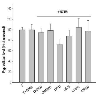
Effect of P-gp blockers. Addition of the P-gp blocker S9788 in the culture medium blocked completely the effect of rrCNTF at 250 ng/ml (-1.5 vs. +75.7 %), rhLIF at 30 ng/ml (-11.5 vs. + 54.3%) and rhCT-1 at 30 ng/ml (-2.5 vs. +26.1%) on P-gp expression in astrocytes.
BMC Cell Biology 2002, 3 http://www.biomedcentral.com/1471-2121/3/20
would be difficult to explain in case of a direct transcrip-tional effect. Second, our results demonstrate that the con-trol of P-gp expression requires a modulation of the intracellular content of the cytokine. Third, the stimula-tion of P-gp expression by cytokines was blocked with an-tagonists of the activity of the transporter. This indicates that the regulation of P-gp depends upon its own activity and suggests the existence in astrocytes of a positive feed-back of the molecule upon its own expression. Such a sug-gestion is concordant with the well demonstrated positive
feedback of P-gp activity upon its expression in various tu-mor cells, in the presence of non-organic substrates like cytotoxic drugs [37,38].
In a mechanistic model based upon this hypothesis, en-dogenous astroglial CNTF would most likely not appear as a usual cytokine activating signalling pathways after binding to a transmembrane receptor system, but as a "sensor", itself modulated by extrinsic cues. The changes induced in the intracellular concentration of this "sensor" might affect the activity of the P-gp transmembrane trans-porter and, eventually, lead to a modulation of its gene ex-pression (Fig. 8). Such a scheme suggests a physiological role for CNTF, a cytokine which, because of the absence of a signal peptide and the lack of a quantifiable release in the culture medium of cells that synthesise it [32,39,40], was not thought to play a physiological role in the ab-sence of a lesion [41]. Whether P-gp is the only target of the effects of CNTF, or just the first one identified is obvi-ously an open question at this point.
Figure 6
Evidence of another P-gp-CNTF-independent-regula-tion pathway. a) AddiP-gp-CNTF-independent-regula-tion of IFNγ at 300 U/ml in the cul-ture medium induces an increase of the P-gp cellular level (+48.42% ± 30). This increase remains unchanged by the addition of the hLIF05, partial LIF receptor inhibitor at two concentrations (+69% ± 35 at 0.5 µg/ml and +76% ± 30 at 5
µg/ml). b) The same effect is observed in CNTF-deficient astrocyte culture. Addition of IFNγ at the same concentra-tion induces a major significant increase of P-gp cellular level (+252% ± 68, factorial ANOVA significant at 95%, t-test p < 0.01**).
Figure 7
Effect of cytokines on CNTF-intracellular content astrocytes. Among all the molecules tested, only LIF and CT-1 induced a CNTF-modulation in long term astrocyte culture. Addition of rhLIF during 24 h elicited a significant increase of P-gp expression both at 10 and 30 ng/ml (+56.12 % ± 25, factorial ANOVA significant at 95%, t-test p < 0.05*
and +33.8 % ± 8, factorial ANOVA significant at 95%, t-test p < 0.01** respectively). The same pattern of regulation was
observed with CT-1 (+44.5 % ± 12, factorial ANOVA signifi-cant at 95%, t-test p < 0.01** and +15.1 % ± 5, factorial
One directly related issue is the role of P-gp in astrocytes. Taking into account the functions of P-gp in other cells, in the protection against potentially harmful chemicals and metabolites, it is plausible that the transporter plays an important role in the response against cell stress [42], as suggested by its increase in reactive astrocytes. In addition, one may consider another role that has been demonstrat-ed for P-gp and other transporters of the mdr family, namely their ability to transport various organic mole-cules through cell membranes, including various cy-tokines. Indeed, P-gp has been shown to transport interleukin-2 (IL-2) and interleukin-4 (IL-4) through cell membranes [9,21,43]. Whether, among other substrates, P-gp can similarly transport CNTF out of astrocytes is a tempting hypothesis that will require further studies.
Conclusions
In this study, we have demonstrated a close interaction be-tween two proteins, CNTF and P-gp. These results suggest another role for the CNTF in which it could not be neces-sary for it to be secreted. Indeed, we have shown that an intrinsic astrocyte P-gp-regulation pathway, in which CNTF has a predominant role, can trigger biochemical changes in astrocytes. This pathway is directly related to the modification of the CNTF-intracellular concentration. Whether P-gp is the only target of CNTF, or just the first one identified and whether, among other substrates, P-gp
can similarly transport CNTF out of astrocytes remains to be seen.
Methods
Astroglial cultures were prepared from cerebral hemi-spheres of neonatal Swiss mice (Iffa Credo, France) or CNTF knockout mice (BRL, Switzerland). Cultures were grown at confluence, for 14 days, thus defining "mature" cultures as previously described [25].
Recombinant rat CNTF (rrCNTF, Boehringer Mannheim, Germany), recombinant human Leukemia Inhibitory Fac-tor and human Cardiotrophin-1 (rhLIF and rhCT-1, Dr Gascan, Angers), Tumor Necrosis Factor alpha (TNFα), Nerve Growth Factor (NGF, Promega, France), Retinoic Acid (RA, Sigma Aldrich, France), dibutyryl cyclic AMP (dBcAMP, Sigma Aldrich), bacterial lipopolysaccharides (LPS, Sigma Aldrich), Transforming Growth Factor alpha (rhTGFα, Promega, France), recombinant human Inter-leukin-1β (rhIL-1β, Sigma), Interferon-β (IFN-β, Prome-ga, France), recombinant human Interferon-γ (rhIFN-γ, R&D systems, United Kingdom), recombinant mouse In-terleukin-6 (rmIL-6, R&D systems, United Kingdom), re-combinant human soluble Il-6 receptor (R&D systems) were used in these experiments.
Culture conditions
Astroglial cultures were prepared as previously described [44]. After 14 days in culture, the astrocytes had formed a confluent monolayer. Serum containing medium (Mini-mal Essential Medium containing 2 mM Glutamin, Essen-tial Amino acids, 0.03% glucose, Penicillin-Streptomycin, foetal calf serum -FCS- 10%) was removed and serum free medium added for 24 h. To investigate the effects of vari-ous factors on P-gp expression, the following compounds were added for 24, 48 or 72 h: rrCNTF (30, 100, 250 ng/ ml); rhLIF (10, 30 ng/ml); rhCT-1 (10 ng/ml), TNFα (10, 100 ng/ml), NGF (10 ng/ml), retinoic acid (10-7 M, 10-8
M), dBcAMP (0.5 mM), IFNβ (600 U/ml), LPS (1 µg/ml), TGFα (50 ng/ml), rhIL-1β (10, 50 ng/ml), rmIL-6 (10, 20, 30, 40, 50 ng/ml) with or without rhsIL-6R (100, 200, 300, 400, 500 ng/ml), rhIFN-γ (300 U/ml). In the experi-ments with soluble CNTFRα, 200 ng/ml of myc-sCNTFRα (kindly provided by Ralph Laufer, IRBM, Italy) were add-ed to the madd-edium without FCS after 14 days in vitro. Thirty minutes later, rrCNTF was added at 0, 10 (4.4 × 10⁻¹⁰ M),
30, 50 (2.2 × 10⁻⁹ M), 100 or 250 ng/ml (10-8 M).
Verapamil (5 µM, Sigma) and S9788 (10 pM, Servier, France) were used as blockers of P-gp. They were added in the culture medium 12 h before rrCNTF, rhCT-1 and rh-LIF, used at the concentrations indicated above.
Since our preliminary results suggested that ligands of the LIF-receptor (LIFR) may modulate P-gp, a specific inhibi-Figure 8
Tentative mechanistic model of P-gp regulation by CNTF based upon the results of the present study.
BMC Cell Biology 2002, 3 http://www.biomedcentral.com/1471-2121/3/20
tor of this receptor (hLIF05) [45] was added to the culture medium in some experiments (0.5 and 5 µg/ml), 30 min before addition of potential stimulating agents. MES-SA/ MX2 cells (ATCC, Biovalley, France) were used as a con-trol. This cell line is a mitoxanthrone-resistant derivative of the human uterine sarcoma cell line MES-SA, that dis-plays features of overexpression of the two classical multi-drug resistance P-gps. The cells were cultured in a medium containing 1:1 mixture of Waymouth's MB 752/1 medi-um and McCoy's 5 a medimedi-um, 90%, foetal bovine sermedi-um, 10%.
In all cases, the medium was removed at the end of the ex-periments and each dish was rinsed three times with HBSS (Hank's Balanced Salt Solution, Seromed, Germany). The cells were collected by scraping into 62.5 mM Tris HCl (pH 6.8), 2 mM EDTA, 2 mM phenylmethylsulfonyl fluo-ride, 0.5 % Triton X-100 and 2.3 % sodium dodecyl sul-fate.
Biochemical analysis
Total protein content was determined by the BCA protein assay kit (Pierce, Illinois, USA) with bovine serum albu-min as a standard. The proteins were analysed by Western blotting. Briefly, samples were boiled for 5 min after addi-tion of 10 % glycerol, 5 % mercaptoethanol (or 5 % Dithi-otreitol for the MAPK-P) and 5 % bromophenol blue, then lysates were electrophoresed on 7.5% SDS polyacry-lamid gels. Gels were blotted on nitrocellulose, blocked for one hour in 5 % non fat dry milk in TBS-T (20 mM Tris, pH 7.5/ 500 mM NaCl/0.1% Tween 20) and then probed overnight at 4°C, first with a polyclonal anti-P-glycoprotein antibody (mdr, Ab-1, Immunotech, France, 1/200), then with a monoclonal anti-α tubulin antibody (Sigma-Aldrich, France, 1/5000). After washing with TBS-T, membranes were incubated with a horseradish peroxi-dase-conjugated donkey anti-rabbit secondary antibody followed by the enhanced chemiluminescent reaction (Amersham, Sweden), according to the manufacturer's in-structions. The levels of P-gp were measured by densitom-etry and normalised to the total protein loaded in each lane.
To limit variations in their processing extracts from the control and all experimental conditions were treated in parallel, on a single sheet, for each specific experiment. In addition, to evaluate the variability between specific ex-periments, control extracts were subsequently reloaded together on a single nitrocellulose membrane and proc-essed together. Statistical analysis used one-factor ANOVA and unpaired t-test.
PCR experiment
For RT-PCR, RNA isolation was performed with the Trizol method (Life technologies, Cergy-Pontoise, France). PCR
was carried out with cDNA derived from 2 µg of RNA, 2.5 unit of AmpliTaq Polymerase and reaction kits (Super-script preamplification systems, Gibco BRL, France) in a final volume of 50 µl. Each cycle of PCR included 30 sec of denaturation at 94°C, 1 min of primer annealing at 55°C, and 2 min of extension/synthesis at 72°C and one cycle of 72°C for 10 min. MDR1-specific sequences were amplified by using the sense-strand primer CCCATCATT-GCAATAGCAGG (residues 2596–2615) and the anti-sense-strand primer GTTCAAACTTCTGCTCCTGA (residues 2733–2752) [46], which yield a 167-bp prod-uct. Each primer was added at 10 µM per reaction.
Authors' contributions
CM participated in the conception of this work, carried out the molecular and biological experiments and drafted the manuscript. CF carried out the study with the interfer-on gamma. AMF, VD supplied the probes and carried out the PCR. CG participated in the IL6 cytokines study. AV carried out hLIF preparation. HG participated to the de-sign of the study. MP conceived the study, participated to its co-ordination and writing.
Acknowledgements
These studies have been supported by INSERM and Association Française contre les Myopathies. The authors gratefully acknowledge Mrs Véronique Ribeil for her help.
References
1. Cordon-Cardo C, O'Brien JP, Casals D, Rittman-Grauer L, Biedler JL, Melamed R, Bertino J: Multidrug resistance gene P-glycoprotein
is expressed by endothelial cells at blood-barrier sites. Proc
Nat Acad Sci 1989, 86:695-698
2. Thiebaut F, Tsuruo T, Hamada H, Gottesman M, Pastan I, Willingham M: Immunohistochemical localization in normal tissues of
different epitopes in the multidrug transport protein P170: evidence for localization in brain capillaries and crossreactiv-ity of one antibody with a muscle protein. J Histochem Cytochem
1989, 37:159-164
3. Hegmann EJ, Bauer IIC, Kerbel RS: Expression and functional
ac-tivity of P-glycoprotein in cultured cerebral capillary en-dothelial cells. Cancer Res 1992, 52:6969-6975
4. Tatsuta T, Naito M, Oh-hara T, Sugawara I, Tsuoro T: Functional
in-volvement of P-glycoprotein in blood-brain barrier. J Biol
Chem 1992, 267:20383-20391
5. Zhang L, Ong WY, Lee T: Induction of P-glycoprotein
expres-sion in astrocytes following intracerebroventricular kainate injections. Exp Brain Res 1999, 126:509-516
6. Juranka PF, Zastawny RL, Ling V: P-glycoprotein:
multidrug-re-sistance and a superfamily of membrane-associated trans-port proteins. FASEB J 1989, 3:2583-2592
7. Hamada H, Tsuoro T: Functional role for the 170- to 180-kDa
glycoprotein specific to drug resistant tumor cells as re-vealed by monoclonal antibodies. Proc. Nat. Acad. Sci 1986, 83:7785-7789
8. Schinkel A: The physiological functions of drug-transporting
P-glycoproteins. Sem Cancer Biol 1997, 8:161-170
9. Drach J, Gsur A, Hamilton G, Zhao S, Angerler J, Fiegl M, Zojer N, Raderer M, Haberl M, Huber H: Involvment of P-glycoprotein in
the transmembrane transport of interleukin-2 (IL-2), IL-4 and interferon-γ in normal human T lymphocytes. Blood 1996, 88:1747-1754
10. Oude-Elferink RPJ: The role of mdr2 P-glycoprotein in
hepato-boliary lipid transport. FASEB J 1997, 11:19-28
11. Fojo AT, Ueda K, Slamon DJ, Poplack DG, Gottesman MM, Pastan I:
Expression of a multidrug-resistance gene in human tumors and tissues. Proc Nat Acad Sci 1987, 84:265-269
Publish with BioMed Central and every scientist can read your work free of charge
"BioMedcentral will be the most significant development for disseminating the results of biomedical research in our lifetime."
Paul Nurse, Director-General, Imperial Cancer Research Fund Publish with BMC and your research papers will be:
available free of charge to the entire biomedical community peer reviewed and published immediately upon acceptance cited in PubMed and archived on PubMed Central yours - you keep the copyright
editorial@biomedcentral.com Submit your manuscript here:
http://www.biomedcentral.com/manuscript/
BioMedcentral.com
12. Jette L, Tetu B, Beliveau R: High levels of P-glycoprotein
detect-ed in isolatdetect-ed brain capillaries. Bioch and Bioph Acta 1993, 1150:147-154
13. Greenwood J, Pryce G, Devine L, Male DK, dos Santos WC, Calder VL, Adamson P: SV40 large T immortalised cell lines of the rat
blood-brain and blood-retinal barriers retain their phenotyp-ic and immunologphenotyp-ical characteristphenotyp-ics. J Neuroimmunol 1996, 71:51-63
14. Biegel D, Spencer DD, Pachter JS: Isolation and culture of human
brain microvessel endothelial cells for the study of blood-brain barrier properties in vitro. Brain Res 1995, 692:988-995
15. Matsuoka Y, Okasaki M, Kitamura Y, Taniguchi T: Developmental
expression of P-glycoprotein (multidrug resistance gene product) in the rat brain. J Neurobiol 1999, 39:383-392
16. Sugawara I, Hamada H, Tsuruo T, Mori S: Specialized localization
of P-glycoprotein recognized by MRK 16 monoclonal anti-body in endothelial cells of the brain and spinal cord. Japanese
J Cancer Res 1990, 81:727-730
17. Pardridge WM, Golden PL, Kang YS, Bickel U: Brain microvascular
and astrocyte localization of P-glycoprotein. J Neurochem 1997, 68:1278-1285
18. Golden PL, Pardridge WM: P-glycoprotein on astrocyte foot
processes of unfixed isolated human brain capillaries. Brain
Res 1999, 819:143-146
19. Decleves X, Regina A, Laplanche JL, Roux F, Boval B, Launay JM, Scherrmann JM: Functional expression of P-glycoprotein and
multidrug resistance-associated protein (Mrp1) in primary cultures of rat astrocytes. J Neurosci Res 2000, 60:594-601
20. Frank MH, Pomer S: Interferon alpha2b differentially affects
proliferation of two human renal cell carcinoma cell lines dif-fering in the P-glycoprotein-associated multidrug-resistant phenotype. J Cancer Res Clin Oncol 1999, 125:117-120
21. Stein U, Walther W, Shoemaker RH: Modulation of mdr1
expres-sion by cytokines in human colon carcinoma cells: an ap-proach for reversal of multidrug resistance. Brain J Cancer 1996, 74:1384-1391
22. Puddu P, Fais S, Luciani F, Gherardi G, Dupuis ML, Romagnoli G, Ra-moni C, Cianfriglia M, Gessani S: Interferon-gamma up-regulates
expression and activity of P-glycoprotein in human peripher-al blood monocyte-derived macrophages. Lab Invest 1999, 79:1299-1309
23. Hirsch-Ernst KI, Ziemann C, Foth H, Kozian D, Schmitz-Salue C, Kahl GF: Induction of mdr1b mRNA and P-glycoprotein
expres-sion by tumor necrosis factor alpha in primary rat hepato-cyte cultures. J Cell Physiol 1998, 176:506-515
24. Scala S, Budillon A, Zhan Z, Cho-Chung YS, Jefferson J, Tsokos M, Bates SE: Downregulation of mdr-1 expression by 8-Cl-cAMP
in multidrug resistant MCF-7 human breast cancer cells. J Clin
Invest 1995, 96:1026-1034
25. Monville C, Coulpier M, Conti L, De-Fraja C, Dreyfus P, Fages C, Riche D, Tardy M, Cattaneo E, Peschanski M: Ciliary neurtrophic
factor may activate mature astrocytes via binding with the leukemia inhibitory factor receptor. Mol Cell Neurosci 2001, 17:373-384
26. Stöckli KA, Lillien LE, Näher-Noé M, Britfeld G, Hugues RA, Raff MC, Thoenen H, Sendtner M: Molecular cloning, developmental
changes, and cellular localization of CNTF-mRNA and pro-tein in rat brain. J Cell Biol 1991, 115:447-459
27. Ip NY, Wiegan SJ, Morse JR: Injury-induced regulation of ciliary
neurotrophic factor mRNA in the adult rat brain. Eur J
Neuro-sci 1993, 5:25-33
28. Carroll P, Sendtner M, Meyer M, Thoenen H: Rat ciliary
neuro-trophic factor (CNTF)- gene structure and regulation of mRNA levels in glial cell cultures. Glia 1993, 9:176-87
29. Rudge JS, Morrissey D, Lindsay RM, Pasnikowski EM: Regulation of
ciliary neurotrophic factor in cultured rat hippocampal as-trocytes. Eur J Neurosci 1994, 6:218-229
30. Ridet JL, Malhotra SK, Privat A, Gage FH: Reactive astrocytes:
cel-lular and molecular cues to biological function. Trends Neurosci
1997, 20:570-577
31. Raivich G, Bohatschek M, Kloss CU, Werner A, Jones LL, Kreutzberg GW: Neuroglial activation repertoire in the injured brain:
graded response, molecular mechanisms and cues to physio-logical function. Brain Res Brain Res Review 1999, 30:77-105
32. Lisovoski F, Akli S, Peltekian E, Vigne E, Haase G, Perricaudet M, Dreyfus PA, Kahn A, Peschanski M: Phenotypic alteration of
as-trocytes induced by ciliary neurotrophic factor in the intact adult brain, as revealed by adenovirus-mediated gene trans-fer. J Neurosci 1997, 17:7228-7236
33. Alderson RF, Pearsall D, Lindsay RM, Wong V: Characterization of
receptors for ciliary neurotrophic factor on rat hippocampal astrocytes. Brain Res 1999, 818:236-251
34. DiStefano PS, Curtis R: Receptor mediated retrograde axonal
transport of neurotrophic factors is increased after peripher-al nerve injury. Prog Brain Res 1994, 103:35-42
35. Davis S, Aldrich TH, Stahl N, Pan L, Taga T, Kishimoto T, Ip NY, Yan-copoulos GD: LIFR beta and gp130 as heterodimerizing signal
transducers of the tripartite CNTF receptor. Science 1993, 260:1805-8
36. Bonni A, Brunet A, West AE, Datta SR, Takasu MA, Greenberg ME:
Cell survival promoted by the Ras-MAPK signaling pathway by transcription-dependent and -independent mechanisms.
Science 1999, 286:1358-1362
37. Su G, Davey M, Davey R, Kidman A: Development of extended
multidrug resistance in HL-60 promyelocytic leukemia cells.
Br J Haematol 1994, 88:566-574
38. Vollrath V, Wietlandt A, Acuna C, Duarte I, Andrade L, Chianale J:
Ef-fects of colchicine and heat shock on multidrug resistance gene and P-glycoprotein expression in rat liver. J Hepatol 1994, 21:754-763
39. Thoenen H: The changing scene of neurotrophic factors. Trends Neurosci 1991, 14:165-70
40. Sendtner M, Carroll P, Holtmann B, Hughes RA, Thoenen H: Ciliary
neurotrophic factor. J Neurobiol 1994, 25:1436-53
41. Sendtner M, Gotz R, Holtmann B, Thoenen H: Endogenous ciliary
neurotrophic factor is a lesion factor for axotomized mo-toneurons in adult mice. J Neurosci 1997, 15:6999-7006
42. Sukhai M, Piquette-Miller M: Regulation of the multidrug
resist-ance genes by stress signals. J Pharm Pharm Sci 2000, 3:268-280
43. Tambur AR, Markham PN, Gebel HM: IL-4 inhibits P-glycoprotein
in normal and malignant NK cells. Hum Immunol 1998,
59:483-487
44. Bardakdjian J, Tardy M, Pimoule C, Gonnard P: GABA metabolism
in cultured glial cells. Neurochem Res 1979, 4:517-527
45. Vernallis AB, Hudson KR, Heath JK: An antagonist for the
leuke-mia inhibitory factor receptor inhibits leukeleuke-mia inhibitory factor, cardiotrophin-1, ciliary neurotrophic factor, and on-costatin M. J Biol Chem 1997, 272:26947-52
46. Noonan KE, Beck C, Holzmayer TA, Chin JE, Wunder JS, Andrulis IL, Gazdar AF, Willman CL, Griffith B, Von Hoff DD, et al: Quantitative
analysis of MDR1 (multidrug resistance) gene expression in human tumors by polymerase chain reaction. Proc Natl Acad Sci