HAL Id: inserm-00266546
https://www.hal.inserm.fr/inserm-00266546
Submitted on 26 Mar 2008
HAL is a multi-disciplinary open access
archive for the deposit and dissemination of sci-entific research documents, whether they are pub-lished or not. The documents may come from teaching and research institutions in France or abroad, or from public or private research centers.
L’archive ouverte pluridisciplinaire HAL, est destinée au dépôt et à la diffusion de documents scientifiques de niveau recherche, publiés ou non, émanant des établissements d’enseignement et de recherche français ou étrangers, des laboratoires publics ou privés.
MHC Class II tetramers and the pursuit of
antigen-specific T cells: define, deviate, delete.
Roberto Mallone, Gerald T Nepom
To cite this version:
Roberto Mallone, Gerald T Nepom. MHC Class II tetramers and the pursuit of antigen-specific T cells: define, deviate, delete.: MHC Class II tetramers. Clinical Immunology, Elsevier, 2004, 110 (3), pp.232-42. �10.1016/j.clim.2003.11.004�. �inserm-00266546�
MHC Class II Tetramers and the Pursuit of Antigen-specific T cells: Define, Deviate, Delete
Roberto Mallone and Gerald T. Nepom
Benaroya Research Institute at Virginia Mason, Seattle, WA 98101 and the Department of
Immunology, University of Washington School of Medicine, Seattle, WA 98101.
Running title: MHC Class II tetramers
Corresponding Author: Roberto Mallone, MD
Benaroya Research Institute at Virginia Mason
1201 Ninth Avenue
Seattle, WA 98101-2795
Phone: 206-223.8812
Fax: 206-223.7638
E-mail: rmallone@vmresearch.org
HAL author manuscript inserm-00266546, version 1
HAL author manuscript
Abstract
Selective expansion and activation of a very small number of antigen-specific CD4+ T cells is a
remarkable and essential property of the adaptive immune response. Antigen-specific T cells were
until recently identified only indirectly by functional assays, such as antigen-induced cytokine
secretion and proliferation. The advent of MHC Class II tetramers has added a pivotal tool to our
research armamentarium, allowing the definition of allo- and autoimmune responses in deeper
detail. Rare antigen-specific CD4+ cells can now be selectively identified, isolated and
characterized. The same tetramer reagents also provide a new mean of stimulating T cells, more
closely reproducing the MHC-peptide/TCR interaction. This property allows the use of tetramers to
direct T cells towards the more desirable outcome, i.e. activation (in malignancies and infectious
diseases) or Th2/T regulatory cell deviation, anergy and deletion (in autoimmune diseases). These
experimental approaches hold promise for diagnostic, prognostic and therapeutic applications.
Keywords: immune tolerance, multimer, oligomer, artificial antigen presenting cell, diabetes,
GAD65.
From MHC Class I to MHC Class II Tetramers: where similarities end
The binding between any given TCR and MHC-peptide complex is fairly specific, but is
characterized by a low affinity and fast off-rates. It is now argued that during T cell recognition,
these low affinity and fast off-rates are necessary to enable serial contacts of each TCR molecule
with multiple MHC-peptide ligands (1). Such characteristics were assumed to be too unfavorable
for direct staining of T cells by means of MHC-peptide reagents. Indeed, fluorescent-labeled single
MHC-peptide molecules are not capable of stable binding to the cell. Such limitation has been
circumvented by multivalently complexing MHC molecules, typically in the form of tetramers. The
low affinity of the single MHC units is thus compensated for by the higher avidity gained by
cooperative binding.
Since their first description in 1996 (2), the innovation of MHC Class I tetramers has revolutionized
our understanding of virus- and tumor-specific T cells. To generate this class of reagents, MHC
Class I molecules are made in Escherichia coli and peptides are introduced during the refolding of
the Class I α chain. The approach is made easier by the fact that only the " chain, coupled with the invariant $2-microglobulin structure, binds the peptide. On the contrary, successful MHC Class II
tetramer production requires interaction of three components – α and β chains (both polymorphic)
and the peptide – making the task more complex.
The group of J. Kappler and P. Marrack first described an approach in designing soluble MHC
Class II molecules for murine alleles, where the peptide of interest is covalently linked to the β
chain of the MHC molecule to ensure its placement in the peptide-binding groove during the
synthesis process (3,4). Peptide-MHC multimers produced in this manner have been used to
identify T cells from mice transgenic for an α/β TCR specific for moth cytochrome c (4). Because
of the introduction of the TCR transgene, the majority of T cells are bound by the Class II tetramer
in this system. In contrast, frequencies of epitope-specific T cells are significantly lower in humans,
necessitating a much more sensitive system to successfully follow CD4+ T cell responses.
Moreover, the main disadvantage is that a separate molecular construct must be produced for every
Class II-peptide tetramer designed.
The drawback of a peptide that had to be engineered into the construct similarly arose with MHC
Class II multimers of the human molecules (5-8). We first reported the production of human MHC
Class II tetramers (9); notably, this construct is expressed in empty form and only subsequently is
the peptide loaded, without any covalent binding. This approach allows greater flexibility, since
different peptides can be loaded in the same MHC molecules. The structure of this tetramer
construct is illustrated in Fig. 1. Recombinant Class II monomeric molecules are produced that
incorporate leucine zipper motifs in place of the native transmembrane and cytosolic domains to
stabilize the α/β complex. Flexible linkers on either side of leucine zippers provide structural
flexibility, which likely allow better clustering of TCRs upon interaction. This molecule is produced
in stably transfected Drosophila cells, purified by affinity chromatography and subsequently
biotinylated on the terminal portion of the MHC β chain (9). The monomers thus obtained can be
stored empty and later loaded with the peptide of interest, using a detergent-facilitated exchange
reaction. The loaded monomers are subsequently assembled into tetramers by the addition of
streptavidin, which has four biotin-binding sites. The use of fluorochrome-labeled streptavidin
(typically phycoerythrin, for its bright emission and limited self-quenching) permits detection of the
binding of the tetramers to target T cells.
MHC Class II Tetramers at work: identifying and characterizing antigen-specific CD4+ T cells
The presence of antigen-specific T cells has traditionally been inferred by functional assays, i.e., as
a readout of the activation induced by the antigen. The most sensitive assays of T cells function rely
on the detection of cytokine synthesis, usually interferon-γ, by means of intracellular cytokine
staining, surface capture or ELISpot. The main disadvantages of these techniques is that they are
indirect and prone to considerable experimental variability.
On the other hand, the main disadvantage of both MHC Class I and Class II tetramers is that only
known MHC-peptide specificities can be analyzed. This limitation is not critical in inbred mouse
strains or human infections for which immunodominant peptides exist, but the problem arises in the
most common situation where a complex set of epitopes is targeted by T cells. Further complexity
is added when unknown epitopes need to be identified to load the appropriate peptide in the
tetramer construct. To this aim, computer-assisted algorithms have been designed that predict
potential MHC-binding epitopes by scanning the aminoacid sequence of whole antigens (10). We
have devised a different approach named tetramer-guided epitope mapping (TGEM) (11,12), where
the ability to load the MHC Class II molecule with different peptides allows to combine tetramer
analysis with peptide array strategies for epitope identification. Different pools of peptides are
loaded on the selected MHC Class II molecule: in this mixture, the peptides binding with higher
affinity preferentially occupy the MHC groove. The corresponding pooled tetramers obtained are
then used to stain T cells. In a second step, peptides from positively staining pooled tetramers are
loaded individually onto MHC Class II molecules, and the staining of T cells with the
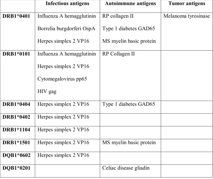
corresponding tetramers is repeated to identify the individual T cell epitope(s). A panel of MHC
Class II tetramers and different antigens containing relevant binding epitopes is shown in Table 1.
The application of human Class I tetramers to study self antigens has been most extensively
developed in studies of tumor antigens. For example, peptides from melanoma-associated antigens
loaded into HLA-A2 tetramers have been used for patient monitoring, phenotyping, and clinical
correlations in patients with melanoma and in cancer vaccine trials (13). Early uses of human MHC
Class II multimers were concentrated primarily in the detection and monitoring of human T cell
responses to infectious antigens, for which the CD4+ T cell response is robust and epitope
specificity is fairly predictable. Antigen-specific T cells from influenza-immune individuals were
detected using Class II tetramers loaded with an immunodominant epitope from hemagglutinin
(HA) (9). The use of tetramer staining to identify antigen-specific cells permits simultaneous
analysis of cells using fluorochrome-labeled antibodies. This additional phenotypic analysis can
provide important information about an antigen-specific response such as the type of T cell
involved, presence of activation or other markers, and cytokine production through intracellular
staining, thus differentiating, for instance, between Th1 and Th2 responses. With this approach,
tetramer-positive cells which had been previously expanded in vitro with peptide-pulsed
antigen-presenting cells were found to be CD3+CD4highCD25+, a phenotype characteristic of activated T
helper cells responding to antigen (9,14). Moreover, the concomitant use of MHC Class II tetramers
and carboxyfluorescein diacetate succinimidyl ester (CFSE) staining allows to calculate precursor
frequencies without the need for limiting dilution analysis. CFSE-labeled cells halve their dye
content each time they divide, resulting in a parallel halving of their corresponding fluorescence.
With each peak of progressively lower fluorescence intensity representing one cell division, the
original number of CD4+ T cell precursors can be derived (9).
Similar approaches have been used in identifying epitope-specific CD4+ T cells from individuals
infected with Herpes simplex virus type 2 (HSV-2) (15). In this latter work, tetramer-positive cells
were subsequently sorted, cloned, and further characterized. The vast majority of the clones retained
tetramer staining and proliferated when challenged with the same peptide used in the tetramer. This
confirms the antigen-specific properties of peripheral blood T cells detected using tetramer staining
(15). Tetramers can thus be used not only to identify antigen-specific cells, but also to isolate these
cells by fluorescence sorting for further characterization. Different T cell clones have been obtained
from the same HSV-2-infected individual with this approach, showing different TCR gene usage
and affinities (16).
The application of MHC Class II tetramer technology to autoimmune diseases faces additional
problems. The number of antigen-specific CD4+ cells present in peripheral blood is even lower than
with allo-responses. Moreover, autoreactive T cells must harbor lower affinity TCRs in order to
escape thymic negative selection. An initial study was conducted in rheumatoid arthritis patients,
taking advantage of synovial fluid sampling, an enriched starting material not available in other
autoimmune diseases. This study failed to directly detect cartilage antigen-specific CD4+ T cells,
finding DR4 tetramer-positive fractions above background only in a marginal subset of individuals
(5). In our study on GAD65-specific T cells in type 1 diabetic patients, preliminary in vitro
expansion of peripheral blood lymphocytes on GAD65-pulsed antigen-presenting cells was
necessary to overcome this problem (17). A large expansion of tetramer-positive cells can thus be
obtained, representing both the accumulation of proliferating antigen-specific cells and the loss of
unrelated T cells lacking appropriate antigenic stimulation during in vitro culture. Similarly,
tetramer staining of gliadin-specific T cells obtained from intestinal biopsies of patients with celiac
disease was accomplished after in vitro expansion and generation of clones (8).
This problem is not unique to autoimmunity studies. The size of clonal expansion is considerably
lower for CD4+ than for CD8+ T cells, resulting in low frequencies of antigen-specific CD4+ T
lymphocytes in peripheral blood, in the range of 1:6,000 to 1:100,000. While it is reasonable to
attempt direct detection strategies in human studies when the antigen challenge is robust, as in
vaccine trials, direct detection of antigen-specific CD4+ T cells has so far yielded very low
tetramer-binding peripheral blood populations even in an infectious context (18). The low number of
antigen-specific CD4+ cells in peripheral blood is beyond the sensitivity limit of flow cytometry:
frequencies of tetramer-positive cells below 0.2% (1:500) significantly overlap with the 0.1%
background staining tipically obtained using tetramers loaded with control irrelevant peptides.
Coupling in vitro amplification with CFSE staining circumvents one problem, allowing to calculate
the original precursor frequency without the need for direct detection (9). Nonetheless, the
requirement for in vitro amplification prior to tetramer detection prompts some caution in
interpreting the data. Changes in T cell phenotype and preferential expansion of cells either with
higher affinity TCRs (in the presence of limited peptide availability) or with lower affinity TCRs (in
the presence of excess peptide concentration) may occur during culture.
Combining tetramer fluorescence sorting with more sensitive techniques such as gene expression
array analysis and real-time PCR might help to fill the gap and to better characterize the functional
profile of antigen-specific cells.
Beyond staining: tickling the TCR with MHC Class II Tetramers
As with monoclonal antibodies (mAbs), MHC Class II tetramers can be used not just as staining
reagents, but also as stimulating tools for T cell activation. There are several ways in which T cells
can be activated for in vitro studies (Table 2): typically, anti-CD3 mAbs (either plate-bound or in
soluble form) have been used for this purpose. These reagents bind to the ε chain of the CD3
complex and supply a potent surrogate signal mimicking the MHC-peptide-TCR trimolecular
interaction. Several features limit the fidelity of this system: 1) the affinity of mAb binding is
approx. five-fold higher than the MHC Class II/TCR interaction, with different on-rate and off-rate
characteristics; 2) anti-CD3 stimulation is not dependent on recognition by the TCR of the
processed antigen in the MHC Class II groove, and 3) there is no CD4 contribution to the signal
delivered. A more physiological approach is to use antigen-presenting cells (APCs) pulsed with the
peptide of interest. This system closely mimics physiology but is subject to the vagaries of less
controllable conditions and lacks the flexibility required to follow the whole range of signals
delivered.
The advent of recombinant MHC Class II molecules has opened new possibilities for T cell
functional studies. Cochran et al. thoroughly investigated the valency required for TCR engagement
in order to initiate signaling, using chemically defined MHC Class II oligomers (6). Monomeric
MHC-peptide complexes did not induce T cell activation, while MHC dimers and tetramers
stimulated T cells (6). Moreover, the extent of activation correlated with the number of TCR
cross-linked, suggesting that clustering of two TCR molecules is necessary and sufficient for signaling.
Another study performed with a series of oligomeric MHC-peptide complexes produced by
streptavidin-mediated cross-linking of biotinylated MHC proteins reported that trimers were the
minimal activating species (19). At odds with these results, some reports have suggested that
monomeric MHC-peptide complexes may not trigger complete T cell activation, but can deliver
early, transient Ca2+ signals (20). Two additional phenomena associated with MHC/TCR
interactions further complicate the interpretation. First, at high concentrations MHC-TCR
complexes show a tendency to aggregate exhibited by neither MHC nor TCR alone, suggesting that
binding of monomeric MHC peptide complexes could trigger oligomerization of TCR on the
surface of T cells (21). Second, at low antigen densities single MHC-peptide complexes can serially
engage multiple TCR molecules (1,22). In principle, such serial triggering could allow a single
MHC molecule on an APC to cluster TCRs on the T cell membrane.
The activation events following the interaction of the TCR with recombinant soluble MHC
molecules have been initially studied by means of dimeric peptide/MHC Class II ligands, obtained
by fusing the MHC extracellular domains with the IgG Fc portion (23-25). In the study by Hamad
et al., IEk-peptide dimers adsorbed on plastic delivered a signal even more potent than anti-CD3
mAb, as assessed by TCR downregulation, IL-2 production and proliferation (23). Similar results
were obtained with a human HLA-DR2-peptide dimer: this construct induced proliferation in
myelin basic protein (MBP)-specific T cell clones when used in soluble form, while the same effect
was not elicited by soluble anti-CD3 mAb (25). Monomeric MHC units coated on plastic have also
been used in place of peptide-pulsed APCs to induce proliferation and support the growth of
antigen-specific T cells (3,17).
More recently, we undertook a comprehensive evaluation of the TCR signaling cascades using
MHC Class II tetramers as activating ligands. Stimulation with these reagents has several
advantages over monomers: 1) tetramer is used in soluble form, allowing the amount of stimulus to
be quantitated rather than just estimated as amount of MHC monomer offered for adsorption; 2) the
signaling events can be correlated with tetramer binding by means of phycoerythrin-labeled
streptavidin; 3) the short timeframes of early transduction steps can be studied more conveniently.
A simplified representation of the signaling pathways elicited by MHC/TCR interaction is depicted
in Fig. 2. At the earliest time points, sequential phosphorylation cascades are activated. These
events also lead to activation of phospholipase C (PLC)-γ1, whose enzymatic activity produces
diacylglycerol (DAG), which triggers protein kinase C (PKC) pathway, and inositol triphosphate
(IP3), which elicits Ca2+ release from intracellular stores. This first Ca2+ burst triggers a second
wave of Ca2+ influx from the extracellular space. All phosphorylation and Ca2+ signal transduction
cascades ultimately lead to the formation of active transcription factor complexes, which initiate the
expression of new genes. These early and late activation events were analyzed in glutamic acid
decarboxylase (GAD65)-specific HLA-DR0401-restricted T cell clones derived from a diabetic
patient (R. Mallone et al., manuscript submitted for publication). The initiation of tetramer-induced
signal transduction was followed by real-time Ca2+ measurements. As shown in Fig. 3, panel A,
optimal concentrations (10-20 ug/ml) of the tetramer loaded with the cognate GAD65 peptide
(TMr-GAD) led to a consistent raise in intracellular Ca2+ concentration, which was paralleled by a
smaller and low-onset signal delivered by the same tetramer loaded with an irrelevant MBP peptide
(TMr-MBP). Fig. 3 panel B shows the Ca2+ fluxes registered with cross-linked anti-CD3 stimulation
as compared to an isotype-matched IgG. The immediate onset of a potent signal reflects the much
higher affinity of antigen/mAb interactions as compared to MHC-peptide-TCR ones. A similar
picture was obtained when protein tyrosine phosphorylation was considered: TMr-GAD was
capable of delivering an efficient signal, although delayed and not as high as the one elicited by
cross-linked anti-CD3 mAb. However, this effect was promiscuous with respect to peptide
specificity, since a smaller, transient shift in phosphorylation was also obtained by the non-cognate
TMr-MBP. This lack of specificity was limited to early signaling events, and MBP-loaded tetramers
were not capable of committing cells to full activation. As exemplified in Fig. 3, panels C-D, CD69
(the earliest newly synthesized surface protein following complete T cell activation) was readily
upregulated by TMr-GAD, but not by TMr-MBP. This upregulation was accompanied by surface
staining for TMr-GAD, while no binding was evident for TMr-MBP.
The early signals delivered by non-cognate tetramers occurred in the absence of any detectable
binding, likely due to fast, transient tetramer/TCR interactions not followed by stable binding and
signal progression. Indeed, approx. 90% of the energy necessary for the interaction with the TCR is
given solely by the MHC molecule (26). In line with a two-step model of TCR recognition (26), the
GAD peptide delivers specificity and full T cell activation by turning this initial association into a
stable, longer binding. Transient peptide-independent TCR engagements are increasingly
recognized as important contributors to effective signaling and antigen sensitivity (20,27,28).
Co-stimulation was not a requirement for these T cell clones, likely due to their memory phenotype
(29) and, possibly, to the high transducing efficiency of tetramers (19,30-32). Despite the
limitations of a direct comparison, tetramer stimulation achieved stronger effects than APC
stimulation, as judged by the induction of cytokine secretion.
The plethora of stimulatory effects observed is even more striking considering the non-saturating
binding of MHC Class II tetramers, which occupied only a small fraction of the TCRs available.
Using a quantitative flow cytometry approach, we estimated that, at optimal signaling
concentrations, only 8-12% of the available TCRs were stably occupied by cognate tetramers. The
potent signals delivered despite binding to so few TCRs could be partly related to a serial
engagement effect as described by Valitutti et al. (1). In other words, each single MHC unit within
the tetramer could transiently contact and scan a large number of TCRs by serial fast interactions.
At the same time, the tetramer as a whole would still be stably bound to the cell surface, due to the
increased avidity achieved by the multiple MHC interactions of the tetramer. In contrast, the high
affinity of anti-CD3 mAbs allows them to bind a very large number of TCRs, but in a static rather
than dynamic fashion.
A promising frontier for T cell activation studies will be the development of so-called “artificial”
APCs. This approach has been mainly developed in the field of adoptive transfer of cytotoxic T
lymphocytes (CTLs) (33,34). The infusion of antigen-specific CD8+ cells is a potential
immunotherapy against selected cancers (e.g., melanoma) and infectious diseases (e.g., HIV and
Cytomegalovirus infections), but its broad use is challenged by the need to generate consistent
numbers of autologous T cells directed against the selected epitopes. While beads coated with
anti-CD3 and anti-CD28 mAbs are capable of supporting the long-term growth of CD4+ T cells,
additional requirements need to be met for CD8+ T cells (34). To circumvent this problem, mouse
fibroblasts have been transfected with single peptide-MHC Class I complexes along with B7.1,
ICAM-1 and LFA-3 costimulatory molecules (33). The induction of fully functional CTLs was
more efficient than that obtained with autologous blood-derived dendritic cells, probably due to a
higher availability of MHC and costimulatory molecules and absence of other MHC alleles in the
artificial APC. Moreover, strong responses were induced not only against flu peptides, but also
against autoantigens in the absence of autoimmune diseases. This suggests that there is not only a
recall effect on primed CTLs, but also activation of naïve T cells present at very low frequencies
(33). For expansion of antigen-specific CD4+ T cells, MHC Class II tetramers have been used to
engineer bead-based artificial APCs capable of activating human CD4+ T cells in an
antigen-specific manner (35). Recombinant MHC Class II molecules have also been incorporated into
liposomes. Compared with bead-based artificial APCs, cell- and liposome-based systems exploit
membrane fluidity to more closely mimic physiological interactions with T cells and have been
shown to induce immunological synapse formation (36).
How much activation is right?
The capability of recombinant MHC Class II reagents for activating CD4+ lymphocytes is a
double-edged sword that can be differently used in distinct disease situations. For malignancies and
infectious diseases, it may be desirable to stimulate these cells, thus allowing better cooperation
between cytotoxic and helper lymphocytes. Trials of adoptive immunotherapy have focused on the
infusion of CD8+ cells as direct effectors of tumor cell killing. However, several studies have shown
that CD4+ cells are required in the optimal induction of human tumor-specific CD8+ cells (37,38).
In murine models, CD4+ cells play an important role in eradicating tumors and can sometimes do so
even in the absence of CD8+ T cells (39). The limited success of trials of adoptive transfer with
CD8+ T cells has been partially attributed to a need for continuing CD4+ T cells help to sustain the
anti-tumor response (40,41).
In the setting of autoimmunity (and of transplantation immunity as well), different goals are
therapeutically pursued. CD4+ T cells can either be skewed towards more protective phenotypes
(e.g., Th2 versus Th1, stimulation of CD4+CD25+ regulatory T cells) or can be turned off. These
outcomes can be achieved by changing the quality or quantity of the signal delivered through the
TCR. Administration of a dimeric peptide/MHC Class II chimera to TCR transgenic mice specific
for an HA peptide in the context of I-Ed induced differentiation of CD4+ cells towards a Th2
response through negative signaling on the STAT4 pathway of Th1 differentiation. This Th2
polarization had subsequent bystander inhibitory effects on CD8+ T cell function as a result of IL-2
deprivation (24). The same dimeric peptide/MHC chimera was shown to have anti-diabetogenic
properties in mice transgenic both for HA/I-Ed-specific TCR and for selective expression of the HA
protein in pancreatic β cells. This treatment prevented autoimmune diabetes and reversed it in
animals that were already diabetic through induction of anergy in autoreactive CD4+ T cells in the
spleen and stimulation of IL-10-secreting T regulatory type 1 cells in the pancreas (42). A similar
induction of IL-2-reversible anergy was obtained in vitro in human MBP-reactive
MHC-DR2-restricted T cells from multiple sclerosis patients by a dimeric DR2/MBP chimera (32). In a murine
model of collagen-induced arthritis, a bivalent form of a single I-Aq chain presenting a peptide from
type II collagen delayed the onset and reduced severity of disease by induction of antigen-specific
hyporesponsiveness (43). Dimeric Class I MHC molecules have also been shown to induce
antigen-specific T cell unresponsiveness in cytotoxic T lymphocytes in vitro and in vivo (44).
Another way to silence autoreactive T cells may be to quantitatively alter the TCR signal to induce
activation-induced cell death (AICD) (45). In our diabetes model of GAD65-specific T cell clones,
sustained stimulation with GAD65-loaded Class II tetramers induced massive apoptosis, as readily
detectable even by morphological parameters alone (Fig. 4, panel A). This apoptotic process was
characteristic of AICD, being inhibited by blocking the Fas/Fas ligand interaction (Fig. 4, panel B)
(R. Mallone et al., manuscript submitted for publication). Gene expression arrays also confirmed
that the apoptosis was mainly antigen-driven, following the classical death receptor pathway (46).
Increased mRNA expression was detected for: 1) members of the tumor necrosis factor (TNF)
ligand family: Fas ligand (47,48), CD40 ligand (49), lymphotoxin α (50) (but not TNF-α, likely for
the short half life of its mRNA) (51); 2) members of the TNF receptor family (52,53): TNF receptor
2 (but not FAS, which is already highly expressed in this clones) and 4-1BB (54); 3) downstream
mediators, more notably IAP-1 (downstream of TNF receptor 2) (53), 3 (46) and
caspase-14 (55); 4) members of the Bcl-2 family: Bcl-x (56), suggesting some contribution of a cytokine
withdrawal mechanism (57) (R. Mallone and E.M. Laughlin, unpublished observations).
Present and future clinical applications: diagnosis, prognosis, therapy
The use of MHC Class II tetramers for diagnostic and prognostic evaluations is currently the focus
of clinical application. In mouse models of infection, Class II tetramers specific for lymphocytic
choriomeningitis virus-derived peptides have been used to track the expansion of CD4+ T cell
populations, showing that the increase in the CD4+ compartment starts later, achieves a lower
maximum level and is less stable than that of CD8+ T cells (58). Results are very promising in
human infectious diseases, where, despite the limited feasibility of direct detection (18), influenza A
and HSV-2 antigen-specific CD4+ T cells have been detected following in vitro expansion
(9,15,16). This area of application is particularly relevant for infectious diseases such as AIDS and
hepatitis C, for which CD4+ T cell responses are crucial to the outcome. The translation of these
tools to the field of tumor immunology could also open new avenues for the development and
monitoring of cancer vaccines and, at the same time, for better understanding the relative
contribution of CD4+ cells.
Lower precursor frequencies in peripheral blood and lower TCR affinities have made advancement
slower for autoimmune diseases. The recent report by Reijonen et al. filled this gap by
demonstrating the possibility to detect significant numbers of GAD65-reactive T cells in patients
with new-onset type 1 diabetes (17). GAD65-loaded MHC DR0401 and DR0404 tetramers were
capable of detecting 4-28% antigen-specific CD4+ T cells from peripheral blood lymphocytes
previously expanded in vitro on GAD65-pulsed APCs. More importantly, GAD-reactive cells were
found in all type 1 diabetic patients and some at risk subjects, but not in normal control subjects
(17). The prognostic significance of tetramer staining in at risk subjects, i.e., whether they have an
increased probability of developing diabetes in the short term, deserves further study. Indeed, the
prediction of diabetes development obtained using MHC Class I tetramers to quantify β cell
epitope-specific CD8+ T cells in the peripheral blood of NOD mice is encouraging (59).
The relevance of the functional properties of recombinant MHC Class II reagents in stimulating T
cells is not limited to in vitro activation studies. The possibility of exploiting these properties for
therapeutic purposes is intriguing. While in oncologic and infectious diseases the aim would be to
bolster the CD4 response in order to provide sustained help for the cytotoxic effector phase (40,41),
the more promising preclinical studies using immunomodulatory MHC Class II multimers come
from autoimmunity (24,32,42,43). In this field, immune intervention has proved to be a formidable
task, so far rewarded with little success. The recent publication of the first clinical trial using a
non-activating (60) humanized form of OKT3 mAb in new-onset type 1 diabetes is a notable exception
(61). This treatment obtained a consistent, although transient, improvement in β cell function, likely
acting through Th2 polarization and induction of CD4+CD25+ regulatory T cells (62,63). The
possibility of using immunodominant peptides or altered peptide ligands for more antigen-specific
immunomodulatory therapies has been extensively explored. Several animal studies have shown
that prevention of experimental autoimmune encephalomyelitis (a murine model of multiple
sclerosis) or diabetes can be achieved by the downregulation of autoreactive T cells after
administration of immunodominant peptides derived from MBP (64,65) or from the major β cell
antigens, i.e., insulin B chain (66), GAD65 (67), heat-shock protein (hsp)60 (68). However, some
studies have shown a lack of protection by peptide-based therapy, and the translation to clinical
trials using different routes of administration has been disappointing at best (69-73). This inefficacy
may partly be due to the fast proteolytic degradation of peptides, with lifespans in the order of
0.5-10 minutes in the bloodstream (74). The threat of exacerbating autoimmunity rather than inducing
tolerance adds further uncertainty (75). New clinical trials with insulin B chain and GAD65
peptides are under way, supported by the Immune Tolerance Network
(http://www.immunetolerance.org).
MHC Class II multimers could represent an appealing therapeutic alternative, endowed with
remarkable specificity and better bioavailability (with half lives of approx. 50 h in vivo) (76). The
selective nature of this approach is a potential drawback because it requires knowledge of the
relevant T cell epitopes of several target proteins. It may also require intervention early in the
disease process, before extensive epitope spreading has occurred (77). Dominant peptide epitopes
are likely to change during the progression of autoimmune disease (78), and successful tolerance
induction will depend on correlating antigen hierarchy with disease progression. In this respect, a
combined approach of disease “immune staging” and tolerogenic therapy could be envisioned.
Periodic screening of at risk or newly-diagnosed individuals with the same tetramers, which could
be conveniently loaded with any relevant peptide, may allow to choose the best timing for specific
immunomodulatory intervention.
Acknowledgments
Work supported by the Juvenile Diabetes Research Foundation International and by grant DK
53004 from the National Institute of Health. R.M. is a student at the Postgraduate School of Internal
Medicine, University of Turin, Turin, Italy.
References
1. Valitutti, S., Muller, S., Cella, M., Padovan, E., and Lanzavecchia, A., Serial triggering of many T-cell receptors by a few peptide-MHC complexes. Nature 375, 148-151, 1995.
2. Altman, J.D., Moss, P.A., Goulder, P.J., Barouch, D.H., McHeyzer-Williams, M.G., Bell, J.I., McMichael, A.J., and Davis, M.M., Phenotypic analysis of antigen-specific T lymphocytes.
Science 274, 94-96, 1996.
3. Kozono, H., White, J., Clements, J., Marrack, P., and Kappler, J., Production of soluble MHC class II proteins with covalently bound single peptides. Nature 369, 151-154, 1994.
4. Crawford, F., Kozono, H., White, J., Marrack, P., and Kappler, J., Detection of antigen-specific T cells with multivalent soluble class II MHC covalent peptide complexes. Immunity. 8, 675-682, 1998.
5. Kotzin, B.L., Falta, M.T., Crawford, F., Rosloniec, E.F., Bill, J., Marrack, P., and Kappler, J., Use of soluble peptide-DR4 tetramers to detect synovial T cells specific for cartilage antigens in patients with rheumatoid arthritis. Proc. Natl. Acad. Sci. U. S. A 97, 291-296, 2000.
6. Cochran, J.R., Cameron, T.O., and Stern, L.J., The relationship of MHC-peptide binding and T cell activation probed using chemically defined MHC class II oligomers. Immunity. 12, 241-250, 2000.
7. Cameron, T.O., Cochran, J.R., Yassine-Diab, B., Sekaly, R.P., and Stern, L.J., Cutting edge: detection of antigen-specific CD4+ T cells by HLA-DR1 oligomers is dependent on the T cell activation state. J. Immunol. 166, 741-745, 2001.
8. Quarsten, H., McAdam, S.N., Jensen, T., Arentz-Hansen, H., Molberg, O., Lundin, K.E., and Sollid, L.M., Staining of celiac disease-relevant T cells by peptide-DQ2 multimers. J.
Immunol. 167, 4861-4868, 2001.
9. Novak, E.J., Liu, A.W., Nepom, G.T., and Kwok, W.W., MHC class II tetramers identify peptide-specific human CD4(+) T cells proliferating in response to influenza A antigen. J.
Clin. Invest 104, R63-R67, 1999.
10. Sturniolo, T., Bono, E., Ding, J., Raddrizzani, L., Tuereci, O., Sahin, U., Braxenthaler, M., Gallazzi, F., Protti, M.P., Sinigaglia, F., and Hammer, J., Generation of tissue-specific and promiscuous HLA ligand databases using DNA microarrays and virtual HLA class II matrices. Nat. Biotechnol. 17, 555-561, 1999.
11. Novak, E.J., Liu, A.W., Gebe, J.A., Falk, B.A., Nepom, G.T., Koelle, D.M., and Kwok, W.W., Tetramer-guided epitope mapping: rapid identification and characterization of
immunodominant CD4+ T cell epitopes from complex antigens. J. Immunol. 166, 6665-6670, 2001.
12. Kwok, W.W., Gebe, J.A., Liu, A., Agar, S., Ptacek, N., Hammer, J., Koelle, D.M., and Nepom, G.T., Rapid epitope identification from complex class-II-restricted T-cell antigens.
Trends Immunol. 22, 583-588, 2001.
13. Monsurro, V., Nagorsen, D., Wang, E., Provenzano, M., Dudley, M.E., Rosenberg, S.A., and Marincola, F.M., Functional heterogeneity of vaccine-induced CD8(+) T cells. J. Immunol. 168, 5933-5942, 2002.
14. Novak, E.J., Masewicz, S.A., Liu, A.W., Lernmark, A., Kwok, W.W., and Nepom, G.T., Activated human epitope-specific T cells identified by class II tetramers reside within a CD4high, proliferating subset. Int. Immunol. 13, 799-806, 2001.
15. Kwok, W.W., Liu, A.W., Novak, E.J., Gebe, J.A., Ettinger, R.A., Nepom, G.T., Reymond, S.N., and Koelle, D.M., HLA-DQ tetramers identify epitope-specific T cells in peripheral blood of herpes simplex virus type 2-infected individuals: direct detection of
immunodominant antigen-responsive cells. J. Immunol. 164, 4244-4249, 2000.
16. Reichstetter, S., Ettinger, R.A., Liu, A.W., Gebe, J.A., Nepom, G.T., and Kwok, W.W., Distinct T cell interactions with HLA class II tetramers characterize a spectrum of TCR affinities in the human antigen-specific T cell response. J. Immunol. 165, 6994-6998, 2000. 17. Reijonen, H., Novak, E.J., Kochik, S., Heninger, A., Liu, A.W., Kwok, W.W., and Nepom,
G.T., Detection of GAD65-specific T-cells by major histocompatibility complex class II tetramers in type 1 diabetic patients and at-risk subjects. Diabetes 51, 1375-1382, 2002. 18. Meyer, A.L., Trollmo, C., Crawford, F., Marrack, P., Steere, A.C., Huber, B.T., Kappler, J.,
and Hafler, D.A., Direct enumeration of Borrelia-reactive CD4 T cells ex vivo by using MHC class II tetramers. Proc. Natl. Acad. Sci. U. S. A 97, 11433-11438, 2000.
19. Boniface, J.J., Rabinowitz, J.D., Wulfing, C., Hampl, J., Reich, Z., Altman, J.D., Kantor, R.M., Beeson, C., McConnell, H.M., and Davis, M.M., Initiation of signal transduction through the T cell receptor requires the multivalent engagement of peptide/MHC ligands [corrected]. Immunity. 9, 459-466, 1998.
20. Irvine, D.J., Purbhoo, M.A., Krogsgaard, M., and Davis, M.M., Direct observation of ligand recognition by T cells. Nature 419, 845-849, 2002.
21. Reich, Z., Boniface, J.J., Lyons, D.S., Borochov, N., Wachtel, E.J., and Davis, M.M., Ligand-specific oligomerization of T-cell receptor molecules. Nature 387, 617-620, 1997.
22. Valitutti, S., and Lanzavecchia, A., Serial triggering of TCRs: a basis for the sensitivity and specificity of antigen recognition. Immunol. Today 18, 299-304, 1997.
23. Hamad, A.R., O'Herrin, S.M., Lebowitz, M.S., Srikrishnan, A., Bieler, J., Schneck, J., and Pardoll, D., Potent T cell activation with dimeric peptide-major histocompatibility complex class II ligand: the role of CD4 coreceptor. J. Exp. Med. 188, 1633-1640, 1998.
24. Casares, S., Zong, C.S., Radu, D.L., Miller, A., Bona, C.A., and Brumeanu, T.D., Antigen-specific signaling by a soluble, dimeric peptide/major histocompatibility complex class II/Fc chimera leading to T helper cell type 2 differentiation. J. Exp. Med. 190, 543-553, 1999. 25. Appel, H., Gauthier, L., Pyrdol, J., and Wucherpfennig, K.W., Kinetics of T-cell receptor
binding by bivalent HLA-DR.Peptide complexes that activate antigen-specific human T-cells.
J. Biol. Chem. 275, 312-321, 2000.
26. Wu, L.C., Tuot, D.S., Lyons, D.S., Garcia, K.C., and Davis, M.M., Two-step binding mechanism for T-cell receptor recognition of peptide MHC. Nature 418, 552-556, 2002.
27. Stefanova, I., Dorfman, J.R., and Germain, R.N., Self-recognition promotes the foreign antigen sensitivity of naive T lymphocytes. Nature 420, 429-434, 2002.
28. Wulfing, C., Sumen, C., Sjaastad, M.D., Wu, L.C., Dustin, M.L., and Davis, M.M.,
Costimulation and endogenous MHC ligands contribute to T cell recognition. Nat. Immunol. 3, 42-47, 2002.
29. London, C.A., Lodge, M.P., and Abbas, A.K., Functional responses and costimulator dependence of memory CD4+ T cells. J. Immunol. 164, 265-272, 2000.
30. Goldstein, J.S., Chen, T., Brunswick, M., Mostowsky, H., and Kozlowski, S., Purified MHC class I and peptide complexes activate naive CD8+ T cells independently of the CD28/B7 and LFA-1/ICAM-1 costimulatory interactions. J. Immunol. 1998.
31. Bluestone, J.A., New perspectives of CD28-B7-mediated T cell costimulation. Immunity 2, 555-559, 1995.
32. Appel, H., Seth, N.P., Gauthier, L., and Wucherpfennig, K.W., Anergy induction by dimeric TCR ligands. J. Immunol. 166, 5279-5285, 2001.
33. Latouche, J.B., and Sadelain, M., Induction of human cytotoxic T lymphocytes by artificial antigen-presenting cells. Nat. Biotechnol. 18, 405-409, 2000.
34. Maus, M.V., Thomas, A.K., Leonard, D.G., Allman, D., Addya, K., Schlienger, K., Riley, J.L., and June, C.H., Ex vivo expansion of polyclonal and antigen-specific cytotoxic T lymphocytes by artificial APCs expressing ligands for the T-cell receptor, CD28 and 4-1BB.
Nat. Biotechnol. 20, 143-148, 2002.
35. Maus, M.V., Riley, J.L., Kwok, W.W., Nepom, G.T., and June, C.H., HLA tetramer-based artificial antigen-presenting cells for stimulation of CD4(+) T cells. Clin. Immunol. 106, 16-22, 2003.
36. Mallet-Designe, V.I., Stratmann, T., Homann, D., Carbone, F., Oldstone, M.B., and Teyton, L., Detection of low-avidity CD4+ T cells using recombinant artificial APC: following the antiovalbumin immune response. J. Immunol. 170, 123-131, 2003.
37. Baxevanis, C.N., Voutsas, I.F., Tsitsilonis, O.E., Gritzapis, A.D., Sotiriadou, R., and
Papamichail, M., Tumor-specific CD4+ T lymphocytes from cancer patients are required for optimal induction of cytotoxic T cells against the autologous tumor. J. Immunol. 164, 3902-3912, 2000.
38. Marzo, A.L., Kinnear, B.F., Lake, R.A., Frelinger, J.J., Collins, E.J., Robinson, B.W., and Scott, B., Tumor-specific CD4+ T cells have a major "post-licensing" role in CTL mediated anti-tumor immunity. J. Immunol. 165, 6047-6055, 2000.
39. Hung, K., Hayashi, R., Lafond-Walker, A., Lowenstein, C., Pardoll, D., and Levitsky, H., The central role of CD4(+) T cells in the antitumor immune response. J. Exp. Med. 188, 2357-2368, 1998.
40. Pardoll, D.M., and Topalian, S.L., The role of CD4+ T cell responses in antitumor immunity.
Curr. Opin. Immunol. 10, 588-594, 1998.
41. Wang, R.F., The role of MHC class II-restricted tumor antigens and CD4+ T cells in antitumor immunity. Trends Immunol. 22, 269-276, 2001.
42. Casares, S., Hurtado, A., McEvoy, R.C., Sarukhan, A., von Boehmer, H., and Brumeanu, T.D., Down-regulation of diabetogenic CD4+ T cells by a soluble dimeric peptide-MHC class II chimera. Nat. Immunol. 3, 383-391, 2002.
43. Zuo, L., Cullen, C.M., DeLay, M.L., Thornton, S., Myers, L.K., Rosloniec, E.F., Boivin, G.P., and Hirsch, R., A single-chain class II MHC-IgG3 fusion protein inhibits autoimmune arthritis by induction of antigen-specific hyporesponsiveness. J. Immunol. 168, 2554-2559, 2002. 44. O'Herrin, S.M., Slansky, J.E., Tang, Q., Markiewicz, M.A., Gajewski, T.F., Pardoll, D.M.,
Schneck, J.P., and Bluestone, J.A., Antigen-specific blockade of T cells in vivo using dimeric MHC peptide. J. Immunol. 167, 2555-2560, 2001.
45. Critchfield, J.M., Racke, M.K., Zuniga-Pflucker, J.C., Cannella, B., Raine, C.S., Goverman, J., and Lenardo, M.J., T cell deletion in high antigen dose therapy of autoimmune
encephalomyelitis. Science 263, 1139-1143, 1994.
46. Budd, R.C., Activation-induced cell death. Curr. Opin. Immunol. 13, 356-362, 2001. 47. Dhein, J., Walczak, H., Baumler, C., Debatin, K.M., and Krammer, P.H., Autocrine T-cell
suicide mediated by APO-1/(Fas/CD95). Nature 373, 438-441, 1995.
48. Brunner, T., Mogil, R.J., LaFace, D., Yoo, N.J., Mahboubi, A., Echeverri, F., Martin, S.J., Force, W.R., Lynch, D.H., Ware, C.F., and Green, D.R., Cell-autonomous Fas (CD95)/Fas-ligand interaction mediates activation- induced apoptosis in T-cell hybridomas. Nature 373, 441-444, 1995.
49. Laman, J.D., Claassen, E., and Noelle, R.J., Functions of CD40 and its ligand, gp39 (CD40L).
Crit Rev. Immunol. 16, 59-108, 1996.
50. Ruddle, N.H., Tumor necrosis factor (TNF-alpha) and lymphotoxin (TNF-beta). Curr. Opin.
Immunol. 4, 327-332, 1992.
51. English, B.K., Weaver, W.M., and Wilson, C.B., Differential regulation of lymphotoxin and tumor necrosis factor genes in human T lymphocytes. J. Biol. Chem. 266, 7108-7113, 1991. 52. Screaton, G., and Xu, X.N., T cell life and death signalling via TNF-receptor family members.
Curr. Opin. Immunol. 12, 316-322, 2000.
53. Chan, F.K., Siegel, R.M., and Lenardo, M.J., Signaling by the TNF receptor superfamily and T cell homeostasis. Immunity 13, 419-422, 2000.
54. Vinay, D.S., and Kwon, B.S., Role of 4-1BB in immune responses. Semin. Immunol. 10, 481-489, 1998.
55. Ahmad, M., Srinivasula, S.M., Hegde, R., Mukattash, R., Fernandes-Alnemri, T., and
Alnemri, E.S., Identification and characterization of murine caspase-14, a new member of the caspase family. Cancer Res. 58, 5201-5205, 1998.
56. Boise, L.H., Gonzalez-Garcia, M., Postema, C.E., Ding, L., Lindsten, T., Turka, L.A., Mao, X., Nunez, G., and Thompson, C.B., bcl-x, a bcl-2-related gene that functions as a dominant regulator of apoptotic cell death. Cell 74, 597-608, 1993.
57. Adams, J.M., and Cory, S., Life-or-death decisions by the Bcl-2 protein family. Trends
Biochem. Sci. 26, 61-66, 2001.
58. Homann, D., Teyton, L., and Oldstone, M.B., Differential regulation of antiviral T-cell immunity results in stable CD8+ but declining CD4+ T-cell memory. Nat. Med. 7, 913-919, 2001.
59. Trudeau, J.D., Kelly-Smith, C., Verchere, C.B., Elliott, J.F., Dutz, J.P., Finegood, D.T., Santamaria, P., and Tan, R., Prediction of spontaneous autoimmune diabetes in NOD mice by quantification of autoreactive T cells in peripheral blood. J. Clin. Invest 111, 217-223, 2003. 60. Herold, K.C., Bluestone, J.A., Montag, A.G., Parihar, A., Wiegner, A., Gress, R.E., and
Hirsch, R., Prevention of autoimmune diabetes with nonactivating anti-CD3 monoclonal antibody. Diabetes 41, 385-391, 1992.
61. Herold, K.C., Hagopian, W., Auger, J.A., Poumian-Ruiz, E., Taylor, L., Donaldson, D., Gitelman, S.E., Harlan, D.M., Xu, D., Zivin, R.A., and Bluestone, J.A., Anti-CD3 monoclonal antibody in new-onset type 1 diabetes mellitus. N. Engl. J. Med. 346, 1692-1698, 2002. 62. Masteller, E.L., and Bluestone, J.A., Immunotherapy of insulin-dependent diabetes mellitus.
Curr. Opin. Immunol. 14, 652-659, 2002.
63. Chatenoud, L., CD3-specific antibody-induced active tolerance: from bench to bedside. Nat.
Rev. Immunol. 3, 123-132, 2003.
64. Higgins, P.J., and Weiner, H.L., Suppression of experimental autoimmune encephalomyelitis by oral administration of myelin basic protein and its fragments. J. Immunol. 140, 440-445, 1988.
65. Smilek, D.E., Wraith, D.C., Hodgkinson, S., Dwivedy, S., Steinman, L., and McDevitt, H.O., A single amino acid change in a myelin basic protein peptide confers the capacity to prevent rather than induce experimental autoimmune encephalomyelitis. Proc. Natl. Acad. Sci. U. S. A 88, 9633-9637, 1991.
66. Daniel, D., and Wegmann, D.R., Protection of nonobese diabetic mice from diabetes by intranasal or subcutaneous administration of insulin peptide B-(9-23). Proc. Natl. Acad. Sci.
U. S. A 93, 956-960, 1996.
67. Tian, J., Clare-Salzler, M., Herschenfeld, A., Middleton, B., Newman, D., Mueller, R., Arita, S., Evans, C., Atkinson, M.A., Mullen, Y., Sarvetnick, N., Tobin, A.J., Lehmann, P.V., and Kaufman, D.L., Modulating autoimmune responses to GAD inhibits disease progression and prolongs islet graft survival in diabetes-prone mice. Nat. Med. 2, 1348-1353, 1996.
68. Elias, D., and Cohen, I.R., Peptide therapy for diabetes in NOD mice. Lancet 343, 704-706, 1994.
69. Bielekova, B., Goodwin, B., Richert, N., Cortese, I., Kondo, T., Afshar, G., Gran, B., Eaton, J., Antel, J., Frank, J.A., McFarland, H.F., and Martin, R., Encephalitogenic potential of the
myelin basic protein peptide (amino acids 83-99) in multiple sclerosis: results of a phase II clinical trial with an altered peptide ligand. Nat. Med. 6, 1167-1175, 2000.
70. Kappos, L., Comi, G., Panitch, H., Oger, J., Antel, J., Conlon, P., and Steinman, L., Induction of a non-encephalitogenic type 2 T helper-cell autoimmune response in multiple sclerosis after administration of an altered peptide ligand in a placebo-controlled, randomized phase II trial. The Altered Peptide Ligand in Relapsing MS Study Group. Nat. Med. 6, 1176-1182, 2000. 71. Chaillous, L., Lefevre, H., Thivolet, C., Boitard, C., Lahlou, N., Atlan-Gepner, C., Bouhanick,
B., Mogenet, A., Nicolino, M., Carel, J.C., Lecomte, P., Marechaud, R., Bougneres, P., Charbonnel, B., and Sai, P., Oral insulin administration and residual beta-cell function in recent-onset type 1 diabetes: a multicentre randomised controlled trial. Diabete Insuline Orale group. Lancet 356, 545-549, 2000.
72. Raz, I., Elias, D., Avron, A., Tamir, M., Metzger, M., and Cohen, I.R., Beta-cell function in new-onset type 1 diabetes and immunomodulation with a heat-shock protein peptide
(DiaPep277): a randomised, double-blind, phase II trial. Lancet 358, 1749-1753, 2001. 73. Diabetes Prevention Trial-Type 1 Diabetes Study Group, Effects of insulin in relatives of
patients with type 1 diabetes mellitus. N. Engl. J. Med. 346, 1685-1691, 2002.
74. Ishioka, G.Y., Adorini, L., Guery, J.C., Gaeta, F.C., LaFond, R., Alexander, J., Powell, M.F., Sette, A., and Grey, H.M., Failure to demonstrate long-lived MHC saturation both in vitro and in vivo. Implications for therapeutic potential of MHC-blocking peptides. J. Immunol. 152, 4310-4319, 1994.
75. Genain, C.P., and Zamvil, S.S., Specific immunotherapy: one size does not fit all. Nat. Med. 6, 1098-1100, 2000.
76. Casares, S., Bona, C.A., and Brumeanu, T.D., Enzymatically mediated engineering of multivalent MHC class II-peptide chimeras. Protein Eng 14, 195-200, 2001.
77. Vanderlugt, C.L., and Miller, S.D., Epitope spreading in immune-mediated diseases: implications for immunotherapy. Nat. Rev. Immunol. 2, 85-95, 2002.
78. Mallone, R., Ortolan, E., Pinach, S., Volante, M., Zanone, M.M., Bruno, G., Baj, G.,
Lohmann, T., Cavallo-Perin, P., and Malavasi, F., Anti-CD38 autoantibodies: characterisation in new-onset type I diabetes and latent autoimmune diabetes of the adult (LADA) and
comparison with other islet autoantibodies. Diabetologia 45, 1667-1677, 2002.
Tables
Table 1. Human MHC Class II tetramers and relevant antigens
Infectious antigens Autoimmune antigens Tumor antigens DRB1*0401 Influenza A hemagglutinin
Borrelia burgdorferi OspA
Herpes simplex 2 VP16
RP collagen II
Type 1 diabetes GAD65
MS myelin basic protein
Melanoma tyrosinase DRB1*0101 Influenza A hemagglutinin Herpes simplex 2 VP16 Cytomegalovirus pp65 HIV gag RP Collagen II
DRB1*0404 Herpes simplex 2 VP16 Type 1 diabetes GAD65
DRB1*0402 Herpes simplex 2 VP16 DRB1*1104 Herpes simplex 2 VP16
DRB1*1501 Herpes simplex 2 VP16 MS myelin basic protein
DQB1*0602 Herpes simplex 2 VP16
DQB1*0201 Celiac disease gliadin
RP, relapsing polycondritis; MS, multiple sclerosis
Table 2. In vitro T cell activation systems
- Anti-CD3 monoclonal antibodies
- Peptide-pulsed antigen-presenting cells
- Plate-bound or soluble MHC dimers
- Plate-bound MHC monomers
- Soluble MHC tetramers
- Artificial antigen presenting cells
Figure Legends
Fig. 1. Structure of MHC Class II tetramers.
Fig. 2. TCR signaling events triggered following TCR/MHC interaction. TCR ligation leads to
phosphorylation of Lck, which then phosphorylates ITAM domains on the ζ chains of the CD3
complex. Once phosphorylated, ITAM domains function as docking sites for ζ-associated protein
(ZAP)-70, which is activated through phosphorylation by Lck and in turn phosphorylates adaptor
proteins such as LAT. Further phosphorylation cascades are triggered, which interact with
co-stimulatory signals and also lead to activation of phospholipase C (PLC)-γ1. The enzymatic activity
of PLC- γ1 produces diacylglycerol (DAG), which triggers protein kinase C (PKC) pathway, and
inositol triphosphate (IP3), which elicits Ca2+ release from intracellular stores. This first Ca2+ burst
triggers a second wave of Ca2+ influx from the extracellular space. All phosphorylation and Ca2+
signal transduction cascades ultimately lead to the formation of active transcription factor
complexes, which initiate gene expression.
Fig. 3. Signals delivered upon tetramer (TMr) stimulation on GAD-specific DR0401-restricted T
cell clones. (A-B) Ca2+ mobilization, as assessed by the fluorescence shift of the Ca2+-sensitive dye
Fluo-3. The indicated stimuli were added at t=0; the dotted lines indicate the basal Ca2+ level before
stimulation. (C-D) CD69 upregulation. Cells were stimulated with the indicated tetramers for 3 h at
37ºC. Insets in panels show density plots of CD69 upregulation relative to tetramer binding.
Fig. 4. Cognate tetramer-induced apoptosis on GAD-specific DR0401-restricted T cells. (A)
Three-dimensional plots of forward (FSC) and side scatter (SSC) distribution among cells treated with the
same DR0401 tetramer loaded with either cognate GAD or non-cognate MBP peptide. (B) The
apoptotic effect of the cognate GAD is due to activation-induced cell death: inhibition of
TMr-GAD-induced apoptosis by a blocking anti-Fas ligand mAb.