HAL Id: hal-01707767
https://hal-amu.archives-ouvertes.fr/hal-01707767
Submitted on 13 Feb 2018
HAL is a multi-disciplinary open access archive for the deposit and dissemination of sci-entific research documents, whether they are pub-lished or not. The documents may come from teaching and research institutions in France or abroad, or from public or private research centers.
L’archive ouverte pluridisciplinaire HAL, est destinée au dépôt et à la diffusion de documents scientifiques de niveau recherche, publiés ou non, émanant des établissements d’enseignement et de recherche français ou étrangers, des laboratoires publics ou privés.
Correlates of HPV: a cross-sectional study in women
with normal cytology in north-central Morocco
Bahia Bennani, Sanae Bennis, Chakib Nejjari, l’Houcine Ouafik, Moulay
Abdelilah Melhouf, Karima El Rhazi, Kaoutar Znati, Hikmat Chaara,
Chahrazed Bouchikhi, Afaf Amarti Riffi
To cite this version:
Bahia Bennani, Sanae Bennis, Chakib Nejjari, l’Houcine Ouafik, Moulay Abdelilah Melhouf, et al.. Correlates of HPV: a cross-sectional study in women with normal cytology in north-central Morocco. Journal of Infection in Developing Countries, Independent, 2012, 6 (7), pp.543-550. �hal-01707767�
Correlates of HPV: a cross-sectional study in women with normal cytology
in north-central Morocco
Bahia Bennani
1,2, Sanae Bennis
3, Chakib Nejjari
4, L’Houcine Ouafik
5,
Moulay Abdelilah Melhouf
6, Karima El Rhazi
4, Kaoutar Znati
3, Hikmat Chaara
6,
Chahrazed Bouchikhi
6, Afaf Amarti Riffi
31Laboratoire de Biologie des Cancers, Equipe Microorganismes et Facteurs Oncogènes, Faculté de Médecine et de
Pharmacie de Fès, Fès Maroc
2Laboratoire de Microbiologie, Faculté de Médecine et de Pharmacie de Fès, Fès Maroc
3Laboratoire de Biologie des Cancers, Equipe d’Anatomie Pathologique, Faculté de Médecine et de Pharmacie de
Fès, Fès Maroc
4Laboratoire d’Epidémiologie, Faculté de Médecine et de Pharmacie de Fès, Fès Maroc 5Inserm UMR 911-CRO2, Faculté de Médecine Timone, Marseille, France
6Service de Gynécologie, Centre Hospitalier Universitaire Hassan II, Fès Maroc.
Abstract
Introduction: Epidemiological studies have shown the association between risk of developing cervical cancer and the persistence of high-risk papillomavirus types in addition to some co-factors. However, little is known about co-factors associated with human papillomavirus (HPV) infection, especially in developing countries. This study aims to determine HPV status and associated risk factors in women with normal cytology living in the north-central area of Morocco.
Methodology: From February 2007 to December 2008, a total of 925 women consulting in the gynaecological department of Fez University Hospital were asked about sociodemographic characteristics and reproductive and sexual health. Cervical samples were collected for cytological examination and HPV DNA detection. Data collected from 751 women with normal cytology were used in this study to assess the correlation between HPV infection and potential risk factors.
Results: High prevalence of HPV infection was detected (42.5%). The highest infection rate was observed in women aged >45 years and in those with history of abortion (OR:3.76; 95%CI[1.77-7.98]) fibroma, polyp or cysts (OR:1.68; 95%CI[1.07-2.65]). No significant association was detected with other reproductive health and risk factors including oral contraception.
Conclusion: In spite of the insignificant association of HPV infection with age, health authorities should seriously consider and implement strategies to increase and maintain a cervical cancer screening programme in women aged 45 and above. More attention must be given to women with gynaecological history (abortion, fibroma, polyp or cysts) since these events may be predictors of HPV infection. Investigations on partner sexual behaviour and some specific hygienic habits, especially public Turkish bath use, are needed to clarify the HPV incidence in this region.
Key words: HPV infection; risk factors; normal cytology; PCR diagnosis
J Infect Dev Ctries2012; 6(7):543-550.
(Received 21 October 2010 - Accepted 24 October 2011)
Copyright © 2012 Bennani et al. This is an open-access article distributed under the Creative Commons Attribution License, which permits unrestricted use, distribution, and reproduction in any medium, provided the original work is properly cited.
Introduction
Human papillomavirus (HPV) infection is associated with different clinical pathologies in the lower genital tract. Persistent infection with some genotypes of this sexually transmitted virus can induce abnormal epithelial changes in the genital tract. Those abnormalities were reported to occasionally progress from low-grade dysplasia to cervical cancer [1]. HPV can be grouped to high-risk (such as HPV-16, -18, -31, and -45) and low-risk HPV types (such as 6, 11, 42, 43, and 44) [4].
High-risk HPV types are present in most cervical cancer tissues and high-grade cervical intraepithelial neoplasia.
In addition to this main etiologic agent [2], several epidemiological factors are associated with the development of cervical cancer, including multiple sexual partners, number of pregnancies, tobacco smoking, oral contraceptives and dietary factors [3]. In many countries, 20% to 40% of sexually active young women have detectable latent HPV infection [4,5]. The frequency of high-risk types
Bennani et al. - HPV prevalence and correlates J Infect Dev Ctries2012; 6(7):543-550.
544 may vary according to geographic regions [5-7] and
to demographic and clinical/pathological factors [8-10]. Nevertheless, the prevalence of HPV infection and associated risk factors are still very little studied. The common determined risk factor associated with HPV infection is multiple sexual partners. However, results obtained on reproductive characteristics (age at first sexual intercourse and parity) use of oral contraceptives, and smoking were inconsistent [10-17].
Cervical cancer has a natural history with prolonged pre-invasive stages that are easily detected and treated. In Fez, in the central region of northern Morocco, women who attend public health-care clinics are far from early detection and survey for HPV. In addition, there is no epidemiological data available on HPV infection in this region and, as such, the prevalence of this infection remains unknown. With this cross-sectional analysis, we report the prevalence of HPV infection and associated risk factors in a group of women with normal cervical cytology in north-central Morocco, a geographical area with high incidence of cervical cancer (according to the Casablanca Cancer Register) [18].
Methodology
This cross-sectional study was conducted during the period from February 2007 to December 2008. It was approved by the Ethical Committee of the University Hospital of Fez. Only consenting women attending the Gynecology and Obstetrics Department at the hospital either for a different health complaint or just for simple gynecological control were recruited. Sexually active women, aged 15 years or older, have been included in this study. Virgin and pregnant women were excluded. A personal interview and a gynaecological examination were conducted for all women enrolled in the study. Socio-demographic characteristics (age, socio-economic and marital status), reproductive health and sexual life variables (number of pregnancies, types of contraceptives used, and number of sexual partners), and medical history were taken.
Cervical samples for cytological and molecular analysis to test for HPV infection were obtained from a total of 925 women. Cell samples were collected by scraping the uterine endocervix with a cytobrush. The samples were collected in 1.5 ml sterile phosphate-buffer saline and saved at -20°C until DNA extraction. The cell suspension was vigorously shaken, and 120 µl was treated with 40 µl of
proteinase K (200 µg/ml) in 3% Triton X-100 overnight at 37°C. The proteinase K was inactivated by heating at 92°C for 10 minutes; the supernatant was directly used for PCR. To detect HPV, DNA samples were amplified in the PCR with the largely used consensus primers MY09/MY11, directed to the highly conserved region of the L1 gene as described by Manos et al. with minor modifications [19]. Briefly, PCR was performed in a 50 µl volume with 10 mM Tris-HCl (pH 8.3), 50 mM KCl, 2.5 mM MgCl2, 200 µM each of four dNTPs, 0.5 µM of each primer, 7µl of DNA sample and 0.4U of Taq polymerase (Invitrogen, Foster City, California, USA).
Samples were subjected to 40 cycles in a Techne thermocycler (Staffordshire, United Kingdom). The initial denaturation step was at 94°C for 5 minutes, and the repeat cycle consisted of denaturation at 94°C for 30 seconds, annealing for 60 seconds at 55°C, followed by an extension at 72°C for 60 seconds. The last extension time was lengthened to 10 minutes. Each experiment was performed with separate positive and negative PCR controls. The positive control consisted of DNA from cells known to have HPV DNA integrated; the negative control contained DNA from cells known not to have HPV DNA integrated; and beta-globin was used as the internal control. PCR products were fractionated on 1.5% agarose gels and visualised under ultraviolet light.
For cytological analysis, conventional one-slide Papanicolau (Pap) smears, obtained with the use of cervical Ayre spatulas, were used to prepare the slides in accordance with the standard screening practice. In each case, a conventional Pap smear was made. A descriptive cytological classification based on the Bethesda System 2001 was used [20].
Statistical analysis was performed using SPSS Version 17.0 (IBM, Chicago, USA). A descriptive analysis was conducted for all variables and possible association between HPV infection and all potential variables were determined by parametric statistics tests (Chi-square test and Student’s t test). A logistic regression was performed to determine factors associated with HPV infection adjusting for other covariates. The odds ratio (OR) and the 95% confidence interval (CI) were measured. A stepwise selection procedure was applied to determine the covariates included in the final multivariate models beginning with all variables with p < 0.25 in the univariate analysis. A test was considered as significant if p < 0.05.
Results
Ninety-five samples from the 925 specimens of recruited women did not have sufficient cells for HPV DNA or cytological detection tests. Analysis was therefore performed on 830 cervical specimens. Of these, 79 had abnormal cytology according to the Bethesda system classification as follows: 83.54 % had squamous intraepithelial lesions (SIL); 11.39 % had atypical squamous cells of undetermined significance (ASCUS) and 5% had atypical glandular cells. After these exclusions, 751 women who showed normal cytology remained for the analysis presented here.
Characteristics of the study population
The characteristics of the study population are summarised in Table 1. Most of the participating women were aged 35 to 44 years (median age 40 years [17-81 years]) from a low-income class (91.8% (690/751)). The majority were housewives (88.4% (664/751)) and had had their first sexual intercourse (first regular sexual partner) at the age of 16 to 25 years (65.7%; 437/665). All women (96.5% of total participant) who were not divorced or widowed reported a single regular lifelong sexual partner, her husband. The divorced or widowed did not have any sexual partner since their husbands divorced them or died. According to the interview, 4.6% (23/502) of the husbands had had multiple sexual partners.
More than half of patients (64%; 478/746) did not use any contraceptive, 28.1% (210/746) used oral contraceptives (OC), 4.92% (37/746) used an intrauterine device (IUD), and 2.13% (16/746) used condoms. No data were available for five patients. Sterile women represented 7.4% of the participants (55/741). When excluding those women, more than half of the women had less than five full-term pregnancies (59.2%; 376/635). Only 0.02% of the women had a past self-reported history of sexually-transmitted infection, particularly syphilis or Chlamydia infection. Neither alcohol consumption nor HIV infection was reported. The percentage of women with persistent exposure to smoking (active or passive) was 22.9% (172/751). According to their answers, 17.04% of the participating women (128/751) had reported past gynaecological anomalies. Women with past history of abortion were 5% (38/751). Ninety cases (12% (90/751)) of women with past history of uterine fibroma, cervical polyp, or ovarian cyst were grouped together.
HPV DNA detection and associated factors
HPV DNA was detected in 319 specimens of the 751 cases, corresponding to 42.5%. The association between HPV infection and several variables was tested. The analysis showed that HPV prevalence was 46.8% among women aged 45 years or more and about 39.5% among women below age 34 years, but association with age was not significant.
However, a significant association (p < 0.05) was observed between HPV infection and past history of gyneacological events (miscarriage, fibroma, cysts, and others). No other factors were clearly associated with overall risk for HPV infections in our study population (Table 2). The educational level, profession, age of the first sexual intercourse, number of pregnancies, smoking, OC usage, marital status and infertility had no significant association with the HPV positivity.
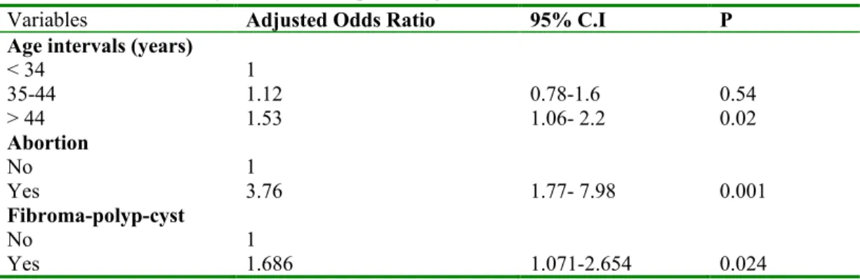
All variables with p < 0.25 in univariate analysis (infertility, age, past history of abortion, and past history of fibroma, polyp, cyst or lesion) were included in the final model. The multivariate-analysis showed a significant association between the past history of gynaecological disorders and HPV infection (Table 3). Effectively, groups of women with gynaecological antecedent of abortion or fibroma, polyps or cysts were with the highest risk of infection (OR: 3.76; 95%CI [1.77- 7.98]) and (OR:1.68; 95%CI [1.07 – 2.65]) respectively). In spite of non-significant association, women aged 45 years and more and infertile women tended to have a higher risk of infection.
Discussion
Cervical cancer remains a major public health problem in Morocco. The main purpose of this study was to determine the prevalence and the factors influencing HPV infection in a population with normal cytology, living in the centre north of Morocco. To our knowledge, the screening of 751 patients with normal cytology for HPV infection is the first investigation of this type performed in Morocco. An overall HPV DNA prevalence of about 42.5 % (n = 319) was found, which is very high when compared to what has been reported internationally. It is higher than the prevalence found among women with normal cytology in several Latin American countries, such as Costa Rica (16%), Mexico (14.5%), Colombia (14.9%), and Chile (14.0%), but similar to other countries such as Honduras (51%) and southern Italy (45.9%) [21-26]. Such a wide variation in HPV rates can be explained by
Bennani et al. - HPV prevalence and correlates J Infect Dev Ctries2012; 6(7):543-550.
546
Variables Number of women (%)
Age intervals (years) n = 727
≤ 34 185 25.4 35-44 290 39.8 ≥ 45 252 34.6 Educational level n = 745 Illiterate 514 69.0 Primary school 121 16.2 Secondary school 110 14.8
Age of first intercourse intervals (years) n = 665
≤ 15 138 20.7 16-25 437 65.7 ≥ 26 90 13.5 Number of pregnancies n = 635 1-4 376 59.2 ≥5 259 40.7
Oral contraception use n = 746
No 536 71.8
Yes 210 28.1
Past history of fibroma, polyp, cyst or lesions n = 751
No 661 88
Yes 90 11.98
Past history of abortion n = 751
No 713 94.9 yes 38 5 Infertility n = 741 No 686 92.5 Yes 55 7.4 Smoking exposure n = 751 No 579 77 Yes 172 22.9
Number of sexual partners n = 727
0 25 3.4
1 702 96.5
Table 1. Data description of women participants in the study
variables N° PCR positive (%) p
Age intervals (years)
≤ 34 73 (39.5%) 0.237 35-44 119 (41%) ≥ 45 118 (46.8%) Educational level Illiterate 221 (43%) 0.93 Primary school 50 (41.3%) Secondary school 46 (41.8%) Profession No 290 (42.2%) 0.551 yes 29 (46%)
Age of first intercourse intervals (years)
≤ 15 61 (44.2%) 0.898 16-25 187 (42.8%) ≥ 26 37 (41.1%) Number of pregnancies 1-4 156 (41.5%) 0.527 ≥ 5 114 (44%)
Oral contraception use
No 223 (41.6%) 0.505
Yes 93 (44.3%)
Past history of fibroma, polyp, cyst or lesions
No 271 (41%) 0.026
Yes 48 (53.3%)
Past history of abortion
No 293 (41.1%) 0.001 yes 26 (68.4%) Infertility No 287 (41.8%) 0.109 Yes 28 (50.9%) Smoking exposure No 247 (42.7%) 0.852 Yes 72 (41.9%)
Number of sexual partners
0 11 (44%) 0.889
1 299 (42.6%)
Bennani et al. - HPV prevalence and correlates J Infect Dev Ctries2012; 6(7):543-550.
548 differences in the age range of the studied groups as
well as the sensitivity of the DNA assay for detection of HPV infection. In our study, the women’s ages were well distributed though the study group does not represent the whole population of Moroccan women. The study power would be increased by combining groups coming from different regions in Morocco. The high HPV prevalence in our cases may be explained by the low socio-economic level of the studied population associated with other determined co-factors (gynaecological past history). As in most developing countries, some cultural factors can be related to this high HPV prevalence, especially misconceptions and beliefs that constrain people from discussing diseases of the genital tract.
As demonstrated by our data, the very high prevalence was associated with gynaecological past history. Nevertheless, no significant relation has been detected between HPV infection and smoking, number of pregnancies, age of the first intercourse, marital status, educational level, and OC usage. The highest prevalence of HPV infection was observed in women older than 45 years. Women less than 34 years of age had significantly low risk of HPV infection in univariate and multivariate analysis. The literature data regarding the potential relationship between age and HPV infection are conflicting [27-30]. In a recent meta-analysis study, De Sanjosé et al. reported that HPV prevalence, in women with normal cytology, declines with increasing age, but the authors estimated an increase of HPV infection for women aged 45-52 [28]. This observation was also reported by Chan et al. in their recent study where they examined the age-specific prevalence of HPV infection among 2,604 women in Hong Kong [29]. The inverse age dependency has been observed in the Kingdom of Bahrain study where women positive for HPV were significantly older than women negative
for HPV [30]. The increase among older women was also observed in a cohort study conducted in Guanacaste, a rural province of Costa Rica [21]. In our study, this trend in the age was observed, which can be explained by higher exposure to HPV of older women when they were young or reactivation of latent HPV infections because of decreased immune surveillance or hormonal factors associated with older age. The high rate of HPV infection observed in menopausal women without cytological alterations may be attributed to HPV persistence, especially the low-risk types. Due to funding limitations, genotyping of HPV was not possible. However, the observations reported in this study confirm the importance of continuous surveillance of women even after menopause, as they could be at risk to develop cervical cancer.
It is important to note that all women who participated in the study had a single, long-term sexual partner (their husbands). Therefore, the risk of infection associated with multiple sexual partners is ignored in our study.
As demonstrated previously [11], we found no significant association between HPV infection and the number of pregnancies. However, when evaluating other pregnancy outcomes including abortion or others, we observed a very high association between HPV infection and those events. More than half of the women in this study (68.4%) who had one abortion or more were HPV infected (p = 0.001), and the rate of infection was 3.76 times higher in this group compared to the others. Our data raises interesting questions on genotype association with abortion and if abortion can be a predictive factor for HPV infection. Few studies were conducted on this issue, and some of them suggested that HPV may be an etiologic agent of at least some spontaneous abortions [12,31]. Our study confirms
Variables Adjusted Odds Ratio 95% C.I P
Age intervals (years)
< 34 1 35-44 1.12 0.78-1.6 0.54 > 44 1.53 1.06- 2.2 0.02 Abortion No 1 Yes 3.76 1.77- 7.98 0.001 Fibroma-polyp-cyst No 1 Yes 1.686 1.071-2.654 0.024
this hypothesis since a significant association between HPV infection and abortion has been noted. Interestingly, half of the infertile patients in our series were HPV infected. This confirms the results reported previously that infertility may be mediated by prior infection with an STI and that history of infertility was associated with increased risk for CIN II/III [32]. It is necessary to survey those patients because of the high risk to develop carcinoma in this group of women. Our results must be verified on a larger series of sterile women and must also include HPV typing.
A significant association has been observed among polyp, fibroma, cysts and HPV infection, with a rate of 53.3% (p = 0.024) in this group and a 1.68 times higher risk.
None of the risk factors related to HPV infection determined in most of the international studies was identified in our study. Only gynaecological antecedent (abortion, fibroma, cyst, polyp) seem to determine risk among our subjects. No other hypothesized risk factor was clearly associated with the risk of HPV infection. However, in this area with a low-level socioeconomic population, where all women had a single, long-term sexual partner, the high rate of infection can be associated with the male’s role in the transmission of HPV infections to women. Unfortunately, no information was available on the sexual behaviour of the partners and on the difference in ages between the women and their husbands. It is also important to note that hygienic habits can also play important role in HPV infection and more investigations are needed to clarify the hygienic factors, particularly the frequent use of public bath houses.
In conclusion, factors that showed clear association with HPV infection included past gynaecological history, abortion, fibroma, and polyps in particular. We have also observed that the prevalence of HPV infection is high in menopausal women, which deserves further research into the epidemiology and the natural history of HPV infection in similar groups of women in Morocco. In light of the high levels of HPV infection detected, Moroccan health authorities should seriously consider and implement specific strategies to increase and maintain a screening programme in women aged 45 and above. Cervical cancer could be prevented if diagnosed and treated early. The molecular HPV detection and typing associated with conventional one-slide Pap smears in sexually active women can
offer greater protection than the conventional Pap smear test alone.
Acknowledgements
The authors gratefully acknowledge Dr. M. Amrani for his help on study management. We also thank the staff of the Pathology Clinic for technical support and data collection.
References
1. Ferenczy A and Franoco E (2002) Persistent human papillomavirus infection and cervical neoplasia. Lancet Oncol 3: 11-16.
2. Van Tine BA, Kappes JC, Banerjee NS, Knops J, Lai L, Steenbergen RD, Meijer CL, Snijders PJ, Chatis P, Broker TR, Moen PT Jr, Chow LT (2004) Clonal selection for transcriptionally active viral oncogenes during progression to cancer. J Virol 78: 11172-11186.
3. Castellsague X, Bosch FX, Munoz N (2002) Environmental co-factors in HPV carcinogenesis. Virus Res 89: 191-199. 4. Oliveira LH, Rosa ML, Pereira CR, Vasconcelos GA, Silva
RA, Barrese TZ, Carvalho MO, Abi GM, Rodrigues EM, Cavalcanti SM (2006) Human papillomavirus status and cervical abnormalities in women from public and private health care in Rio de Janeiro State, Brazil. Rev Inst Med Trop Sao Paulo 48: 279-285.
5. Bosch Fx, Manos MM, Munoz N, Sherman M, Jansen AM, Peto J, Schiffman MH, Moreno V, Kurman R, Shah KV (1995) Prevalence of human papillomavirus in cervical cancer: a worldwide perspective. International biological study on cervical cancer (IBSCC) study group. J nat Cancer Inst 87: 796-802.
6. Giuliano AR, Papenfuss M, Abrahamsen M, Denman C, de Zapien JG, Henze JL, Ortega L, Brown de Galaz EM, Stephan J, Feng J, Baldwin S, Garcia F, Hatch K (2001) Human papillomavirus infection at the unated states-Mexico border: implication for cervical cancer prevention and controle. Cancer Epidemiol Biomarkers Prev 10: 1129-1136. 7. Clifford GM, Gallus S, Herrero R, Muñoz N, Snijders PJ, Vaccarella S, Anh PT, Ferreccio C, Hieu NT, Matos E, Molano M, Rajkumar R, Ronco G, de Sanjosé S, Shin HR, Sukvirach S, Thomas JO, Tunsakul S, Meijer CJ, Franceschi S; IARC HPV Prevalence Surveys Study Group (2005) Worldwide distribution of human papillomavirus types in cytologically normal women in the International Agency for Research on Cancer HPV prevalence surveys: a pooled analysis. Lancet 366: 991-998.
8. Lo KW, Cheung TH, Chung TK, Wang VW, Poon JS, Lam P, Wong YF (2001) Clinical and prognostic significance of human papillomavirus in chinese population of cervical cancers. Gyn Obst Investigation 51: 202-207.
9. Muñoz N, Méndez F, Posso H, Molano M, van den Brule AJ, Ronderos M, Meijer C, Muñoz A (2004) Incidence, duration, and determinants of cervical human papillomavirus infection in a cohort of Colombian women with normal cytological results. J Infect Dis 190: 2077-2087. 10. Sukvirach S, Smith JS, Tunsakul S, Muñoz N, Kesararat V, Opasatian O, Chichareon S, Kaenploy V, Ashley R, Meijer CJ, Snijders PJ, Coursaget P, Franceschi S, Herrero R (2003) Population-based human papillomavirus prevalence in Lampang and Songkla, Thailand. J Infect Dis 187: 1246-1256.
Bennani et al. - HPV prevalence and correlates J Infect Dev Ctries2012; 6(7):543-550.
550
11. Vaccarella S, Herrero R, Dai M, Snijders PJ, Meijer CJ, Thomas JO Hoang Anh PT, Ferreccio C, Matos E, Posso H, de Sanjosé S, Shin HR, Sukvirach S, Lazcano-Ponce E, Ronco G, Rajkumar R, Qiao YL, Muñoz N, Franceschi S (2006) Reproductive factors, oral contraceptive use, and human papillomavirus infection: pooled analysis of the IARC HPV prevalence surveys. Cancer Epidemiol Biomarkers Prev 15: 2148-2153.
12. Kŭlvachev Z, Draganov P, Minkov L, Ivanov I, Dimova D, Shishkova S (2007) HPV and spontaneous abortion: results from a virological study. Akush Ginekol 46: 8-12.
13. Herrero R, Castle PE, Schiffman M, Bratti MC, Hildesheim A, Morales J, Alfaro M, Sherman ME, Wacholder S, Chen S, Rodriguez AC, Burk RD (2005) Epidemiologic profile of type-specific human papillomavirus infection and cervical neoplasia in Guanacaste, Costa Rica. J Infect Dis 191: 1796-1807.
14 . Deacon JM, Evans CD, Yule R , Desai M, Binns W, Taylor C, Peto J (2000) Sexual behaviour and smoking as determinants of cervical HPV infection and of CIN3 among those infected: a case-control study nested within the Manchester cohort. Br J Cancer 83: 1565-1572.
15. Green J, Berrington de González A, Smith JS, Franceschi S, Appleby P, Plummer M, Beral V (2003) Human papillomavirus infection and use of oral contraceptives. Br J Cancer 88: 1713-1720.
16. Bauer HM, Hildesheim A, Schiffman MH, Glass AG, Rush BB, Scott DR, Cadell DM, Kurman RJ, Manos MM (1993) Determinants of genital human papillomavirus infection in low-risk women in Portland, Oregon. Sex Transm Dis 20: 274-278.
17. Hildesheim A, Gravitt P, Schiffman MH, Kurman RJ, Barnes W, Jones S, Tchabo JG, Brinton LA, Copeland C, Epp J (1993) Determinants of genital human papillomavirus infection in low-income women in Washington, D.C. Sex Transm Dis 20: 279-285.
18. Registre de la région du Grand Casablanca. Casablanca,
Maroc, 2004. Available:
http://www.scribd.com/doc/56672406/1/Registre-des-Cancers-de-la-Region-du-Grand-Casablanca-RCRC. Last accessed 04/07/2011.
19. Manos Mm, Ting Y, Wright Dk, Lewis AI, Broker TR, Wolinsky SM (1989) Use of polymerase chain reaction amplification for detection of genital papillomavirus. Cancer Cells 7: 209-214.
20. Cochand-Priollet B, Le Galès C, de Cremoux P, Molinié V, Sastre-Garau X, Vacher-Lavenu MC, Vielh P, Coste J; 20 Monolayers French Society of Clinical Cytology Study Group (2001) Cost effectiveness of monolayers and human papillomavirus testing compared to that of conventional pap smears for cervical cancer screening. Protocol of the study of the french society of clinical cytology Diagn Cytopathol 24: 412-420.
21. Herrero R, Hildesheim A, Bratti C, Sherman M, Hutchinson M, Morales J, Balmaceda I, Greenberg MD, Alfaro M, Burk RD, Wacholder S, Plummer M, Schiffman M (2000) A population-based study of human papillomavirus infection and cervical neoplasia in rural Costa Rica. J Natl Cancer Inst 92: 464-474.
22. Lazcano E, Herrero R, Munoz N (2001) Epidemiology of HPV infection among Mexican women with normal cervical cytology. Int J Cancer 91: 412-420.
23. Molano M, Posso H, Weiderpass E, Van den Brule AJ, Ronderos M, Franceschi S, Meijer CJ, Arslan A, Munoz N; HPV Study Group. (2002) Prevalence and determinants of HPV infection among Colombian women with normal cytology. Br J Cancer 87: 324-333.
24. Ferreccio C, Prado RB, Luzoro AV, Ampuero SL, Snijders PJ, Meijer CJ, Vaccarella SV, Jara AT, Puschel KI, Robles SC, Herrero R, Franceschi SF, Ojeda JM (2004) Population-based prevalence and age distribution of human papillomavirus among women in Santiago, Chile. Cancer Epidemiol Biomarkers Prev 13(: 2271-2276.
25. Tabora N, Bakkers J M J, Quint WGV. Massuger L FAG, Matute JA, Melchers WJG, Ferrera A (2009) Human papillomavirus infection in honduran women with normal cytology. Cancer Causes Control 20: 1663-1670.
26. Menegazzi P, Barzon L, Pal G, Reho E, Tagliaferro L (2009) Human Papillomavirus Type Distribution and Correlation with Cyto-Histological Patterns inWomen from the South of Italy. Infect Dis Obstet Gynecol, Epub 2010 Jan 24: 198425.
27. Woodman CB, Collins S, Winter H, Bailey A, Ellis J, Prior P, Yates M, Rollason TP, Young LS (2001) Natural history of cervical human papillomavirus infection in young women: a longitudinal cohort study. Lancet 357: 1831-1836. 28. De Sanjosé S, Diaz M, Castellsagué X, Clifford G, Bruni L, Muñoz N, Bosch FX (2007) Worldwide prevalence and genotype distribution of cervical human papillomavirus DNA in women with normal cytology: a meta-analysis. Lancet Infect Dis 7: 453-459.
29. Chan PK, Chang AR, Yu MY, Li WH, Chan MY, Yeung AC, Cheung TH, Yau TN, Wong SM, Yau CW, Ng HK (2010) Age distribution of human papillomavirus infection and cervical neoplasia reflects caveats of cervical screening policies. Int J Cancer 126: 297-301.
30. Hajjaj AA, Senok AC, Al Mahmeed AE, Issa AA, Arzese AR, Botta GA (2006) Human papillomavirus infection among women attending health facilities in the Kingdom of Bahrain. Saudi Med J 27: 487-491.
31. Hermonat PL, Han L, Wendel PJ, Quirk JG, Stern S, Lowery CL, Rechtin TM (1997) Human papillomavirus is more prevalent in first trimester spontaneously aborted products of conception compared to elective specimens. Virus Genes 14: 13-17.
32. Zhang LD, Zhang HM, Pei J, He GR, Sun XF, Li B (2007) Investigation on HPV viral load and high risk HPV types infection among patients with infertility. Zhonghua Shi Yan He Lin Chuang Bing Du Xue Za Zhi 21:1.
Corresponding author Bahia Bennani, PhD
Laboratoire de Biologie des cancers
Equipe des microorganismes et facteurs oncogènes Faculté de Médecine et de pharmacie de Fès BP. 1893; Km 2.200 Route Sidi Harazem-Fès Maroc Telephone: +212661730763; Fax: +212535619321 Email: bahia_bc@yahoo.fr