HAL Id: hal-00686616
https://hal.archives-ouvertes.fr/hal-00686616
Submitted on 24 Apr 2012HAL is a multi-disciplinary open access archive for the deposit and dissemination of sci-entific research documents, whether they are pub-lished or not. The documents may come from teaching and research institutions in France or abroad, or from public or private research centers.
L’archive ouverte pluridisciplinaire HAL, est destinée au dépôt et à la diffusion de documents scientifiques de niveau recherche, publiés ou non, émanant des établissements d’enseignement et de recherche français ou étrangers, des laboratoires publics ou privés.
Biomechanical wall properties of human intracranial
aneurysms resected following surgical clipping
Vincent Costalat, Mathieu Sanchez, Dominique Ambard, L. Thines, Nicolas
Lonjon, Franck Nicoud, H. Brunel, J.P. Lejeune, Henri Dufour, P. Bouillot, et
al.
To cite this version:
Vincent Costalat, Mathieu Sanchez, Dominique Ambard, L. Thines, Nicolas Lonjon, et al.. Biome-chanical wall properties of human intracranial aneurysms resected following surgical clipping. Jour-nal of Biomechanics, Elsevier, 2011, 44 (15), pp.2685-2691. �10.1016/j.jbiomech.2011.07.026�. �hal-00686616�
1
Biomechanical wall properties of human intracranial
1
aneurysms resected following surgical clipping
2
(IRRAs Project*)
3
4
5
V. Costalat, M. Sanchez, D. Ambard, L. Thines, N. Lonjon, F. Nicoud, H. Brunel, J.P. Leje-6
une, H. Dufour, P. Bouillot, JP Lhaldky, K. Kouri, F. Segnarbieux, CA Maurage, K. Lobote-7
sis, M.C. Villa-Uriol, C. Zhang, A.F. Frangi, G. Mercier, A. Bonafé, L. Sarry, and F. Jourdan. 8
9
* The research consortium “Individual Risk Rupture Assessment of Intracranial aneurysm” 10
(IRRAs) was founded by 4 clinical centers, and 3 European laboratories in France, and Spain. 11
12
13
Classification = Original Research 14
15
Word count: Main Text and Abstract = 2829 16 17 18 BM-D-11-00205 19 20 21 Contact information: 22
Vincent Costalat, MD, PhD (vincentcost@hotmail.com) 23
CHU Montpellier, Interventional Neuroradiology, Av Augstin Fliche, Montpellier, France. 24
25 26
2 27 28
All authors have made substantial contributions to all of the following: 29
(1) the conception and design of the study; VC;DA;FJ;FN;MS 30
(2) acquisition of data; VC;MS; LT;NL;JPL;FS;HD;HB;PB;JPH;CAM;KK;AB 31
(3) analysis and interpretation of data; GM;VC;FJ;DA;MS 32
(4) drafting the article or revising it critically for important intellectual content; 33
VC;MS;FJ;KL;DA;FN;LS;AFF 34
3 35 36 37
4 38
Abstract: 39
Background and Purpose— Individual rupture risk assessment of intracranial aneurysms is a 40
major issue in the clinical management of asymptomatic aneurysms. Aneurysm rupture occurs 41
when wall tension exceeds the strength limit of the wall tissue. At present, aneurysmal wall 42
mechanics are poorly understood and thus, risk-assessment involving mechanical properties is 43
inexistent. Additionally, aneurysmal computational hemodynamics usually makes the as-44
sumption of rigid walls, an arguable simplification. We therefore aim to assess mechanical 45
properties of ruptured and unruptured intracranial aneurysms in order to provide the founda-46
tion for future patient-specific aneurysmal risk assessment. This work will also challenge 47
some of the currently held hypotheses in computational flow hemodynamics research. 48
Methods—A specific conservation protocol was applied to aneurysmal tissues following clip-49
ping and resection in order to preserve their mechanical properties. Sixteen intracranial an-50
eurysms (11 female, 5 male) underwent mechanical uni-axial stress tests under physiological 51
conditions, temperature, and saline isotonic solution. These represented 11 unruptured and 5 52
ruptured aneurysms. Stress/strain curves were then obtained for each sample, and a fitting 53
algorithm was applied following a 3-parameter (C10, C01, C11) Mooney-Rivlin hyperelastic 54
model. Each aneurysm was classified according to its biomechanical properties and 55
(un)rupture status. 56
Results— Tissue testing demonstrated three main tissue classes: Soft, Rigid, and Intermedi-57
ate. All unruptured aneurysms presented a more Rigid tissue than ruptured or pre-ruptured 58
aneurysms within each gender subgroup. Wall thickness was not correlated to aneurysmal 59
status (ruptured/unruptured). An Intermediate subgroup of unruptured aneurysms with softer 60
tissue characteristic was identified and correlated with multiple documented risk factors of 61
rupture. 62
Conclusion: A significant biomechanical properties modification between ruptured an-63
eurysm, presenting a soft tissue and unruptured aneurysms, presenting a rigid material 64
was observed. This finding strongly supports the idea that a biomechanical-based risk 65
factor can and should be developed in the near future to improve the therapeutic deci-66
sion making. 67
5 68
69
Introduction: 70
The prevalence of unruptured intracranial aneurysms in the general population, as reported by 71
a recent review,1 ranges between 3% and 6.6%. The incidence of ruptured aneurysms is how-72
ever, low, with approximately 0.5% per year suggesting that very few aneurysms rupture. 73
Subarachnoid hemorrhage is the consequence of aneurysm rupture and approximately 12% of 74
patients die before receiving medical attention, 40% of hospitalized patients die within one 75
month after the event, and more than one third of those who survive have major neurological 76
deficits. In contrast endovascular treatment of unruptured aneurysms is safe with less than 1% 77
mortality rate2. Unruptured intracranial aneurysms represent a dilemma for the physicians.
78
The risks of aneurysm rupture with respect to its natural history against the risk of morbidity 79
and mortality from an endovascular or surgical repair has to be carefully balanced. With brain 80
imaging being more frequently and widely used, a growing number of intracranial aneurysms 81
are being diagnosed, posing the problem of which aneurysms harbor a sufficiently high risk of 82
rupture to merit endovascular or surgical repair. Recent publications have addressed this issue 83
and have demonstrated that, among other variables affecting the natural history of aneurysms, 84
aneurysm size and location represent independent predictors of rupture risk 3. Other
parame-85
ters, such as irregular aneurysm shape and, in particular, the presence of blebs4, 5 are recog-86
nized as high risk factors. 87
Rupture of an aneurysm occurs when wall tension exceeds the strength limit of the wall tis-88
sue. The ideal approach to risk assessment would therefore be to determine tension and mate-89
rial strength limits of the tissue6 in the aneurysmal wall. Individual aneurysmal material 90
strength is impossible to measure non-invasively, but wall tension can be estimated using 91
computational simulation7. Over the last decade, a number of authors have shown that com-92
putational fluid dynamic simulations based on patient-specific anatomical models derived 93
from medical imagery may be used to assess wall shear stress (WSS) and pressure in the Cir-94
cle of Willis8 9, 10. Others have used the same approach to analyze cerebral aneurysms, with
95
particular focus on WSS, which is thought to be associated with aneurysm formation and risk 96
rupture.11 12, 13 Although simulation of aneurysmal wall tension is now feasible in principle,
97
real human aneurysmal wall biomechanical properties are still rarely explored in the litera-98
ture.14, 15 Computational simulations must be based on reliable material properties and
6
boundaries. As opposed to hemodynamic boundary conditions that may be assessed by Phase 100
Contraste-MRI or transcranial Doppler, in vivo, patient specific measurements of tissue prop-101
erties are not feasible yet. .Most of the current research on computational hemodynamics 102
assumes uniform wall thickness and wall properties based on average values extracted from 103
the scarce available literature. Although some of these assumptions may be acceptable in 104
practice, no in-depth study has demonstrated their validity thus casting shadows on the accu-105
racy of the current computational hemodynamics work. For this purpose a research consor-106
tium (IRRAs for Individual Risk Rupture Assessment of Intracranial Aneurysm) was 107
founded between 3 research Laboratories and 4 French neurosurgical centers (Montpellier, 108
Lille, Nimes and Marseille) and involving corresponding departments of neuroradiology and 109
anatomopathology. The first purpose of the IRRAs consortium is to build a database of aneu-110
rysmal biomechanical parameters. Such database will be instrumental in supporting new 111
computational strategies to determine the risk of rupture in cerebral aneurysms based on pa-112
tient-specific imaging data and domain-specific biomechanical knowledge. 113
The purpose of this study is to explore the biomechanical behavior characteristics of 114
ruptured and unruptured aneurysms. Building such database is a mandatory to estab-115
lish the idea that the rheology of the tissue correlates with the status of the aneurysm 116
and that a biomechanical-based risk rupture factor can indeed be developed. 117
7 118
Materials and methods 119
Surgical technique and aneurysm selection 120
Eighteen patients treated for ruptured or unruptured aneurysms by surgical clipping were re-121
cruited by 4 French neurosurgical teams. The research study protocol was approved by the 122
local ethical committee in each center. A consent form was signed by patients with normal 123
neurological status, or by the relatives in all other cases. Following surgical clipping (Fig.1) 124
angio fluoroscopy imaging was performed by the neurosurgeon to control aneurysm sac ex-125
clusion. Once the distal aneurysm was confirmed to be safely excluded from the circulation, 126
the neurosurgeons removed it in one piece. In this way, 18 intact aneurysms samples were 127
then extracted from 17 patients. 128
Clinical and Radiological Data 129
For each patient, clinical, and radiological information was collected concerning age, 130
gender, aneurysm status (ruptured/unruptured), size (measured from the dome of the 131
aneurysm and the neck represented by the communication of the aneurysm with the 132
parent artery), the “dome to neck” ratio (aneurysm size/neck length), location on the 133
Willis circle, morphological evaluation (classified "simple shaped" for regular unilobu-134
lated aneurysm and "complex shaped" for multilobulated and irregular aneurysm), as 135
well as documented rupture risk factors; multiple aneurysms, previous ruptured aneu-136
rysm, positive family history of ruptured intracranial aneurysm, autosomal dominant 137
polycystic kidney disease, hypertension, alcohol and tobacco. A possible mycotic intracra-138
nial aneurysm etiology was considered an exclusion criterion, as well as any previous history 139
of endocarditis and inflammatory disease. All documented risk factors were then recorded 140
in order to be related to the biomechanical behavior of each aneurysm. 141
Aneurysm Sample Conservation protocol 142
In order to conserve the mechanical properties of the aneurysm wall, a specific conservation 143
protocol was applied in each center by means of a dedicated histopathological removal kit 144
available in the neurosurgical operating room. The resected aneurysm was initially inserted in 145
a tube containing a Ringer lactate, 10% DMSO solution. This first tube was then placed in a 146
larger second one containing isopropanol. This combination of the two tubes was placed in a 147
freezer (-80°C). The sample was progressively frozen due to the surrounding isopropanol so-148
lution in order to maintain its biomechanical properties16. Frozen samples were then stored in 149
8
the anatomopathology department of the neurosurgical center, before mechanical testing was 150
carried out. 151
152
Biomechanical testing methodology 153
One hour before mechanical testing, aneurysms sample were thawed at ambient temperature.
154
Under microscopy, the aneurysmal wall samples were dissected in a meridional manner in
155
order to obtain a regular rectangular piece (Fig.2). Only the meridional axis of the 156
aneurysm was chosen in order to preserve maximum length of the aneurysmal tissue in 157
the sample given the very small size of each specimen and the fragility of the tissue. The
158
aneurysm strips were physically measured and then glued on each extremity to aluminum
159
grips. Meanwhile, physiological isotonic liquid was warmed to 40°C inside the traction test
160
machine. A uniaxial stretch test was carried out on the sample within the warmed
161
physiological liquid in order to simulate the in vivo conditions (Fig. 3a). This testing device
162
was composed of a Texture Analyzer (TA-XT2, Stable Microsystems, UK) with a 50 N load
163
cell and an optical microscope (ZEISS) equipped with a digital video camera .
164
The uniaxial stretch test consisted of a sequence of 10% length displacement of the sample, in 165
5 repeated cycles (Fig. 3b), while registering the traction force applied. This 10% value was 166
initially calculated by Karmonik et al.17 in an in-vivo wall motion MRI study of 7 aneurysms.
167
In accordance to standard mechanical testing protocol for biological tissue, the speci-168
mens were first preconditioned18 during the first four cycles..The extension rate was 0.01 169
mm/s and the tension load was recorded every 0.01 s. Velocity of the solicitations was small 170
enough to not consider viscous phenomena. A baseline tension of 0 Newton was applied to 171
the strip before starting each test, and two cameras were orthogonally placed and focused on 172
the sample after a calibration test 173
During the test, the two subset cameras were used to record the displacement of the sample. 174
These images were subsequently used to determine the exact dimensions of the strips (resolu-175
tion of 4µm/pixel). A Force/Displacement graph was obtained from each sample testing al-176
lowing tissue characterization 177
9
Post processing 178
Only the measurements from the last elongation cycle were used in order to obtain more real-179
istic mechanical characterization of the data. Note however that except for the very first cycle, 180
the force/displacement graph was roughly cycle independent (fig 3 b).. In order to tune the 181
parameters of an equivalent hyperelastic model, the force/displacement graph described 182
above was converted in a strain/stress graph. For this calculation, the length, thickness and 183
width of each strip were considered (Fig. 3b). A baseline for this aneurysmal strip dimen-184
sions were obtained at 0-Newton traction in the third cycle of each test. Using the assumption 185
that the specimen was subjected to a uniform traction and presented a constant section during 186
the test, the Cauchy stress was computed, and the engineering strain registered19, 20. 187
During the cycles some permanent deformation in the traction phase was observed causing
188
slight compression of the sample in the rest phase. This is reflected by the negative values of
189
the curve origin in Figure 3b and is in accordance to the elasto-plastic behavior of the tissue.
190
Since we consider an hyperelastic model to represent the tissue, the (moderate) plastic effects
191
cannot be represented and only the positive part of the curve was used to identify material
192
behavior.
193
Once the strain/stress graph was obtained, we proceed to a mathematical matching using a
194
Sequential Least Squares Programming algorithm in order to determine the corresponding
195
hyperelastic model and its coefficients. In our cases, the best match was obtained with a 3
196
parameters Mooney-Rivlin model21, 22. Let F be the measured load, S0 the initial section and ! 197
the elongation of the sample, the behavior law is given by equation (1)..
198 (1) 2( )
(
(3 2 3 3 3 1))
11 1 01 10 2 0 ! ! ! ! ! ! + + + ! = " " c c " c " " " S F 199where the material parameters are C10, C01 and C11. The values of each of these 200
coefficients for each aneurysm are gathered in Table 1 together with strip dimensions 201
and relevant clinical factors.
202
Statistical analysis
203
The aneurysmal wall characteristics were presented using median and range for continuous 204
variables and frequencies and proportions for categorical variables. Groups (defined by 205
biomechanical status and material property) were compared using non-parametric Wilcoxon 206
10
rank test for continuous variables and Fisher exact test for categorical ones. Statistical 207
significance threshold was set at 5%. Statistical analyses were performed using SAS version 208
9.1 (SAS Institute, Cary, North Carolina). 209 210 Results: 211 Population: 212
Eleven unruptured and five ruptured aneurysms were included in the study. In one unruptured 213
aneurysm case, pre-rupture symptoms with acute headache, and recent vision loss secondary 214
to optic nerve compression was reported and highlighted in the table 1. Mean age was 46.7 215
(min 32 – max 64). Location was middle cerebral artery (MCA) in 9 cases (56.6%), anterior 216
communicant artery (AComA) in 3 cases (18.7%), posterior communicant artery (PComA) in 217
2 cases (12.5%), and internal carotid artery (ICA) in 2 cases (6%). Aneurysm size is ranging 218
from 4.2 to 13 mm. 219
Sample and Mechanical testing: 220
Out of the 16 surgically clipped aneurysms that were subjected to mechanical uniaxial strain 221
tests, 11 were from female and 5 from male patients (Sex Ratio = 0.45). Mean strip length 222
was 4.8 mm (ranging from 1.3 to 8), with a mean thickness of 370 !m (ranging from 170 to 223
680 !m), and a mean section surface of 0.62 mm2 (ranging from 0.29 to 1.6 mm2).
224
The coefficient C10 value ranged from 0 to 0.9 Mpa with a mean value of 0.19 Mpa, C01
225
ranged from 0 to 0.13 Mpa with a mean value of 0.024, C11 ranged from 0.124 to 32 Mpa
226
with a mean value of 7.87. 227
To facilitate analysis, aneurysms were classified according to their status Rup-228
tured/Unruptured and their biomechanical behavior Rigid/Soft/Intermediate (see Tables 1 and 229
2). 230
231
Comparison of biomechanical parameters value among ruptured and unruptured aneu-232
rysms are summarized in tables 3, 4, and 5. 233 234 235 236 237 Discussion 238 Results analysis 239
11
A recent case-control study 23 on 4000 patients based on genetic variation suggests 240
that the underlying mechanism for intracranial aneurysm pathogenesis may differ between 241
male and female subjects, underlining the importance of a stratified analysis between genders. 242
A significant difference in biomechanical parameters between ruptured and unruptured aneu-243
rysms was observed in our data within each gender group (Tables 3, 4, 5), therefore support-244
ing the above statement. 245
Interestingly, the C11 coefficient, which represents the main curvature of each graph was the
246
most representative parameter of this observed biomechanical difference (Table 3, p<0.004). 247
The first two coefficients C01, C10 presented a low value (near 0) and were not significantly
248
different (p=0.4 and p=0.7) regarding aneurysmal status (ruptured/unruptured). All C11 values
249
were observed below 1.2 MPa (mean 0.37 MPa) in ruptured or pre ruptured female aneu-250
rysms, and below 3.2MPa (mean 3.1MPa) in male ruptured aneurysms. 251
In the female group, all the unruptured aneurysms presented a more rigid behavior 252
than the ruptured or pre-ruptured aneurysms (C11 = 0.48 vs 11.7 MPa; p<0.001, Table 4). Be-253
tween the unruptured aneurysms, we can distinguish two subgroups; the Unruptured/Rigid 254
patients (#2-#7-#9) and the Unruptured/Intermediate patients (#8-#10-#11-#15). In this last 255
subgroup representing an unruptured aneurysm with a softer aneurysmal wall, three out of 256
four presented either documented major epidemiologic risk factors or a radiologically high 257
risk shape (multilobulated). Only one aneurysm (#15) was simple shaped without further as-258
sociated risk factors in this subgroup. One single patient (#16) presenting an unruptured aneu-259
rysm was classified as Soft with similar material properties with the Ruptured group. In this 260
particular case, pre-rupture symptoms with major headache, and recent optic nerve compres-261
sion were recorded few days before surgery and motivated urgent treatment. Therefore, in this 262
case, the mechanical test findings of Soft may not be as paradoxical as thought of at the first 263
glance. 264
In the male group, all the unruptured aneurysms tended have Rigid material than the 265
ruptured ones (C11 = 11.95 vs 3.17 MPa; p=0.058, Table 5). Similarly to the female, unrup-266
tured aneurysms can be split in two subgroups, with 2 aneurysms classified Unrup-267
tured/Intermediate (Aneurysm #12 and #14), and one aneurysm classified Unruptured/Rigid. 268
Among the Intermediate/Unruptured aneurysms one presented a major documented risk factor 269
with multiple aneurysms, in accordance to a possible underlying connective tissue disease. 270
Interestingly, in the male group, the ruptured aneurysms (Aneurysms #5 and #13) had a 271
thicker wall compared to the Intermediate/Unruptured aneurysms, in contradiction to common 272
conceptions, and in accordance to previous histoptahologic work24. 273
12
Rheological data relevant to aneurysmal tissue are very scarce in the literafture. 274
To the authors’ knowledge, the only previous study is by Toth et al. 22 who also gained 275
insights about the tissue rheology from 1D traction testing. Unfortunately, no subgroup 276
analysis among ruptured or unruptured status was performed by Toth et al. and the 277
significant parameter C11 was not calculated. Still, the initial mean tangent modulus they
278
obtained for their hyerelastic model can be compared to the C01+C10 parameter from the
279
previous study. For the women group, this initial tangent modulus was evaluated from 280
2.79 102 to 22.4 102 Mpa vs whereas it is in the range 1.905 10-2 to 77.05 10-2 in our 281
study. For the men group, this value was measured from 2 102 to 25.3 102 Mpa vs 282
from 10.15 10-2 to 93.74 10-2 in our study. From the results presented in this study, re-283
producible material characteristics were observed among the ruptured and pre-284
ruptured aneurysms with similar biomechanical behavior, suggesting a same vulnerable 285
status of the aneurysmal wall in a rupture or pre-rupture status. These two behaviors 286
were significantly different from the unruptured aneurysms, presenting a rigid tissue. At 287
the same time, there was no statistical correlation observed between the thickness of the wall, 288
the aneurysm size and the Ruptured/Unruptured aneurysms status. This observation supports 289
the hypothesis that the aneurysm wall vulnerability is directly related to the tissue microstruc-290
ture25, and not to a progressive wearing of the wall. 291
The evolution of aneurysmal biomechanical properties was modeled by Watton et al.et 292
al.26 who performed numerical simulations of aneurysmal initiation and growth and postulated 293
a degradation model of the elastin layer. Furthermore, a previous histopathology study25 dem-294
onstrated that prior to rupture, the wall of cerebral aneurysm undergoes morphological 295
changes associated with degeneration and repair. 296
The main contribution of our study has been to demonstrate a very significant 297
biomechanical property difference between pre-rupture or rupture aneurysms with a 298
soft tissue and unruptured aneurysm with a Rigid tissue (Fig.6). 299
13 300 301 302
Limitation of the study 303
Because of the surgical resection necessary to obtain these samples, only aneurysms easily 304
and safely accessible were selected, introducing a potential selection bias in this study. Hence, 305
the MCA location was over-represented. There may be also a bias induced by the differences 306
between cases with indication of surgical treatment compared to those indicated for interven-307
tional therapy. Nevertheless we observed aneurysm size ranging from 4.2 to 13 mm represent-308
ing the majority of aneurysms treated to date. Within the statistical limitations of our sample, 309
we could not observe any significant difference in mechanical properties of the aneurysmal 310
wall according to the location. Interestingly, in the patient harboring two aneurysms (aneu-311
rysm #10 and #11) in different locations (ICA and MCA, respectively), we observed similar 312
mechanical properties. 313
Uniaxial strain/stress testing is not representative of the anisotropicbehavior of the aneurysm 314
wall in vivo. Although, bi-axial testing was contemplated in this study, it is technically 315
challenging to carry out. In our experiments the main limitation was the small size of the 316
strips ranging from 1.2 to 8 mm. Work by Toth et al.15 also confirms that bi-axial testing
317
does not generate reliable and reproducible results. MacDonald et al. 30 investigated the 318
molecular strength of the collagen fibers layers in 4 aneurysms. When comparing directional 319
tissue strength, an anisotropy was demonstrated by a factor of 2. In our study, aneurysm 320
samples were selected in a meridional direction in both groups demonstrating a differ-321
ence between ruptured and unruptured aneurysm in this direction. A bi-axial testing of 322
the sample would have been a response to this high level of anisotropy, but these tests 323
were not possible in our experience due to the very small size of the aneurysms. 324
Furthermore, the probability of consistently and systematically slicing the aneurysm 325
sample in the weakest direction in the ruptured aneurysms, and the strongest direction in un-326
ruptured aneurysms was unfeasible. 327
328
Conclusion 329
Gender stratification was necessary to interpret the biomechanical testing. Within each gender 330
subgroup; ruptured aneurysms presented lower Rigidity than ruptured aneurysms, supporting 331
the hypothesis that there is a change in the biomechanical properties of the aneurysm wall 332
14
preceding rupture. Secondly, wall thickness was not correlated to ruptured/unruptured status. 333
An Intermediate subgroup of unruptured aneurysms characterized by material properties cor-334
responding to softer tissue was identified, and associated with multiple well documented risk 335
factors of aneurysm rupture. Further studies about the biomechanical properties of cerebral 336
aneurysms can help elucidating the biomechanical conditions preceding rupture. With the 337
recent progress in in vivo aneurysm wall motion estimation17, 31 it may be possible soon to 338
estimate in vivo mechanical material properties of cerebral aneurysms32, 33. Such biomechani-339
cal parameters might be themselves good predictors of aneurysm rupture or might be inte-340
grated within a more comprehensive pipeline for image-based patient-specific simulations of 341
fluid-wall structure interaction that renders a personalized estimate of the presence of vulner-342
able aneurysmal wall tissue and a potential increased rupture risk. 343
344 345
15 345 346
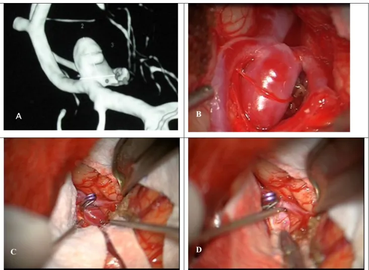
347
Fig. 1. (A) Three-dimensional rotational angiography imaging from Aneurysm #9. (B) Surgical view of this aneurysm in Middle Cerebral Artery location before clipping. (C)Aneurysm sac after surgical clipping. (D) Aneurysm resection.
348
349
A B
16 349
Fig.2: Strips were cut out from the aneurysm sac following a meridional axis. 350
17 351
A.
B.
Fig.3: Set-up and results from the uni-axial biomechanical testing. A. Side view of a wall strip from aneurysm n°4 during strain test, in a Ringer Lactacte solution at 40°C. B. Strain/Stress graph. The hysteresis is observed between the first load (*) and the five cycles. A slight compression was observed during the rest phase due to micro structural changes of the sample during the first cycles leading to a permanent elongation. When coming back to initial size of the sample, a slight compression is then observed on the graph (arrow).
352 353
18 353
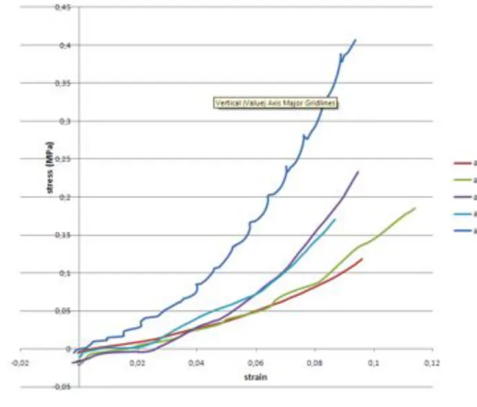
Fig. 4: Plots of the strain/stress relationships measured for men (R=ruptured, U=unruptured, 354
ACA = Anterior Cerebral Artery, ACM=Middle Cerebral Artery) 355 356 357 358 359 360 361 362 363 364 365 366 367 368
19 369 370 371 372
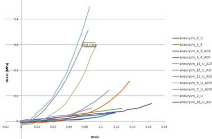
Fig. 5: Plots of the strain/stress relationships measured for women. (R=ruptured, 373
U=unruptured, ACA = Anterior Cerebral Artery, ACM=Middle Cerebral Artery, ACI= Inter-374
nal Carotid Artery, CPA= Cerebral Posterior Artery) 375
376 377 378
20 378 379
380
Fig.6: Mean strain/stress curves representing the biomechanical tissue classification. 381
382 383
21 383
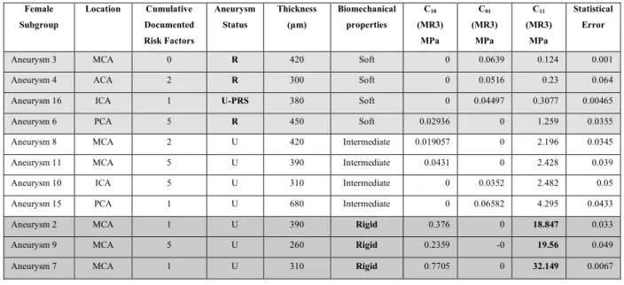
Table 1: Summary of clinical, anatomical and biomechanical data of the sixteen cases studied. 384
(U=Unruptured, R=Ruptured, U-PRS = Unruptured with pre-rupture symptoms, MCA 385
=Middle Cerebral Artery, PComA = Posterior Communicating Artery, ICA = Internal Carotid 386
Artery).The tissue classification (Soft/Rigid/Intermediate) is done according to the graphs 387 shown in fig.4 et 5. 388 389 Female Subgroup Location Cumulative Documented Risk Factors Aneurysm Status Thickness (!m) Biomechanical properties C10 (MR3) MPa C01 (MR3) MPa C11 (MR3) MPa Statistical Error
Aneurysm 3 MCA 0 R 420 Soft 0 0.0639 0.124 0.001 Aneurysm 4 ACA 2 R 300 Soft 0 0.0516 0.23 0.064 Aneurysm 16 ICA 1 U-PRS 380 Soft 0 0.04497 0.3077 0.00465 Aneurysm 6 PCA 5 R 450 Soft 0.02936 0 1.259 0.0355 Aneurysm 8 MCA 2 U 420 Intermediate 0.019057 0 2.196 0.0345 Aneurysm 11 MCA 5 U 390 Intermediate 0.0431 0 2.428 0.039 Aneurysm 10 ICA 5 U 310 Intermediate 0 0.0352 2.482 0.05 Aneurysm 15 PCA 1 U 680 Intermediate 0 0.06582 4.295 0.0433 Aneurysm 2 MCA 1 U 390 Rigid 0.376 0 18.847 0.033 Aneurysm 9 MCA 5 U 260 Rigid 0.2359 -0 19.56 0.049 Aneurysm 7 MCA 1 U 310 Rigid 0.7705 0 32.149 0.0067
390 391 392 393 Male Sub-group Location Cumulative Documented Risk Factors Aneurysm Status Thickness (!m) Biomechani-cal proper-ties C10 (MR3) MPa C01 (MR3) MPa C11 (MR3) MPa Stastisti-cal Error
Aneurysm 5 ACA 1 R 330 Soft 0.1803 0 3.241 0.016 Aneurysm 13 ACA 1 R 290 Soft 0.9374 0 3.101 0.0267 Aneurysm 12 MCA 2 U 170 Intermediate 0.1951 0 14.987 0.0457 Aneurysm 14 MCA 1 U 200 Intermediate 0.1015 0 7.9232 0.04 Aneurysm 1 MCA 1 U 620 Rigid 0.2569 0 12.265 0.035
394 395 396 397
22 397 398 399
Table 2: Comparison of clinical and biomechanical parameters between the three identified tissue 400
subgroups in the overall population. 401 n = 16 Aneurysmal wall biomecha-nical classifica-tion n Mean p
Risk Factors INTERMEDIATE 6 2.6 0.456
SOFT 6 1.6 .
RIGID 4 2 .
Wall Thickness INTERMEDIATE 6 360 0.994
SOFT 6 361 . RIGID 4 392 . C10 INTERMEDIATE 6 0.0598 0.057 SOFT 6 0.1911 . RIGID 4 0.3916 . C01 INTERM 6 0.0168 0.792 SOFT 6 0.0267 . RIGID 4 0.0322 . C11 INTERM 6 5.71 <.001** SOFT 6 1.37 . RIGID 4 20.87 . 402 403 404 405 406 407 408 409 410 411 412 413 414 415
23 416 417 418
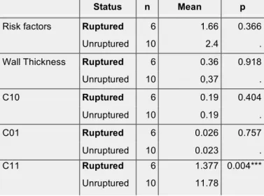
Table 3: Comparison of clinical data and biomechanical parameters between Ruptured and 419
Unruptured aneurysms in the overall population (male and female). 420
421
Status n Mean p
Risk factors Ruptured 6 1.66 0.366
Unruptured 10 2.4 .
Wall Thickness Ruptured 6 0.36 0.918
Unruptured 10 0,37 . C10 Ruptured 6 0.19 0.404 Unruptured 10 0.19 . C01 Ruptured 6 0.026 0.757 Unruptured 10 0.023 . C11 Ruptured 6 1.377 0.004*** Unruptured 10 11.78 422 423
24 423 424
Table 4: Comparison of clinical data and biomechanical parameters between Ruptured and 425
Unruptured aneurysms in the female group. 426
Status n Mean p
Risk factors Ruptured 4 2 0.516
Unruptured 7 2.85 .
Wall Thickness Ruptured 4 388 0.861
Unruptured 7 392 . C10 Ruptured 4 0.00734 0.200 Unruptured 7 0.20637 . C01 Ruptured 4 0.04012 0.279 Unruptured 7 0.01443 . C11 Ruptured 4 0.48 0.001** Unruptured 7 11.70 427 428
25 428
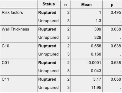
Table 5: Comparison of clinical data and biomechanical parameters between Ruptured and 429
Unruptured aneurysms in the male group. 430
Status n Mean p
Risk factors Ruptured 2 1 0.495
Unruptured 3 1.3 .
Wall Thickness Ruptured 2 309 0.638
Unruptured 3 329 . C10 Ruptured 2 0.558 0.638 Unruptured 3 0.160 . C01 Ruptured 2 -0.0001 0.638 Unruptured 3 0.043 . C11 Ruptured 2 3.17 0.058 Unruptured 3 11.95 . 431 432 433 434 435 436 437 438 439 440 441 442 443 444 445 446 447 448
26 449 450 REFERENCES: 451 452
1. Wardlaw JM, White PM. The detection and management of unruptured intracranial 453
aneurysms. Brain. 2000;123 ( Pt 2):205-221 454
2. Sluzewski M, Bosch JA, van Rooij WJ, Nijssen PC, Wijnalda D. Rupture of intracranial 455
aneurysms during treatment with guglielmi detachable coils: Incidence, outcome, and risk 456
factors. J Neurosurg. 2001;94:238-240 457
3. Unruptured intracranial aneurysms--risk of rupture and risks of surgical intervention. 458
International study of unruptured intracranial aneurysms investigators. N Engl J Med. 459
1998;339:1725-1733 460
4. Asari S, Ohmoto T. Natural history and risk factors of unruptured cerebral aneurysms. 461
Clin Neurol Neurosurg. 1993;95:205-214 462
5. Wiebers DO, Whisnant JP, Sundt TM, Jr., O'Fallon WM. The significance of unruptured 463
intracranial saccular aneurysms. J Neurosurg. 1987;66:23-29 464
6. Kyriacou SK, Humphrey JD. Influence of size, shape and properties on the mechanics of 465
axisymmetric saccular aneurysms. J Biomech. 1996;29:1015-1022 466
7. Isaksen JG, Bazilevs Y, Kvamsdal T, Zhang Y, Kaspersen JH, Waterloo K, Romner B, 467
Ingebrigtsen T. Determination of wall tension in cerebral artery aneurysms by numerical 468
simulation. Stroke. 2008;39:3172-3178 469
8. Alnaes MS, Isaksen J, Mardal KA, Romner B, Morgan MK, Ingebrigtsen T. Computation 470
of hemodynamics in the circle of willis. Stroke. 2007;38:2500-2505 471
9. Cebral JR, Castro MA, Appanaboyina S, Putman CM, Millan D, Frangi AF. Efficient 472
pipeline for image-based patient-specific analysis of cerebral aneurysm hemodynamics: 473
Technique and sensitivity. IEEE Trans Med Imaging. 2005;24:457-467 474
10. Radaelli AG, Augsburger L, Cebral JR, Ohta M, Rufenacht DA, Balossino R, Benndorf G, 475
Hose DR, Marzo A, Metcalfe R, Mortier P, Mut F, Reymond P, Socci L, Verhegghe B, 476
Frangi AF. Reproducibility of haemodynamical simulations in a subject-specific stented 477
aneurysm model--a report on the virtual intracranial stenting challenge 2007. J Biomech. 478
2008;41:2069-2081 479
11. Hoi Y, Meng H, Woodward SH, Bendok BR, Hanel RA, Guterman LR, Hopkins LN. 480
Effects of arterial geometry on aneurysm growth: Three-dimensional computational fluid 481
dynamics study. J Neurosurg. 2004;101:676-681 482
12. Cebral JR, Mut F, Weir J, Putman C. Quantitative characterization of the hemodynamic 483
environment in ruptured and unruptured brain aneurysms. AJNR Am J Neuroradiol. 484
2011;32:145-151 485
13. Cebral JR, Mut F, Weir J, Putman CM. Association of hemodynamic characteristics and 486
cerebral aneurysm rupture. AJNR Am J Neuroradiol. 2011;32:264-270 487
14. Scott S, Ferguson GG, Roach MR. Comparison of the elastic properties of human 488
intracranial arteries and aneurysms. Can J Physiol Pharmacol. 1972;50:328-332 489
15. Toth BK, Nasztanovics F, Bojtar I. Laboratory tests for strength paramaters of brain 490
aneurysms. Acta Bioeng Biomech. 2007;9:3-7 491
16. Masson I, Fialaire-Legendre A, Godin C, Boutouyrie P, Bierling P, Zidi M. Mechanical 492
properties of arteries cryopreserved at -80 degrees c and -150 degrees c. Med Eng Phys. 493
2009;31:825-832 494
17. Karmonik C, Diaz O, Grossman R, Klucznik R. In-vivo quantification of wall motion in 495
cerebral aneurysms from 2d cine phase contrast magnetic resonance images. Rofo. 496
2010;182:140-150 497
18. Ogden GAHaRW. Biomechanical modelling at the molecular, cellular and tissue levels. 498
CISM Courses and Lectures, Springer: Wien, New York. 2009;No. 508: 259-343 499
19. Marc André Meyers P-YC, Albert Yu-Min Lin, Yasuaki Seki. Biological materials: 500
Structure and mechanical properties, review article. Progress in Materials Science. 501
2008;Volume 53:1-206 502
27
20. Oliver A. Shergold NAFaDR. The uniaxial stress versus strain response of pig skin and 503
silicone rubber at low and high strain rates. International Journal of Impact Engineering. 504
2006;Volume 32:1384-1402 505
21. Mooney M. A theory of large elastic deformation. Journal of Applied Physics. 506
1940;11:582-592. 507
22. Rivlin RS. Large elastic deformations of isotropic materials. I. Fundamental concepts, 508
Philosophical Transactions of the Royal Society of London.Series A, Mathematical and 509
Physical Sciences. 1948; 240:459-490. 510
23. Low SK, Zembutsu H, Takahashi A, Kamatani N, Cha PC, Hosono N, Kubo M, Matsuda 511
K, Nakamura Y. Impact of limk1, mmp2 and tnf-alpha variations for intracranial 512
aneurysm in japanese population. J Hum Genet. 2011 513
24. Crawford T. Some observations on the pathogenesis and natural history of intracranial 514
aneurysms. J Neurol Neurosurg Psychiatry. 1959;22:259-266 515
25. Frosen J, Piippo A, Paetau A, Kangasniemi M, Niemela M, Hernesniemi J, Jaaskelainen J. 516
Remodeling of saccular cerebral artery aneurysm wall is associated with rupture: 517
Histological analysis of 24 unruptured and 42 ruptured cases. Stroke. 2004;35:2287-2293 518
26. Watton PN, Ventikos Y, Holzapfel GA. Modelling the growth and stabilization of cerebral 519
aneurysms. Math Med Biol. 2009;26:133-164 520
27. Krings T, Willems P, Barfett J, Ellis M, Hinojosa N, Blobel J, Geibprasert S. Pulsatility of 521
an intracavernous aneurysm demonstrated by dynamic 320-detector row cta at high 522
temporal resolution. Cen Eur Neurosurg. 2009;70:214-218 523
28. Hayakawa M, Katada K, Anno H, Imizu S, Hayashi J, Irie K, Negoro M, Kato Y, Kanno 524
T, Sano H. Ct angiography with electrocardiographically gated reconstruction for 525
visualizing pulsation of intracranial aneurysms: Identification of aneurysmal protuberance 526
presumably associated with wall thinning. AJNR Am J Neuroradiol. 2005;26:1366-1369 527
29. Ishida F, Ogawa H, Simizu T, Kojima T, Taki W. Visualizing the dynamics of cerebral 528
aneurysms with four-dimensional computed tomographic angiography. Neurosurgery. 529
2005;57:460-471; discussion 460-471 530
30. MacDonald DJ, Finlay HM, Canham PB. Directional wall strength in saccular brain 531
aneurysms from polarized light microscopy. Ann Biomed Eng. 2000;28:533-542 532
31. Zhang C, Villa-Uriol MC, De Craene M, Pozo JM, Frangi AF. Morphodynamic analysis of 533
cerebral aneurysm pulsation from time-resolved rotational angiography. IEEE Trans Med 534
Imaging. 2009;28:1105-1116 535
32. Balocco S, Camara O, Vivas E, Sola T, Guimaraens L, Gratama van Andel HA, Majoie CB, 536
Pozo JM, Bijnens BH, Frangi AF. Feasibility of estimating regional mechanical properties 537
of cerebral aneurysms in vivo. Med Phys. 2010;37:1689-1706 538
33. Zhao X, Raghavan ML, Lu J. Identifying heterogeneous anisotropic properties in cerebral 539
aneurysms: A pointwise approach. Biomech Model Mechanobiol. 2011;10:177-189 540
541 542 543
28 543 544
Conflict of interest statement 545
All authors do not have any financial and personal relationships with other people or
organi-546
sations that could inappropriately influence this work.
547 548 549
29 549
Acknowledgement: 550
The authors would like to thank Philips™, Ansys™, for funding part of this research and pro-551
viding free research software licenses. These sources of funding were not involved in the
552
study design, in the collection, analysis and interpretation of data; in the writing of the
manu-553
script; and in the decision to submit the manuscript for publication.
554 555