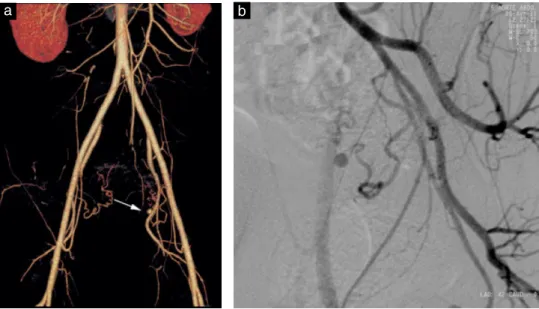
Pitfalls in diagnosis of uterine artery pseudoaneurysm after Cesarean section
Texte intégral
Figure


Documents relatifs
On discutait à présent comme deux vieux amis… Elle m’expliqua qu’elle fréquentait depuis six mois un homme marié, que le type avait le cul entre deux chaises, que
[r]
[r]
la démarche et de ses effets sur le personnel avec questionnaires ou bilan basé sur les commentaires selon l’état d’avancement des travaux 7 rencontres annuelles de
Results show that in our corpus, lexical diversity metrics only capture an increase in vocabulary from the first to the third production of each student, whereas vocabulary
This study in a referral hospital in Sana’a, Yemen investigated the outcome of vaginal birth after caesarean section in 357 women who had one prior caesarean section and were
F stimmen Sie zu, dass die Prognose entscheidend von der Dauer und der Intensität der Hyperammonämie abhängt und daher eine rasche Verlegung des Patienten in ein
[r]
