ATOM ABSTRACTION IN THE INTERACTION
OF F2 AND XeF2 WITH
Si(100)
byDAVID GOSALVEZ-BLANCO
B.S. THE UNIVERSITY OF CALIFORNIA AT BERKELEY
(1992)
SUBMITTED TO THE DEPARTMENT OF CHEMISTRY IN PARTIAL FULFILLMENT OF THE REQUIREMENTS
FOR THE DEGREE OF DOCTOR OF PHILOSOPHY
AT THE
MASSACHUSSETTS INSTITUTE OF TECHNOLOGY
SEPTEMBER 1997
© Massachusetts Institute of Technology 1997 All rights reserved
Signature of Author Department of Chemistry July 22, 1997 Certified by Accepted by Sylvia T. Ceyer '/ iThesis Supervisor Dietmar Seyferth Chairman, Department Committee
This doctoral thesis has been examined by a Committee of the Department of Chemistry as follows:
Professor Robert J. Silbey
Chairman
Professor Sylvia T. Ceyer
Thesis Supervisor
ATOM ABSTRACTION IN THE INTERACTION
OF F2 AND XeF2 WITH
Si(100)
byDAVID GOSALVEZ-BLANCO
Submitted to Department of Chemistry on July 22, 1997 in partial fulfillment of the requirements for the Degree of Doctor of Philosophy
in Chemistry
ABSTRACT
The chemisorption dynamics of thermal energy F2 and XeF2 interacting with Si(100) are investigated in an ultra-high vacuum molecular beam scattering chamber. The apparatus is equipped with a differentially pumped line-of-sight quadrupole mass spectrometer which enables the detection of highly reactive radical species produced during the gas-surface interaction. The interaction of thermal energy F2 with Si(100) 2x1 can lead to
three different outcomes: unreactive scattering, two-atom adsorption, and single-atom abstraction. The absolute probabilities of each of these reaction channels are determined. Since both the scattered F and F2 ionize to produce F+, the F product is primarily
distinguished on the basis of its different velocity and dependence on exposure. A detailed mass balance of the incident and scattered fluorine allows for the determination of the absolute reaction probabilities and the fluorine coverage on the Si surface as a function of F2 exposure. The unreactive scattering probability, Po, is 0.05±0.01 on the
clean surface but rapidly increases with surface coverage, reaching unit probability at saturation coverage. The two-atom adsorption probability, P2, exhibits an initial value of 0.85±10.03 on the clean surface and drops linearly with coverage, vanishing at saturation coverage. The single-atom abstraction probability starts at a value of 0.10±0.03 for the clean surface, then goes through a maximum value of 0.3±0.85 at a coverage of approximately 0.5 ML, and finally drops to zero on the fluorine saturated surface. The total reaction probability is 0.95±+0.04. The fluorine coverage obtained by integrating the exposure dependent reaction probabilities saturates at a value of 1.06±+0.05 ML. The value of the saturation coverage coupled with helium diffraction measurements on the clean and fluorinated Si(100) 2x1 surface confirm that the Si dangling bonds are both the abstraction and adsorption sites, and that no Si-Si dimer or bulk bonds are cleaved during the chemisorption of F2. Within the detection sensitivity of the apparatus, no silicon containing etch products are observed with thermal F2 incident on a 250 K Si
Three phenomenological models based on the lattice-gas formalism are shown to reproduce the major features of the data and provide some insight into the adsorption and abstraction mechanisms. Atom abstraction occurs when an incident F2 molecule finds an
empty site onto which to adsorb, but its complementary F atom does not. The orientation of the bond axis of the incident F2 with respect to the surface is one factor that affects the ability of the second F atom to find a reactive site onto which to bind. The occupation of the Si atoms surrounding the initial abstraction site, is a second factor which determines the likelihood that the complementary F atom is ejected to the gas phase. Following an abstraction event, the ejected F atom may find a reactive site and also adsorb on the surface. The linear dependence of the two-atom adsorption probability with fluorine coverage suggests that a single Si dimer pair is most likely responsible for the adsorption of two fluorine atoms from a single incident F2 molecule.
Single-atom abstraction is also demonstrated in the interaction of XeF2 with Si(100) by the mass spectrometric identification of XeF ejected to the gas phase. The identification of XeF is complicated by the dissociative ionization of unreactively scattered XeF2 and by the ionization of Xe arising from the two-atom adsorption process. The exposure dependence as well as the velocity and angular distributions ofXeFf, XeF2 +, Xe+ and F' are used to confirm the abstraction a single F atom from an incident XeF2 molecule. The exposure dependence of the XeF' signal is reminiscent of that observed for the F atoms ejected in the interaction of F2 with Si(100). The XeF product is primarily observed at scattering angles near the surface normal, with its intensity rapidly decreasing with increasing scattering angle. The unusual angular and exposure dependence of the ejected XeF product are exploited to deconvolute the mass spectrometer signals into the neutral products giving rise to them. The XeF fragment ejected from the surface gains some of the reaction's exothermicity as evidenced by its translational excitation, which is confirmed by time-of-flight measurements. The exothermicity of the reaction is also observed to induce the gas phase dissociation of the ejected XeF fragment. The chemically induced dissociation of XeF is inferred from the observation of very fast F* atoms thought to arise from the gas phase decomposition of vibrationally or electronically excited XeF*.
Thesis Supervisor: Sylvia T. Ceyer Title: Professor of Chemistry
ACKNOWLEDGEMENTS
The work presented in this thesis is the product of the combined efforts of over a dozen people for the last ten years. I would like to thank the first generation of "BiMPs" because their careful design and construction of the apparatus afforded me the chance to work with one of the best instruments in the business. In addition, many of the key ideas and experiments that laid down the foundation of the F2 work must be credited to them. From my early years in the project I would like to thank Dr. Julius Yang, Dr. Yulin Li and Dr. David Pullman for teaching me the intricacies of the apparatus. Dave was responsible for innumerable improvements to the apparatus as well as for the design of many of the experiments aimed at calibrating the flux of the incident beam and scattered products. I benefited greatly from Dave's extensive experience with molecular beams and vacuum technology as well as from his programming ability. Most of the computer code utilized for the data acquisition and analysis, which greatly simplified my work, must be credited to him. His dedication to the project, his unlimited patience and endless
good humor made him an ideal coworker during many late night runs.
My contributions to the project are shared with Dr. Thanos Tsekouras and Matthew Tate. Thanos relentlessly challenged all my bad ideas and helped turn them into reasonable ones. I could always count on him to catch and correct my many careless mistakes both in dealing with the apparatus and analyzing the experimental results. His uncanny ability to fix and improve the apparatus kept us up and running through many vacuum mishaps. I have certainly missed Thanos' oversight during the preparation of my thesis. I can assure the reader that the many inaccuracies and typographical errors found in this work would have certainly been eradicated by Thanos. Matthew Tate joined the project and rapidly took the helm. It is customary for the senior students to teach the new members of the team, but in the Matthew's case, he quickly learned the few things that I could teach him and then proceeded to teach me the many things I did not know. His addition to the project catalyzed the completion of the F2 experiments and has added new dimensions to the XeF2 work of which he has now taken charge. It has been a pleasure working with Matthew over the past three years, and wish him the best of luck in his future scientific endeavors.
The latest additions to the project include Stacey Eckman and Dr. Massimo Bertino. Stacey brings and endless supply of friendly smiles and a positive attitude to the group. Her constant words of encouragement and occasional candy bar kept me going through the long writing sessions. Massimo has quickly become a close friend, and I regret not being able to spend more time with him before my departure. I wish him luck in his
scientific career and hope that we will cross paths again in the future.
I have also enjoyed keeping up a friendly rivalry with the "LiMP" team. Kerstin Haug and Thomas Btirgi deserve credit for putting up with my constant teasing while they tried to tune their EELS spectrometer. I am grateful to Judson Holt for the use of his personal computer during a fatal hard drive crash in the last stages of the writing of this thesis, and for putting up with all my bad jokes about Rice University.
In a more personal note, I would like to thank my close friends and family. My first years at MIT were marked by close friendships with Chris Murray, Ted Trautman, Marc Wefers and Sean Daley. We had some great times together and I hope that we keep in
touch in the years to come. Fernando Bergasa has also been a close friend over the past six years, and I have enjoyed our many conversations about subjects ranging from the paradoxes of quantum mechanics or the intricacies of protein folding, to the right amount of vodka to add to a sangria.
Cathy and Victoria have given me the motivation to keep working through the hard times. I love you both more than life itself, and would like to thank Cathy for all her love, support and encouragement over the past five years. My parents deserve my most heartfelt gratitude for their generosity and unwavering support in all my endeavors. After a long 10 year absence from their side I look forward to spending some quality time with them. I thank Sherilyn, Jim and Jaime Harrison for their boundless generosity and support of Cathy, Victoria and me. Charles Edgar and Carolyn McCarthy have been a constant source of support and have provided some very welcomed hospitality during our Cape Cod escapades. I am greatly indebted to the Fordyce family for adopting me during my college years and continued support after I left California. I hope we can keep up our wonderful friendship in spite of the long distance between us.
Finally, I would like to thank my advisor Sylvia Ceyer. Her uncompromising commitment to involve herself in only the highest quality scientific research is solely responsible for her international prestige. She has pushed me for the last six years to strive for the best possible work and patiently waited for the results that she knew could be achieved. Without her determination, the quality of the work here presented would have greatly suffered from my haste. I am also very grateful to Sylvia for her extensive help in editing and proofreading the many revisions of this manuscript. This work is as much hers as it is mine.
TABLE OF CONTENTS ABSTRACT ... .. ... .. ... ... 3 ACKNOWLEDGEMENTS ... ... 5 LIST OF FIGURES... ... ... 9 LIST OF TABLES ... ... 11 PREFACE ... ... ... ... 12
1 CHAPTER I: THE INTERACTION OF F2 WITH Si (100) ... 16
1.1 INTRODUCTION ... ... ... 17
1.2 EXPERIMENTAL A PPARATUS ... ... 21
1.2.1 Description ... ... 21
1.2.2 Modifications to the Apparatus... 27
1.2.2.1 Attachment of Crystal Temperature Thermocouple ... ... 27
1.2.2.2 Changes to Detector Box Turbo Molecular Pumps...29
1.2.2.3 Detector Box Pin-hole ... ... 30
1.3 SUMMARY OF EXPERIMENTAL RESULTS ... 33
1.3.1 Time-of-flight Measurements of the Scattered F and F2 ... .. . .. . .. . .. . . .... 33
1.3.2 Exposure Dependence of the Scattered F and F2 . . . .. . .. . . . . . ... 42
1.3.3 Thermal Desorption Measurements ... ... 47
1.3.4 Helium Atom Diffraction Measurements ... 53
1.4 EXPOSURE DEPENDENCE OF THE FLUORINE COVERAGE... 60
1.4.1 The Probabilities of the Reaction Channels... 60
1.4.1.1 Measurement of the Electron Impact Ionization Cross-sections ... 68
1.4.1.2 Determination of the Quadrupole Transmission Function ... ... 72
1.4.2 Scattering Angle Dependence of the Reaction Probabilities ... . 74
1.4.3 Calculation of the Exposure Dependent Surface Coverage... 79
1.4.4 Determination of Molecular Beam Fluxes... ... 82
1.4.4.1 Determination of the Flux of Single Component Molecular Beams... 82
1.4.4.2 Determination of the Flux of Seeded Molecular Beams... 86
1.4.4.3 Pumping Speed Measurements... ... 89
1.4.4.4 Measurement of the Reaction Chamber Volume ... ... 92
1.4.4.5 Ion Gauge Sensitivity Correction Factor ... ... 93
1.5 DISCUSSION ... ... 97
1.5.1 Physical Picture Resulting from the Experimental Results... 97
1.5.2 Qualitative Features of the F2 + Si (100) Potential Energy Surface... 99
1.5.3 The Dependence of the Reaction Probabilities on Fluorine Coverage ... 104
1.5.3.1 Lattice-gas Model for the Dissociative Chemisorption of F2 on Si(100) ... 107
1.5.3.2 Three-state Lattice-gas Model ... ... 121
1.5.3.3 Extended Three-state Lattice-gas Model ... 135
1.6 CONCLUSIONS ... ... ... 140
2 CHAPTER H. THE INTERACTION OF Si(100) WITH XeF2 ... . . .. . .. . . .. .. . . .. . 141
2.1 INTRODUCTION ... ... ... 142
2.1.1 An Overview of Previous Experimental Work... ... 142
2.1.1.1 Measurements of the Si Etch Rates... 143
2.1.1.2 Identification of the Gas-phase Etch Products ... 144
2.1.1.3 Composition and Growth of the Fluorinated Surface Layer ... 145
2.1.2 Atom Abstraction in the Interaction of XeF2 with Si(100) ... . 148
2.1.3 Background on XeF2 .... . . . . .. . .. . .. . .. . . .. .. . . .. . . 150
2.1.3.2 Physical Properties of XeF2... .. ... .. .. ... .. .. ... ... . . . ... . . ..... ... 152
2.1.3.3 Thermodynamic Properties of XeF2 ... .. .. ... .. .. ... .. .. ... . . . .. ... 152
2.1.3.4 Mass Spectrometry of XeF2 .. . . . ... .. .. ... .. .. ... . ... . 154
2.1.3.5 XeF2 Effusive Beams... ... ... ... ... 155
2.2 EXPERIMENTAL ... ... . ... 157
2.2.1 Production of Seeded Supersonic XeF2 Beams ... 157
2.2.1.1 X eF2 M ixing Cylinders ... ... 157
2.2.1.2 Passivation Issues ... ... 159
2.2.2 Characterization of Seeded Supersonic XeF2 Beams ... 163
2.2.2.1 Mass Spectra of XeF2 Beams ... ... 163
2.2.2.2 Van der Waals Clustering in the Beam ... 166
2.2.2.3 Velocity Distribution of Seeded XeF2 Beams... 175
2.2.3 Fragmentation of XeF2 by Electron Impact Ionization ... 178
2.2.3.1 Scattering from an Inert SiO Surface... ... 179
2.3 RESULTS ... ... ... ... .. 188
2.3.1 Observation of Unreactively Scattered XeF2.... . . . .. .. . .. . .. . .. . .. . .... .... . . .... 192
2.3.2 Identification of Atom Abstraction by Observation of XeF ... 197
2.3.3 Observation of Scattered Xe Product ... ... 205
2.3.4 Other Evidence for the Reaction ofXeF2 with Si(100)...223
2.3.4.1 Thermal Desorption Products and Surface Fluorine Coverage ... 223
2.3.4.2 Identification of the Order of the Adsorbed Products by Helium Diffraction ... 230
2.3.5 Preliminary Results at Higher XeF2 Incident Energy ... ... 235
2.3.5.1 XeF2/He Scattered from Si(100)... 235
2.4 D Iscus SIo N ... 24 1 2.5 CONCLUSIONS ... ... ... 257
3 APPENDICES ... 258
APPENDIX A: ERROR ANALYSIS...259
APPENDIX B: UNCERTAINTY IN CROSS-CORRELATION TOF ... 276
APPENDIX C: ATTENUATION OF THE MOLECULAR BEAM... ... 280
APPENDIX D: RELATIVE TDS PRODUCT YIELD...285
LIST OF FIGURES
Figure 1.1 Schematic Diagram of the UHV Beam-surface Scattering Apparatus ... 26
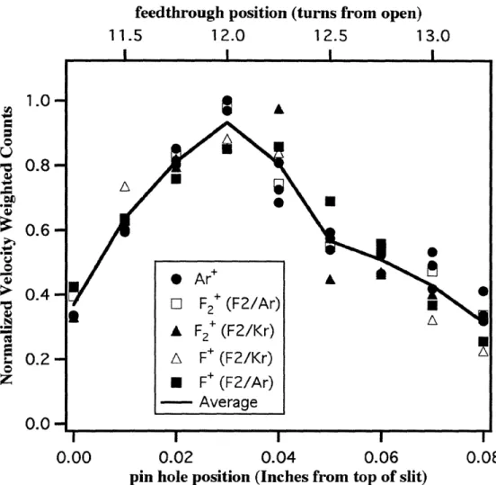
Figure 1.2 Straight-Through Signal as a Function of Pin-hole Position...32
Figure 1.3 Time-of-Flight Spectra of Scattered 1 %F2/Kr...38
Figure 1.4 F2 and SiF2 Time-of-flight Spectra from a 1000 K Si(100) Surface ... 39
Figure 1.5 Time-of-flight Spectra of F Atoms Ejected from a 1000K Si(100)...40
Figure 1.6 Energy Distributions of F-atoms from 250K and 1000 K Surfaces ... 41
Figure 1.7 Exposure Dependence of Scattered F' and F2 Signal...46
Figure 1.8 Thermal Desorption Spectra of Si(100) Exposed to 1%F2/Ar ... 51
Figure 1.9 Total Fluorine Thermal Desorption Yield as a Function of F2 Exposure ... 52
Figure 1.10 Structure of Unreconstructed and Reconstructed Si(100)...58
Figure 1.11 He Diffraction Spectra of a Clean and Fluorinated Si(100) ... 59
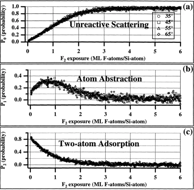
Figure 1.12 Exposure Dependence of F2 Reaction Probabilities ... 67
Figure 1.13 Angular Distribution of Scattered F and F2...... ... .... . . . .77
Figure 1.14 Reaction Probabilities Measured at Various Scattering Angles ... 78
Figure 1.15 Fluorine Coverage as a Function of F2 Exposure ... 80
Figure 1.16 Coverage Dependence of F2 Reaction Probabilities ... 81
Figure 1.17 Pumping Speed Curves for Ar and Ne... ... 91
Figure 1.18 Ar Expansion Measurement for Ion Gauge Calibration ... 96
Figure 1.19 Reaction Probabilities Predicted by the Lattice-gas Model ...119
Figure 1.20 Lattice-gas Model Probabilities as a Function of Coverage... 120
Figure 1.21 Reaction Probabilities Predicted by Three-state Model... 133
Figure 1.22 Three-state Model Probabilities as a Function of Coverage ... 134
Figure 1.23 Extended Three-state Model Probabilities as a Function of Coverage... 139
Figure 2.1 Initial Time Evolution of the XeF2 Fragments from the Clean Nozzle... 162
Figure 2.2 Mass Spectrum of Xe Isotopes from a Xe/Ar Molecular Beam... 164
Figure 2.3 Mass Spectrum of XeF2/He Molecular Beam ... 165
Figure 2.4 Mass Spectrum of XeF2/Kr Molecular Beam... 170
Figure 2.5 High and Low Resolution Mass Spectra of XeF2/Ar Molecular Beam... 171
Figure 2.6 Fit of XeF2/Ar Signal to a XeF2 and [Xe--Ar]+ Superposition ... 172
Figure 2.7 Mass Spectra of Xe/Ar and XeF2/Ar Molecular Beams ... 173
Figure 2.8 Mass Spectra Demonstrating the Existence of XeF2--Ar in the Beam...174
Figure 2.9 Time-of-flight Spectra of XeF2/Kr and XeF2/Ar Beams... 176
Figure 2.10 Time-of-flight Spectra of XeF2/He/Ar Beams... 177
Figure 2.12 Thermal Desorption Products from Inert Surface ... 185
Figure 2.13 Thermal Desorption Products from Inert Surface ... 186
Figure 2.14 Angular Distribution of Scattered XeF2 from a Silicon Oxide Surface ... 187
Figure 2.15 Scattered Products as a Function of Exposure to a XeF2/Ar Beam... 190
Figure 2.16 Scattering Angle Dependence of the XeF2/Si Reaction Products ... 191
Figure 2.17 Angular Distribution of Scattered XeF2 ... ... 195
Figure 2.18 Time-of-flight Spectra of XeF2 Unreactively Scattered from Si(100) ... 196
Figure 2.19 XeF from Atom Abstraction Detected as XeF+ ... 201
Figure 2.20 Exposure Dependence XeF Product at Eight Scattering Angles ... 202
Figure 2.21 Angular Distribution of XeF Product ... 203
Figure 2.22 Time-of-flight Spectra of Scattered XeF' Signal ... 204
Figure 2.23 Partial Deconvolution of Xe' Signal detected at 00... 213
Figure 2.24 Partially Deconvoluted Xe÷ as a Function of Scattering Angle ... 214
Figure 2.25 Deconvolution of Xe and XeF Contributions to the Xe' Signal ... 215
Figure 2.26 Comparison of XeF Product Detected as XeF+ and Xe. ... 216
Figure 2.27 Time-of-flight Spectra of Scattered Xe' Signal ... 217
Figure 2.28 Time-of-flight Spectra of Scattered F+ Signal... 218
Figure 2.29 Angular Distribution of SiF3 ... 221
Figure 2.30 Time-of-flight Spectra of Scattered SiF' Signal ... 222
Figure 2.31 Thermal Desorption Products from Si(100) Exposed to XeF2 ... 227
Figure 2.32 Comparison of TD Products from F2 and XeF2 Fluorination ... 228
Figure 2.33 TD Product Yield as a Function of Exposure to XeF2/Ar ... 229
Figure 2.34 He Diffraction Spectra of Clean and XeF2 Exposed Si(100)... 232
Figure 2.35 He Diffraction Spectra as a Function of XeF2/Ar Exposure... 233
Figure 2.36 Peak He-diffraction Intensity as a Function of XeF2/Ar Exposure ... 234
Figure 2.37 Scattered Products as a Function of Exposure to 0.2% XeF2/He... 238
Figure 2.38 Scattered Products as a Function of Exposure to 0.05% XeF2/He... 239
Figure 2.39 TD Product Yield as a Function of Exposure to XeF2/He... 240
Figure 2.40 Exposure Dependence of the XeF2 and Etch Products... 253
Figure 2.40 Exposure Dependence of Xe and XeF Products ... 254
Figure 2.41 Total Reaction Probability of a XeF2/Ar Beam Exposed to Si(100)... 255
Figure 2.42 Comparison of Angular Distribution of XeF÷ and Xe÷ Signals ... 256
Figure 3.1 Main Chamber Pressure as a Function of Beam Stagnation Pressure... 284
LIST OF TABLES
Table 1-1 Parameters Describing Velocity Distribution of F2 in Seeded Beams ... 25
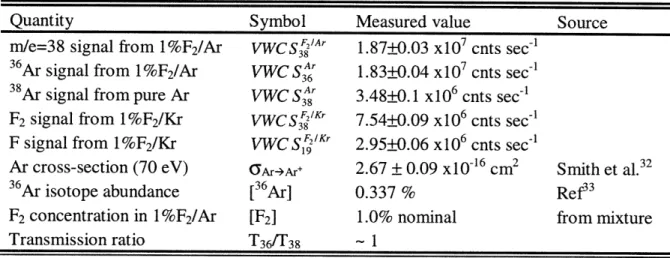
Table 1-2 Absolute Values Required for Calculation of Reaction Probabilities ... 66
Table 1-3 Values Required for Calculation of F2 Ionization Cross-sections ... 71
Table 1-4 Values Required for Calculation of Relative Transmissivity...74
Table 1-5 Values Required for Determination of Fluxes by Steady-state Method...84
Table 1-6 Values Required for Determination of Fluxes by the Stagnant Method...85
Table 1-7 Flux of Pure Ar and Ne Molecular Beams ... ... 86
Table 1-8 Values Required for Determination of Seeded Beam Fluxes...88
Table 1-9 Flux of Seeded M olecular Beams... 89
Table 1-10 Pum ping Speeds... ... 90
Table 1-11 Values Required for the Determination of Ion Gauge Correction Factors ....95
Table 2-1 Some Physical Properties of XeF2 ... ... . . . ... ... . 154
Table 2-2 Parameters Describing Velocity Distribution of XeF2 in Seeded Beams... 175
Table 2-3 Cracking Ratios for XeF2 ... . . ... ... ... ... . 183
Table 3-1 Estimated Uncertainties in the Determination of the Pure Beam Flux...265
Table 3-2 Estimated Uncertainties in the Seeded Beam Flux Determination...268
Table 3-3 Estimated Uncertainty in the Determination of F2 and F Velocity Ratio ... 269
Table 3-4 Estimated Uncertainties for the Determination of Transmission Ratio...272
Table 3-5 Estimated Uncertainties for the Determination of Cross-section Ratio...273
Table 3-6 Values Required for Determination of Expected Pressure Ratio ... 283
Preface
PREFACE
The dry etching of silicon is an essential reaction in the manufacturing of semiconductor based electronic devices in very large scale integrated circuits (VLSI).
Dry etching involves the reaction of silicon with an ignited plasma typically containing CF4 as well as inert buffer gases. The thermodynamic driving force behind the etching reaction is the large Si-F bond energy (-140 kcal/mol) and the ultimate formation of stable but volatile SiFx products (i.e. SiF4, Si2F6 , Si3Fs...). The most active chemical species in the plasma is believed to be the F atoms which, due to their open shell electronic structure readily attack the silicon surface. Emphasis has also been placed on
the importance of "chemical sputtering", an ill-defined superposition of physical
sputtering and chemical reactions, in which the combination of chemically reactive
neutrals and ionic species present in the plasma are believed to enhance cooperatively the
etch rate of the Si surface. Many mechanisms have been proposed for this reaction, but no definitive experimental confirmation has yet been obtained.
Extensive work has been performed over the last twenty years on simplified model
systems. These studies include numerous measurements of the etch rates and reaction
kinetics as well as molecular beam studies aimed at clarifying the dynamics of the
interaction. A number of experiments designed to investigate this reaction have used F2 and XeF2 as convenient sources of fluorine atoms for the reaction. An interesting, yet puzzling result is that, at room temperature, XeF2 etches silicon at a rate approximately 10,000 times faster than F2. In addition, it is found that the etch rate of Si with XeF2 is at least one order of magnitude faster than that of F atoms. This observation casts some
doubt on the importance of the open shell nature of the F atom, and calls for an
explanation of the unusually large etch rates obtained with the less thermodynamically
Preface
Previous work in the Ceyer group has established some important facts about the reactivity of F2 molecules with the Si(100) surface which may help to understand the underlying reasons for the unusually high XeF2 reactivity, and may also shed new light on the mechanism for the plasma reaction. Low energy, molecular F2 is found to
dissociatively chemisorb onto a clean Si(100) 2x1 reconstructed surface. Helium diffraction results show that the incoming F2 exclusively fluorinates the Si dangling bonds (1 dangling bond/Si atom) producing a monolayer structure which retains the clean surface (2x1) periodicity. This observation implies that low energy fluorine can break neither the Si surface dimer bonds nor the bonds between the top and second layer of Si atoms. This monolayer saturation coverage is consistent with the low etching rate
measured for F2, since etching requires cleavage of the dimer and second layer Si bonds. In addition, the existence of a novel mechanism by which the F2 molecule adsorbs on the dangling bonds has been demonstrated. Briefly, this new mechanism, denoted "F atom abstraction", consists of the abstraction of a single fluorine atom by a Si dangling bond
from a fluorine molecule impinging on the surface, with the subsequent ejection of the
complementary F atom. Depending on the orientation of the F2 molecule as it approaches the surface, the complementary atom may be ejected either away from or towards the
surface. In the latter case, if the ejected atom encounters an empty dangling bond site, it may also adsorb on the surface. Although the ejected atom gains a significant amount of
translational energy from the exothermicity of the bond breaking step, and may be
propelled into the Si with higher than thermal velocities, it is found not to break dimer or
second layer Si bonds, as verified by helium diffraction.
The first half of the present investigation extends the study of the interaction of F2
with Si(100), corroborating the inability of F2 to fluorinate the Si surface beyond a saturation coverage of 1 ML. A careful quantitative analysis of F2 scattering data allows
Preface coverage is calculated as a function of F2 exposure. The measurement of the reaction probability from mass spectrometric data requires the determination of various electron ionization cross-sections as well as the careful calibration of reactant flux and mass spectrometer sensitivity. The experimental details of the absolute F2 reaction probability measurement, and its subsequent use to measure the surface coverage are the primary subjects of Chapter I. Determination of the saturation coverage as 1 ML is consistent with the helium diffraction results, and confirms the inability of F2 to cleave Si-Si dimer bonds and thus to etch Si.
In contrast, studies have shown that XeF2 can fluorinate the Si surface well beyond the monolayer limit encountered in the F2 reaction. This result is consistent with the high etch rates, since it is believed that the etching proceeds via a thick (-7 ML) SiFx reaction overlayer. The main difference between F2 and XeF2 must lie in the ability of the latter to break surface and lattice bonds and thus fluorinate the silicon surface beyond the single-monolayer limit. A hypothesis is proposed that XeF2 is able to fluorinate the surface to higher coverages than F2 because the XeF molecule is able to transfer, due to its large mass, a large fraction of its incident kinetic energy to the silicon lattice, thus creating local excitations or distortions of the lattice which activate the reaction of F with the lattice. In the case of F2, the hypothesis notes that F2 is too light to transfer a large enough fraction of its energy to the surface to induce the lattice distortions which activate the reaction. The importance of kinetic energy transfer is also supported by the observation that XeF2 has a faster etching rate than the much more reactive, but lighter, fluorine atom, which cannot vibrationally excite the surface. The role of energy transfer may be additionally important because it could account for the enhanced reactivity observed under plasma conditions, since many of the very energetic ionic species present in the plasma do transfer a substantial amount of momentum to the lattice and thus maintain a supply of vibrationally excited reactive sites on the silicon surface.
Preface
Although the experimental confirmation of the energy transfer hypothesis is beyond the scope of the current investigation, an important step towards demonstrating its validity is currently undertaken. The first step in assessing the validity of the proposed
hypothesis is the corroboration of the existence of the F atom abstraction mechanism in
the interaction of XeF2 with a Si(100). The work presented in Chapter II establishes the operability of the F atom abstraction mechanism in the XeF2/Si system by the mass spectrometric identification of the ejected XeF fragment. Furthermore, the velocity
distribution of the XeF product is measured and found to be consistent with a large
degree of translational excitation of the ejected fragment, likely arising from the
exothermicity of the abstraction reaction. A XeF fragment propelled towards the
fluorinated Si surface has both the large of momentum required to induce local
vibrational excitation of the lattice, and the thermodynamic instability of the weakly
bound F atom. The challenge of assessing the relative importance of momentum transfer
and reactant stability in the unusually high reactivity of XeF2 towards Si remains ahead' 2.
SM. R. Tate, Massachusetts Institute of Technology, in preparation.
1.1 Introduction
1.1 INTRODUCTION
An important function of a surface in heterogeneous catalysis or chemical vapor deposition is to cleave a bond of an incident gas phase molecule. This cleavage process is commonly referred to as dissociative chemisorption, and results in the chemical binding of the incident molecule to the surface as two separate adsorbates. Because the surface-to-adsorbate bonds in the majority of gas-surface interactions are weaker than the internal bond of the impinging molecule, the energetic cost required to break the internal bond in the impinging molecule necessitates the formation of two adsorbate-surface
bonds in order to render the overall process exothermic.
Based on the investigation of the reactivity of gas phase F2 incident on a Si(100) 2x1
surface, Ceyer et al.3 have demonstrated a new mechanism for dissociative chemisorption termed atom abstraction. Atom abstraction differs from the classic dissociative
chemisorption process in that only one of the two molecular fragments of the incoming
species binds to the surface. In the specific case of the interaction of F2 with a Si, a single Si-F bond is formed at the surface with the subsequent release of the
complementary F atom into the gas phase. Since the abstraction mechanism is
thermodynamically allowed only if enough energy is released upon formation of a single
gas-surface bond to offset the energetic cost incurred to break the original bond, it is only
expected to occur in systems in which a very strong bond can form between the surface
and a gas phase molecular fragment. The interaction of F2 with the Si(100) surface is such a system.
Ab initio calculations4 of the binding of a gas phase F atom and a Si surface dangling bond estimate the Si-F bond energy to be 6.4 eV. This bond strength is very large
3 Y. L. Li, D. P. Pullman, J. J. Yang, A. A. Tsekouras, D. B. Gosalvez, K. B. Laughlin, Z. Zhang, M. T. Schulberg, D. J. Gladstone, M. McGonigal and S. T. Ceyer, Phys. Rev. Lett. 74, 2603 (1995)
Chapter I: The Interaction of F2 with Si(100) compared to the 1.4 eV binding energy of the gas phase F2 molecule. The energy released upon formation of a single Si-F bond would then greatly exceed the energy needed to break the F-F bond giving a net 4.8 eV exothermicity for the abstraction process. Simultaneous formation of two Si-F bonds is clearly not thermodynamically necessary for the dissociation of F2, and thus a single F atom can be abstracted by the surface with the concomitant release of the complementary F atom into the gas phase. Similar abstraction mechanisms have been well documented in gas phase reactions, but they have never been experimentally corroborated in gas-surface interactions prior to the work of Ceyer et a13. Simultaneously with the first experimental observation, abstraction was observed for the F2/Si reaction by molecular dynamics trajectory calculations performed by Stillinger and Weber5. The occurrence of F atom abstraction by the Si dangling bonds means that in the plasma environment used in commercial Si etching applications, the surface reaction is contributing to the production of reactive F atoms which are believed to be the most reactive species. Kinetic models of the plasma etching environment should therefore take into account the rate of production of F atoms by the abstraction mechanism.
Experimental confirmation of the proposed F-atom abstraction mechanism involves the detection of the scattered F atom after it fails to form a bond to the silicon surface. The reactive nature of the ejected F atoms is likely responsible for the failure of numerous published studies of the interaction of fluorine and fluorinated hydrocarbons with silicon to detect the atom abstraction mechanism. Undoubtedly, if a scattered F atom is allowed to collide with the reaction chamber walls before detection, it will adsorb and hence not be observed. In addition, although this new mechanism for dissociative chemisorption is a general one and must be occurring in other exothermic molecule-surface systems, it has also gone undetected in all other systems. The successful
1.1 Introduction
identification of the atom abstraction mechanism in the case of the interaction of F2 with Si is directly attributable to the unique features of the molecular beam-surface scattering apparatus which is briefly described in Section 1.2.1. In particular, the use of a beam coupled with a line-of-sight, triply-differentially-pumped mass spectrometer allows the detection of the highly reactive F radicals produced by the abstraction reaction.
Direct observation of scattered F atoms was first achieved by time-of-flight velocity measurements of a low energy F2 molecular beam scattered from a Si(100) surface.
Helium atom diffraction studies of the structure of the fluorinated surface coupled with the determination that the abstraction mechanism ceases at 1 ML fluorine coverage strongly suggest that not only are the dangling bonds on each Si atom the adsorption sites, but that they are also the abstraction sites. However, the complementary F atom does not necessarily scatter into the gas phase. It may be trapped by a second reactive site encountered during its outgoing trajectory and bind. Whether the scattered F atom is propelled away from the surface or towards a reactive site depends on the orientation of the incident F2 molecule. Measurement of the evolution of the reaction products as a function of exposure to the F2 molecular beam yields information about the coverage dependence of the abstraction mechanism. For example, an elaborate analysis of these measurements as a function of exposure yields a quantitative determination of the absolute reaction probability for each channel as a function of coverage. Armed with the knowledge of the absolute reaction probabilities, the evolution of the fluorine coverage with F2 exposure and a saturation coverage of 1 ML are determined. The unusual
dependencies of the reaction probabilities on coverage suggest a strong dependence of the F2 reaction probability on both the orientation of the incident F2 molecule and on the
dimer structure of the Si(100) 2x1 surface.
Section 1.2 gives a brief description of the experimental apparatus used for this investigation as well as of some minor modifications implemented since the apparatus
Chapter I: The Interaction ofF2 with Si(100) was last described6. Section 1.3 presents a summary of the results of time-of-flight measurements, exposure dependence of scattering products, thermal desorption spectroscopy, and helium diffraction measurements needed to establish the existence of the atom abstraction mechanism. A more detailed discussion of these experiments has been given by Yang6. Section 1.4 concentrates on the additional measurements, such as the incident beam flux and the F2 ionization cross-sections necessary to obtain
quantitative information about the coverage dependence of the abstraction mechanism. Section 1.5 presents a discussion of the qualitative aspects of the potential energy surface derived from the experimental results, as well as three empirical models aimed at understanding the dynamical and chemical interactions that dictate the coverage dependence of the reaction probabilities.
1.2 Experimental Apparatus
1.2 EXPERIMENTAL APPARATUS
1.2.1 Description
All experiments presented in this work were conducted in an ultra-high vacuum (UHV) molecular beam surface scattering apparatus. A detailed description of this apparatus as well as its design criteria have been given elsewhere'78,'910. Only a brief description of its essential components is presented here.
The single crystal Si(100) sample is positioned inside an UHV main chamber with a base pressure of approximately 6x10 11 Torr. This low pressure is essential to ensure that the silicon surface remains free of adsorbed contaminants during the duration of the experiments. Reactants (i.e. F2) are introduced via a differentially pumped, supersonic molecular beam precisely coupled to the UHV main chamber. The molecular beam impinges on the Si surface where the etching reaction takes place. The scattered molecules are then detected by a line-of-sight, triply differentially pumped, electron impact ionization, quadrupole mass spectrometer. The mass spectrometer is rotatable about the center of the crystal, thereby allowing the angular distribution of molecules scattered from the surface to be measured. A pseudo-random slotted chopper wheel
mounted at the entrance to the first differential pumping stage of the mass spectrometer modulates the scattered molecules for the purpose of velocity analysis using a
time-of-flight technique. A schematic representation of the apparatus is given in Figure 1.1. The supersonic molecular beam is produced by expanding a dilute mixture of either F2 or XeF2 seeded in a noble gas carrier through a 100 gm diameter orifice. The center of
7 S. T Ceyer, D. J. Gladstone, M. McGonigal, and M. T. Schulberg, Physical Methods of Chemistry, edited by B. W. Rossitier and R. C. Baetzold (Wiley, New York, 1933), 2 nd ed., Vol. IXA, p. 383
8 M. McGonigal, Ph.D. Thesis, Massachusetts Institute of Technology, (1989)
9 D. J. Gladstone, Ph.D. Thesis, Massachusetts Institute of Technology, (1989)
Chapter I: The Interaction of F2 with Si(100) the expanded beam passes through a skimmer placed 5.6 mm from the nozzle. Two
differential pumping chambers, separated by collimating apertures, ensure a collisionless expansion environment and minimize the effusive gas contribution into the main chamber. The collimating apertures define a rectangular beam cross-section of 6.4 mm by 4.5 mm at the center of the silicon crystal. The nozzle to sample distance is 13.6 cm. Typical stagnation pressures behind the nozzle range from 100 to 600 Torr leading to beam fluxes in the range of 1016 to 1018 cm-2 sec-1 (10-1000 ML sec'-1). These seeded
supersonic beams allow the reactants to reach the surface with a well defined angle of incidence and translational energy. The angle of incidence can be varied from 0' to 900 with respect to the surface normal. The range of translational energies attainable by seeding depends on the kind and relative amounts of seed and carrier gas. For the case of F2, translational energies can be varied between 0.1 and 14 kcal mole-'. Table 1-1 gives a summary of the velocity and translational energy of the F2 beams used in this
investigation.
The Si(100) samples used are approximately 2.5 cm in diameter and 525 jt thick. They are cut out of 10 cm diameter wafers supplied by either Monsanto or Sematech. The samples provided by Monsanto are lightly n-doped with a resistivity of 8-12 g2-cm, while the Sematech ones are p-type with similar resistivity. No dependence on Si doping is observed in any of the experiments conducted during this investigation. The
machining and mounting procedure has been described by Yang". In order to obtain a good surface quality, the machined samples are wet etched in dilute HF following the
procedure described by Shiraki et al.12 before insertion into the vacuum chamber.
Neutral products scattering or desorbing from the surface must be ionized before
they can be detected. Non-selective, but efficient ionization of the reaction products is
11 J. J. Yang, Ph.D. Thesis, Massachusetts Institute of Technology, p. 37, (1993)
1.2 Experimental Apparatus performed by use of a Brink's'3,14 type electron bombardment ionizer. The neutral product beam is ionized by crossing a shower of 70 eV electrons produced by heating a tungsten filament. The ions are born inside a cylindrical grid at a potential of 45 eV above the filament bias voltage and are then extracted out of the ionization region and focused by an Einzel lens onto the entrance of the quadrupole mass filter'5. Only ions of a pre-selected mass-to-charge ratio emerge at the exit of the quadrupole field were they are counted by a channel electron multiplier. The overall efficiency of this ionization and detection scheme is estimated to be approximately one in 106 particles entering the
ionization region.
In order to limit detection to products arising exclusively from the reaction at the silicon surface, the entire detector is placed inside a housing containing three
differentially pumped chambers separated by small rectangular collimating apertures. The
detector housing collimating slits are designed so that only a portion of the Si surface
exposed to the incoming molecular beam is imaged onto the ionization region, ensuring
that the detector's line-of-sight is limited to products traveling in a straight line from the
surface. Any products having undergone further reaction by collisions with the chamber walls are not likely to be detected. The differential pumping scheme maintains a
collisionless gas phase environment and minimizes the effusive gas load onto the
ionization region by pumping away particles not directly in the line-of-sight of the
detector. In order to further reduce the background gas load in the ionization region, and
to prevent excessive outgassing from hot metal surfaces near the filament, the walls of
the ionization chamber are cooled to liquid nitrogen temperatures. Line-of-sight detection
coupled with the maintenance of low background pressures in both the reaction chamber
13 Y. T. Lee, J. D. MacDonald, P. R. Le Breton, and D. R. Herschbach, Rev. of Sci. Instr. 40, 1402 (1969)
14 Gilbert O. Brink, Rev. of Sci. Instr. 37, 857 (1966)
Chapter I: The Interaction ofF2 with Si(100) and inside the detector housing enables the detection of unstable or reactive products. For example, detection of the highly reactive F radicals expected to arise from an abstraction event is made possible by the line-of-sight alignment of the triply-differentially pumped spectrometer housing. This arrangement prevents the F radicals from being depleted through collisions with the chamber walls or background gas before they can be detected.
Detection of molecules desorbed or scattered from the surface is not limited to the narrow acceptance angle defined by the collimating slits on the detector housing. The entire distribution of molecules scattered from the surface is measurable by rotation of the detector and its housing around the center of the crystal, allowing the angular distribution of the molecules scattered in the plane defined by the molecular beam and the line-of-sight of the detector to be collected. Symmetry considerations and reasonable
assumptions about the out-of-plane scattering must be used to account for the complete
product distribution about a hemispherical shell centered on the sample surface.
The entrance to the detector chamber is fitted with a cross-correlation chopper wheel for measurement of the velocity distribution of the scattered molecules by a time-of-flight
technique. The wheel modulates the scattered beam into short pulses of gas with a well defined starting position and time. The time necessary for the molecules in each pulse to travel the known distance between the chopper and the ionizer is measured, thus yielding
the velocity of the scattered molecules. To maximize the signal, a pseudo-random
modulation sequence of slits is carved on the chopper wheel, which has a 50% duty cycle. The high duty cycle of the modulation sequence causes some overlap of the scattered gas pulses as they travel towards the detection region. The measured spectrum
must then be deconvoluted using the known modulation pattern to yield the time-of-flight
time-of-1.2 Experimental Apparatus
flight measurements has been given by Comsa et al16
The most important capabilities of the molecular beam-surface scattering apparatus
used in this investigation are summarized as follows: 1) allows control of the energy and
angle of incidence of the reactants impinging on the surface. 2) maintains a low base
pressure in the reaction chamber to ensure surface cleanliness and a collisionless gas
phase environment. 3) provides line-of-sight detection capable of collecting even the
most reactive radicals scattered from the surface. 4) renders the complete angular
distribution of reaction products. 5) gives the translational energy distribution of those
products.
Table 1-1 Parameters Describing Velocity
600 Torr Beam Average Velocity
1% F2/Kr 384±0.1m sec-'
1% F2/Ar 547±0.1 m secl'
0.25% F2/He 1684±0.3 m secl'
Pure Ne 812±5 m sec-'
For explanation of the uncertainties see the section on Appendix A.
Distribution of F2 in Seeded Beams
Beam Temp. Average F2 Energy
1.59+0.02 K 0.67_+0.005 kcal mol'
2.13±+0.02 K 1.36_+0.005 kcal mol1 -28.4±0.18 K 12.9±+0.004 kcal mol-1
13.1+0.71 K 1.59+0.01 kcal mo' 1
error analysis of seeded beam fluxes presented in
16 R. David, K. Kern, P. Zeppenfeld and G. Comsa, Rev. Sci. Instrum. 57, 2771 (1986)
r-Chapter I: The Interaction ofF2with Si(100)
XeF
2
or F
2
Molecular Beam
1.2 Experimental Apparatus 1.2.2 Modifications to the Apparatus
1.2.2.1 Attachment of Crystal Temperature Thermocouple
The temperature of the silicon crystal mounted inside the UHV chamber can be
varied from 120 K up to its melting point by a combination of heating and cooling. The
lowest temperatures are achieved by connecting the sample support to a liquid nitrogen
filled reservoir via a thick copper braid. The sample is resistively heated by applying a
voltage difference across the support clamps and hence running a current across the silicon wafer. The desired temperature is obtained by an optimal control feedback circuit
described elsewhere17. The feedback circuit requires accurate real-time measurements of
the crystal temperature, which is measured by an Omega Instruments type-C
(W-5%Re/W-26%Re) thermocouple attached to back of the silicon wafer. Good thermal
contact between the thermocouple and the silicon substrate is essential for accurate and
reproducible temperature measurements. Two different approaches have been used in
these investigation to ensure proper attachment of the thermocouple to the crystal.
As a first approach, the thermocouple junction produced by spot welding the 0.005"
thick Re-W wire leads was glued to the back of the crystal. Since the sample is routinely
heated to temperatures above 1000 K and rapidly cooled to 250 K in a UHV
environment, the glue must have some very special properties. Aside from the obvious
UHV compatibility issues, the glue must possess the following properties: 1) good adherence to the silicon substrate, 2) its coefficient of thermal expansion must be similar
to that of silicon so as not to fracture under the stress of thermal cycling, 3) it must
withstand temperatures in excess of 1300 K, 4) it must efficiently conduct heat from the
silicon substrate to the thermocouple junction.
The glue of choice was Aremco Ceramabond Ultratemp 516, a zirconium silicate
Chapter I: The Interaction of F2 with Si(100) base cement which satisfies all the above listed properties. A small dab of glue was placed on the back of the silicon wafer and the thermocouple junction was pressed against it until the glue hardened. The glue was then cured in accordance to the manufacturer's specifications. The curing schedule required introducing the end of the manipulator and the sample holder into a small oven for several hours. Proper curing of the glue led to well-attached thermocouples that gave accurate and reproducible surface temperature measurements. The gluing and curing procedure, however, proved to be hard to reproduce. The success rate of this attachment method was approximately 30 to 50 %. Unsuccessful gluing attempts, in which the thermocouple separated from the crystal, would become apparent only after investing several days to bake the chamber and
clean the silicon crystal. An alternate, more reliable method of attaching the
thermocouple to the sample was then devised.
The alternate attachment method does not involve the use of glue. Instead, a pair of
screws is used to securely pin two thermocouples to the back of the silicon sample. First,
each thermocouple junction is spot welded to a small square tantalum tab of
approximately 2.5 mm on each side and 75 pm thick. A bracket spot-welded across the back of the sample holder provides two threaded holes for the attachment screws. A
piece of insulating ceramic material (silica) is placed between the screw tip and the
tantalum tab which is then sandwiched against the back of the silicon sample. The
ceramic piece electrically insulates the thermocouple junction from the grounded bracket
and screw. The screw must provide a secure hold to insure good thermal contact between
the thermocouple and the silicon, but care must be taken not to over tighten it since
excess force may cause the silicon wafer to bulge and crack upon heating.
The mechanical attachment of a thermocouple with a screw gives adequate
temperature readings for the feedback of the temperature control circuit, and has proven
1.2 Experimental Apparatus thermal desorption experiments performed with mechanically attached thermocouples are however, typically lower than the peak temperatures obtained by gluing. In addition, the reproducibility of thermal desorption temperatures was significantly better for glued than for screwed thermocouples. These observations suggest that the thermal contact between the silicon and the screwed thermocouple is not as good as that obtained by using the zirconium glue. Although screwing the thermocouple to the back of the crystal allows for an easy and durable way to control the crystal temperature, it is not ideal for measurements that require an accurate and reproducible measurement of the absolute surface temperature.
1.2.2.2 Changes to Detector Box Turbo Molecular Pumps
The differential pumping scheme used for the rotatable mass spectrometer has been previously described in detail7',s,0. Some changes have been made to the pumps during the course of this investigation and are briefly described here. In order to increase the pumping speed of the first differential pumping region of the mass spectrometer housing,
the existing ion pump was replaced with a Balzers TPU 330 turbomolecular pump
identical to those used in the second and third pumping stages. A Balzers TPC 121
controller is used to drive the new pump. The foreline of this new pump was connected
to the common foreline of the two existing turbo pumps. The Balzers TPU 110
turbomolecular pump used to provide backing pressure for the large turbo pumps was
replaced by a Balzers TMH 065 turbomolecular drag pump. Pneumatically actuated
butterfly valves (Key High Vacuum Products models QBV-75-P-SS16 and
QBV-150-P-SS40 ) were placed at both ends of the drag pump. All forelines between the turbo pumps and the backing turbo pump were shortened to approximately one foot and
widened to a diameter of one inch so as to maximize the pumping speed. A
thermocouple vacuum gauge tube was added at the inlet of the mechanical pump that
Chapter I: The Interaction of F2 with Si(100) to monitor and protect all four turbomolecular pumps. Details of this interlock circuit are presented elsewhere1 .
1.2.2.3 Detector Box Pin-hole
The line-of-sight of the mass spectrometer can be rotated to coincide with the molecular beam axis. This configuration is denoted as a "straight-through" position, and is necessary for measuring the velocity distribution and intensity of the incident molecular beams. However, the apertures in the detector chamber are designed to maximize the signal from the scattered molecules. So, when the detector is aligned in the straight-through configuration the gas load becomes too large, causing excessive ion densities in the ionization region. While the ion densities can be easily reduced by lowering the ionization current, the quantitative measurements described in Section 1.4
require the incident beam intensity to be measured under the exact ionization conditions
used in the scattering experiments. A way is then needed to reduce the gas load admitted
into the detector chamber so that the spectrometer can be operated at higher ionization
currents without the detrimental effects of excessive ion density.
In order to reduce the gas load, an additional limiting aperture was placed in front of
the detector housing entrance. The new aperture was fabricated from a commercially
available 12.5 g pin-hole at the center of a 3.3 mm disk which was spot welded around a
larger hole in a piece of tantalum foil. The tantalum foil is attached to a 1 mm thick
rectangular tantalum plate measuring 3x1 cm, which is in turn bolted to the detector-box
beam valve support rod. The use of this limiting aperture ensures that the gas load in the
straight-through configuration is approximately equal to the gas load arising from a beam
scattered off the surface.
The position of the pin-hole aperture with respect to the detector entrance can be
1.2 Experimental Apparatus
controlled by rotating the detector valve feedthrough. A profile of the mass spectrometer signal as a function of the beam valve feedthrough position is shown in Figure 1.2. The
signal intensity is determined by recording and integrating a time-of-flight spectrum at
each pinhole position. The maximum of the intensity profile corresponds to the position
at which the pin-hole is aligned with the centerline of the ionization region, which is obtained by turning the detector valve 12 turns from its fully open position. Note that the
detection sensitivity rapidly decreases as the pin-hole moves away from the ionizer's
centerline. It is therefore very important to ensure the pin-hole position is accurately
Chapter I: The Interaction of F2with Si(100)
feedthrough position (turns from open)
11.5 12.0 12.5 13.0 1.0 0.8 0.6 0.4 0.2 0.0 0.00 0.02 0.04 0.06 0.08
pin hole position (Inches from top of slit)
Figure 1.2 Straight-Through Signal as a Function of Pin-hole Position
Optimal pin-hole alignment is obtained by turning the detector valve feedthrough 12 turns from its fully open position. The onset of the signal seen at approximately 11 turns corresponds to the top edge of the rectangular limiting aperture at the entrance to the detector chamber. The 12 turn position corresponds to having the pin-hole centered, 0.03" from the top edge of the 0.078" high aperture.
1.3 Summary of Experimental Results 1.3 SUMMARY OF EXPERIMENTAL RESULTS
1.3.1 Time-of-flight Measurements of the Scattered F and F2
When a F2 molecule impinges upon an adsorbate-free Si surface three pathways are possible: 1) The molecule may unreactively scatter back to the gas phase without any chemical change taking place. 2) The F2 molecule could undergo an abstraction reaction in which one F atom remains bound to the surface while its partner scatters back to the gas phase. 3) The incident F2 can dissociatively chemisorb by leaving both fluorine atoms bound to the surface. The first pathway is readily identifiable by detection of either the parent ion F2 with a mass-to-charge ratio of 38, or the fragment ion F' with m/e =19 of unreactively scattered F2 molecules with the electron impact ionization mass
spectrometer. The second pathway, involving atom abstraction is in principle identifiable
by mass spectrometric detection of F atoms as the parent F+ ion at m/e=19. However,
their signal would be superimposed on the m/e=19 signal produced from the cracking of
F2 in the electron bombardment ionizer.
A challenge then arises in distinguishing the ejected fluorine atoms from the fraction
of the unreactively scattered F2 that gets fragmented to F' during ionization. Fortunately, the two neutrals giving rise to the superimposed signals can be differentiated on the basis
of their different velocities. The unreactively scattered F2 is expected to inelastically scatter or even partially accommodate on the surface upon collision. It would then scatter
back to the gas phase with a broad velocity distribution. On the other hand, an F atom
ejected during the abstraction event may be translationally excited from the large amount
of energy released by the exothermicity of the reaction. The ejected fluorine atoms
would then leave the surface with a faster and narrower velocity distribution than the
thermalized, unreactively scattered F2.
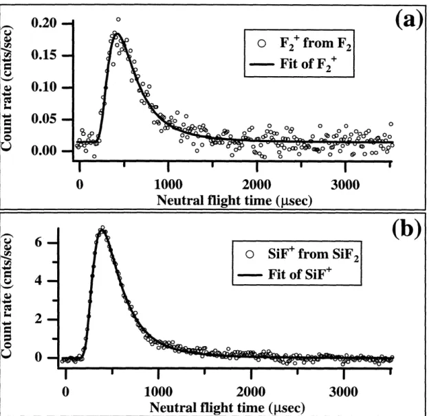
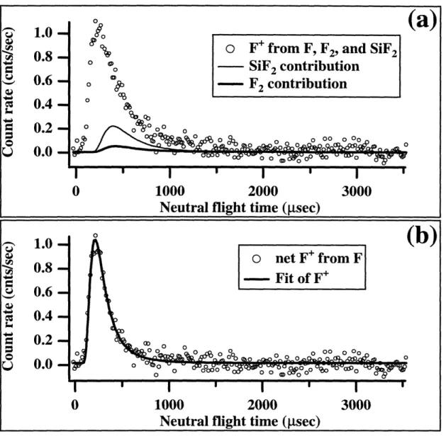
Chapter I: The Interaction of F2 with Si(100) scattered reaction products. Figure 1.3 (a) shows time-of-flight spectra of products scattered from a 250 K Si(100) surface exposed to a 600 Torr, 1% F2/Kr beam (Ei=0.66 kcal mol'), incident at 0o for a total exposure to 0.2 ML of fluorine atoms. The scattered products are collected as m/e=38 and 19 at a detection angle of 350 with respect to the surface normal. The chopper is spun at 280 Hz and the data are collected into 255 channels each with a width of 14 psec.
The filled circles correspond to signal detected at m/e=38, and therefore can be attributed to the parent ion of the unreactively scattered F2. The solid line represents a fit to a Maxwell-Boltzmann distribution with a characteristic temperature of 240 K, which is
approximately equal to the temperature of the surface, indicating an almost complete accommodation of the incident F2 translational energy. The hollow circles in Figure 1.3 (a) correspond to signal detected at m/e=19, which arises from the superposition of directly ionized ejected F atoms and unreactively scattered F2 molecules that fragment to
F+ upon ionization. The spectrum displays a bimodal distribution consisting of a fast, narrow peak at early arrival times followed by a broader, slow component at later arrival
times. The broad, slow peak is attributed to unreactively scattered F2 while the sharper,
fast feature is identified as F atoms ejected in the abstraction process. There is good agreement between the velocity distribution of the F2 signal and the slow contribution to the F' signal as should be expected since they both arise from the same neutral molecule,
namely unreactively scattered F2. Figure 1.3 (b) shows the time-of-flight distribution obtained by point-by-point subtraction of the two distributions presented in panel (a).
The net distribution is attributed to ejected F atoms. This observation constitutes the first
direct confirmation of the fluorine atom abstraction mechanism.
The ejected F atom signal is fitted to a supersonic velocity distribution19 with
1.3 Summary of Experimental Results average velocity of 1100 m sec- corresponding to an average translational energy,
1 m v , of 3.2 kcal mo1-1. Given that the average translational energy of the impinging F2
beam is known to be 0.66 kcal mol-1, it is clear that the scattered F atoms possess a greater translational energy than available in the incident beam. The high translational energy of the ejected F suggests that part of the exothermicity of the abstraction reaction is channeled into translational energy of F. However, the exothermicity released upon formation of a single Si-F bond is calculated to be 110 kcal mo'-1, implying that only about 3% of the available energy released in the reaction is channeled into translational excitation of the ejected F fragment.
To confirm the origin of the translational excitation of the scattered F atoms, the time-of-flight measurements are repeated while holding the crystal temperature at 1000 K. A 3.8% F2/Kr beam (0.67 kcal mole-l) is incident on the hot surface at 0O, while the scattered signals are detected at 35o. The total fluorine exposure is estimated to be about
240 ML of F atoms per Si on the surface. Figure 1.4 (a) shows the unreactively scattered
F2 signal (m/e=38) fitted to a Maxwell-Boltzmann distribution with a characteristic temperature of 495 K, which corresponds to an average velocity of 619 m sec-1 (- mv2 1.97 kcal molP) for the scattered F- 2. Comparison of the energy of the unreactively scattered F2 to the average energy expected for F2 desorbing from a 1000 K surface
(2kT=3.98 kcal mol') reveals that F2 gains some translational energy upon collision with
the hot surface, but it does not fully accommodate with the crystal surface. Figure 1.4 (b)
shows the time-of-flight distribution of the SiF2 etch product detected as SiF' at m/e=47. As expected, the SiF' etch product desorbs with a thermal velocity distribution at 1004 K,
reflecting the temperature of the crystal, and with a corresponding average velocity of
669 m sec- .
The F' signal (m/e=19), demonstrating the existence of F atoms ejected from the hot