Differential Regulation of Genes and Transcription Factors in Vascular Endothelial Cells Exposed to
Uniform and Disturbed Laminar Shear Stress MIT LIBRARIES
DEC
Tobi Elaine Nagel
Bachelor of Science with Distinction, Stanford University, 1989
1997
SCHERINGSubmitted to the Harvard-MIT Division of Health Sciences and Technology in Partial Fulfillment of the Requirements for the Degree of
DOCTOR OF PHILOSOPHY IN MEDICAL ENGINEERING at the
Massachusetts Institute of Technology June, 1997
C © 1997 Massachusetts Institute of Technology
All rights reserved
Signature of Author
Certified by
Certified by
Accepted 1
Harvard-MIT Division of Health Science and&rechnology
A, I-y, 1997
Michael A. Gin rrne, Jr. Professor of Pathology, Harvard MedicaT School Thesis Supervisor - A _.-n
Robert Langer Professor of Chemical and Biomedical Engineering, MIT Chairman, Thesis Committee
by
.v
% I
v Martha L. Gray Associate Professor of Electrical Engineering and C mputer Science, MIT Interim Co-Director, Harvard-MIT Division of Health Sci nces and Technology
f~.I
• . • z•?: :'.. :,,- •, .. .. . . .. ,
DIFFERENTIAL REGULATION OF GENES AND TRANSCRIPTION FACTORS IN VASCULAR ENDOTHELIAL CELLS EXPOSED TO
UNIFORM AND DISTURBED LAMINAR SHEAR STRESS
by
Tobi Elaine Nagel
Submitted to the Harvard-MIT Division of Health Sciences and Technology on May 23, 1997, in Partial Fulfillment of the Requirements for the Degree of
Doctor of Philosophy in Medical Engineering
ABSTRACT
Hemodynamic shear stress has been shown to induce various functional changes in vascular endothelium, many of which reflect alterations at the level of gene expression. We have utilized a class of endothelial-leukocyte adhesion molecules, including Intercellular Adhesion Molecule-i (ICAM- 1), Vascular Cell Adhesion Molecule-i (VCAM- 1), and E-selectin (ELAM- 1) -- which are coordinately induced by soluble stimuli such as cytokines -- as model genes with which to investigate the shear stress parameters which regulate endothelial gene expression. These studies indicate that ICAM-1, but not VCAM-1 or E-selectin, is upregulated in endothelial cells exposed to uniform laminar shear stress, and this induction occurs at the levels of transcriptional activation, steady state message, and functional cell surface protein. In addition, using a disturbed laminar shear stress model -- designed to mimic in vivo flow patterns at sites predilected for atherosclerotic development, such as arterial bifurcations and curvatures -- we have shown that cytokine induction of VCAM-1, in contrast to ICAM-1 or E-selectin, is inhibited by preconditioning with disturbed, but not uniform laminar shear stress. Thus, this differential regulation of adhesion molecule expression by fluid shear stresses, in contrast to the coordinate pattern of induction by humoral stimuli, indicates that regional differences in biomechanical forces may represent important local modulators of endothelial gene regulation.
Work in our laboratory and others has identified several "shear stress response elements" within the promoters of certain endothelial genes which mediate the shear stress induction of those genes by functionally interacting with previously identified transcription factors, including nuclear factor-,B (NF-KB), early growth response-i (Egr-1), and activator protein-i (AP-1, comprised of c-Jun/c-Jun and c-Jun/c-Fos protein dimers). With the aid of newly developed image analysis techniques, we have demonstrated that endothelial cells exposed to disturbed laminar shear stress exhibit increased levels of nuclear localized NF-KB, Egr-1, c-Jun, and c-Fos, as compared with cells subjected to uniform flow and static conditions. Taken together, these data suggest that the spectrum of biomechanical forces encountered in the circulatory system may represent important stimuli that are relevant to both the physiology and pathology of vascular endothelium.
Thesis Committee:
Michael A. Gimbrone, Jr., Professor of Pathology, Harvard Medical School (Adviser)
Robert Langer, Professor of Chemical & Biomedical Engineering, MIT (Committee Chairman) C. Forbes Dewey, Jr., Professor of Mechanical Engineering, MIT
ACKNOWLEDGMENTS
As I complete the chapters of this thesis, I also complete a significant chapter in my life, and it has been a very rich one, mostly because of the people in my life. I am indebted to my adviser, Dr. Michael Gimbrone, for guiding me in my development as a scientist and for providing me with a wealth of resources, in terms of both laboratory supplies and outstanding colleagues with whom to interact. I am also deeply grateful to Dr. Nitzan Resnick, who served as my direct mentor during the first few years of my thesis research and who taught me so much about bench-top science as well as how to balance that science with other aspects of life. Dr. Forbes Dewey, in his co-advisory role, provided me with guidance from an engineering perspective, which helped to round out my training in this interdisciplinary field. Dr. Bob Langer served as both my academic adviser and thesis committee chairman, and I have greatly appreciated the counsel he has given me throughout my graduate career. I am also thankful to Dr. Rakesh Jain for his encouragement and insights as a member of my thesis committee.
Many members of the Vascular Research Division at Brigham and Women's Hospital have helped to make my experiments possible and the work enjoyable at the same time. Bill Atkinson worked side-by-side with me during my initial shear stress experiments and the development of the image analysis system, and I have also appreciated his serving as a great sounding board in my various decision-making processes. Kay Case provided me with endothelial cell cultures too numerous to count, especially single cord preparations, and I am thankful to both Kay and Bill for letting me take temporary residence in their laboratory and office during the final stages of my research. I am grateful to Alice Callahan for her diligence and sense of humor in helping me with all of the administrative paperwork that comes with being an MIT student conducting research at Brigham and Women's Hospital. Dick Fenner (as a consultant from MIT), together with Keith Anderson, provided invaluable assistance in maintaining the shear stress apparatuses. Dr. Jamie Topper allowed me to bounce numerous questions and ideas off of him, and I enjoyed working closely with both him and Scott Wasserman on our related shear stress projects. It was also a delight to share the laboratory for several years with Jeanne Kiely, Dr. Tony Rosenzweig, and Dr. Masayuki Yoshida. George Stravrakis was helpful with many of my staining protocols. I am thankful to Dr. Tucker Collins for educating me regarding the intricacies of gene regulation. Dr. Margaret Read was particularly helpful in teaching me about NF-KlB regulatory mechanisms, as well as serving as an unofficial mentor.
I have been amazed at the personal support I have received from many friends throughout my graduate school experience. I have appreciated hours of laughter and encouragement shared with Anita Erler, and I am glad that she was a medical resident during the past few years, so that I could page her anytime of the day or night. Chi Huang has been particularly supportive during the final stages of my thesis, helping to keep me going during some long work days and boosting my spirits with his sense of humor. I have also appreciated the support of Don and Mary Martin, Anne O'Donnell, Lois Barndt, and Andrea Kirazian (with whom I will celebrate the completion of this thesis with a trip to Greece!), and many other friends too numerous to list. In addition, I have enjoyed the camaraderie of fellow HST students, especially Ann Celi, Chris Chen, Brian Benda, and Edwin Ozawa.
My family members have been a constant source of support for me. When I was in fifth grade, my parents stood at the edge of the soccer field and cheered their hearts out, and they have remained my greatest cheerleaders throughout this period of my life. I have appreciated the support and strength of my grandmother and, together with her, I wish my grandfather could have been here to share the final few years of my graduate experience. I am also thankful for my relationships with my aunt Coletta and uncle John, who have been like paternal grandparents to me.
Finally, I believe that the presence of all of these wonderful people in my life has not been just a chance occurrence, and I am thankful to God who has blessed me with these relationships and given me the opportunity to pursue this significant life experience exploring the intricacies of biological systems.
TABLE OF CONTENTS
Abstract ... 2
Acknowledgm ents ... 3
Table of Contents ... . ... 4
Chapter 1: Introduction: Regulation of Endothelial Cell Structure and Function by Fluid Shear Stress ... 6
Hemodynamics and the Endothelium ... 7
In Vitro Responses of Endothelial Cells to Shear Stress ... 8
Mechanisms of Endothelial Gene Regulation by Shear Stress ... 10
Objectives ... . ... 11
Chapter 2: Differential Regulation of Endothelial-Leukocyte Adhesion Molecules by Uniform Laminar Shear Stress ... 13
Introduction: Endothelial-Leukocyte Adhesion Molecules in Vascular Biology ... 14
M ethods ... 17
R esults ... 20
Discussion ... 28
Chapter 3: Differential Regulation of Endothelial-Leukocyte Adhesion Molecules by Disturbed Laminar Shear Stress ... 37
Introduction: Disturbed Laminar Shear Stress In Vivo ... 38
M ethods ... ... 41
Results ... ... ... 42
Discussion ... ... ... 49
Chapter 4: Differential Regulation of Transcription Factors in Endothelial Cells Exposed to Uniform and Disturbed Laminar Shear Stress ... 56
Introduction: The Roles of Nuclear Factor-KB, Early Growth Response-1, c-Jun, and c-Fos in Vascular Biology ... ...57
Methods ... 63
Results ... 64
D iscussion ... 80
Chapter 5: Conclusions and Future Directions ... 88
Appendices: Oncor Image Macros ... 109 Appendix A: SetStageList.tip ... 111 Appendix B: GetStrip.tip ... 113 Appendix C: mTimeLapseStart_Tobi.tip ... 114 Appendix D: NLTF.tip ... 116 Appendix E: Confl.tip ... 119 Appendix F: NLhisto.tip ... 122
Chapter 1
INTRODUCTION:
REGULATION OF ENDOTHELIAL CELL STRUCTURE AND FUNCTION BY FLUID SHEAR STRESS
Hemodynamics and the Endothelium
The pulsatile blood flow through the mammalian vascular network imparts several different forms of biomechanical stresses on the vascular wall, including hydrostatic pressure, cyclic stretch, and tangential fluid shear stress [1]. The pressure and stretch components are borne by cellular and matrix constituents throughout the full thickness of the vessel wall. In contrast, the fluid shear stresses are borne primarily by the endothelium, which comprises the monolayer of cells lining the luminal wall of the vessel. Indeed, the endothelium appears uniquely positioned to sense variations in the shear stress patterns and transduce those signals into biological responses through its production of an impressive repertoire of soluble effector molecules and the display of various cell surface-associated receptors [2]. While these hemodynamically induced responses can have significant physiologic and pathophysiologic implications, the mechanisms through which endothelial cells sense and respond to their biomechanical environment have yet to be fully elucidated.
Initial studies by Fry and colleagues in 1968 demonstrated that extreme elevations in shear stress levels can lead to physical disruption of the endothelial monolayer [3]. However, subsequent analyses have shown that more subtle shear stress variations, within the physiological range, can significantly alter endothelial cell structure and function, in the absence of frank injury. Under physiologic conditions, blood flow patterns and shear stress levels vary along the arterial tree, and regional differences in endothelial cell shape reflect these local hemodynamic differences [4, 5]. In relatively straight, unbranched regions of the vasculature, the blood flow is primarily unidirectional, and endothelial cells are elongated and aligned in the direction of flow. Near bifurcations and curves, complex disturbed flow patterns prevail, with the endothelial cells displaying less aligned, more polygonal shapes. These morphological patterns can be replicated by surgical modifications leading to changes in blood flow patterns, indicating that the endothelium can truly act as a flow sensor of the local fluid mechanical environment [6-8].
While the direct transmission of fluid shear stresses is limited to the endothelial monolayer, alterations in blood flow patterns, mediated through the endothelium, can affect the entire vessel wall. Thus, alterations in blood velocity lead to both acute (vasodilation or vasoconstriction) and chronic (vascular remodeling) endothelial-dependent adaptations within the vessel wall which serve to return the wall shear stress levels to original values [9]. These vascular responses involve several different cell types and are likely mediated through the shear-induced elaboration of various endothelial-derived vasoactive substances (histamine, prostacyclin, endothelin-1, nitric oxide) and growth factors (platelet derived growth factors-A and -B, basic fibroblast growth factor, transforming growth factor) [10-17]. In addition, sites predilected for atherosclerotic development
in vivo -- including arterial bifurcations, curves and ostial openings -- are associated with the
presence of a unique flow pattern, identified as disturbed laminar shear stress [18]. Since endothelial dysfunction is considered an initiating event in atherogenesis [2], endothelial cell responses to the local fluid mechanical environment may play pivotal roles in the focal development of this disease. Undoubtedly, hemodynamically-induced alterations in the endothelium are important in many other physiologic and pathophysiologic processes, including development, angiogenesis, inflammation, and metastatic progression. Many of these processes may reflect a complex balance of shear stress-mediated alterations in gene expression within the endothelial cell.
In vitro Responses of Endothelial Cells to Shear Stress
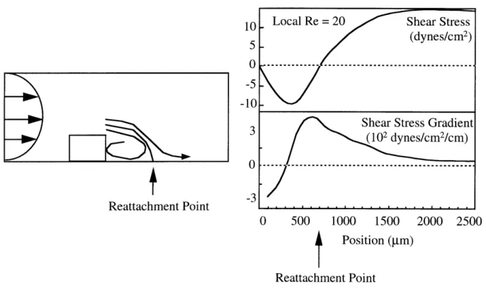
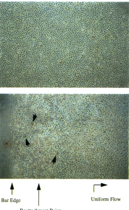
In order to more precisely analyze the shear stress parameters (e.g., amplitude, duration and spectral properties of the applied force) which regulate endothelial cell structure and function, different types of in vitro apparatuses have been developed. The two most common designs are the cone and plate device, described by Dewey et al. in 1981 [19], and the parallel plate flow system, first utilized by Eskin et al. in 1984 [20]. Both can generate defined fluid flow over cultured monolayers of endothelial cells isolated from animal or human blood vessels. The cone-plate system, utilized in the studies reported here, is comprised of a rotating cone over a stationary base plate which supports the endothelial monolayer (Figure 1.1). By adjusting cone angle, medium viscosity, and cone rotation speed, a broad dynamic range of shear stresses can be generated, in both laminar and turbulent flow regimes [21]. Endothelial monolayers may be plated either on multiple small coverslips mounted at different positions in the base plate -- which allows for analysis of replicate samples -- or on a single large plastic plate, providing ample numbers of cells for molecular biological analyses. Addition of a barrier in the flow streamline, in the form of a small step on each coverslip, results in regions of disturbed laminar shear stress downstream. The regions of flow reversal and reattachment thus created have large spatial shear stress gradients which mimic disturbed flow patterns that occur in vivo [22, 23]. Similar uniform and disturbed laminar shear stress patterns may be produced within a parallel plate apparatus [22, 24]. Also, by coupling a parallel plate device to a microscopic image analysis system, live-time analyses of endothelial cell responses to shear stress may be performed. However, unlike the cone and plate apparatus, parallel plate systems generally allow analysis on only one coverslip at a time and require constant recirculation of culture medium.
Initial experiments with these in vitro flow apparatuses confirmed in vivo shear stress-associated endothelial cell shape changes [25-28] and analyzed alterations in other complex cell functions, such as cytoskeletal reorganization [29, 30], cell stiffness [31], proliferation [32-34],
Shear Stress
r
= cone angular velocity = radial position
= cone angle = viscosity
= kinematic viscosity
Figure 1.1. The in vitro cone and plate shear stress apparatus. Endothelial
monolayers are exposed to defined fluid mechanical conditions within the cone and plate system. Flow is created by rotating the cone over the stationary base plate, and the shear stress, t, is a function of the viscosity of the culture medium contained between the cone and plate, the cone angle and rotation speed, and the dimensionless parameter, R.
[Adapted from Davies et al, 1984, J Clin Invest, 73:1121-1129.] a
= 1 - 0.4743R2)
r 2JO2
1r2 12v
migration [33], and pinocytosis [35]. In 1985 Frangos et al. and Grabowski et al. demonstrated the enhanced secretion of a distinct endothelial-derived molecule, prostacyclin, by endothelial monolayers exposed to laminar shear stress [12, 36]. Subsequent analyses by several groups revealed the shear stress regulation of other endothelial-derived effector molecules, including coagulation factors (tissue plasminogen activator, thrombomodulin) [37-40], vasoactive substances (histamine, endothelin-1, nitric oxide) [11, 13, 14, 41, 42], cytokines (interleukin-l and -6) [43], and growth factors (platelet-derived growth factors-A and -B, heparin-binding EGF-like growth factor, basic fibroblast growth factor) [15, 16, 44]. Certain of the acutely regulated events involve alterations in substrate availability and rate-limiting enzymatic activities in metabolic pathways. Other more chronic effects reflect direct alterations in gene expression, due to transcriptional as well as post-transcriptional events. The variety of shear stress responses in endothelial cells may involve a growing number of intracellular signal transduction mediators that are also modulated by shear stress (intracellular Ca", arachidonic acid and phosphoinositide metabolites, G-proteins, mitogen-activated protein kinases, reactive oxygen intermediates) [45-51].
Mechanisms of Endothelial Gene Regulation by Shear Stress
In 1993, Resnick and colleagues in our laboratory provided the first evidence of a specific transcriptional mechanism through which shear stress can regulate endothelial gene expression [52]. Transfection of deletional promoter constructs of the platelet-derived growth factor-B (PDGF-B) gene into endothelial cells exposed to uniform laminar shear stress led to the identification of a six base pair nucleotide sequence (GAGACC), termed the "shear stress response element" (SSRE), within the promoter of PDGF-B that was required for the shear stress induction of this gene. Subsequent studies revealed that nuclear factor-icB (NF-icB), a DNA-binding transcription factor, could functionally interact with the SSRE within the PDGF-B promoter [53]. Recent data from our laboratory and others have identified three additional "SSREs" which mediate the shear stress induction of other endothelial cell genes by binding specific, previously defined transcription factors. A TPA (tetra-decanoyl phorbol acetate) response element (TRE) mediates the upregulation of the monocyte chemotactic protein-1 gene by interacting with activator protein-i (AP-1), a transcription factor composed of protein dimers of c-Jun and c-Fos [54]. The shear-induction of the platelet-derived growth factor-A (PDGF-A) gene requires functional interactions between another transcription factor, early growth response-1 (Egr-1), and its DNA binding sequence in the PDGF-A promoter [55]. Finally, upregulation of the tissue factor gene by shear stress involves activation of the transcription factor, Sp I, by phosphorylation [56]. The diversity
of molecular mechanisms through which these SSREs and associated transcription factors exert their regulatory effects suggest that the local promoter architecture and regional associations between distinct transcription factors may be important variables in the shear stress activation of
different endothelial genes.
Objectives
One of the best studied paradigms of endothelial cell gene activation is the cytokine induction of a class of cell surface receptors, the endothelial-leukocyte adhesion molecules, which includes Intercellular Adhesion Molecule-1 (ICAM-1), Vascular Cell Adhesion Molecule-1 (VCAM-1), and E-selectin (ELAM-1). All three genes are coordinately upregulated, with varying kinetics, in endothelial cells exposed to soluble cytokines such as interleukin-1B (IL-113) and tumor necrosis factor-oa (TNFa), and elevated expression levels have been detected in a number of disease states, particularly inflammation and atherosclerosis [2]. The molecular regulatory mechanisms through which cytokines induce ICAM-1, VCAM-1, and E-selectin have been extensively studied and involve several transcription factors and DNA binding sites, certain of which have been identified as shear responsive in other genes [57]. However, at the time the work presented here was initiated, the potential shear stress regulation of endothelial adhesion molecules
had not yet been examined. As such, the specific aims of this research were as follows:
* Investigate the potential regulatory effects of uniform laminar shear stress on ICAM-1, VCAM-1, and E-selectin expression in cultured human endothelial cells.
This regulation was explored at multiple levels, including cell surface protein, functional interactions with leukocytes, steady state mRNA, and transcriptional activation.
* Extend these findings on adhesion molecule regulation by uniform laminar shear stress to a disturbed laminar shear stress model, which incorporates regions of both uniform and disturbed flow.
The disturbed flow model also was utilized to examine the effects of shear stress pre-conditioning on subsequent cytokine induction of ICAM-1, VCAM-1, and E-selectin.
Examine the effects of uniform and disturbed laminar shear stress on the induction and nuclear localization of transcription factors (NF- B, Egr-1, c-Jun, c-Fos) which bind to identified SSREs.
These analyses required the assemblage of a customized image analysis system and the development of specialized image analysis algorithms.
Chapter 2
DIFFERENTIAL REGULATION OF ENDOTHELIAL-LEUKOCYTE ADHESION MOLECULES BY UNIFORM LAMINAR SHEAR STRESS
INTRODUCTION: ENDOTHELIAL-LEUKOCYTE ADHESION MOLECULES IN VASCULAR BIOLOGY
The adhesive properties of the endothelium are central to its role in physiological and pathophysiological processes. Of particular importance are its adhesive interactions with blood-borne leukocytes, since leukocytes must emigrate from the bloodstream through the endothelial lining of blood vessels in order to perform their functions in host defense at extravascular sites. Endothelial cells coordinate leukocyte recruitment via the regulated expression of a number of cell surface adhesion molecules (endothelial-leukocyte adhesion molecules, ELAMs) which bind to specific counter-receptors on leukocytes (Table 2.1). A large body of both in vivo and in vitro experimental evidence has identified a sequence of adhesive interactions in this process [58, 59]. First, a loose attachment is initiated between the two cell types, which serves to decelerate leukocyte velocity and results in their rolling along the endothelial surface. This process, which is generally mediated by adhesion molecules of the selectin family (E-selectin and P-selectin on endothelial cells) interacting with sialylated counter-receptors (expressed on different leukocyte types), involves rapid bond formation at the leading edge of the leukocyte together with bond dissociation at the trailing edge [60-63]. Cell rolling is followed by leukocyte activation mediated by a variety of factors (cytokines, chemokines, chemoattractants), with resulting upregulation of secondary adhesion molecules. Firm attachment is then established via the interaction of these secondary adhesion molecules, such as B, and 12 integrins expressed by leukocytes (e.g., very late antigen-4/VLA-4, leukocyte function-associated antigen-1/LFA-1, Mac-I) with members of the immunoglobulin gene superfamily (VCAM-1, ICAM-1) on the activated endothelial surface. Finally, the leukocyte migrates through cell-cell junctions in the endothelial monolayer and into the extravascular tissue. Thus, the regulation of expression of ELAMs, such as E-selectin, VCAM-1, and ICAM-1, constitutes an important control mechanism in leukocyte recruitment.
The significance of ELAMs in inflammatory processes is emphasized by the complications encountered by patients with the Type 1 Leukocyte Adhesion Deficiency Syndrome. These patients have reduced levels of functional B2 integrin subunits and experience recurrent bacterial
infections and impaired wound healing [66]. ELAMs also play significant roles in pathophysiologic processes other than inflammation. A number of disease-associated cells utilize adhesion receptors for pathologic functions. For example, ICAM-1 has been identified as the major cell surface receptor for at least two pathogens: rhinovirus and Plasmodium falciparum (the microorganism responsible for malaria) [67-70]. Certain malignant cells, including carcinomas, melanomas and lymphomas, express LFA-1, VLA-4, and ligands for E-selectin, and increased expression of ICAM-1 on endothelial cells has been correlated with metastatic spread [59, 71-75].
Table 2.1
Endothelial Adhesion Molecules and Their Leukocyte Counter-Receptors [64, 65]
Endothelial Cell ICAM-1
VCAM-1
Leukocyte 12- integrins: LFA-1 (CD11a/CD18) Mac-i (CD 1 lb/CD 18) p150 (CD11 c/CD18) 1,- and 137 - integrins: VLA-4 (ca4B1) Sialyl-Lewisx E-selectinThus, the adhesive properties of ELAMs are important in a diverse set of disease processes, including inflammation, infection, and metastatic progression. In addition, many adhesive interactions are intimately associated with signaling mechanisms, transducing information linking intracellular and extracellular events. For example, ICAM-1 functions as a costimulatory molecule to activate T-cells, and upregulated endothelial expression of ICAM-1 and VCAM-1 has been demonstrated in a number of autoimmune diseases, including Graves' disease, Hashimoto's thyroiditis, and experimental autoimmune neuritis [76-78]. Recent evidence from our laboratory has indicated that leukocyte binding to E-selectin on the endothelial cell surface induces transmembrane cytoskeletal linkage of this adhesion molecule, a process which may have important implications for cell-cell signaling as well as mechanical anchoring during leukocyte binding [79]. Therefore, both the adhesive and signaling functions of ELAMs may be important mechanisms in pathophysiologic processes.
Atherosclerosis, the leading cause of death in the United States, involves alterations in ELAM expression. One of the earliest morphologically detectable events in atherogenesis is the adherence of circulating blood monocytes to the endothelial wall [80-82]. These cells then migrate across the endothelium and into the arterial intima where they transform into resident macrophages and accumulate cholesterol esters to become "foam cells" [83]. In addition to accumulating lipids, foam cells elaborate a number of soluble effector molecules such as cytokines, growth factors, chemotactic factors, and reactive oxygen species. Many of these products result in the recruitment and proliferation of additional monocytes from the lumen as well as smooth muscle cells from the media. The resulting intimal hyperplasia, together with depositions of extracellular matrix proteins synthesized by intimal smooth muscle cells, characterizes the developing atherosclerotic plaque
[66].
The first in vivo demonstration of lesion-selective ELAM expression showed enhanced endothelial VCAM-1 covering aortic atherosclerotic lesions at various stages of development in atherogenic rabbit models [84]. Several subsequent studies have revealed elevated levels of VCAM-1, ICAM-1, E-selectin, and P-selectin on endothelium in histologic sections of advanced human atherosclerotic plaques [85-87]. What is most striking, in terms of hemodynamics factors, is the recent finding that ICAM-1 expression, but not VCAM-1 or E-selectin, was significantly upregulated on histologic sections from the lateral walls of carotid sinuses, both in normal (lesion-free) and advanced atherosclerotic vessels [85]. This anatomic position is the frequent site of atherosclerotic development, a finding correlated with its exposure to disturbed shear stress patterns.
as other disease processes, we chose to investigate the potential regulation of ICAM-1, VCAM-1, and E-selectin in endothelial cells exposed to laminar shear stress. Endothelial activation by soluble mediators (e.g., cytokines and bacterial endotoxins), most commonly detected as enhanced ELAM expression, has been extensively examined. Indeed, biochemical stimulation constitutes a well-established model of endothelial cell activation. In contrast, biomechanical forces are now being recognized as an emerging paradigm of endothelial activation [88] and, at the time the work presented here was initiated, no comparative study of endothelial-leukocyte adhesion molecule regulation by hemodynamic forces had been reported.
METHODS
Cell Culture. Pooled primary cultures of human umbilical vein endothelial cells (HUVEC) were established from normal term human umbilical cords as previously described [89]. For experimental use, second passage cells were plated on tissue culture treated polystyrene (Costar, Cambridge, MA; and Modem Plastics, Peabody, MA) coated with 0.1% gelatin (Difco Laboratories, Detroit, MI), and grown to confluence in Medium 199 (with 25 mM HEPES; Gibco Laboratories, Gaithersburg, MD) supplemented with 10% fetal bovine serum (FBS) (Gibco Laboratories), 2 mM glutamine, 100 U/ml penicillin, 100 jig/ml streptomycin, 25 mg/ml endothelial cell growth supplement (Collaborative Research, Bedford, MA), 50 mg/ml heparin (Sigma Chemical Company, St. Louis, MO), and 250 ng/ml amphotericin B (Fungizone@, Gibco Laboratories). Bovine aortic endothelial cells (BAEC) were isolated from calf descending thoracic aortas as previously described [89], and were cultured in Dulbecco's modified Eagles' medium (Gibco Laboratories) supplemented with 10% bovine serum (BioWhittaker, Walkersville, MD), 2 mM glutamine, 100 U/ml penicillin, and 100 jgg/ml streptomycin. Suspension cultures of human JY lymphocytic cells [90], kindly provided by Dr. T. Springer (Center for Blood Research, Boston, MA), were maintained in RPMI-1640 medium (with 25 mM HEPES; BioWhittaker) supplemented with 10% FBS, 20 mM glutamine, 100 U/ml penicillin, and 100 mg/ml streptomycin. In certain experiments, endothelial cells were activated by treatment with recombinant human interleukin-lB (IL-1B; Biogen, Cambridge, MA), as indicated.
Shear Stress Apparatus. The cone and plate flow apparatus used to expose cultured endothelial monolayers to defined fluid shear stresses has been described in detail previously [19, 21]. The essential components consist of a stainless steel cone rotating over a stationary base plate, which supports either twelve 12 mm diameter polystyrene coverslips or a single 17.8 cm diameter polystyrene plate (see Figure 1.1). The culture medium present between the cone and base plate (15 ml total volume) is gradually exchanged (0.5 ml/min) without recirculation. The
entire apparatus is maintained at 370C in a humidified 5% CO2 and 95% air atmosphere. The shear
stress on the surface of the plate, r, can be described by:
=
(1 - 0.4743f2),where r2 2
12v
and where gi is medium viscosity, co is angular velocity of the cone, a is cone angle, r is radial location on the plate, and v is kinematic viscosity of the medium. The flow is laminar for R << 1 [21]. For the experiments reported here, fluid mechanical parameters were adjusted such that endothelial monolayers were subjected to laminar shear stress of 2.5 - 46 dynes/cm2 for variable time intervals. Dextran (476,000 MW, 1% w/v; Sigma Chemical Company) was utilized to alter the medium viscosity, when required, and the viscosity was measured with a coaxial cylinder viscometer at 370C (Haake, Berlin, Germany). Shear stressed and static monolayers were cultured in the same medium for each experiment. Control monolayers on coverslips were maintained in cell culture dishes (Costar) under static (no flow) conditions at 370C in a humidified 5% CO
2 and
95% air atmosphere for equivalent time intervals.
Fluorescence Immunobinding Assay (FIA). Endothelial monolayers were incubated on ice for 1 hour in RPMI plus 10% FBS with saturating concentrations of monoclonal antibodies (mAbs) specific for human endothelial-leukocyte adhesion molecules (Hu 5/3, purified IgG, anti-human ICAM-1; E 1/6, ascites, anti-anti-human VCAM-1; H18/7, purified IgG, anti-anti-human E-selectin), followed by a FITC-labeled F(ab')2 anti-mouse IgG (14 jgg/ml in PBS plus 1% FBS, 1
hour; Caltag Laboratories, South San Francisco, CA), then lysed with a 0.01% NaOH/O. 1% SDS solution and the fluorescence was measured in a Pandex plate reader (Travenol Laboratories, Mundelein, IL).
Immunocytochemistry. For microscopic visualization of cell surface associated proteins, endothelial monolayers were fixed in 2% paraformaldehyde at 40C, and incubated for 1 hour with
specific mAbs directed to human endothelial-leukocyte adhesion molecules, as described above, followed by biotinylated anti-mouse IgG (15 gg/ml in PBS plus 2% bovine serum, 1 hour; Vector Laboratories, Burlingame, CA), a peroxidase conjugated biotin-avidin complex (Vectastain Elite ABC Kit, 1 hour; Vector Laboratories), and finally an amino-ethyl-carbazole developing reagent (Peroxidase Substrate Kit, 8 minutes; Vector Laboratories). All incubations were performed at ambient temperature (250C).
Northern Blot Analysis. Total cellular RNA was extracted from endothelial monolayers by the acid guanidinium thiocyanate-phenol-chloroform method (Cinna/Biotecx Laboratories International, Inc., Friendswood, TX). Samples (15 mg each) were electrophoresed through 1% agarose gels containing formaldehyde, transferred to nitrocellulose membranes (Schleicher & Schuell, Inc., Keene, NH) and hybridized with human ICAM-1, VCAM-1, or E-selectin cDNA probes labeled by [x-32P] dCTP (Amersham Corporation, Arlington Heights, IL), using random
hexanucleotide primers (Pharmacia, Inc., Piscataway, NJ). The cDNA fragments used were as follows: a 1.8 kb SalI-KpnI fragment of ICAM-1 cDNA from pGEM4, kindly provided by Dr. T. Springer (Center for Blood Research, Boston, MA); a 1.0 kb EcoRI/BamHI fragment of VCAM-1 cDNA from pBSM13, kindly provided by Dr. T. Collins (Brigham and Women's Hospital, Boston, MA); and a 1.0 kb XbaI fragment of E-selectin cDNA from pCDM8.
Leukocyte Adhesion Assay. Endothelial monolayers on 12 mm coverslips were coincubated for 15 minutes at room temperature under static conditions in 24-well cell culture plates (Costar) with JY cells (1.5 x 10⁵ cells/ml), a human lymphocytic cell line which expresses an ICAM-1 ligand, LFA-1 (CDlla/CD18) [90], in 75 ml of RPMI-1640 plus 10% FBS. To remove unattached JY cells, each coverslip was washed by immersion three times in RPMI-1640 plus 1% FBS, then once in phosphate buffered saline (PBS), and fixed in 2% paraformaldehyde at 4°C. After staining with hematoxylin, adherent JY cells were counted in 5 randomly selected high power microscopic fields (100X) on each of 2 coverslips for each controlled variable.
Plasmid Vectors. The ICAM-1 deletional promoter constructs were kindly provided by Dr. M. Gerritsen (Bayer Corporation, West Haven, CN) and were made by cloning sequences (-716 to -12 bp or -617 to -12 bp) from the ICAM-1 promoter into the KpnI and BglII sites of the luciferase reporter vector pGL2-Basic (Promega, Madison, WI). CMV-CAT and CMV-1-gal (the kind gifts of Dr. T. Collins, Brigham and Women's Hospital) contain the human cytomegalovirus (CMV) immediate-early enhancer/promoter coupled to the chloramphenical acetyl transferase (CAT) and 13-galactosidase (13-gal) genes, respectively, and are constitutively expressed, non-shear-regulated reporter genes which were utilized for normalization of transfection efficiency and cell number.
Transient Transfections. BAEC (passages 2-5) grown on 100 mm culture dishes (Corning Glass Works, Coming, NY) at 70% confluence were transiently co-transfected by the calcium phosphate precipitation method with either of the ICAM-1 reporter gene constructs (22.5 - 26.25 gjg) and either CMV-CAT or CMV-B-gal (3.75 - 7.5 gg). The cells were then incubated in a 3% CO2 humidified atmosphere at 370C for at least 16 hours, washed three times with fresh culture medium, allowed to recover 6 - 10 hours in a 5% CO2 atmosphere, washed again, then
hours later, the confluent monolayers of BAEC were exposed to laminar shear stress or were maintained under static conditions. For the CMV-CAT experiments, each sample was split for separate luciferase (Enhanced Luciferase Assay Kit, Analytical Luminescence Laboratory, San Diego, CA) and CAT (as previously described) assays [91]. In the CMV-B-gal experiments, luciferase and B-galactosidase assays were performed consecutively on the same sample (Dual-Light Kit, Tropix, Inc., Bedford, MA) using a Monolight 2010 luminometer (Analytical Luminescence Laboratory).
RESULTS
Shear stress upregulates ICAM-1 surface expression in a time-dependent and force-independent manner. ICAM-1 expression on the surface of HUVEC monolayers which had been either incubated under static (no flow) conditions or subjected to laminar shear stress of 10 dynes/cm2 for 4, 8, 24, and 48 hours was measured quantitatively by a fluorescence immunobinding assay (FIA). Table 2.2 summarizes the results obtained from a total of 12
separate experiments in which confluent endothelial monolayers on multiple coverslips were simultaneously exposed to the same fluid shear stress or static conditions. Significant elevations in cell surface immunoreactive ICAM-1 were observed as early as 8 hours after the onset of shear stress, and increased progressively up to 48 hours. It is important to note the significant variability in basal expression of ICAM-1 in different HUVEC cultures. In particular, in cultures with high basal levels, shear stress induction of ICAM-1 was seldom observed. The maximum level of ICAM-1 expression induced by shear stress was comparable to that observed in HUVEC activated with a maximally effective concentration of recombinant human interleukin-13 (10 U/ml) (see Figure 2.3 below). Conditioned effluent medium collected from the shear stress apparatus, at intervals ranging from 15 minutes to 24 hours, failed to induce ICAM-1 upregulation when incubated with static HUVEC monolayers for 3 and 24 hours (data not shown).
As illustrated in Figure 2.1, immunocytochemical staining of paraformaldehyde-fixed HUVEC monolayers subjected to laminar shear stress of 10 dynes/cm2 also demonstrated the progressive upregulation of cell surface ICAM-1 at 24 and 48 hours. Morphological responses of cells, namely elongation and alignment in the flow direction, were clearly evident in these shear stressed monolayers at both time points. However, HUVEC monolayers exposed to a lower level of shear stress (3 dynes/cm2) for 24 hours displayed a similar increase in surface ICAM-1
expression, without any detectable elongation or alignment of the cells. In both of these relatively high (10 dynes/cm2) and low (3 dynes/cm2) shear stress conditions, ICAM-1 induction was not uniform across the monolayer. To further investigate the potential force-dependence of ICAM-1
Table 2.2
Shear Stress Induces Cell Surface Expression of ICAM-1 in Cultured Human Umbilical Vein Endothelial Cells
Cell Surface Immunobinding (Fluorescence Units)
Experiment Duration Shear Stress /
(hours) Static Shear Stress Static
1974 ± 109 886 ± 180 2819 ± 116 1618 ± 319 599 673 2443 207 767 395 997 944 48 98 371 144 154 132 72 19 2259 ± 545 838 ± 88 3689 ± 223 1590 ± 160 1427 1333 2753 3413 2419 1080 2474 941 327 268 597 406 111 71 283 637
Confluent HUVEC monolayers were either exposed to shear stress (10 dynes/cm2) or maintained
under static conditions for the times indicated. Cell surface expression of ICAM-1 was measured using a fluorescence immunobinding assay, as described in Methods. Data are expressed as mean ± standard deviation; n = 2-4 replicate coverslips in each separate experiment; *p < 0.01 and *p < 0.05 shear stress versus static control (Student's t-test).
1.1 1.0 1.3* 1.0 2.4* 2.0* 1.1 16.5* 3.2* 2.7* 2.5* 1.0
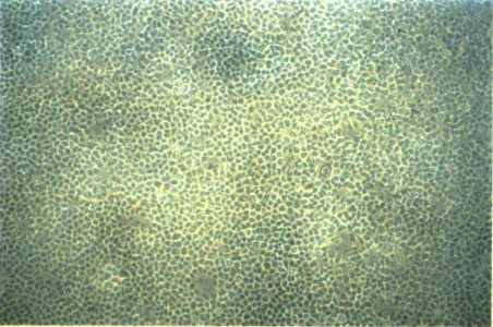
Figure 2.1. Immunocytochemistry of ICAM-1 surface expression on HUVEC exposed to shear stress (following page). HUVEC monolayers were either maintained under static conditions or subjected to high (10 dynes/cm2) or low (3 dynes/cm2) laminar shear stress for variable time periods: (A) static, (B) high shear stress, 24 hours, (C) high shear stress, 48 hours, or (D) low shear stress, 24 hours. The monolayers were then fixed with 2% paraformaldehyde and immunocytochemically stained using an ICAM-1-specific mAb, as described in Methods. Original magnification = 45X.
4~ -~i
C"CrK..ifr
F5·sian
j ·%9
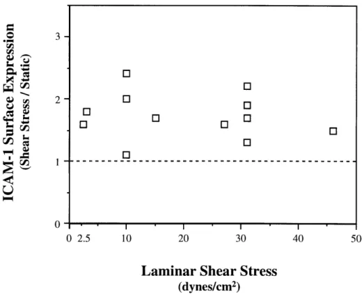
D 65:" r .?a "la:("~ B~"i~.*·i~-"1~ )i II&IJ -· n;·l~' r,, I·B:,F *' QFI,; .~t, I, Ci·L L~ c.:6 ~""; i~ * ~ 9 "- ''~;" islrl~-"r: "~~ ~, ": ·"~~1;~:~ jg"o %~i: ~I· ~ ~··r·59 i i F !iinduction, confluent HUVEC monolayers were exposed for 24 hours to a broad range of laminar shear stresses (2.5 -46 dynes/cm2), comparable to those encountered in vivo in large vessels [92].
In a series of 12 experiments, as illustrated in Figure 2.2, significant increases in immunoreactive surface ICAM-1 expression, measured by FIA, were observed at each level of shear stress tested; however, the amount of induction appeared to be independent of the magnitude of the applied force.
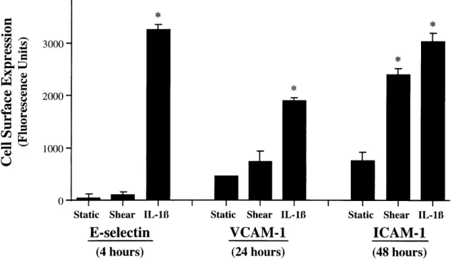
E-selectin and VCAM-1 are not upregulated in HUVEC by shear stress. In contrast to the induction of ICAM-1 by shear stress, no significant changes were observed in either E-selectin or VCAM-1 expression, as measured by cell surface FIA, at any time point (4 - 48 hours) or shear stress level (2.5 -46 dynes/cm2) studied (data not shown). In particular, as seen in Figure 2.3, at time points when IL-18 treated HUVEC cultures displayed peak levels of E-selectin (4 hours) and VCAM-1 (24 hours), no changes in the cell surface expression of these molecules were observed in HUVEC cultures subjected to 10 dynes/cm2 of laminar shear stress.
In order to test whether shear stressed endothelial cells were still responsive to other activating stimuli, such as cytokines, we subjected HUVEC monolayers to 24 hours of laminar shear stress (10 dynes/cm2) followed by a 4 or 24 hour treatment with IL-1B (10 U/ml) under static
conditions. Immunocytochemical staining revealed marked upregulation of E-selectin (after 4 hours) and VCAM-1 (after 24 hours) comparable to that observed in HUVEC that were not exposed to shear stress (data not shown).
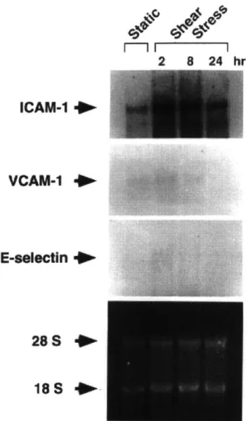
Laminar shear stress selectively upregulates ICAM-1 mRNA. Northern blot analysis of RNA extracted from HUVEC revealed a marked increase in ICAM-1 transcript levels as early as 2 hours after the onset of shear stress, which was sustained at 8 hours and still elevated above static HUVEC at 24 hours (Figure 2.4). Rehybridization of the membrane with cDNA probes for VCAM-1 and E-selectin did not show induction of these genes at any time point examined, consistent with the FIA measurements of cell surface protein (see Figure 2.3).
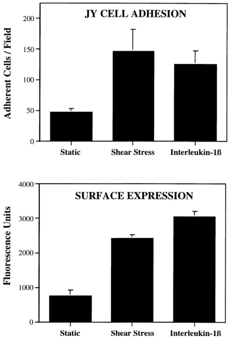
Enhanced leukocyte adhesion to shear stressed HUVEC monolayers. The shear stress-induced surface expression of immunoreactive ICAM-1 suggested that HUVEC monolayers subjected to laminar shear stress might be more adhesive for leukocytes expressing ligands for ICAM-1. To test this hypothesis, HUVEC were exposed to 48 hours of shear stress and then coincubated in a standard monolayer adhesion assay with JY cells, a human lymphoblastoid cell line which expresses the ICAM-1 ligand, LFA-1 (CD 11 a/CD 18), as measured by flow cytometry (data not shown). A 3-fold increase in JY cell adhesion was observed to shear stressed HUVEC versus static monolayers, which was comparable to the upregulated ICAM-1 expression detected by FIA. These increases in leukocyte adhesion were comparable in magnitude
o
3-o
2-
0-0 2.5 10 20 30 40 50
Laminar Shear Stress
(dynes/cm2)
Figure 2.2. Upregulation of ICAM-1 by a range of laminar shear stress levels (2.5 -46 dynes/cm2). HUVEC monolayers were either maintained under static conditions or subjected to various levels of laminar shear stress, as indicated, for 24 hours. Cell surface protein was determined using a fluorescence immunobinding assay, as described in Methods, and displayed as the ratio (shear stress / static) for each experiment.
O O [EJ O O O O0 0I 0O I
I Static Shear IL-1B
E-selectin
(4 hours)
I1
Static Shear IL-1B
VCAM-1
(24 hours)
Static Shear IL-18 ICAM-1
(48 hours)
Figure 2.3. Peak cell surface expression of endothelial-leukocyte adhesion molecules in
HUVEC subjected to shear stress or IL-1B. HUVEC monolayers were maintained under static conditions, exposed to laminar shear stress (10 dynes/cm2), or treated with a maximally effective concentration of IL-1B (10 U/ml) for time periods corresponding to the peak cytokine-induced surface expression of E-selectin (4 hours), VCAM-1 (24 hours), or ICAM-1 (48 hours), respectively. Cell surface protein was measured using a fluorescence immunobinding assay, as described in Methods. n = 3-4 replicate coverslips for each controlled variable; data expressed as mean ± standard deviation, *p < 0.01 stimulus versus
static (Student's t test). 4000- 3000-2000 -1000
--r
MT
I I m m m m2
8 24
ICAM-1
VCAM-1
E-selectin
*I
28S
*
Figure 2.4. Northern blot analysis of adhesion molecule induction by shear stress in HUVEC. HUVEC monolayers were either maintained under static conditions or exposed to laminar shear stress (10 dynes/cm2) for 2, 8, or 24 hours. Total cellular RNA was isolated for Northern blot analysis as described in Methods, and each lane was loaded with 15 gig aliquots (bottom panel, ethidium bromide staining of 18S and 28S ribosomal RNA). The membrane was sequentially hybridized with radiolabeled cDNA probes of ICAM-1, VCAM-1, and E-selectin.
to those seen in the same experiment with maximally IL-13B activated (10 U/ml, 24 hours) HUVEC monolayers (Figure 2.5).
ICAM-1 is induced by shear stress at the level of transcription. The correlation of the selective shear stress inducibility of ICAM-1 with the presence of the SSRE in its promoter suggested that this regulatory element might be functional in the shear stress induction of ICAM-1. In order to investigate the potential role of the SSRE (located at position -644 bp in the ICAM-1 promoter) in the transcriptional regulation of ICAM-1, we utilized two ICAM-1 deletional promoter/reporter gene constructs, one which contains the SSRE (-716 to -12 bp) and one which lacks it (-617 to -12 bp), as depicted in Figure 2.6. Cultured BAEC were transfected with either of these constructs and subjected to laminar shear stress (10 dynes/cm2, 3 hours) or maintained under static conditions. Normalization for both transfection efficiency and cell number was accomplished by co-transfecting with either CMV-CAT or CMV-B-gal, both of which are constitutively expressed and non-shear-regulated. Table 2.3 summarizes the results obtained from 5 separate experiments in which transfected endothelial monolayers on multiple coverslips were simultaneously exposed to the same fluid shear stress or static conditions. The promoter construct which contains the SSRE exhibited significant transcriptional activation by shear stress in 4 of 5 experiments (p < 0.05 versus static), whereas the construct lacking the SSRE displayed significant induction in only 1 experiment.
DISCUSSION
At the time they were performed, these studies provided the first evidence that physiologically relevant levels of laminar shear stress can differentially regulate the expression of endothelial-leukocyte adhesion molecules. This differential pattern of adhesion molecule expression elicited by laminar shear stress is in contrast to the coordinate activation profile typically observed in cultured HUVEC with cytokines such as TNFcx, IL-1B, or bacterial endotoxins [93-95], thus suggesting that distinct transduction pathways and/or transcriptional and post-transcriptional regulatory mechanisms are involved. In addition, both the lack of E-selectin upregulation by shear stress and the fact that conditioned media from shear stressed monolayers failed to induce adhesion molecule expression on static monolayers, strongly argue against the action of an endogenous cytokine (e.g., IL-1B or IL-lo ) as an autocrine mechanism of activation in this system.
The correlation of the shear stress-inducibility of ICAM- 1 with the presence of the SSRE in its promoter region and, conversely, the non-inducibility of E-selectin and VCAM-1, suggested
200 150 100 50 0 4000 3000- 2000- 1000-I Static
Static Shear Stress Interleukin-1B
SURFACE EXPRESSION
Shear Stress Interleukin-lB
Figure 2.5. ICAM-1 induction by shear stress in HUVEC is correlated with an
increased adhesion of JY cells. HUVEC monolayers were exposed to laminar shear stress (10 dynes/cm2, 48 hours) or treated with IL-18 (10 U/ml, 24 hours), and then were incubated
in a standard adhesion assay with human JY cells, a lymphocytic cell line that expresses the ICAM-1 ligand, LFA-1 (CDlla/CD18), as described in Methods. Adherent JY cells (top panel) were counted in five randomly selected high power (100X) fields on each of two coverslips for each controlled variable. ICAM-1 cell surface expression (lower panel) was measured in parallel by a fluorescent immunobinding assay, as described in Methods. Data
are presented as mean ± standard devation.
4000
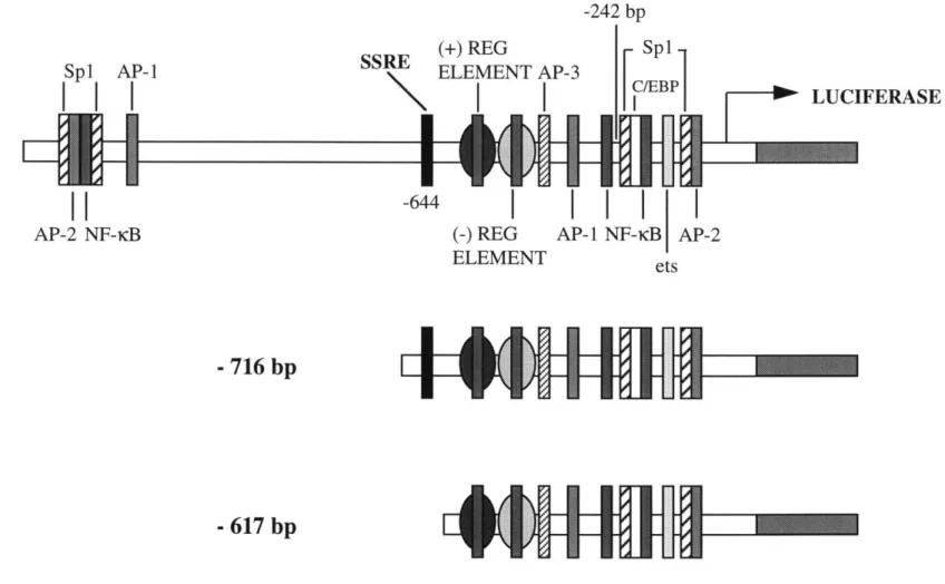
Figure 2.6. ICAM-1 promoter/reporter gene constructs. Deletional ICAM-1 promoter/luciferase constructs were
made by cloning sequences from the ICAM-1 5' untranslated region into the luciferase reporter vector. The SSRE,
located at position -644 bp in the ICAM-1 promoter, is present in the -716 bp construct, but absent in the -617 bp construct. A number of other putative transcriptional regulatory elements are also present in the ICAM-1 promoter, as shown. -242 bp SSRE -644 Spl AP-1
I i
I I
AP-2 NF-dcB(+
El IFERASEI
AP-ELEMIEN I- 716 bp
- 617 bp
etsI
L
(
W-M
I AMr IM flUU1 MEEMTable 2.3
Transcriptional Activation by Shear Stress is Enhanced with an ICAM-1 Deletional Promoter Construct Which Contains the SSRE
+SSRE Construct -SSRE Construct
SS/Static (*p) Static
Shear
Stress SS/Static (*p)
Normalized with CMV-CAT: 2.54±0.06 5.35±0.53 9.22±0.52 11.84±1.26 8.24±1.10 14.39±4.48
Normalized with CMV-fl-gal: .090±.017 .156±.009 .167±.017 .146±.008 2.11 1.28 1.75 1.73 0.87 (0.04)
(0.05)
(0.01)
(0.01)
(0.13) 1.84±0.31 5.51±2.01 10.27±4.08 .040±.003 .046±.008 2.37±0.98 5.91±0.33 9.90±3.82 .056±.005 .034±.009 1.29 1.07 0.96 (0.33) (0.35) (0.39) 1.40 (0.01) 0.74 (0.07) ** p = 0.02Cultured BAEC were transfected with either the ICAM-1 deletional promoter construct containing the SSRE (+SSRE) or the deletional construct lacking the SSRE (-SSRE), and normalization was performed by co-transfecting with either CMV-CAT or CMV-B-gal, as described in Methods. Transfected cells were then exposed to laminar shear stress (10 dynes/cm2) for 3 hours, and the
appropriate reporter gene assays were performed. Data are expressed as mean ± standard deviation; n = 2-6 samples in each experiment; Student's t-tests: * static versus shear stress for individual experiments, ** induction of +SSRE construct versus -SSRE construct for all experiments.
Static
Shear Stress
that this putative genetic regulatory element might be functional in the transcriptional upregulation of ICAM-1. Transfection experiments with ICAM-1 deletional promoter/reporter genes demonstrated that ICAM-1 was indeed upregulated by laminar shear stress at the level of transcription. Subsequent work in our laboratory has confirmed this finding by nuclear run-off analysis [96]. The ICAM-1 reporter gene experiments also revealed significantly greater shear stress induction of the construct which contained the SSRE, indicating that the SSRE may function to enhance the shear stress activation of ICAM-1. However, the fact that the reporter construct lacking the SSRE was still activated by shear stress, albeit in one experiment, indicates that the SSRE may not be necessary for shear stress induction of ICAM-1. Indeed, recent studies in our laboratory have demonstrated that another ICAM-1 deletional promoter construct which lacks the SSRE (-242 to -12 bp) was also shear stress responsive [97]. Thus, any functional shear stress response element(s) for ICAM-1 must be contained within the proximal 242 bp of its promoter. Interestingly, this is also the region most critical for TNF( induction of ICAM-1 in endothelial cells [98]. Figure 2.6 illustrates the presence of several consensus sequences for known DNA-binding transcription factors within this region of the promoter, including Sp 1, C/EBP, AP-2, ets, and NF-dKB [98, 99]. Within this region, a 75 bp section containing the Spl and AP-2 sites is necessary for constitutive ICAM-1 expression [99], and the NF-KB site is required for TNF inducibility [98]. Identification of the functional shear stress responsive element(s) within this gene will require site-directed mutagenesis of these, and potentially other non-consensus, regulatory elements.
Since the initial publication of this work [100], several reports have generally confirmed and extended our findings. Tsuboi et al. demonstrated selective induction of ICAM-1 in HUVEC by shear stress at the levels of both cell surface protein and mRNA, with lack of VCAM-1 induction [101]. However, in contrast to our data, they found that the ICAM-1 upregulation was force-dependent (approximate range: 1 -20 dynes/cm2), in addition to being time-dependent (1 - 24
hours). It is quite probable that the method they utilized to measure cell surface protein, flow cytometry, is more sensitive than the fluorescence immunobinding assay we utilized, thus enabling them to detect the force-dependency. The Tsuboi report showed that the upregulation by shear stress was reversible, since induced surface protein levels (after 24 hours of flow) returned to baseline by 4 hours once the shear stress stimulus was terminated. In addition, experiments with different medium viscosities revealed that the ICAM-1 induction was shear stress- and not shear rate-dependent. Morigi et al. also reported selective upregulation of cell surface ICAM-1, as compared with E-selectin, together with increases in both firmly adherent and rolling leukocytes on
HUVEC monolayers exposed to laminar shear stress [102]. In the adhesion assay used in these experiments, endothelial monolayers which had been either maintained under static conditions or exposed to laminar shear stress (8 dynes/cm2, 6 hours) were placed in a parallel plate flow chamber
which was perfused with a fresh human leukocyte suspension at low flow velocity (0.6 - 3 dynes/cm2) for less than 15 minutes, and endothelial-leukocyte interactions were analyzed with digitized microscopic images. With this system they further demonstrated that the increases in firmly adherent leukocytes could be completely blocked by treatment with an anti-ICAM-1 monoclonal antibody. Additionally, they showed that neither ICAM-1 nor E-selectin levels were affected by turbulent flow. Sampath et al. confirmed the specific ICAM-1 induction, though with different expression kinetics [103]. Peak mRNA levels were seen after 1 hour of shear stress exposure, then were downregulated below baseline at 6 hours. VCAM-1 and E-selectin transcript levels were depressed after both 1 and 6 hours of shear stress. It should be noted, however, that the HUVEC utilized in these studies exhibited relatively high basal levels of VCAM-1 and E-selectin, a finding which generally indicates cellular activation in this cell type [100, 104]. Their experimental conditions, therefore, may not be comparable to those in our studies. Taken together, these reports indicate that ICAM-1 is selectively and reversibly induced by laminar, but not turbulent, shear stress in a time- and force-dependent manner. This upregulation occurs at the levels of transcription, mRNA, and protein, and cell surface ICAM-1 specifically mediates leukocyte adhesion to the endothelial cells. In addition, recent preliminary data from Chappell et al. indicated that cell surface expression of both ICAM-1 and VCAM-1 were significantly induced in HUVEC by oscillatory flow (0 ± 5 dynes/cm2, 24 hours), with concomitant increases in leukocyte adhesion, indicating that regular temporal variations in the shear stress stimulus may be important in cellular activation of adhesion molecule expression [105].
In contrast to HUVEC, cultured mouse lymph node endothelial cells constitutively express VCAM-1 under static conditions. Endothelial cells derived from this source demonstrated downregulation of VCAM-1 by laminar shear stress, both at the levels of protein and mRNA, which was time- and force-dependent as well as reversible when the cells were returned to static conditions [106, 107]. In addition, this downregulation was correlated with decreased lymphocyte adhesion which could be partially inhibited (70.6%) by treatment with an anti-VCAM-1 monoclonal antibody. Very recent studies with deletional VCAM-1 promoter/reporter gene constructs demonstrated that this regulation occurred, at least in part, at the level of transcription and appears to be mediated by two TRE response elements within the VCAM-1 promoter [108]. This finding is particularly intriguing since, as mentioned in Chapter 1, a non-consensus TRE site in the MCP-1 promoter mediates the upregulation of this gene in endothelial cells exposed to shear
stress. The ability of TRE sequences to both increase and decrease transcriptional activation may reflect differences in transcription factor binding between consensus and non-consensus response elements, as well as the importance of local promoter architecture in gene regulation.
The potential interaction of shear stress and biochemical stimuli in the regulation of endothelial adhesion molecule expression has also been investigated by other workers. Preliminary data has indicated that IL-11 induction of VCAM-1 was significantly inhibited in HUVEC previously exposed to uniform laminar shear stress (5 dynes/cm2, 24 hours) [109]. Also,
upregulation of cell surface VCAM- 1 by either oxidized LDL, or a combination of LPS and TNFx, was suppressed in human aortic endothelial cells pre-conditioned with only 4 hours of laminar shear stress (12 dynes/cm2); these effects were potentially mediated by shear stress-induced NO production, since exposure of cells to nitro-L-arginine abolished the suppression [110]. ICAM-1 induction by IL-113, oxidized LDL, or LPS plus TNFao was not affected by shear stress pre-conditioning in any of these experiments.
In addition to regulating endothelial-leukocyte interactions by altering the expression of cell surface adhesion molecules, shear stress also modulates leukocyte adhesion by imparting a physical force opposing adhesive bonds formed between the two cell types. Gonzales et al. investigated the interrelationship of these dual effects by first exposing HUVEC monolayers to extended periods of laminar flow (on the order of hours), then perfusing leukocytes (U937 cells) over the monolayer under low shear stress conditions and assaying the number of leukocytes firmly adherent to the monolayer [111]. As documented previously by numerous reports, leukocyte adherence decreased as the cellular perfusate flow velocity increased, and this was true for all experimental conditions examined. Pre-conditioning with both low and medium level shear stresses (2 and 10 dynes/ cm2, respectively) for 2 hours enhanced overall adhesion, whereas pre-conditioning with high shear stress (30 dynes/cm2) had little effect. Surprisingly, the increased adhesion could be blocked with an anti-VCAM-1 antibody, and VCAM-1 surface protein was upregulated, albeit to a small extent (38%). In our laboratory, differential display analysis of mRNA transcripts in shear stressed endothelial cells has indicated that many genes, including VCAM-1, exhibit a brief upregulation immediately following onset of flow, with return to basal levels at longer time points (Dr. J. Topper, unpublished observations).
In vivo findings with acutely altered shear stress levels in experimental animal models have correlated well with in vitro data of adhesion molecule regulation. Average shear stresses within rabbit carotid arteries were increased (170%) in the right artery and decreased (73%) in the left artery by surgical manipulation, and subsequent alterations in ICAM-1 and VCAM-1 expression