Alternans in a Canine Left-Ventricular Myocyte
The MIT Faculty has made this article openly available.
Please share
how this access benefits you. Your story matters.
Citation
Armoundas, A.A. “Discordant Calcium Transient and Action
Potential Alternans in a Canine Left-Ventricular Myocyte.”
Biomedical Engineering, IEEE Transactions on 56.9 (2009):
2340-2344. © 2009 Institute of Electrical and Electronics Engineers
As Published
http://dx.doi.org/10.1109/tbme.2009.2023671
Publisher
Institute of Electrical and Electronics Engineers
Version
Final published version
Citable link
http://hdl.handle.net/1721.1/52424
Terms of Use
Article is made available in accordance with the publisher's
policy and may be subject to US copyright law. Please refer to the
publisher's site for terms of use.
Discordant Calcium Transient and Action Potential
Alternans in a Canine Left-Ventricular Myocyte
Antonis A. Armoundas, Senior Member, IEEE
Abstract—Electrocardiographic alternans is known to
predis-pose to increased susceptibility to life threatening arrhythmias and sudden cardiac death. While this decreased level of cardiac elec-trical stability is often due to the presence of discordant action potential (AP) alternans in the heart, the mechanism of discordant cardiac alternans remains unknown. This study presents a case report of cellular discordant cardiac alternans between AP and [Ca2 +]
i and employs a novel reverse engineering approach that
applies a simultaneous AP and [Ca2 +]i clamp of experimentally
obtained data to a left-ventricular canine myocyte model, to probe its underlying mechanism. The model results indicate that dur-ing alternans, the increased sarcoplasmic reticulum Ca2 +, triggers multiple ryanodine receptor (RyR) channel openings and delayed Ca2 +release, which subsequently triggers an inward depolarizing current, a subthreshold early after-depolarization, and AP prolon-gation. The amplitude of [Ca2 +]iplays a critical role in defining
the concordant or discordant relationship between the [Ca2 +]
iand
AP at the myocyte level. In conclusion, the results presented in this study support the idea that aberrant RyR openings on alternate beats are responsible for the [Ca2 +]
i alternan-type oscillations,
which, in turn, give rise to an in- or out-of-phase relationship be-tween [Ca2 +]
iand AP alternans.
Index Terms—Cellular alternans, model, myocyte, ryanodine
re-ceptor (RyR), sarcoplasmic reticulum (SR).
I. INTRODUCTION
T
-WAVE alternans has been associated with an increased risk to arrhythmias [1] and sudden cardiac death (SCD) [2], [3].However, at the myocyte level, it is still unclear what triggers action potential (AP) alternans. It was recently shown that the morphology of the AP (through its modulation by sarcolemmal Ca2+[4] and K+[5], [6] currents) has a significant effect on the stability of the Ca2+handling processes and the transition from normal intracellular Ca2+([Ca2+]i) to stable [Ca2+]i
alter-nans [7]. However, other studies have suggested that [Ca2+]i
al-ternans give rise to AP alal-ternans [7]–[15]; thus, according to this hypothesis, [Ca2+]ialternans resulting from stress-induced [1],
[8] deficiencies in any number of Ca2+transport processes may, in turn, give rise to AP alternans. Irrespective of the proposed hypothesis, common characteristic of all studies was that the
Manuscript received December 14, 2008; revised February 17, 2009. First published June 2, 2009; current version published August 14, 2009. This work was supported by the American Heart Association awards under Beginning Grant-in-Aid 0365304U and under Scientist Development Grant 0635127N.
The author is with the Cardiovascular Research Center, Massachusetts Gen-eral Hospital, Boston, MA 02129 USA, and also with Massachusetts Institute of Technology, Cambridge, MA 02139 USA (e-mail: aarmoundas@partners.org).
Digital Object Identifier 10.1109/TBME.2009.2023671
[Ca2+]
i and AP oscillated in phase (concordant alternans; for
definitions, see Section II).
This study presents the first report of discordant (out of phase) [Ca2+]i and AP alternans at the myocyte level and employs a
novel hybrid experimental–computational approach to probe the mechanisms underlying the relationship between [Ca2+]i and
AP alternans.
II. METHODS
A. Definitions
Concordant and discordant alternans at the whole heart as well as the myocyte level are defined as follows.
1) Large/small [Ca2+]i during alternans, refers to [Ca2+]i
amplitude that is larger/smaller than its immediately preceding [Ca2+]i.
2) Long/short AP duration (APD) during alternans, refers to APD that is longer/shorter than its immediately preceding APD.
3) Concordant APD alternans between two areas in the heart refer to APD oscillations that are in-phase, i.e., both areas exhibit either long or short APDs.
4) Discordant APD alternans between two areas in the heart refer to APD oscillations that are out-of-phase, i.e., one area exhibits long APDs, while the other exhibits short APDs.
5) Concordant alternans between [Ca2+]iand AP at the
sin-gle myocyte refer to oscillations of these signals that are
in-phase, i.e., a large [Ca2+]icorresponds to a long APD
and vice versa.
6) Discordant alternans between [Ca2+]iand AP at the single
myocyte refer to oscillations of these signals that are
out-of-phase, i.e., a large [Ca2+]icorresponds to a short APD
and vice versa.
B. Myocyte Isolation and Electrophysiological Studies
A canine-isolated left-ventricular myocyte [15] was whole-cell patch-clamped at 37◦C in a heated chamber on the stage of an inverted fluorescence microscope (Olympus IX70). Borosil-icate glass pipettes of 3–5 MΩ tip resistance were used for whole-cell recording of APs or membrane currents with an Ax-opatch 200B amplifier digitized via a Digidata 1200A (Axon Instruments) personal computer interface.
A xenon arc lamp was used to excite indo-1 fluorescence at 365 nm (390 nm dichroic mirror), and the emitted fluorescence was recorded using a dual-channel photomultiplier tube assem-bly (ESP associates, Toronto, ON, Canada) at wavelengths of 405 and 495 nm. Cellular autofluorescence at both emission
wavelengths was recorded before rupturing the cell-attached patch. The ratio of indo-1 fluorescence (R = F405 nm/F495 nm)
was determined after subtraction of cellular autofluorescence, and was used to calculate free intracellular Ca2+according to the equation [Ca2+]i= Kdβ[(R− Rm in)/(Rm ax − R)], using
a Kd of 844 nmol/L [16]. The Rm in, Rm ax, and β for the
flu-orescence system were determined to be 0.45, 2.4, and 3.8, respectively [16]. Electrophysiological and fluorescence signals were acquired simultaneously and analyzed offline.
The myocyte was patched using physiological extracellu-lar solution containing (in millimoles per liter): NaCl 138, KCl 4, MgCl21, CaCl22, NaH2PO40.33, glucose 10, and
4-(2-hydroxyethyl)-1-piperazineethanesulfonic acid, (HEPES) 10; pH 7.4 with NaOH, and intracellular solution containing (in millimoles per liter): potassium glutamate 130, KCl 9, NaCl 10, MgCl20.5, MgATP 5, and HEPES 10; pH 7.2 with KOH and 50 µmol/L indo-1 (Molecular Probes). The pipette-to-bath liq-uid junction potential was−17 mV, and was corrected.
The myocyte was stimulated in current clamp at progressively faster frequencies until alternans was elicited.
C. Canine Left-Ventricular Myocyte Model
A simultaneous [Ca2+]i- and AP-clamp variation of the
ca-nine left-ventricular myocyte computer model [15]–[18] was developed, which permitted to input experimentally obtained records of [Ca2+]iand AP as a driving function, and compute,
in isolation, the intracellular compartments’ Ca2+concentration and fluxes as well as the underlying membrane currents (i.e., the
L-type current, the sodium–calcium exchanger, etc).
III. RESULTS
A. Concordant and Discordant[Ca2+]iand AP Alternans
Fig. 1(a) presents sustained concordant (in phase) [Ca2+]i
and AP alternans recorded from an epicardial left-ventricular myocyte stimulated every 0.8 s. After some period of sustained concordant [Ca2+]i and AP alternans in the same data record,
in Fig. 1(b), one notices examples of discordant [Ca2+]iand AP
alternans; specifically, one observes that the in-phase relation-ship between [Ca2+]iand AP is altered at point (a) to become
discordant (out-of-phase), becomes again concordant at point (b), changes to discordant at point (c), and finally, becomes concordant at (d).
To investigate the complex interplay between the sarcolem-mal potential and ionic currents with the sarcoplasmic retic-ulum (SR) Ca2+ fluxes (uptake and release), a simultaneous
AP- and [Ca2+]i-clamp approach was applied to a previously
de-scribed left-ventricular canine myocyte model [16]. The model was clamped with the experimentally recorded [Ca2+]iand AP
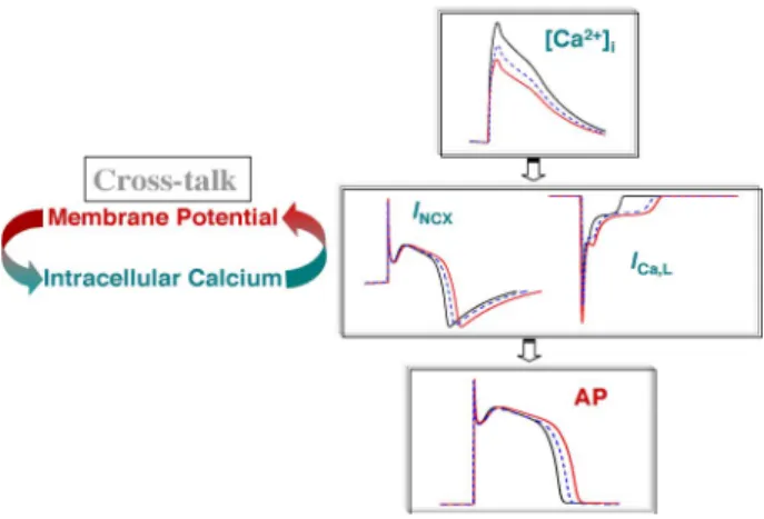
data, a segment of which is presented in Fig. 1(b), and the resulting sarcolemmal ionic currents and SR Ca2+ fluxes are presented in Fig. 2.
Thus, in Fig. 2(a), the left axis presents the APD90 and the
right axis presents the ∆[Ca2+]i(defined as systolic minus
dias-tolic [Ca2+]i) of the beat-to-beat fluctuations of the
correspond-ing beats in Fig. 1(b), which were used to clamp the model. In
Fig. 1. (a) Representative example of concordant [Ca2 +]iand AP alternans in
ventricular myocyte stimulated every 0.8 s; (b) representative example of phase
transitions between [Ca2 +]iand AP alternans indicated at points (a) to (d), in
the same data record. In-phase (concordant) [Ca2 +]
i and AP alternans leads
to out-of-phase (discordant) alternans indicated at points (a) and (c), and back again to in-phase alternans indicated in beats (b) and (d). “A” and “B” denote large and small [Ca2 +]ior long and short APD respectively; the peak [Ca2 +]i
and APD for these beats are also shown. The arrows in the voltage tracing of (b) indicate a sub-threshold after early after-depolarization.
Fig. 2(b), the left axis presents the time-dependent beat-to-beat current attributed to both the L-type calcium channel (LTCC) (IC a,L) and the Na/Ca exchanger (IN C X), while the right axis
presents the ryanodine receptor (RyR) open probability of the same beats presented in Fig. 1(b); in this figure, the choice of lumping the two currents is based on the hypothesis that if [Ca2+]i and AP alternans are linked, it is likely that the
bal-ance (reflected in the sum) of these two currents that control the depolarization versus repolarization of the membrane, will determine their effect on the APD.
One observes that the APD prolongation observed in Fig. 2(a) is associated with the larger inward depolarizing current at-tributed to the LTCC and the NCX seen in Fig. 2(b); this depo-larizing current is a result of secondary, much smaller SR Ca2+ release events, reflected at the RyR state-1 open probability PO 1
[see Fig. 2(b)] on an every other beat basis indicated by an “∗,” while the primary SR Ca2+release events are represented by an “↑.” This depolarizing current is associated with a small deflec-tion on the AP [also seen after careful inspecdeflec-tion in Fig. 1(b)],
Fig. 2. [Ca2 +]i and AP experimental data of Fig. 1(b) were used to
simultanously [Ca2 +]
i- and voltage-clamp the canine myocyte model. (a) Left
ordinate presents the beat-to-beat experimentally measured APD (in black) and
the right ordinate presents ∆[Ca2 +]i (expressed as systolic minus diastolic
[Ca2 +]
i, and is shown in red); data are plotted in pairs to demonstrate the
al-ternans beat-to-beat oscillations. (b) Left ordinate presents the sum (black line)
of the IC a , L (blue line) and IN C X(red line), and the right ordinate the RyR
state 1 open probability PO 1 (magenta line). (c) the sarcoplasmic reticulum
([Ca2 +]S R) is presented for the same beats in (a) and (b). (↑): indicates primary
RyR openings; (∗): indicates secondary RyR openings.
which is a subthreshold early after-depolarization (sEAD). This secondary Ca2+release also results in a longer time for [Ca2+]i
to reach its peak value, and is seen only in beats associated with a large [Ca2+]ithat is on an every other beat basis [see Fig. 2(b)].
Significantly, in Fig. 2(b), after sustained concordant alter-nans [seen in Fig. 1(b)] between the ∆[Ca2+]
i and APD in
which a large/small (indicated as A/B) ∆[Ca2+]i corresponds
to a long/short (indicated as A/B) APD, in beat 3, one observes that a phase reversal between ∆[Ca2+]i and APD occurs (i.e.,
a small ∆[Ca2+]i is associated with a long APD). It is likely
that what causes the phase reversal between [Ca2+]i and APD
is the ∆[Ca2+]iamplitude, which in this beat is larger than the
amplitude of the previous small ∆[Ca2+]i, and is smaller than
the previous large ∆[Ca2+]i [see Fig. 1(b), and also Fig. 3(a)
and (b)]; ∆[Ca2+]i influences the balance of the depolarizing
and repolarizing currents through the LTCC and NCX, and their effect on APD.
In beat 4, one observes that the ∆[Ca2+]iand APD continue
to be out of phase, in which case, a large ∆[Ca2+]i coincides
with a short APD. In beats 5 and 6, one sees that the ∆[Ca2+]i
and APD are again in-phase, but because ∆[Ca2+]
iis still within
a critical range (see Fig. 3(a) and (b) later) between a small and a large ∆[Ca2+]i(i.e., of the first, eighth, ninth, and tenth beat),
the APD can be either short or long, which is likely to result in a phase reversal between ∆[Ca2+]i and APD. Indeed, in beat
7, there is a phase reversal between ∆[Ca2+]iand APD, that is
a large ∆[Ca2+]i(larger than that in beat 6), corresponds to a
short APD.
Thereafter, in beat 8, phase reversal again results in in-phase alternans of ∆[Ca2+]iand APD, which is maintained in
subse-quent beats.
Fig. 3. Presence of phase transitions between [Ca2 +]
i and AP (red pixels)
in experimentally obtained ∆[Ca2 +]i and APD time series. (a) The
relationship between the APD and ∆[Ca2 +]
i (systolic minus diastolic
[Ca2 +]i), which indicates that phase reversal occurs when the APD falls
within a small range of its minimum and maximum values (113%≤
APDre v/APDm in≤ 117% and 81% ≤ APDre v/APDm a x≤ 84%). (b)
Rela-tionship between the ∆[Ca2 +]
i of the (N + 1)st-beat versus the ∆[Ca2 +]i
of the (N )th-beat, also indicates that phase reversal in consecutive beats
oc-curs when ∆[Ca2 +]
i falls within a small range of its minimum and
max-imum values (214%≤ ∆[Ca2 +]i , re v/∆[Ca2 +]i , m in≤ 232%, and 37% ≤
∆[Ca2 +]
i , re v/∆[Ca2 +]i , m a x≤ 41%), as indicated by the red symbols. Where
APDre vis the APD value in which a phase reversal occurs, APDm inis the
min-imum APD value in the APD time series, APDm a x is the maximum APD
value in the APD time series, ∆[Ca2 +]
i , re vis the ∆[Ca2 +]ivalue in which a
phase reversal occurs, ∆[Ca2 +]i , m inis the minimum ∆[Ca2 +]i value in the
∆[Ca2 +]
itime series, and ∆[Ca2 +]i , m a xis the maximum ∆[Ca2 +]ivalue in
the ∆[Ca2 +]itime series.
Since reduced SR Ca2+ uptake alone could lead to smaller Ca2+release and [Ca2+]i, the beat-to-beat ∆[Ca2+]SR (defined
as the diastolic minus the systolic [Ca2+]SR) oscillations have
been quantified from the computer-generated data (using the same simultaneous AP- and [Ca2+]i-clamp approach). It was
observed that ∆[Ca2+]SRalternated between beats
correspond-ing to small and large [Ca2+]
i during alternans [see Fig. 2(c)];
however, ∆[Ca2+]SR was almost constant in beats
correspond-ing to nonchangcorrespond-ing [Ca2+]i.
B. Relationship of Beat-to-Beat [Ca2+]iand APD
During Alternans
Analysis of the beat-to-beat ∆[Ca2+]iand APD for the
whole-data record revealed that discordant alternans occurred four times [red pixels in Fig. 3(a) and (b)]. Interestingly, similarly to the example presented in Fig. 1(b), all four phase transitions of concordant to discordant alternans were transient, which re-sulted into discordant alternans to be reverted back to sustained concordant alternans, within a few beats.
Fig. 3(a) demonstrates the relationship between the ∆[Ca2+]i
and APD. One observes that phase reversal occurs for APD values that fall within 113%≤ APDrev/APDm in ≤ 117% and
81%≤ APDrev/APDm ax ≤ 84%, where APDrev is the APD
value in which a phase reversal occurs, and APDm in/APDm ax
are the minimum/maximum APD value, respectively, in the APD time series.
On the other hand, Fig. 3(b) which presents the relation-ship between the ∆[Ca2+]i of the (N + 1)st-beat versus
the ∆[Ca2+]i of the (N )th-beat, indicates that phase
re-versal occurs when ∆[Ca2+]i falls within a small range of
Fig. 4. Schematic of the apparent dependence of the shape of the AP waveform on the NCX that contributes either a depolarizing or repolarizing current during the AP and the LTCC that contributes a depolarizing current. Since both the
NCX and LTCC are directly mediated by [Ca2 +]i, a large calcium transient
(in black) first causes the NCX to reverse earlier, thus contributing a smaller repolarizing current, and thus, contributing to AP prolongation, and second
causes acceleration of the LTCC-Ca2 +-mediated inactivation that would cause
AP shortening; one expects the opposite results for a small calcium transient (in red). Thus, the effect of [Ca2 +]
ion the membrane potential is defined by the
net balance of these two currents.
series (214%≤ ∆[Ca2+]i,rev/∆[Ca2+]i,m in≤ 232% and
37%≤ ∆[Ca2+]i,rev/∆[Ca2+]i,m ax≤ 41%), as indicated by
the red symbols; where ∆[Ca2+]i,rev is the ∆[Ca2+]i value in
which a phase reversal occurs, and ∆[Ca2+]i,m in/∆[Ca2+]i,m ax
are the minimum/maximum ∆[Ca2+]i values, respectively, in
the ∆[Ca2+]itime series.
Thus, one sees that the ∆[Ca2+]iand APD oscillations change
phase between each other, when ∆[Ca2+]i is within a critical
range between a small and a large ∆[Ca2+]
i.
IV. DISCUSSION
The present study presents the first report of discordant [Ca2+]iand AP alternans in the cardiac myocyte, and attempts to
explore the mechanisms underlying the discordant relationship between [Ca2+]iand AP alternans.
The hybrid, experimental–computational approach used al-lowed the opening of the closed-loop system of the subcellular Ca2+homeostatic mechanisms, and study their individual effect on the AP, without inadvertently perturbing the system under study. Specifically, it resulted in the following novel findings: 1) elevated SR Ca2+results in aberrant SR Ca2+release and in [Ca2+]ialternans and 2) it established the presence of discordant
alternans between the [Ca2+]iand AP at the myocyte level, and
the importance of [Ca2+]
i in defining the in- or out-of-phase
relationship between [Ca2+]iand AP.
The apparent inter-dependence of the shape of the AP waveform on the NCX which contributes either a depolar-izing or repolardepolar-izing current during the AP, and the LTCC which contributes a depolarizing current, and their effect on [Ca2+]i and AP alternans, is presented in Fig. 4. Since both
the NCX and LTCC are directly mediated by [Ca2+]i, a
large calcium transient (in black) first causes the NCX to reverse earlier, thus contributing a smaller repolarizing cur-rent, and thus, contributing to AP prolongation, and second
causes acceleration of the LTCC Ca2+-mediated inactivation that causes AP shortening; one expects the opposite results for a small calcium transient (in red). Thus, the effect of [Ca2+]ion
the membrane potential is defined by the net balance of these two currents.
Overall, the biophysical observations of this and previous studies [15] are consistent with experiments in heart cells [19], in planar lipid bilayers [20], and cardiac vesicles [21] that con-firm that Ca2+in the SR lumen influences RyR gating such that RyRs are more likely to be triggered by cytosolic Ca2+when
SR luminal Ca2+is elevated, which increases spontaneous SR Ca2+release [22] and the delayed after-depolarization (DAD) amplitude threshold to trigger an AP [23]. Furthermore, these findings agree with those of Diaz et al. [9] in which, during al-ternans, secondary releases and biphasic [Ca2+]iwere observed
in tetracaine-treated myocytes. These secondary releases were attributed to the increased spatial and temporal desynchroniza-tion of SR Ca2+release, during which, in a given region of a myocyte, SR Ca2+release propagated as a wave, the amplitude of which could alternate on a beat-to-beat basis.
At the whole-heart level, optical mapping studies in normal hearts have shown that discordant AP alternans (reflecting two areas in the heart that oscillate with opposite phase) were asso-ciated with a state of marked cardiac electrical instability, since ventricular fibrillation was always preceded by discordant and never by concordant AP repolarization alternans [1], [14]. This pattern of inhomogeneity was consistently induced at a critical threshold heart rate, and was largely independent of the pac-ing site [1], suggestpac-ing that it was caused by heterogeneities of cellular repolarization properties rather than heterogeneous propagation delay. Interestingly, in this study, alternans most commonly involved the slope of the AP plateau and the onset of final repolarization.
In summary, SR Ca2+ overload that results in spontaneous SR Ca2+ release and stimulates additional Ca2+ extrusion via the NCX which in turn produces an inward, depolarizing current and sub-threshold triggered depolarizations, are the underlying events for [Ca2+]i and AP alternans. Furthermore, the finding
that the [Ca2+]iand APD can oscillate in an uncorrelated manner
is likely to constitute the ventricular myocyte as the smallest unit underlying cardiac alternans and increased susceptibility to arrhythmogenesis.
REFERENCES
[1] K. R. Laurita, J. M. Pastore, and D. S. Rosenbaum, “How restitution, repolarization, and alternans form arrhythmogenic substrates: Insights from high-resolution optical mapping,” in Cardiac Electrophysiology:
From Cell to Bedside, D. P. Zipes and J. Jalife, Eds., 2nd ed. Philadelphia, PA: Saunders, 1999, pp. 239–248.
[2] A. A. Armoundas, G. F. Tomaselli, and H. D. Esperer, “Pathophysiolog-ical basis and clin“Pathophysiolog-ical application of T-wave alternans,” JACC, vol. 40, pp. 207–217, 2002.
[3] A. A. Armoundas, S. H. Hohnloser, T. Ikeda, and R. J. Cohen, “Can microvolt T-wave alternans testing reduce unnecessary defibrillator im-plantation?” Nat. Clin. Pract. Cardiovasc. Med., vol. 2, pp. 522–528, 2005.
[4] A. Mahajan, D. Sato, Y. Shiferaw, A. Baher, L. H. Xie, R. Peralta, R. Olcese, A. Garfinkel, Z. Qu, and J. N. Weiss, “Modifying L-type cal-cium current kinetics: Consequences for cardiac excitation and arrhythmia dynamics,” Biophys. J., vol. 94, pp. 411–423, 2008.
[5] F. Hua, D. C. Johns, and R. F. Gilmour Jr., “Suppression of electrical alternans by overexpression of HERG in canine ventricular myocytes,”
Amer. J. Physiol. Heart Circ. Physiol., vol. 286, pp. H2342–H2351,
2004.
[6] J. J. Fox, J. L. McHarg, and R. F. Gilmour, Jr., “Ionic mechanism of electrical alternans,” Amer. J. Physiol. Heart Circ. Physiol., vol. 282, pp. H516–H530, 2002.
[7] P. N. Jordan and D. J. Christini, “Action potential morphology influences intracellular calcium handling stability and the occurrence of alternans,”
Biophys. J., vol. 90, pp. 672–680, 2006.
[8] J. I. Goldhaber, L. H. Xie, T. Duong, C. Motter, K. Khuu, and J. N. Weiss, “Action potential duration restitution and alternans in rabbit ventricular myocytes: The key role of intracellular calcium cycling,” Circ. Res., vol. 96, pp. 459–466, 2005.
[9] M. E. Diaz, D. A. Eisner, and S. C. O’Neill, “Depressed ryanodine re-ceptor activity increases variability and duration of the systolic Ca2+ transient in rat ventricular myocytes,” Circ. Res., vol. 91, pp. 585–593, 2002.
[10] E. Chudin, J. Goldhaber, A. Garfinkel, J. Weiss, and B. Kogan, “Intra-cellular Ca(2+) dynamics and the stability of ventricular tachycardia,”
Biophys. J., vol. 77, pp. 2930–2941, 1999.
[11] J. Kockskamper, A. V. Zima, and L. A. Blatter, “Modulation of sarcoplas-mic reticulum Ca2+ release by glycolysis in cat atrial myocytes,” J.
Physiol., vol. 564, pp. 697–714, 2005.
[12] J. H¨user, Y. G. Wang, K. A. Sheehan, F. Cifuentes, S. L. Lipsius, and L. A. Blatter, “Functional coupling between glycolysis and excitation– contraction coupling underlies alternans in cat heart cells,” J. Physiol.
(Lond.), vol. 524, pp. 795–806, 2000.
[13] J. M. Pastore, S. D. Girouard, K. R. Laurita, F. G. Akar, and D. S. Rosenbaum, “Mechanism linking T-wave alternans to the genesis of car-diac fibrillation,” Circulation, vol. 99, pp. 1385–1394, 1999.
[14] J. M. Pastore and D. S. Rosenbaum, “Role of structural barriers in the mechanism of alternans-induced reentry,” Circ. Res., vol. 87, pp. 1157– 1163, 2000.
[15] A. A. Armoundas, “Mechanism of abnormal sarcoplasmic reticulum ccium release in canine left ventricular myocytes results in cellular al-ternans,” IEEE Trans. Biomed. Eng., vol. 56, no. 2, pp. 220–228, Feb. 2009.
[16] A. A. Armoundas, I. A. Hobai, G. F. Tomaselli, R. L. Winslow, and B. O’Rourke, “Role of sodium–calcium exchanger in modulating the ac-tion potential of ventricular myocytes from normal and failing hearts,”
Circ. Res., vol. 93, pp. 46–53, 2003.
[17] R. L. Winslow, J. Rice, S. Jafri, E. Marban, and B. O’Rourke, “Mech-anisms of altered excitation–contraction coupling in canine tachycardia-induced heart failure, II: Model studies,” Circ. Res., vol. 84, pp. 571–586, 1999.
[18] J. L. Greenstein, R. Wu, S. Po, G. F. Tomaselli, and R. L. Winslow, “Role of the calcium-independent transient outward current I(to1) in shaping action potential morphology and duration,” Circ. Res., vol. 87, pp. 1026–1033, 2000.
[19] Y. Li, E. G. Kranias, G. A. Mignery, and D. M. Bers, “Protein kinase A phosphorylation of the ryanodine receptor does not affect calcium sparks in mouse ventricular myocytes,” Circ. Res., vol. 90, pp. 309–316, 2002. [20] I. Gyorke and S. Gyorke, “Regulation of the cardiac ryanodine receptor
channel by luminal Ca2 + involves luminal Ca2 +sensing sites,” Biophys.
J., vol. 75, pp. 2801–2810, 1998.
[21] N. Ikemoto, M. Ronjat, L. G. Meszaros, and M. Koshita, “Postulated role of calsequestrin in the regulation of calcium release from sarcoplasmic reticulum,” Biochemistry, vol. 28, pp. 6764–6771, 1989.
[22] H. Satoh, L. A. Blatter, and D. M. Bers, “Effects of [Ca2+]i, SR Ca2+ load, and rest on Ca2 +spark frequency in ventricular myocytes,” Amer.
J. Physiol., vol. 272, pp. H657–H668, 1997.
[23] C. H. Orchard, D. A. Eisner, and D. G. Allen, “Oscillations of intracellular Ca2+ in mammalian cardiac muscle,” Nature, vol. 304, pp. 735–738, 1983.