The Development of Brain Systems Associated
with Successful Memory Retrieval of Scenes
The MIT Faculty has made this article openly available.
Please share
how this access benefits you. Your story matters.
Citation
Ofen, N. et al. “The Development of Brain Systems Associated with
Successful Memory Retrieval of Scenes.” Journal of Neuroscience
32.29 (2012): 10012–10020.
As Published
http://dx.doi.org/10.1523/jneurosci.1082-11.2012
Publisher
Society for Neuroscience
Version
Final published version
Citable link
http://hdl.handle.net/1721.1/76734
Terms of Use
Article is made available in accordance with the publisher's
policy and may be subject to US copyright law. Please refer to the
publisher's site for terms of use.
Behavioral/Systems/Cognitive
The Development of Brain Systems Associated with
Successful Memory Retrieval of Scenes
Noa Ofen,
1,3,4Xiaoqian J. Chai,
1,3Karen D. I. Schuil,
1,3,5Susan Whitfield-Gabrieli,
1,3and John D. E. Gabrieli
1,2,3 1Department of Brain and Cognitive Sciences,2Harvard-MIT Division of Health Sciences and Technology, and3McGovern Institute for Brain Research, Massachusetts Institute of Technology, Cambridge, Massachusetts, 02139,4Institute of Gerontology and Department of Pediatrics, Wayne State University, Detroit, Michigan 48202, and5Institute of Psychology, Erasmus University Rotterdam, 3000 DR Rotterdam, The NetherlandsNeuroanatomical and psychological evidence suggests prolonged maturation of declarative memory systems in the human brain from
childhood into young adulthood. Here, we examine functional brain development during successful memory retrieval of scenes in
children, adolescents, and young adults ages 8 –21 via functional magnetic resonance imaging. Recognition memory improved with age,
specifically for accurate identification of studied scenes (hits). Successful retrieval (correct old–new decisions for studied vs unstudied
scenes) was associated with activations in frontal, parietal, and medial temporal lobe (MTL) regions. Activations associated with
suc-cessful retrieval increased with age in left parietal cortex (BA7), bilateral prefrontal, and bilateral caudate regions. In contrast, activations
associated with successful retrieval did not change with age in the MTL. Psychophysiological interaction analysis revealed that there were,
however, age-relate changes in differential connectivity for successful retrieval between MTL and prefrontal regions. These results
suggest that neocortical regions related to attentional or strategic control show the greatest developmental changes for memory retrieval
of scenes. Furthermore, these results suggest that functional interactions between MTL and prefrontal regions during memory retrieval also
develop into young adulthood. The developmental increase of memory-related activations in frontal and parietal regions for retrieval of scenes
and the absence of such an increase in MTL regions parallels what has been observed for memory encoding of scenes.
Introduction
Declarative or explicit memory is comprised of three successive
stages: (1) encoding, (2) storage or consolidation, and (3)
re-trieval. Neuroimaging studies have highlighted the involvement
of medial temporal lobe (MTL), prefrontal, and parietal regions
in memory retrieval in adults (Fletcher et al., 1998; Buckner and
Wheeler, 2001; Kahn et al., 2004; Wagner et al., 2005; Konishi et
al., 2000; Spaniol et al., 2009), but little is known about the
devel-opment of the functional neural correlates for the retrieval of
declarative memories. Structurally, there is prolonged anatomic
maturation in prefrontal and parietal regions (Giedd et al., 1999;
Sowell et al., 2003, 2004; Gogtay et al., 2004, 2006). Behavioral
evidence suggests that retrieval strategies continue to mature
through adolescence (Bauer, 2008). This study investigated the
maturation of the neural correlates of memory retrieval from
childhood through adolescence and into young adulthood.
There is growing evidence about the development of memory
systems in the human brain and the different maturational
tra-jectories of specific brain regions and memory processes (Menon
et al., 2005; Chiu et al., 2006; Ofen et al., 2007; Paz-Alonso et al.,
2008; Chai et al., 2010; Ghetti et al., 2010; Maril et al., 2010).
There is prolonged functional maturation of prefrontal and
pa-rietal cortices for successful encoding of vivid memories for
scenes (recollection), but early maturation of those regions for
encoding of less vivid memories for scenes (familiarity) (Ofen et
al., 2007). The MTL appears to mature early in its contribution to
both vivid and less vivid memories for simple scenes (Ofen et al.,
2007), but mature more slowly in its support for vivid memories
of complex scenes (Chai et al., 2010) and contextually rich
mem-ories (source memory) (Ghetti et al., 2010).
Only one study has examined the development of neural
sys-tems associated with successful retrieval of memories
(Paz-Alonso et al., 2008). This study examined veridical and false
memories for highly related words in children and adults and
found age-related differences in MTL, prefrontal, and parietal
activations. Successful memory retrieval with this sort of task
involves a complex interaction between semantic knowledge of
words, retrieval, and strategic monitoring (Roediger et al., 2001).
Here we examined the development of successful memory
re-trieval for scenes, a domain of memory that is less complicated by
parallel language and strategic development.
The objective of this study was to discover whether brain
re-gions that are functionally associated with successful memory
retrieval for scenes change across development from ages 8 to 21.
We hypothesized that, similar to successful memory encoding,
activations in prefrontal and parietal regions associated with
suc-cessful retrieval would increase with age. A hypothesis about
Received March 1, 2011; revised May 16, 2012; accepted May 24, 2012.
Author contributions: N.O., X.J.C., and J.D.E.G. designed research; N.O., X.J.C., and K.D.I.S. performed research; N.O., X.J.C., K.D.I.S., and S.W.-G. analyzed data; N.O., X.J.C., S.W.-G., and J.D.E.G. wrote the paper.
This work was supported by NIH Grant RO1-MH-080344 to J.D.E.G. We thank Christina Triantafyllou, Sheeba Arnold, and Steve Shannon at the Martinos Center for help with imaging protocols; Ayesha Hameed, Kai Qiu, Larissa Estrada, Tania Abedian, Sudhandra Sudanderam, and Natalie Rubinstein for help with data collection; and the participants and their families.
The authors declare no competing financial interests.
Correspondence should be addressed to Noa Ofen, Institute of Gerontology, 257 Knapp Building, 87 East Ferry Street, Detroit, MI 48202. E-mail: noa.ofen@wayne.edu.
DOI:10.1523/JNEUROSCI.1082-11.2012
MTL activations was less clear, because previous reports did not
find age-related change in MTL activations for successful
encod-ing of scenes (Ofen et al., 2007), but did find age-related
increas-ing activations for retrieval of highly related words (Paz-Alonso
et al., 2008). We also examined whether there are age-related
changes in retrieval-related connectivity between the MTL and
neocortical regions associated with the maturation of successful
memory retrieval.
Materials and Methods
Participants. Eighty volunteers, ages 8 –21 years old, were recruited from the local community and provided informed consent as required by a Massachusetts Institute of Technology Institutional Review Board-approved protocol. Data from three participants (8-year-old male, 8-year-old female, and 11-year-old male) were removed due to excessive movement that resulted in them not being able to contribute a complete data set in the scanning session. Data from other participants were ex-cluded due to (1) behavioral noncompliance (i.e., performance was so poor that it appeared that participants were not performing or under-standing the task; three participants, 14-year-old female, 17-year-old female, and 21-year-old male), (2) not completing the study (two 8-year-old males), and (c) technical difficulties when collecting behavioral re-sponses in the scanner (three participants, 8-year-old female, 12-year-old female, and 14-year-old male). Neuroimaging data are thus presented for 69 participants (35 males; age range, 8 –21 years old; mean age, 14.7⫾ 4.2 years) who met data quality criteria. All participants were right handed, had normal or corrected to normal visual acuity, and were screened for histories of psychiatric or developmental disorders. IQ estimates were obtained for each participant based on the Kaufman Brief Intelligence Test (second edition). The mean composite standard (normed to control for chronological age) IQ score across participants was 117.2⫾ 14.3 (SD). Importantly, participants’ age and IQ did not correlate (r⫽ 0.03, p⫽ 0.80), indicating that in this sample of participants there was no confound of differences in IQ between younger and older participants. Participants 18 years or older were paid $20 per hour for their participa-tion. Participants younger than 18 years received a bookstore gift certif-icate with a value of $20 per hour for their participation.
Materials. Two hundred and eighty pictures of indoor and outdoor scenes were used and divided into lists of 28 pictures (each comprised of 14 indoor and 14 outdoor scenes). Five lists were presented during the study, and the remaining five lists were presented as foils during the scanned recognition-memory test. List presentation order was counter-balanced across participants, such that pictures were presented equally often as study and test items across participants. Eight additional cartoon images of animals were used in a short practice period before the begin-ning of study.
Memory task. Participants studied 140 pictures of indoor and outdoor scenes. Each picture was presented for 2.5 s. Participants were explicitly instructed to memorize the scenes for a later memory test. During the study phase, participants judged whether each picture depicted an in-door or outin-door scene, and indicated their judgment by pressing one button on the computer keyboard with their right index finger to indicate an indoor scene or another button with their left index finger to indicate an outdoor scene. The instructions (indoor/outdoor) appeared on the bottom of the screen, prompting participants to use the appropri-ate button press. Immediappropri-ately following the study phase, participants were placed in an MRI scanner and given a recognition test consisting of 140 old and 140 new pictures presented on a screen. Each scene was presented on the screen for 3 s, followed by a 1 s fixation cross. Variable intertrial intervals (2– 8 s) were used to increase fMRI measurement reliability. For each picture, participants judged whether they had seen it before in the study phase (old) or not (new). Participants were instructed to take as much time as they needed to make their most accurate re-sponse, but to try to respond to each of the trials as soon as they had reached a decision. Intentional memory instructions were used to mini-mize the possibility that the older, compared to younger participants, would expect a later memory test and more voluntarily use intentional mnemonics strategies. Furthermore, the instructions emphasized
accu-racy to assure that the younger participants attempt to reach an accurate retrieval decision. Studied items that elicited an old response were cate-gorized as a HIT; studied items that elicited a new response were catego-rized as a MISS. New items (foils) that elicited a new response were categorized as a correct rejection (CR); new items that elicited an old response were categorized as a false alarm (FA). Participants were highly accurate (96.9%) in making indoor/outdoor judgments during the study phase (on average, 135.6⫾ 4.2 recorded correct responses from 140 trials). There was, however, a positive correlation between age and accu-racy in the encoding task (r⫽ 0.50, p ⬍ 0.001), suggesting that younger participants were not properly attending to some of the items or pressing the wrong buttons for some items. To minimize potential age-related effects of attention during encoding from being carried over to the rec-ognition test, only studied items that were correctly judged as indoor or outdoor were used in the imaging analysis. This minimized the influence of age on accuracy in the encoding task from influencing recognition test analyses. Reaction times for correct indoor/outdoor judgments during encoding (1001⫾ 218 ms) did not change with age (r ⫽ 0.10, p ⫽ 0.43). Younger participants were more likely not to respond (r⫽ 0.43, p ⬍ 0.001) or to press two different buttons for an item (r⫽ 0.32, p ⫽ 0.006) during the recognition test. The items for which either no response or more than one response occurred were omitted from the imaging analysis.
MRI data acquisition. Data were acquired on a 3T Tim Trio Siemens scanner using a 12-channel head coil. Before the functional scans, we acquired T1-weighted whole-brain anatomy images (MPRAGE se-quence, 256⫻ 256 voxels, 1 ⫻ 1.3 mm in-plane resolution, 1.3 mm slice thickness). Functional images were obtained in 32 4-mm-thick slices (with 0.8 mm gap) oriented parallel to the line connecting the anterior and posterior commissures, covering the entire brain (echoplanar T2*-weighted images; 3.1 mm in-plane resolution; repetition time, 2 s; echo time, 30 ms; flip angle, 90, 64⫻ 64 voxels). To minimize effects of scanner signal stabilization, the first four images were omitted from all analysis. The memory task was imaged in five functional runs, each with 56 picture stimuli. Functional runs lasted 4 min and 56 s, in which 144 images were collected. Foam pads were used to restrict head movement. Imaging data analyses. SPM5 (Wellcome Department of Imaging Neu-roscience, London, UK; http://www.fil.ion.ucl.ac.uk/spm/software/spm5) was used in all analyses. Images went through motion correction using sinc interpolation and normalized to the T2 Montreal Neurological In-stitute (MNI) template. Normalization to a common stereotactic space was used to allow comparison of activations across participants; such normalization has been shown to be appropriate for children age 7 and older (Burgund et al., 2002). Finally, images were spatially smoothed with an isotropic Gaussian kernel of 6 mm full-width at half-maximum. All functional data were subjected to artifact detection using custom software (http://web.mit.edu/swg/software.htm). From each participant, an image was defined as an outlier if movement from a preceding image exceeded 0.5 mm in any of the three translation directions and/or 0.01 radians of rotation. An image was also defined as an outlier if the global mean intensity in the image was⬎3 SDs from the mean image intensity for the run in which it was collected. Overall, 2.3⫾ 3.0% (mean ⫾ SD) of the images were defined as outliers (with a maximum 16.1% in one participant). A single regressor for each outlier image was included in the first level model (described below) to control for outlier images effects on estimated activations. Analysis of the movement parameters obtained during realignment (three translation and three rotation, mean of the means of each of five runs) showed that movement increased with age, as evident in a negative correlation between age and two movement dimen-sions (x-axis translation, r⫽ ⫺0.27, p ⫽ 0.03; y-axis translation, r ⫽ ⫺0.32, p ⫽ 0.01; movement in the z-axis translation and in the three rotational dimensions did not change reliably with age, 0.03⬍ 兩r兩 ⬍ 0.13, p values⬎0.29). Movement parameters were not correlated with stimu-lus presentation across participants (for any of the six movement param-eters with either HIT or CR; mean,兩r兩 values ⬍ 0.03; max, 兩r兩 values of 0.17). Therefore, to minimize artifacts due to differences in movement across ages, movement parameters (the six movement parameters as-sessed during realignment) were included as nuisance covariates in par-ticipants’ statistical models (first-level analysis). Inclusion of motion
parameters ensured that the variance related to residual head motion was explicitly modeled, and hence would not result in false positives.
Individual general linear model-based analyses were conducted in MNI space. Models included regressors of interest generated by convolv-ing task events (HIT, CR) with a canonical model of the homodynamic response function (HRF) as implemented in SPM5, including temporal derivative. A canonical model of the HRF were used, as previous studies found minimal differences in HRF time courses between children and adults (Kang et al., 2003). Additional regressors and their temporal de-rivatives modeled other task events (MISS, FA) and invalid trials (incor-rect response during encoding, more than one or no response during encoding). Additional regressors modeled motion (as described above) and outlier images (one regressor per outlier image). Individual subject’s effects were estimated using a fixed-effects model across the five runs. Linear combination of the HIT minus CR regressors was used in defining contrasts of interest for successful retrieval. Memory-related activations were also assessed using a linear combination of HIT minus MISS, FA minus CR, and CR minus FA contrasts. Group analyses that included FA were restricted to participants having⬎17 FA trials (n ⫽ 56; 30 males; age range, 8 –21 years old; mean age, 15.0⫾ 4.1 years). Contrasts constructed at the single participant level were then input into a second-level group analysis using a random-effects model. Reported clusters survived thresholds of an uncorrected p⬍ 0.001 at the voxel level and a familywise error (FWE) correction (as implemented in SPM, using Gaussian Field Theory) of p⬍ 0.05 at the cluster level. A one-sample t test was per-formed on the contrast HIT⬎ CR. Next, age effects for successful re-trieval (HIT⬎ CR) were investigated across the whole brain regressing age as a continuous variable. In addition, age effects were also examined at a more lenient threshold of p⬍ 0.001 and a cluster extent of 50 contiguous voxels. In a complementary analysis, we used functionally defined ROIs derived from the group-level HIT⬎ CR activations map to examine, across participants, the correlations of successful retrieval acti-vations (HIT⬎ CR) with age and recognition memory accuracy. In defining ROIs from the group-level map of HIT⬎ CR, we used a cor-rected voxel-level FEW threshold of 0.001, which allowed for identifying limited extent clusters as ROIs.
Because of the known importance of MTL regions for memory, and known problems of susceptibility in the MTL region, we tested the a priori hypotheses that these regions are involved in memory retrieval. ROIs within the MTL were defined by small volume correction on the group HIT⬎ CR activation map using an anatomically defined MTL mask comprising the left and right hippocampi and left and right para-hippocampal gyri (using the Wake Forest University Pickatlas tool). Re-ported clusters survived an uncorrected p⬍ 0.001 at the voxel-level and FWE correction of p⬍ 0.05 at the cluster-level. In ROI analyses, data were extracted from individual participant’s contrast images and sub-jected to a test of correlation with age as a continuous variable.
To examine possible developmental changes in functional connectiv-ity of MTL during successful memory retrieval, we conducted a psycho-physiological interaction (PPI) analysis (Friston et al., 1997). We identified MTL seed ROIs for the PPI analysis based on MTL regions that were active in the group HIT⬎ CR map (described above). Seed ROIs were created as 4 mm spheres centered on peak activations in left para-hippocampal gyrus (PHG;⫺18, ⫺30, ⫺14), right PHG (18, ⫺34, ⫺10), and left anterior MTL (⫺16, 2, ⫺16). The PPI analysis compared the temporal correlation (or functional “coupling”) between the MTL seed regions and other brain regions (physiological component) during dif-ferent memory conditions (psychological component). The PPI design matrix contained three columns of variables: (1) a “psychological” vari-able that represents the contrast of two conditions (Hit⬎ CR), (2) a time-series “physiological ” variable that represents the time course of the source (seed) region, and (3) an interaction variable that represents the interaction of the psychological and physiological, respectively, variables (1) and (2). The regression coefficient for the interaction term provides a measure of PPI; a correlation (or covariance) in activation between the MTL seed region and the identified “coupled ” region that is significantly different between memory conditions (HIT⬎ CR) yields a significant PPI effect. Individual images of the interaction term were entered into a second-level whole-brain correlation analysis with age as a regressor
variable to identify developmental changes in MTL PPI effects. Whole-brain PPI analyses of the three MTL seeds were constrained using a binary mask from the whole-brain analysis of age-related changes in successful retrieval (HIT⬎ CR). This procedure allowed us to test for developmental influences on PPI effects between MTL seeds and neo-cortical regions that showed developmental effects in successful re-trieval activations. Small volume correction was performed using this mask (FWE cluster-level threshold of p⬍ 0.05). We also examined age-related changes in PPI effects of the MTL seeds at the whole-brain level ( p⬍ 0.001, voxel-level uncorrected; cluster extent, ⬎50 contig-uous voxels).
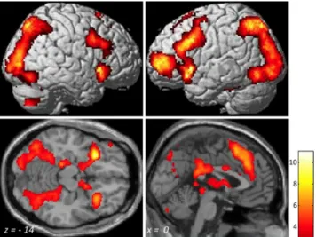
Figure 1. Successful retrieval activation (HIT⬎ CR) across all 69 participants (ages 8–21 years). Activation maps are rendered on images from a standard brain right and left lateral views (top) and on horizontal and sagittal sections (bottom). MNI coordinates are presented at the bottom of each section. Voxel-level threshold, p⬍0.001;correctedattheclusterlevelwith few, p⬍ 0.05. A t value scale is presented at the bottom right.
Table 1. Successful retrieval effects
BA MNIcoordinates Peak t value No. of voxels x y z
Across all participants
Left caudate n.a. ⫺10 12 0 11 8983
Left inferior frontal gyrus 47 ⫺30 20 ⫺8 10.71
Right caudate n.a. 8 12 2 9.76
Right posterior cingulate/preuneus 30 20 ⫺58 24 9.75 18929
Left posterior cingulate/preuneus 30 ⫺14 ⫺60 16 9.03
Right precuneus n.a. 16 ⫺68 44 8.82
Right inferior frontal gyrus/insula 47/11 32 24 0 8.92 1044 Left/right medial/superior frontal gyrus 9/8 ⫺4 34 34 8.4 2920
Right cerebellum n.a. 6 ⫺82 ⫺30 6.79 581
Left cerebellum n.a. ⫺10 ⫺72 ⫺30 6.14
Right inferior frontal gyrus 9 42 6 32 6.29 927
Right middle frontal gyrus 46 48 22 30 5.25
Right/left cingulate gyrus 24 6 4 30 5.47 179
Right cerebellum n.a. 36 ⫺68 ⫺50 5.39 244
Within the MTL
Left parahippocampal gyrus 35/28/19 ⫺18 ⫺30 ⫺14 6.09 184
Right parahippocampal gyrus 35/36/37 18 ⫺34 ⫺10 4.98 117
Left entorhinal cortex/amygdala 34/28 ⫺16 2 ⫺16 4.24 58
Increased with age
Left middle/superior frontal gyrus 6 ⫺32 10 64 4.43 54a
Left inferior frontal gyrus 47 ⫺46 24 ⫺10 4.38 95a
Right inferior frontal gyrus 47 56 22 0 4.14 53a
Left superior parietal lobule 7/40 ⫺24 ⫺70 54 4.06 179
Left/right caudate n.a. ⫺2 ⫺6 ⫺4 3.95 60a
Right inferior frontal gyrus 47 34 24 ⫺18 3.59 52a
Results
Behavior
Across all participants, 53
⫾ 13% (mean ⫾ SD) of the studied
scenes were recognized as old (HIT rate), and 24
⫾ 12% of the
new scenes presented at the test (foils) were falsely classified as old
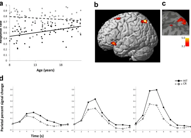
(FA rate). HIT rate increased significantly with age (r
⫽ 0.43, p ⬍
0.001), whereas CR rate did not change reliably with age (r
⫽ ⫺0.18,
p
⫽ 0.14) (see Fig. 2a). The difference between the age-related
cor-relations for HIT and CR was significant (z
⫽ 5.7, p ⬍ 0.001,
two-tailed). Participants’ response times for HIT (1612.1
⫾ 226.6 ms),
MISS (1684.7
⫾ 287.1 ms), CR (1630.7 ⫾ 240.0 ms), and FA
(1794.2
⫾ 319.9 ms) did not change significantly with age (兩r兩 values
⬍0.19, p values ⬎0.12).
Imaging
Successful retrieval across all participants
Across all participants, successful retrieval (contrast HIT
⬎ CR)
was associated with significant activations in prefrontal, parietal,
and temporal regions, as well as in subcortical regions (Fig. 1;
Table 1). Across all participants, three regions within the MTL
showed successful retrieval activations (Table 1).
Age-related changes in successful retrieval
A significant age-related increase in activation for successful
re-trieval was found in left parietal cortex with peak coordinates in
the superior parietal lobule (SPL; p
⬍ 0.001, voxel level;
cluster-corrected FWE, p
⬍ 0.05) (Table 1; Fig. 2c). The majority of
voxels in this parietal cluster were within the superior parietal
lobule (72% in 7A, 13% in 7P; a small portion were within the
superior part of the inferior parietal cortex, PGa, 12%; PFm,
0.6%) as identified using probabilistic cytoarchitectonic maps
from the SPM Anatomy Toolbox (Eickhoff et al., 2005). In
addi-tion, at a more liberal threshold, activations associated with
suc-cessful retrieval increased with age in bilateral prefrontal
neocortices and the left caudate ( p
⬍ 0.001, voxel level; cluster
extent,
⬎50 contiguous voxels) (Fig. 2b,c; Table 1). HRF time
courses from the left superior parietal lobule demonstrate
in-creased differentiation between HIT and CR from children
(ages 8 –12) to adolescents (ages 13–17) to adults (ages 18 –21)
(Fig. 2d).
In a complementary investigation of age effects, we used
func-tionally defined ROIs form the group HIT
⬎ CR activation map.
In a left parietal ROI (peak coordinates,
⫺14, ⫺60, 16)
activa-Figure 2. Age-related changes in recognition memory and brain activations associated with successful retrieval. a, HIT rate improved significantly with age (individual values in closed circles; regression with age, solid line), and CR accuracy did not change significantly with age (individual values in open circles; regression with age, dashed line). b, Activations associated with retrieval success (rendered on a standard brain’s left lateral view) increased with age in left parietal and frontal regions as indicated by regression analysis using age as a continuous variable across participants. Voxel-level threshold, p⬍ 0.001; cluster extent, ⬎50 contiguous voxels. c, For visual presentation, parietal activations depicted in b were overlaid on a surface based representation of the MNI canonical brain using the SPM SurfRend Toolbox (http://spmsurfrend.sourceforge.net). A t value scale is presented at bottom right. d, Time courses for the HIT and CR conditions in left superior parietal cortex were extracted for each participant and averaged across participants in three age groups corresponding to children (left; n⫽ 27; 8–12 years old), adolescents (middle; n ⫽ 24; 13–17 years old), and adults (right; n⫽ 18; 18–21 years old). HIT, Closed circles, solid line; CR, open circles, dashed line.
tions significantly increased with age (r
⫽ 0.25, p ⫽ 0.04).
Simi-larly, activation increased with age in the right caudate ROIs
(peak coordinates, 8, 12, 2; r
⫽ 0.30, p ⫽ 0.01), and marginally in
the left caudate (peak coordinates,
⫺10, 12, 0; r ⫽ 0.23, p ⫽ 0.06).
In one ROI in the medial frontal gyrus, activation increased with
age (peak coordinates,
⫺4, 34, 34; r ⫽ 0.24, p ⫽ 0.04). In other left
and right frontal ROIs (peak coordinates, left,
⫺30, 20, ⫺8; ⫺42,
18, 30;
⫺42, 46, 2; right, 32, 24, 0) activations did not change with
age (兩r兩 values ⬍0.18, p values ⬎0.16). Recognition memory
ac-curacy (average HIT rate and CR rate) increased with age (r
⫽
0.30, p
⫽ 0.01), but was not a significant predictor of activation in
any of the ROIs defined by group activations (
兩r兩 values ⬍0.18, p
values
⬎0.14). Age-related increases in HIT ⬎ CR activation
within ROIs remained significant (right caudate, r
⫽ 0.27, p ⫽
0.03) or approached significance (left parietal, left caudate,
medial frontal gyrus;
兩r兩 values ⬎0.20, p values ⬍0.10) when
con-trolling for individual differences in recognition memory
accu-racy. Thus, both whole-brain analysis and ROI methods of
assessing age effects in successful retrieval activations revealed
developmental effects in left superior parietal cortex and caudate,
and whole-brain analysis at a more lenient threshold suggested
developmental effects in bilateral prefrontal cortices.
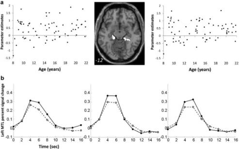
In contrast to the age-related changes described above, there
were no age-related correlations with successful retrieval
activa-tions in MTL regions (兩r兩 values ⬍0.17, p values ⬎0.15) (Fig. 3).
In addition, there were no age-related decreases in activations
associated with successful retrieval.
Retrieval-related functional connectivity between MTL and
prefrontal cortex increased with age
First, we identified age-related increases of differential functional
connectivity assessed with PPI for successful retrieval (HIT vs
CR) between the three MTL seeds and the regions in which
acti-vations for successful retrieval increased with age (MTL PPI
ef-fects restricted to regions of HIT
⬎ CR activations increased with
age). Across all participants, there was significant age-related
change in PPI between the left PHG and a region in the left
inferior frontal gyrus (IFG; r
⫽ 0.46, p ⬍
0.001) (Fig. 4; Table 2). Thus, the PPI
effects demonstrated reliable change in
the pattern of MTL–IFG connectivity
across the HIT and CR trials as a function
of age. Furthermore, we tested age-related
changes in left PHG–left IFG connectivity
separately for HIT and CR trials. Left
PHG–left IFG connectivity during CR
re-sponses significantly decreased with age
(r
⫽ ⫺0.38, p ⫽ 0.001), whereas left PHG–
left IFG connectivity during HIT trials did
not change with age (r
⫽ 0.10, p ⫽ 0.42).
PPI between the right PHG and left IFG
also changed with age (r
⫽ 0.40, p ⬍
0.001). No significant developmental
ef-fects were found in PPI of the left
ante-rior MTL. Second, we examined
age-related changes in the PPI of the three
MTL seeds at the whole-brain level (not
restricted to regions of HIT
⬎ CR
acti-vations increased with age; p
⬍ 0.001,
voxel level; cluster extent,
⬎50
contigu-ous voxels). There was a significant
age-related change in PPI between the left
PHG and the same region in the left
in-ferior frontal gyrus that was identified when we tested for PPI
effects with a mask (described above in this section) (Table 2).
No brain regions showed age-related changes in PPI with the
right PHG, or with the left anterior MTL using this threshold
for the whole-brain-level analysis.
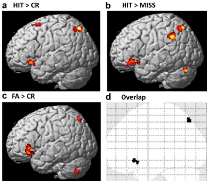
Additional retrieval contrasts
Across all participants, there were significant activations for the
contrast HIT
⬎ MISS (recognition contrast) in prefrontal and
parietal cortical regions and in MTL and subcortical regions
(Ta-ble 3). Activations for HIT
⬎ MISS increased with age in the left
inferior parietal lobule/supramarginal gyrus, left inferior frontal
gyrus, superior parietal cortex, and cerebellum ( p
⬍ 0.001, voxel
level; cluster extent,
⬎50 contiguous voxels) (Table 3). There was
overlap in the left superior parietal and left inferior frontal
clus-ters that showed age-related increase for HIT
⬎ CR and HIT ⬎
MISS (Table 3; Fig. 5). Furthermore, similar to what was found
for HIT
⬎ CR, there were no age-related changes in HIT ⬎ MISS
activations within the MTL (兩r兩 values ⬍ 0.06, p values ⬎ 0.60).
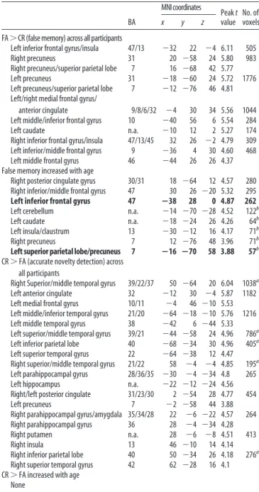
Across all participants, there were significant activations for
the contrast FA
⬎ CR (false memory of new scenes) in prefrontal
and parietal cortical regions and in subcortical regions (Table 4).
Activations for FA
⬎ CR increased with age in the left and right
inferior frontal gyri and in the left posterior cingulate gyrus, left
and right superior parietal cortices, cerebellum, caudate, and left
insula ( p
⬍ 0.001, voxel level; cluster extent, ⬎50 contiguous
voxels) (Table 4). There was overlap in the left superior parietal
and left inferior frontal clusters that showed age-related increases
for HIT
⬎ CR and FA ⬎ CR (Table 4; Fig. 5). Age-related
in-creases in activations for FA
⬎ CR were found in similar regions
that showed age-related increases for HIT
⬎ CR. Although in this
study rates of CR and FA did not change with age, age-related
increases in false memory are commonly found when richly
as-sociative verbal memory is tested (Paz-Alonso et al., 2008). The
overlap of age-related increases for HIT
⬎ CR and for FA ⬎ CR is
in line with an interpretation that age-related increases in
recog-nition memory accuracy coincide with age-related increases in
Figure 3. Activations associated with successful retrieval did not change with age in MTL. a, Left, Right, Plots of correlations of parameterestimatesvaluesextractedfromindividualparticipantsinthecontrastHIT⬎CRfromleftandrightMTLROIs.Middle,Thebinary masks of left and right ROIs within the MTL are depicted on an axial brain image. The MNI z-coordinate is presented in the bottom of the image. b, Time courses of retrieval success activations in. Time courses for the HIT and CR conditions in the left MTL in children, adolescents, and adults are shown. Description of age group and symbols are as described in Figure 2d.
false memory. Across all participants there were significant
acti-vations for the contrast CR
⬎ FA (accurate novelty monitoring)
in the anterior MTL and in parietal and medial frontal regions
that are typically associated with the default network (Table 4).
There were no changes with age for contrast CR
⬎ FA.
The main focus of this study was to identify developmental
effects in activations for retrieval success (contrast HIT
⬎ CR).
Because the other contrasts with FA and MISS trials involved the
HIT and CR trials, it might be expected that similar regions of
activation would be revealed as in the HIT
⬎ CR contrast. These
analyses, however, support the view that similar
developmen-tal factors support both successful objective memory retrieval
(HIT
⬎ CR) and also subjective memory retrieval (Wheeler
and Buckner, 2003) of “oldness” (HIT
⬎ MISS) or “newness”
(FA
⬎ CR).
Discussion
This study revealed the development of
declarative memory systems associated
with successful retrieval from memory for
scenes in children, adolescents, and young
adults. Older participants were more
ac-curate in recognition memory decisions,
and specifically in recognizing previously
presented scenes. Successful retrieval
across all ages was associated with
activa-tions in frontal, parietal, and MTL brain
regions that are consistently activated for
successful retrieval in adults (Konishi et
al., 2000; Montaldi et al., 2006; Spaniol et
al., 2009). Successful retrieval activations
increased with age in left parietal, bilateral
caudate, and bilateral frontal regions. In
contrast, successful retrieval activations in
the MTL did not change with age,
al-though retrieval-related PPI connectivity
between left and right parahippocampal
gyri and the left IFG changed with age.
Although the influence of age on brain
activation was examined only for scenes
that were accurately encoded and
accu-rately retrieved, it is likely that retrieved memories for studied
scenes were less vivid or detailed in younger participants. To
minimize demands on the youngest participants, we did not
col-lect a second measure of memory strength for each response to
distinguish between more vivid, or recollected, memories versus
less vivid, or familiar, memories. In a parallel study of encoding
for similar scenes, there was no developmental influence for
sub-sequent familiarity-based retrieval for scenes, but there were
sig-nificant developmental influences both on accuracy for
recollection-based retrieval for scenes, and also on activations in
left parietal and bilateral prefrontal regions that were associated
with subsequent recollection-based retrieval for scenes (Ofen et
al., 2007). Therefore, the developmental differences found in
pa-rietal and frontal regions in the present study of retrieval are likely
to reflect reduced details or information in the retrieved
memo-ries of younger participants.
Activation for successful retrieval grew across ages in bilateral
ventral prefrontal cortex and left dorsal prefrontal cortex. The
ventral prefrontal cortex is implicated in selection and retrieval of
information (Thompson-Schill et al., 1997; Wagner et al., 2001;
Thompson-Schill, 2003) and the maintenance of retrieved
repre-sentations (Petrides et al., 1993; Rugg et al., 1999; Dobbins et al.,
2002; Dobbins and Han, 2006). Age-related increases in
success-ful retrieval activations in these regions may reflect older
partic-ipants relying more on semantic knowledge or using better
maintenance processes for successful retrieval than younger
par-ticipants. Dorsal prefrontal cortex is also implicated in memory
retrieval in adults (Spaniol et al., 2009) and may specifically
con-tribute to retrieval of pictorial stimuli, rich in spatial information
(Passingham and Sakai, 2004). Activations also increased with
age in the left caudate, a region that is strongly connected with the
left ventral prefrontal cortex (Barnes et al., 2010).
The strongest influence of age occurred in left parietal cortex.
Activation for successful retrieval grew across ages primarily in the
left SPL (BA7). This age-related growth of parietal activation for
retrieval of memory for scenes is similar to developmental findings
of memory for verbal information (Paz-Alonso et al., 2008).
Multi-Figure 4. PPI for successful retrieval (difference between HIT and CR responses) between MTL and prefrontal cortex changed with age. Top left, PHG seed used in the PPI analysis (overlaid on an axial standard brain’s image). Bottom left, Left IFG cluster that exhibited age-related changes in differential functional connectivity for HIT versus CR (PPI) with the PHG seed (rendered on a standard brain’s left lateral view). Right, Parameter estimate of the PPI between left PHG and left IFG, plotted against age. Similar age-related changes in PPI were observed between right PHG and left IFG.
Table 2. MTL connectivity changed with age
BA MNIcoordinates Peak t value No. of voxels x y z
Within regions showing maturation of successful retrieval effects MTL seed region
Left PHG
Left inferior frontal gyrus 47 ⫺44 24 ⫺10 4.39 48
Right PHG
Left inferior frontal gyrus 47 ⫺44 24 ⫺6 3.99 7
Left anterior MTL None Whole brain analysis
MTL seed region Left PHG
Left inferior frontal gyrus 47 ⫺44 24 ⫺10 4.39 83a
Left/right, medial frontal/ precentral 6 12 ⫺28 64 4.39 90a Right PHG None Left anterior MTL None
MTL seed region coordinates are listed in Table 1.
aIncreases with age did not survive FWE cluster-level correction.
ple studies in adults report successful retrieval activations across left
parietal regions in BA7, BA39, and BA40 (Cabeza et al., 2008). Most
of these studies involved verbal stimuli, but activation for scenes
increased during retrieval as a function of self-reported memory
strength in left parietal cortex somewhat ventral to the region
show-ing an age-related change in the present study (Montaldi et al., 2006).
Increasing activation exhibited by adults in the present study may
have reflected the increasing strength of memories retrieved from
childhood to adulthood.
The role of parietal cortex in memory retrieval has been the
focus of intense investigation (Cabeza, 2008; Ciaramelli et al.,
2008; Vilberg and Rugg, 2008; Wagner et al., 2005) motivated by
its ubiquitous activation in neuroimaging studies and by the lack
of neuropsychological evidence for a critical role of parietal
cor-tex in memory (Haramati et al., 2008). Studies in adults have
suggested that left superior regions of the lateral parietal regions
reflect top-down attentional processes for memory retrieval
(Cabeza, 2008; Ciaramelli et al., 2008, 2010; Vilberg and Rugg,
2008). The extent to which these superior parietal activations
reflect primary memory retrieval processes is unclear, because
these regions are sensitive to expectations and decision biases
more generally (O’Connor et al., 2010), but such attentional
and retrieval effects may be separable (Hutchinson et al., 2009;
Sestieri et al., 2010). It has been proposed that activations in
superior parietal cortex (BA7) are associated with
familiarity-based retrieval, whereas activations in inferior parietal cortex
(BA39 and BA40) are associated with recollection-based
re-trieval (Yonelinas et al., 2005; Cabeza et al., 2008; Ciaramelli et
al., 2008; Vilberg and Rugg, 2008). Given prior evidence that
developmental influences are greater on recollection than
fa-miliarity at retrieval, it might have been expected that
devel-opmental differences would have occurred in more inferior
parietal cortex instead of the more superior location, where
the developmental influence was found. Developmental
stud-ies that more precisely control for
recol-lection and familiarity may clarify this
issue.
There were no developmental changes
in MTL activations associated with
suc-cessful retrieval, despite the finding that
such activations were found across ages.
Developmental changes in MTL
activa-tions associated with memory are
incon-sistent
across
studies.
Age-related
reductions in MTL activations were found
during the encoding of scenes (Menon et
al., 2005) and of certain verbal memories
(Maril et al., 2010). Another study found
no developmental changes in MTL
activa-tion for encoding that yielded recogniactiva-tion
familiarity for simple and complex scenes
and recollection for simple scenes, but
age-related increases in activations in
pos-terior parahippocampal gyrus that
sup-ported
subsequent
recollection
of
complex scenes (Chai et al., 2010).
Simi-larly, age-related increases of MTL
activa-tions have been reported for formation
(Ghetti et al., 2010) and retrieval of verbal
memories, and with recognition of
sen-tences in older but not in younger
chil-dren (Chiu et al., 2006). Together, these
findings suggest protracted maturation in
some, but not all, MTL functions related to memory, but it is not
yet clear what principles determine when MTL activations do or
do not increase with age. In general, it appears that memory
processes that involve detailed memories are associated with
age-related increases in MTL activation. The present findings are also
consistent with a proposal of dissociable developmental
trajecto-Figure 5. Similar age-related changes in retrieval contrasts HIT⬎ CR, HIT ⬎ MISS, and FA ⬎ CR. a–c, Activations associated with contrast HIT⬎ CR (successful retrieval; a), contrast HIT ⬎ MISS (accurate recognition; b), and contrast FA ⬎ CR (false memory; c) (rendered on a standard brain’s left lateral view) increased with age in frontal and parietal regions. Voxel-level threshold, p⬍0.001;clusterextent,⬎50contiguousvoxels.d,Overlapofage-relatedincreasesofactivationforcontrastsHIT⬎ CR, HIT⬎ MISS, and FA ⬎ CR in left frontal and parietal regions.
Table 3. HIT > MISS (recognition effects)
BA MNIcoordinates Peak t value No. of voxels x y z
Across all participants
Left inferior frontal gyrus/insula 47/13 ⫺28 20 ⫺8 8.84 7854
Right caudate n.a. 8 12 2 8.38
Left caudate n.a. ⫺10 10 0 8.03
Left occipital lobe/parietal lobe 18/19 ⫺36 ⫺86 ⫺6 7.93 19779
Right precuneus 7 26 ⫺68 30 7.59
Right superior parietal lobe 7 28 ⫺66 46 7.47
Left medial frontal/anterior cingulate 9/8/32 ⫺4 38 32 7.44 1968
Right inferior frontal gyrus/insula 47/13 38 22 ⫺6 7.3 917
Left cerebellum n.a. ⫺10 ⫺74 ⫺30 5.85 211
Right inferior/middle frontal gyrus 46/45/10 56 34 14 5.66 1211
Right inferior frontal gyrus 9 42 6 32 5.02
Right cerebellum 30 ⫺72 ⫺50 4.56 222
Within the MTL
Left entorhinal cortex/amygdala 34/28 ⫺16 2 ⫺16 5.52 315
Left hippocampus n.a ⫺32 ⫺8 ⫺16 4.75
Right parahippocampal gyrus 36/37 36 ⫺36 ⫺16 4.55 44
Right entorhinal cortex/amygdala 34 14 ⫺6 ⫺16 4.33 60
Increased with age
Left supramarginal gyrus/inferior parietal lobe
40 ⫺56 ⫺50 36 5.01 168
Left cerebellum n.a. ⫺46 ⫺74 ⫺26 4.64 71a
Left inferior frontal gyrus 47 ⴚ48 30 ⴚ8 4.34 171 Left superior parietal lobe 7 ⴚ36 ⴚ64 56 4.29 140
Regions in bold are also associated with age-related increases in the contrast HIT⬎ CR. n.a., Not applicable.
ries between two components of memory: (1) a strategic
compo-nent that specifies the role of cognitive control via frontal–
parietal networks that undergo protracted maturation and (2) an
associative component that specifies the role of binding processes
via regions in the MTL and matures early (Shing et al., 2008,
2010). In the present study, there were age-related changes in
frontal and parietal regions often associated with strategic control
and an absence of age-related changes in MTL regions associated
with associative binding.
Although MTL activations associated with successful retrieval
did not change with age, there were age-related changes in MTL–
prefrontal coupling during memory retrieval. With age, there was
a change in the successful retrieval connectivity (hits vs correct
rejections) of the left and right PHG with the left IFG, a region
where activations for successful retrieval increased with age. This
finding is similar to a developmental growth of MTL–prefrontal
functional connectivity during encoding (Menon et al., 2005)
and is consistent with evidence that increased MTL–prefrontal
functional connectivity is associated with better memory
encod-ing (Ranganath et al., 2005; Summerfield et al., 2006). An
unex-pected finding was that the age difference arose from connectivity
(temporal correlations) during correct rejections, which did not
change with age behaviorally, and that there was no age-related
change in connectivity during hits, which did change with age
behaviorally and in parietal activation. Perhaps the connectivity
differences between MTL and prefrontal cortex reflected a
differ-ent kind of retrieval operation than that mediated by the parietal
cortex. In broad terms, declarative memory for recently formed
memories is frequently conceptualized as reflecting an
interac-tion between MTL and neocortical regions, and these findings
suggest that this interaction may change with age during
success-ful retrieval.
The identification of brain regions associated with memory
retrieval is achieved through convergence of neuropsychology
and neuroimaging evidence. Neuropsychological evidence indicates
that the MTL is essential for normal recognition memory
(Zola-Morgan et al., 1986), although lesion studies cannot
dissociate MTL contributions to encoding versus retrieval.
Neuroimaging studies, which can identify regions correlated
with a function but cannot determine the necessity of those
regions for that function, have consistently observed frontal
and parietal activation for recognition memory. Here we
showed that during memory retrieval, frontal and parietal
cor-tical regions and the caudate undergo protracted functional
maturation between midchildhood and adulthood. In
addi-tion, there were age related changes in the differential
func-tional connectivity (PPI) between the MTL and frontal cortex
during successful memory retrieval (hits vs correct rejections).
Future studies will need to characterize what recognition
re-trieval processes are mediated by frontal and parietal regions
and by the basal ganglia and how these may grow in
development.
References
Barnes KA, Cohen AL, Power JD, Nelson SM, Dosenbach YB, Miezin FM, Petersen SE, Schlaggar BL (2010) Identifying basal ganglia divisions in individuals using resting-state functional connectivity MRI. Front Syst Neurosci 4:18.
Buckner RL, Wheeler ME (2001) The cognitive neuroscience of remember-ing. Nat Rev Neurosci 2:624 – 634.
Burgund E, Kang HC, Kelly JE, Buckner RL, Snyder AZ, Petersen SE, Schlaggar BL (2002) The feasibility of a common stereotactic space for children and adults in fMRI studies of development. Neuroimage 17:184 –200. Cabeza R (2008) Role of parietal regions in episodic memory retrieval:
the dual attentional processes hypothesis. Neuropsychologia 46:1813–1827.
Cabeza R, Ciaramelli E, Olson IR, Moscovitch M (2008) The parietal cortex and episodic memory: an attentional account. Nat Rev Neurosci 9:613– 625.
Chai XJ, Ofen N, Jacobs LF, Gabrieli JD (2010) Scene complexity: influence on perception, memory, and development in the medial temporal lobe. Front Hum Neurosci 4:21.
Chiu CY, Schmithorst VJ, Brown RD, Holland SK, Dunn S (2006) Making memories: A cross-sectional investigation of episodic memory encoding in childhood using fMRI. Dev Neuropsychol 29:321–340.
Ciaramelli E, Grady CL, Moscovitch M (2008) Top-down and bottom-up attention to memory: a hypothesis (AtoM) on the role of the pos-terior parietal cortex in memory retrieval. Neuropsychologia 46:1828 –1851.
Ciaramelli E, Grady C, Levine B, Ween J, Moscovitch M (2010) Top-down and bottom-up attention to memory are dissociated in posterior parietal cortex: neuroimagingand and neuropsychological evidence. J Neurosci 30:4943– 4956.
Table 4. Additional retrieval contrasts
BA MNIcoordinates Peak t value No. of voxels x y z FA⬎CR(falsememory)acrossallparticipants
Left inferior frontal gyrus/insula 47/13 ⫺32 22 ⫺4 6.11 505
Right precuneus 31 20 ⫺58 24 5.80 983
Right precuneus/superior parietal lobe 7 16 ⫺68 42 5.77
Left precuneus 31 ⫺18 ⫺60 24 5.72 1776
Left precuneus/superior parietal lobe 7 ⫺12 ⫺76 46 4.81 Left/right medial frontal gyrus/
anterior cingulate 9/8/6/32 ⫺4 30 34 5.56 1044
Left middle/inferior frontal gyrus 10 ⫺40 56 6 5.54 284
Left caudate n.a. ⫺10 12 2 5.27 174
Right inferior frontal gyrus/insula 47/13/45 32 26 ⫺2 4.79 309
Left inferior/middle frontal gyrus 9 ⫺36 4 30 4.60 468
Left middle frontal gyrus 46 ⫺44 26 26 4.37
False memory increased with age
Right posterior cingulate gyrus 30/31 18 ⫺64 12 4.57 280
Right inferior/middle frontal gyrus 47 30 26 ⫺20 5.32 295
Left inferior frontal gyrus 47 ⴚ38 28 0 4.87 262
Left cerebellum n.a. ⫺14 ⫺70 ⫺28 4.52 122b
Left caudate n.a. ⫺18 ⫺24 26 4.26 64b
Left insula/claustrum 13 ⫺30 ⫺12 16 4.17 71b
Right precuneus 7 12 ⫺76 48 3.96 71b
Left superior parietal lobe/precuneus 7 ⴚ16 ⴚ70 58 3.88 57b
CR⬎ FA (accurate novelty detection) across all participants
Right Superior/middle temporal gyrus 39/22/37 50 ⫺64 20 6.04 1038a
Left anterior cingulate 32 ⫺12 30 ⫺4 5.87 1182
Left medial frontal gyrus 10/11 ⫺4 46 ⫺10 5.53
Left middle/inferior temporal gyrus 21/20 ⫺64 ⫺18 ⫺10 5.76 1216
Left middle temporal gyrus 38 ⫺42 6 ⫺44 5.33
Left superior/middle temporal gyrus 39/21 ⫺44 ⫺58 24 4.96 786a
Left inferior parietal lobe 40 ⫺68 ⫺34 30 4.96 405a
Left superior temporal gyrus 22 ⫺64 ⫺38 12 4.47
Right superior/middle temporal gyrus 21/22 58 ⫺4 ⫺4 4.85 195a
Left parahippocampal gyrus 28/36/35 ⫺30 ⫺4 ⫺34 4.8 265
Left hippocampus n.a. ⫺22 ⫺12 ⫺24 4.56
Right/left posterior cingulate 31/23/30 2 ⫺54 28 4.77 454
Left precuneus 7 ⫺2 ⫺58 44 3.88
Right parahippocampal gyrus/amygdala 35/34/28 22 ⫺6 ⫺22 4.57 264
Right parahippocampal gyrus 36 28 ⫺4 ⫺34 4.28
Right putamen n.a. 28 ⫺6 ⫺8 4.51 413
Right insula 13 46 ⫺10 14 4.14
Right inferior parietal lobe 40 50 ⫺34 26 4.18 276a
Right superior temporal gyrus 42 62 ⫺28 16 4.1
CR⬎ FA increased with age None
Regions in bold are also associated with age-related increases in the contrast HIT⬎ CR. n.a., Not applicable.
aRegions also showed activations in the contrasts CR⬎ HIT and/or CR ⬎ MISS. bIncreases with age did not survive FWE cluster-level correction.
Dobbins IG, Han S (2006) Cue- versus probe-dependent prefrontal cor-tex activity during concor-textual remembering. J Cogn Neurosci 18:1439 –1452.
Dobbins IG, Foley H, Schacter DL, Wagner AD (2002) Executive control during episodic retrieval: multiple prefrontal processes subserve source memory. Neuron 35:989 –996.
Eickhoff SB, Stephan KE, Mohlberg H, Grefkes C, Fink GR, Amunts K, Zilles K (2005) A new SPM toolbox for combining probabilistic cy-toarchitectonic maps and functional imaging data. Neuroimage 25:1325–1335.
Fletcher PC, Shallice T, Frith CD, Frackowiak RS, Dolan RJ (1998) The functional roles of prefrontal cortex in episodic memory. II. Retrieval. Brain 121:1249 –1256.
Friston KJ, Buechel C, Fink GR, Morris J, Rolls E, Dolan RJ (1997) Psycho-physiological and modulatory interactions in neuroimaging. Neuroimage 6:218 –229.
Ghetti S, DeMaster DM, Yonelinas AP, Bunge SA (2010) Developmental differences in medial temporal lobe function during memory encoding. J Neurosci 30:9548 –9556.
Giedd JN, Blumenthal J, Jeffries NO, Castellanos FX, Liu H, Zijdenbos A, Paus T, Evans AC, Rapoport JL (1999) Brain development during childhood and adolescence: a longitudinal MRI study. Nat Neurosci 2:861– 863. Gogtay N, Giedd JN, Lusk L, Hayashi KM, Greenstein D, Vaituzis AC,
Nugent TF 3rd, Herman DH, Clasen LS, Toga AW, Rapoport JL, Thompson PM 2004 Dynamic mapping of human cortical develop-ment during childhood through early adulthood. Proc Natl Acad Sci U S A 101, 8174 – 8179.
Gogtay N, Nugent TF 3rd, Herman DH, Ordonez A, Greenstein D, Hayashi KM, Clasen L, Toga AW, Giedd JN, Rapoport JL, Thompson PM 2006 . Dynamic mapping of normal human hippocampal development. Hip-pocampus 16, 664 – 672.
Haramati S, Soroker N, Dudai Y, Levy DA (2008) The posterior parietal cortex in recognition memory: a neuropsychological study. Neuropsy-chologia 46:1756 –1766.
Hutchinson JB, Uncapher MR, Wagner AD (2009) Posterior parietal cortex and episodic retrieval: convergent and divergent effects of attention and memory. Learn Mem 16:343–356.
Kahn I, Davachi L, Wagner AD (2004) Functional-neuroanatomic corre-lates of recollection: implications for models of recognition memory. J Neurosci 24:4172– 4180.
Kang HC, Burgund ED, Lugar HM, Petersen SE, Schlaggar BL (2003) Com-parison of functional activation foci in children and adults using a com-mon stereotactic space. Neuroimage 19:16 –28.
Konishi S, Wheeler ME, Donaldson DI, Buckner RL (2000) Neural corre-lates of episodic retrieval success. Neuroimage 12:276 –286.
Maril A, Davis PE, Koo JJ, Reggev N, Zuckerman M, Ehrenfeld L, Mulkern RV, Waber DP, Rivkin MJ (2010) Developmental fMRI study of epi-sodic verbal memory encoding in children. Neurology 75:2110 –2116. Menon V, Boyett-Anderson JM, Reiss AL (2005) Maturation of medial
tem-poral lobe response and connectivity during memory encoding. Brain Res Cogn Brain Res 25:379 –385.
Montaldi D, Spencer TJ, Roberts N, Mayes AR (2006) The neural system that mediates familiarity memory. Hippocampus 16:504 –520. O’Connor AR, Han S, Dobbins IG (2010) The inferior parietal lobule and
recognition memory: expectancy violation or successful retrieval? J Neu-rosci 30:2924 –2934.
Ofen N, Kao YC, Sokol-Hessner P, Kim H, Whitfield-Gabrieli S, Gabrieli JD (2007) Development of the declarative memory system in the human brain. Nat Neurosci 10:1198 –1205.
Passingham D, Sakai K (2004) The prefrontal cortex and working memory: physiology and brain imaging. Curr Opin Neurobiol 14:163–168.
Paz-Alonso PM, Ghetti S, Donohue SE, Goodman GS, Bunge SA (2008) Neurodevelopmental correlates of true and false recognition. Cereb Cor-tex 18:2208 –2216.
Petrides M, Alivisatos B, Evans AC, Meyer E (1993) Dissociation of human mid-dorsolateral from posterior dorsolateral frontal cortex in memory processing. Proc Natl Acad Sci U S A 90:873– 877.
Ranganath C, Heller A, Cohen MX, Brozinsky CJ, Rissman J (2005) Func-tional connectivity with the hippocampus during successful memory for-mation. Hippocampus 15:997–1005.
Roediger HL 3rd, Watson JM, McDermott KB, Gallo DA (2001) Factors that determine false recall: a multiple regression analysis. Psychon Bull Rev 8:385– 407.
Rugg MD, Fletcher PC, Chua PM, Dolan RJ (1999) The role of the prefron-tal cortex in recognition memory and memory for source: an fMRI study. Neuroimage 10:520 –529.
Sestieri C, Shulman GL, Corbetta M (2010) Attention to memory and the environment: functional specialization and dynamic competition in hu-man posterior parietal cortex. J Neurosci 30:8445– 8456.
Shing YL, Werkle-Bergner M, Li SC, Lindenberger U (2008) Associative and strategic components of episodic memory: a life-span dissociation. J Exp Psychol Gen 137:495–513.
Shing YL, Werkle-Bergner M, Brehmer Y, Mu¨ller V, Li SC, Lindenberger U (2010) Episodic memory across the lifespan: the contributions of asso-ciative and strategic components. Neurosci Biobehav Rev 34:1080 –1091. Sowell ER, Peterson BS, Thompson PM, Welcome SE, Henkenius AL, Toga AW (2003) Mapping cortical change across the human life span. Nat Neurosci 6:309 –315.
Sowell ER, Thompson PM, Leonard CM, Welcome SE, Kan E, Toga AW (2004) Longitudinal mapping of cortical thickness and brain growth in normal children. J Neurosci 24:8223– 8231.
Spaniol J, Davidson PS, Kim AS, Han H, Moscovitch M, Grady CL (2009) Event-related fMRI studies of episodic encoding and retrieval: meta-analyses using activation likelihood estimation. Neuropsychologia 47:1765–1779.
Summerfield C, Greene M, Wager T, Egner T, Hirsch J, Mangels J (2006) Neocortical connectivity during episodic memory formation. PLoS Biol 4:e128.
Thompson-Schill SL (2003) Neuroimaging studies of semantic memory: in-ferring “how” from “where.” Neuropsychologia 41:280 –292.
Thompson-Schill SL, D’Esposito M, Aguirre GK, Farah MJ (1997) Role of left inferior prefrontal cortex in retrieval of semantic knowledge: a reeval-uation. Proc Natl Acad Sci U S A 94:14792–14797.
Vilberg KL, Rugg MD (2008) Memory retrieval and the parietal cortex: a review of evidence from a dual-process perspective. Neuropsychologia 46:1787–1799.
Wagner AD, Pare´-Blagoev EJ, Clark J, Poldrack RA (2001) Recovering meaning: left prefrontal cortex guides controlled semantic retrieval. Neu-ron 31:329 –338.
Wagner AD, Shannon BJ, Kahn I, Buckner RL (2005) Parietal lobe contri-butions to episodic memory retrieval. Trends Cogn Sci 9:445– 453. Wheeler ME, Buckner RL (2003) Functional dissociation among
compo-nents of remembering: Control, perceived oldness, and content. J Neuro-sci 23:3869 –3880.
Yonelinas AP, Otten LJ, Shaw KN, Rugg MD (2005) Separating the brain regions involved in recollection and familiarity in recognition memory. J Neurosci 25:3002–3008.
Zola-Morgan S, Squire LR, Amaral DG (1986) Human amnesia and the medial temporal region: enduring memory impairment following a bilat-eral lesion limited to field CA1 of the hippocampus. J Neurosci 6:2950 – 2967.