HAL Id: hal-00301048
https://hal.archives-ouvertes.fr/hal-00301048
Submitted on 4 Sep 2008HAL is a multi-disciplinary open access archive for the deposit and dissemination of sci-entific research documents, whether they are pub-lished or not. The documents may come from teaching and research institutions in France or abroad, or from public or private research centers.
L’archive ouverte pluridisciplinaire HAL, est destinée au dépôt et à la diffusion de documents scientifiques de niveau recherche, publiés ou non, émanant des établissements d’enseignement et de recherche français ou étrangers, des laboratoires publics ou privés.
Histone deacetylase inhibitors and genomic instability.
Grégory Eot-Houllier, Géraldine Fulcrand, Laura Magnaghi-Jaulin, Christian
Jaulin
To cite this version:
Grégory Eot-Houllier, Géraldine Fulcrand, Laura Magnaghi-Jaulin, Christian Jaulin. Histone deacetylase inhibitors and genomic instability.. Cancer Letters, Elsevier, 2009, 274 (2), pp.169-76. �10.1016/j.canlet.2008.06.005�. �hal-00301048�
Histone deacetylase inhibitors and genomic instability.
Grégory Eot-Houlliera,Géraldine Fulcrandb,
Laura Magnaghi-Jaulin b and Christian Jaulinb,*
a
Groupe Microtubules et Cycle Cellulaire, Institut de Génétique Humaine, CNRS UPR 1142, rue de la cardonille, 34396 Montpellier cedex 5, France.
b
CNRS UMR 6061, Institut de Génétique et Développement, Université de Rennes I, IFR 140, 2 Avenue du Pr Léon Bernard, 35043 Rennes, France.
*
Corresponding author. Tel.: +33 223 23 43 29; Fax +33 223 23 44 78. E-mail address : [email protected]
Abstract:
Histone deacetylase inhibitors (HDACIs) are a promising new class of anticancer drugs. However, their mechanism of action has not been fully elucidated. Most studies have investigated the effect of HDACIs on the regulation of gene transcription. HDAC inhibition also leads to genomic instability by a variety of mechanisms. This phenomenon, which has been largely overlooked, may contribute to the cytotoxic effects of these drugs. Indeed, HDACIs sensitize DNA to exogenous genotoxic damage and induce the generation of reactive oxygen species. Moreover, HDACIs target mitosis resulting in chromosome segregation defects. Here, we review the effects of HDACI treatment on DNA damage and repair, and chromosome segregation control.
Keywords:
Histone deacetylase inhibitors; genomic instability; chromosome segregation; DNA damage and repair.
1. Introduction:
Although the presence of acetyl groups on histones and their role in transcription was first reported more than forty years ago [1; 2], the function of protein acetylation/deacetylation remained a relatively neglected field over the following thirty years. Since the identification of the first histone acetyltransferases (HATs) and histone deacetylases (HDACs) and their respective functions in regulation of transcription in the mid-90s [3-9], interest in previously overlooked protein post-translational modifications has been renewed [10-13]. There is now a growing body of evidence that protein acetylation is involved in a large number of cell signaling pathways, reminiscent of protein phosphorylation [14]. Protein acetylation is catalyzed by HATs, which transfer an acetyl group from acetyl-CoA to the ε-amino group of a lysine; the reverse reaction, involving the deacetylation of acetylated lysines, is mediated by HDACs. Acetylation and deacetylation by HATs and HDACs were first described for histones. However, these processes occur in bacteria [15; 16], which do not have histones. Indeed, a rapidly growing number of non-histone proteins have been found to be regulated by acetylation/deacetylation [17; 18].
There are four classes of HDAC in humans: class I HDACs (HDAC1, 2, 3 and 8) share sequence similarity with the yeast RPD3 deacetylase, are ubiquitously expressed and localized in the nucleus. Class II HDACs (HDAC4, 5, 6, 7, 9 and 10) are homologous to the yeast Hda1 deacetylase, are mainly cytoplasmic (or both nuclear and cytoplasmic) and restricted to certain tissues [19]. Class II HDACs are subdivided into two subclasses, class IIa (HDAC4, 5, 7 and 9) and class IIb (HDAC6 and 10). Class III HDACs are represented by sirtuins (SIRT1 to SIRT7), a family of seven HDACs sharing homology with yeast silent information regulator 2 (Sir2) [20]. In addition to their deacetylase activity, some sirtuins are able to transfer ADP-ribose from NAD+ to an acetylated protein target. Class IV has only one
member, HDAC11, which shares conserved residues with both class I and II HDACs [21]. Class I, II and IV HDACs activity requires Zn2+ and can be inhibited by a variety of different pharmacological HDAC inhibitors (HDACIs) [22], whereas class III enzymes are NAD+-dependent deacetylases with only a limited number of inhibitors described [20; 23]. Class I, II and IV HDAC-specific inhibitors have antiproliferative activity and several are currently being tested in clinical trials, alone or in combination with other therapies, for their antitumor properties [24; 25]. There is now accumulating evidence of anticancer activity in myeloid malignancies and solid tumors for several HDACIs (reviewed in [26; 27]) and the first HDACI to obtain FDA approval for cancer therapy was suberoylanilide hydroxamic acid (SAHA, vorinostat), which was effective for the treatment of cutaneous manifestations of cutaneous T-cell lymphoma in clinical trials [28]. Although HDACIs represent a new class of very promising anticancer drugs, their pleiotropic effects on a variety of cell functions make it difficult to identify the precise mechanism(s) underlying their antiproliferative properties. The most obvious target for HDACIs is gene expression; indeed, previous studies suggest that HDACI treatment (valproic acid, trybutirine, sodium butyrate [29] and SAHA [30]) leads to the reexpression of silenced tumor-suppressor genes, such as the p21(WAF1/CIP1)-encoding gene, in cancer cells. Some other specific targets for particular HDACIs have also been described. For example, treatment with valproic acid, but not trichostatin A (TSA), selectively induce proteasome dependent HDAC2 degradation [31]. However, potential downstream targets of HDACIs include various aspects of the cell cycle, DNA recombination and repair, extrinsic and intrinsic apoptosis pathways, angiogenesis, autophagy and senescence, among others; and it is not clear whether transcription mediates HDACI action in these processes [32]. Only a relatively small proportion of genes are up- or down-regulated following treatment with the HDACIs TSA [33-35], SAHA and MS-275[34] and non-transcriptional targets have been proposed [36; 37].
A substantial number of studies have demonstrated a synergistic effect of conventional and HDACI-based therapies. In particular, HDACI treatments sensitize cycling cells to irradiation and DNA-targeting drugs, possibly resulting from HDACI-induced chromatin remodeling. Additionally, HDACIs seem to impair a variety of cellular processes that ensure genome stability, such as the control of DNA damage and repair, the regulation of reactive oxygen species (ROS) concentration and the progression into mitosis (relevant HDACIs and their known effects on genomic instability are listed on Table 1). Targeting genome integrity in rapidly cycling cells has always been a preferred strategy in cancer therapy; thus, in this review, we will focus on the different aspects of genome instability induced by pharmacological inhibition of Zn2+-dependent HDACs.
2. HDACIs and DNA damage and repair.
There is no evidence suggesting that HDACIs have direct mutagenic or clastogenic properties. However, histone hyperacetylation induced by HDAC inhibition causes profound changes in chromatin structure. These changes may expose sections of DNA that are normally protected from damage in tightly packed chromatin. HDAC inhibition may also affect the expression of DNA repair genes and disrupt their finely-tuned expression timing, leading to improper cellular responses to DNA damage and accumulation of strand breaks and modified bases. Moreover, the activity of certain DNA repair proteins may be directly regulated by acetylation, resulting in a shift of the cellular acetylation/deacetylation balance towards hyperacetylation, and impaired DNA repair function. Thus, HDAC inhibition may indirectly exert clastogenic and mutagenic effects through DNA exposure and abnormal regulation of the DNA repair machinery.
2.1. Sensitization to exogenous DNA damage by HDACI.
Early observations indicated that the HDACI sodium butyrate enhances cell radiosensitivity in
vitro [38-40]. Since these findings and the development of HDACIs as anticancer drugs, the
number of studies reporting synergistic action between ionizing irradiation (IR) and HDACIs in cancer cell lines or animal pre-clinical models has grown exponentially. Various HDACIs, belonging to different chemical classes, appear to sensitize cultured cancer cells to IR (reviewed in [41]). In mouse xenograft models, the combination of HDACI treatment (MS-275 [42], valproic acid [43], LBH589 [44], LAQ824 [45]and AN-9 [46]) with radiation therapy, results in a greater delay in tumor growth than that accounted for by a simple additive effect of the corresponding individual treatments. HDAC inhibition by LBH589 [44], TSA [47], depsipeptide [48] and SAHA [49] stabilizes and enhances IR-induced phosphorylated histone H2AX nuclear foci, classical markers of DSBs, suggesting that HDACIs inhibit DSB repair and/or render DNA more susceptible to IR-induced damage. HDACIs facilitate radiation-induced killing in tumor cells and at the same time appear to protect healthy tissues from cutaneous radiation syndrome. Although this hinders our understanding of the mechanisms involved, it will be a significant benefit for future therapies [50].
HDACIs such as HC-toxin, MS-275, SAHA and TSA also promote the induction of cell death by a variety of DNA-targeting drugs such as mitomycin C, cisplatin, bleomycin, topotecan, doxorubicin, etoposide, 5-fluorouracil and Ara-C [51; 52]. An HC-toxin induced increase in phosphorylated H2AX foci was observed for all drugs that directly, or indirectly, cause DSBs (mitomycin C, cisplatin, bleomycin, topotecan, doxorubicin and etoposide) [52]. This further supports the notion that HDAC inhibition stabilizes DSBs and/or increases DNA sensitivity to DSBs. In fission yeast, strains mutated for components of HDAC complexes are highly sensitive to genotoxic agents, with a massive accumulation of DSBs [53]. The same study
also demonstrated that these strains increase antisense transcripts, suggesting that DSBs could be triggered by the presence of RNA-DNA hybrids, structures known to induce genomic instability [54].
2.2. HDACI and double strand break repair machinery.
Spontaneous endogenous DNA double-strand breaks occur at the surprisingly high rate of 50 DSBs per cell and per generation in normal cells [55]. Given that going through mitosis with a single unrepaired DSB is lethal in most cases, DSB repair mechanisms have to be remarkably efficient. Most DSBs are repaired by non homologous end joining (NHEJ) during the G1 phase of the cell cycle, while the availability of a sister chromatid following DNA replication allows homologous recombination (HR) repair in S and G2 phases [56]. HDACIs alone do not induce DSBs but nevertheless induce a DNA-damage response [57]. Generating unstable DNA structures or making DNA more accesible to genotoxic agents may not be the only mechanism by which HDACIs sensitize cells to DNA damaging agents: histone acetylation/deacetylation also appears to be important for the DNA-damage response. In the yeast S. cerevisiae, HR triggers the transient acetylation of histones H3 and H4 N-terminal tails flanking a DSB. HDACs Rpd3, Sir2 and Hst1 are involved in the following deacetylation step, and RPD3 mutants show sensitivity to DSBs [58]. In addition, RPD3 deacetylates lysine 16 on histone H4 in the vicinity of a DSB that can only be repaired by NHEJ. Consequently, RPD3 mutants also disrupt NHEJ, indicating that HDAC activity is also involved in the NHEJ-mediated DSB repair [59]. Time-course chromatin immunoprecipitation data suggest that HDACs are involved in restoring chromatin to its original conformation during the final steps of the DSB repair process [60]. Changes in the global chromatin configuration induced by HDACI treatment also promote DNA-damage signaling pathways, reflecting the
importance of the HAT/HDAC balance in determining an appropriate cellular response to DNA damage [57; 61]. In yeast, the DNA-damage response may be mediated by histone H3 acetylation at lysine 16 (H3K16) by the HAT, Rtt109 [62; 63]. The sirtuins Hst3 and Hst4 appear to be involved in the regulation of this H3K16-dependent DNA-damage response pathway [64]. In addition to their action on chromatin structure during DSB repair, HDACs can also affect the expression or activity of repair machineries. Treatments with the HDACIs TSA, SAHA, MS-275 and OSU-HDAC42 leads to acetylation of Ku70, a key NHEJ component, and sensitizes prostate cancer cells to DNA-damaging agents. This study showed that Ku70 acetylation does not affect the formation of a complex with its natural partner, Ku80; but, when the authors mimicked acetylation by replacing key lysines with glutamines, the ability of Ku70 to bind DNA extremities was suppressed [65]. HDACIs can also affect the expression of genes encoding HR components, as reported by two recent studies. Cell treatment with TSA induces down-regulation of a panel of genes involved in DNA repair, such as ATR (Ataxia telangiectasia-related, involved in DNA-damage signaling), BLM (Bloom’s syndrome helicase, involved in collapsed replication fork repair), BRCA1 and -2 (Breast Cancer 1 and -2, involved in HR) and NBS1 (Nijmegen breakage syndrome 1, involved in both DSB sensing and repair) [66]. In this study, TSA did not sensitize BRCA1-null cells to irradiation, whereas a cell line complemented with a BRCA1 wild-type allele showed a TSA-dependent enhanced susceptibility to IR, suggesting that down-regulation of BRCA1 is a key event in HDACI-induced radiosensitization [66]. However, the picture is likely to be much more complex since this study did not detect a change in gene expression for the key strand transfer protein RAD51, whereas another group found that it was down-regulated to one third of its normal levels following treatment with PCI-24781, another broad-spectrum HDACI [67]. Consistent with HDACI-induced RAD51 depletion, treated cells exhibited HR repair defects and NHEJ mutant cells were hypersensitive to PCI-24781 [67].
As recentely reported, deletion of a particular HDAC, HDAC3, in mouse embryonic fibroblasts affects S phase progression and causes DNA breaks, perhaps by disrupting DNA repair [68]. Consequentely, HDAC3 may represent an important target for HDACIs in terms of genomic instability.
Thus, HDAC inhibition seems to promote the generation and stabilization of DNA DSBs by a pleiotropic mechanism. It sensitizes DNA to exogenous genotoxic damage, impairs the “DNA-damage histone code” function and down-regulates the activity of DNA repair machinery, either by acting on the expression of the corresponding genes or by directly inactivating the components by acetylation.
2.3. HDACI-induced oxidative stress.
Reactive oxygen species (ROS), such as superoxide anion, hydrogene peroxide or hydroxyl radicals, represent a major source of endogenous DNA damage. Normal cells control ROS concentrations through the actions of enzymatic and non-enzymatic ROS scavengers. However, cells with particularly high ROS concentrations, or with defective ROS scavengers, enter a state of genotoxic oxidative stress which often leads to apoptosis. The most widely studied mechanism of ROS-induced mutagenesis is base oxidation. Oxidized bases are normally removed by glycosylases. The resulting abasic site is then recognized by an AP-endonuclease, which makes an incision on the DNA strand, generating a single-strand break (SSB) (reviewed in [69]). SSBs, which can also result from the direct oxidative damage of the nucleotide’s sugar component [70], are normally efficiently repaired to prevent their conversion into potentially lethal DSBs during DNA replication or transcription [71].
A number of studies have reported that HDAC inhibition by several HDACIs such as SAHA, MS-275, and NCH-51 leads to accumulation of ROS [72-76]. Furthermore, the resulting
oxidative stress seems to be involved in HDACI-induced cytotoxic effects, since co-treatment with ROS scavengers such as N-acetylcysteine decreases HDACI-induced cell death [72; 73; 76]. SAHA treatment upregulates TBP2, an inhibitor of the ROS scavenger thioredoxin, possibly accounting for HDACI-induced accumulation of ROS [75; 77]. Notably, 8-oxoguanine-DNA glycosylase 1 (OGG1) is regulated by acetylation. This enzyme is responsible for the removal of 8-oxoguanine, a common oxidative base lesion. OGG1 activity is stimulated by its acetylation and it associates with class I HDACs, which may be involved in its deacetylation [78]. Consequently, HDACI treatment may enhance oxidative DNA damage by both causing ROS accumulation and disrupting mechanisms involved in the repair of oxidative DNA lesions.
Thus, by acting on chromatin conformation, disrupting the intracellular redox balance and deregulating repair mechanisms, HDACIs may induce extensive oxidative DNA damage. It is possible that the resulting accumulation of SSBs saturates repair mechanisms and that residual SSBs are converted to DSBs following DNA duplex unwinding during replication or transcription. The resulting chromosome fragmentation would then contribute to induction of apoptosis by HDACI.
3. HDACIs and control of chromosome segregation.
Mitosis is a common target in anticancer therapy, as illustrated by the success of microtubule targeting drugs such as vinca alkaloids or taxanes. Inhibition of the main mitotic segregation control mechanism, the spindle assembly checkpoint (SAC), using RNA interference, leads to cell death in a few generations [79]. Thus, forcing rapidly cycling cells towards aneuploidy by damaging chromosome segregation control mechanisms may represent a novel therapeutic strategy. It now clear that HDAC inhibition also impairs the control of mitotic progression,
supporting the potential development of novel HDACI-based anticancer therapy targeting mitosis.
3.1. HDACI effects on mitosis.
Cells treated in culture with HDACI often display mitotic defects. One of the most striking effects of HDACI treatment is the disruption of pericentric heterochromatin associated with chromosomal segregation defects. Long-term incubation in TSA leads to loss of the heterochromatin-specific factor HP1 and to the appearance of lagging chromosomes [80]. HP1 proteins normally bind to a site encompassing histone H3 tri-methylated at lysine 9 by the histone methyl-transferase Suv39h [81; 82]. In fission yeast, HP1 and Suv39h orthologs are required for centromeric chromatid cohesion [83; 84]. It is thus possible that TSA-induced segregation defects result from abnormal acetylation of histone H3 on lysine 9, which would prevent Suv39h tri-methylation and inhibit HP1binding. Failure to bind HP1 proteins would in turn hamper heterochromatin formation and sister chromatid cohesion. However, a recent study demonstrated that, in contrast with fission yeast, the Suv39h-HP1 pathway is not required for chromatid cohesion in mammalian cells [85], suggesting that TSA-induced heterochromatin disruption and segregation defects may be unrelated.
TSA-treated cells also exhibit chromosome condensation defects. Short-term treatment with high doses of TSA induces heterogeneity in chromosome condensation in all mitotic stages, associated with a delay in prophase progression and the occurrence of lagging chromosomes and chromatid bridges in anaphase [86]. In a Xenopus oocyte maturation system commonly used to mimic mitosis, TSA treatment impedes chromosome condensation without preventing meiosis progression [87]. Chromosome condensation is an essential step to eliminate DNA catenation at prophase and prevent chromosome missegregation at anaphase. The
condensation defects observed following HDAC inhibition may thus contribute to the segregation defects reported.
HDAC inhibition may also disrupt mitosis by acting on mitotic kinases or their substrates. The class I histone deacetylase, HDAC3, targets chromosomes and associates with the mitotic kinase, Aurora B, during mitosis. Selective down-regulation of HDAC3 leads to a decrease of Aurora B-dependent phosphorylation of histone H3 on serine 10, and causes G2/M delay and segregation defects. The involvement of HDAC3 in providing a hypoacetylated histone H3 N-terminal tail, the preferred substrate for Aurora B, has been suggested [88]. HDAC3 also seems to be involved in microtubule-kinetochore attachment and, may therefore have critical functions in different functional subcompartments during mitosis [89]. However the picture may be more complex since a recent study reported that, although HDAC3 knockout leads to embryonic lethality, no mitosis defects were observed in HDAC3-/- embryonic fibroblasts while these cells appear to be defective in DSB repair. [68]. Another mitotic kinase often overexpressed in cancer cells, Aurora A, may also represent a target for HDACIs. A recent study showed that the HDACIs LAQ824 and SK-7068 induced tumor-selective mitotic toxicity by depleting Aurora A. Treated cells had spindle defects and the mechanism by which Aurora A was down-regulated following treatment may involve stimulation of proteasome-dependent degradation of Aurora A [90]. Mitotic spindle defects have also been reported using other HDACIs such as SBHA, SAHA, sodium butyrate and TSA[91].
3.2. The spindle assembly checkpoint.
Among their many effects, HDACIs appear to be potent pharmacological inhibitors of an essential segregation control mechanism: the mitotic spindle assembly checkpoint (SAC). The SAC maintains cells in mitosis with attached sister chromatids by preventing cyclin B and
securin proteasome-dependent degradation until all kinetochores have been captured by spindle microtubules and tension is established between sister kinetochores (reviewed in [92]). The SAC can be kept artificially activated by treating cells with the microtubule-disrupting drug, nocodazole. Nocodazole-treated cells accumulate cyclin B and securin and can remain in a prometaphase-like state during several hours. Exit from mitosis in the presence of microtubule-disrupting drugs requires either loss of function of SAC or slow degradation of cyclin B in the presence of an active SAC (“mitotic slippage”) [93]. Nocodazole-arrested cells subsequently treated with HDACIs (SBHA or TSA) fail to accumulate cyclin B and associated cdk1 activity, suggesting that HDAC inhibition can induce exit from mitosis, despite nocodazole-induced SAC activation [94; 95]. Accordingly, in mitosis-arrested cells, TSA or SBHA treatment induces chromosome decondensation, nuclear membrane reformation, loss of phosphorylation on serine 10 of histone H3 and decreases the proportion of cells with 4 N DNA content [91; 95; 96]. Furthermore, key SAC proteins, such as Bub1, BubR1 and Mad2, fail to localize properly at the kinetochores following TSA treatment of nocodazole-arrested cells, suggesting a failure to maintain an active SAC when HDAC functions are disrupted [95-97]. In line with these findings, HDACIs (TSA, apicidin and sodium butyrate) induce premature sister chromatid separation [95], a phenotype consistently observed when the SAC is inhibited by RNA interference or genetic means [98-100]. Securin proteasome-dependent degradation is also observed in TSA treated mitotic cells and most likely accounts for premature sister chromatid separation [95]. A recent study reported that SBHA treatment results in failure to properly recruit the chromosomal passenger complex (CPC) at centromeres. Given that survivin and Aurora B, two essential CPC componants, control SAC protein localization [101-103], it was proposed that the HDACI-induced overriding of SAC results from CPC mistargeting [91]. Consistent with a role for HDACI in inhibiting the SAC, a synergistic effect of a combination of
microtubule-targeting drugs with TSA has been reported [96]. Together, these findings demonstrate that HDAC inhibition leads to SAC dysfunction, providing a possible explanation for the previous observations that HDACIs treatments induce segregation defects and polyploidy [80; 86; 97; 104].
4. Conclusions and future directions.
HDACIs appear to affect genome stability by targeting various cellular mechanisms of genome surveillance (outlined on Figure 1). This pleiotropic effect may explain why combinations of HDACIs and other genome-targeting agents, with different mechanisms of action, often show synergetic effects. The variety of potential genomic HDACI targets and the other effects of HDAC inhibition on cellular functions should be considered when optimizing HDACI-based anticancer therapies. Accordingly, the order in which drugs are administered can be important for combined treatments involving HDACIs. For example, co-treatment with SAHA or TSA and the topoisomerase II (Topo II) inhibitors VP-16 (Etoposide) only has a synergistic effect if HDACIs are administrated before VP-16; indeed, the effect of treatment with these drugs in the reverse order has no greater effect than with each drug alone, suggesting that HDACI-induced chromatin relaxation is required for increased efficacy of Topo II inhibitors [51]. Similarly, targeting mitosis and the SAC with HDACIs to cause gross aneuploidy in cancer cells is predicted to be effective only if the cells are first allowed to reach mitosis. However, many cells respond to HDACI treatment by up-regulation of p21(WAF1/CIP1) and activation of the p38 MAPK checkpoint pathway, resulting in G1 and G2 arrest, respectively [29; 30; 105; 106]. Therefore, HDACI treatment aimed at overriding the SAC to cause a mitotic catastrophe, may require pretreatment with microtubule-targeting drugs, such as vinca alkaloids or taxanes, to accumulate cells in mitosis.
Targeting chromosome segregation to fight cancer may seem an attractive strategy but it must be remembered that forcing cells to chromosome missegregation is a double-edged sword since it appears to be both oncogenic and anti-oncogenic. As a matter of fact, mice heterozygous for the SAC components MAD1 and MAD2 are more susceptible to spontaneous tumors [99; 107], while down regulation of SAC component leads to proliferative cell death in an in vitro assay [79]. It was proposed that moderate chromosome missegregation could be tumorigenic while massive missegregation would lead to tumor suppression[108]. A recent and elegant study reports that mice heterozygous for the SAC component CENP-E had an increase incidence of spleen lymphomas and lung adenomas. However, the same animals had a decreased incidence of spontaneous liver cancers and of cancers chemically or genetically induced [109]. Thus, within the same organism, chromosome missegregation can act both as a tumor promoter and a tumor suppressor, depending on the cell type and the genetic and environmental contexts. This dual potential should be kept in mind when optimizing therapeutic protocols intended to target chromosome segregation for cancer therapy.
Thus, the pleiotropic effect of nonspecific HDAC inhibition may hamper treatment targeting a given cellular function such as transcription, DNA repair or mitosis. As a consequence, the identification of the individual HDACs involved in these functions and the design of specific HDACIs that target single, or a small subset of, HDAC(s) remains a priority.
Acknowledgments:
We apologize to all authors whose work was not cited in this article due to space and knowledge limitations. Our laboratory is supported by La Ligue Contre le Cancer (comités du Languedoc-Roussillon et de l’Aveyron) and by the Fondation Pour la Recherche Médicale. G. E.-H. is supported by INCa-Lilly fellowship; G. F. is supported by a Ligue Contre le Cancer Research PhD scholarship (comité des Côtes d’Armor). C.J. is an investigator of the Institut National de la Santé et de la Recherche Médicale (INSERM).
References:
[1] D.M. Phillips, The presence of acetyl groups of histones, Biochem. J. 87 (1963) 258-263. [2] V.G. Allfrey, Faulkner, R., and Mirsky, A.E., Acetylation and methylation of histones and
their possible role in regulation of RNA synthesis., Proc. Natl. Acad. Sci. USA 51 (1964) 786-794.
[3] J.E. Brownell, J. Zhou, T. Ranalli, R. Kobayashi, D.G. Edmondson, S.Y. Roth, C.D. Allis, Tetrahymena histone acetyltransferase A: a homolog to yeast Gcn5p linking histone acetylation to gene activation, Cell 84 (1996) 843-851.
[4] J.E. Brownell, C.D. Allis, An activity gel assay detects a single, catalytically active histone acetyltransferase subunit in Tetrahymena macronuclei, Proc. Natl. Acad. Sci. USA 92 (1995) 6364-6368.
[5] A.J. Bannister, T. Kouzarides, The CBP co-activator is a histone acetyltransferase, Nature 384 (1996) 641-643.
[6] V.V. Ogryzko, R.L. Schiltz, V. Russanova, B.H. Howard, Y. Nakatani, The transcriptional coactivators p300 and CBP are histone acetyltransferases, Cell 87 (1996) 953-959. [7] S.E. Rundlett, A.A. Carmen, R. Kobayashi, S. Bavykin, B.M. Turner, M. Grunstein,
HDA1 and RPD3 are members of distinct yeast histone deacetylase complexes that regulate silencing and transcription, Proc. Natl. Acad. Sci. USA 93 (1996) 14503-14508.
[8] J. Taunton, C.A. Hassig, S.L. Schreiber, A mammalian histone deacetylase related to the yeast transcriptional regulator Rpd3p, Science 272 (1996) 408-411.
[9] W.M. Yang, C. Inouye, Y. Zeng, D. Bearss, E. Seto, Transcriptional repression by YY1 is mediated by interaction with a mammalian homolog of the yeast global regulator RPD3, Proc. Natl. Acad. Sci. USA 93 (1996) 12845-12850.
[10] A.E. Ehrenhofer-Murray, Chromatin dynamics at DNA replication, transcription and repair, Eur. J. Biochem. 271 (2004) 2335-2349.
[11] S.R. Bhaumik, E. Smith, A. Shilatifard, Covalent modifications of histones during development and disease pathogenesis, Nat. Struct. Mol. Biol. 14 (2007) 1008-1016. [12] T. Kouzarides, Chromatin modifications and their function, Cell 128 (2007) 693-705. [13] B. Li, M. Carey, J.L. Workman, The role of chromatin during transcription, Cell 128
(2007) 707-719.
[14] X.J. Yang, Lysine acetylation and the bromodomain: a new partnership for signaling, Bioessays 26 (2004) 1076-1087.
[15] R.A. Frye, Phylogenetic classification of prokaryotic and eukaryotic Sir2-like proteins, Biochem. Biophys. Res. Commun. 273 (2000) 793-798.
[16] X.J. Yang, The diverse superfamily of lysine acetyltransferases and their roles in leukemia and other diseases, Nucleic Acids Res. 32 (2004) 959-976.
[17] M.A. Glozak, N. Sengupta, X. Zhang, E. Seto, Acetylation and deacetylation of non-histone proteins, Gene 363 (2005) 15-23.
[18] K. Batta, C. Das, S. Gadad, J. Shandilya, T.K. Kundu, Reversible acetylation of non histone proteins: role in cellular function and disease, Subcell. Biochem. 41 (2007) 193-212.
[19] S. Thiagalingam, K.H. Cheng, H.J. Lee, N. Mineva, A. Thiagalingam, J.F. Ponte, Histone deacetylases: unique players in shaping the epigenetic histone code, Ann. NY Acad. Sci. 983 (2003) 84-100.
[20] J.M. Denu, The Sir 2 family of protein deacetylases, Curr. Opin. Chem. Biol. 9 (2005) 431-440.
[21] I.V. Gregoretti, Y.M. Lee, H.V. Goodson, Molecular evolution of the histone deacetylase family: functional implications of phylogenetic analysis, J. Mol. Biol. 338 (2004) 17-31.
[22] M. Dokmanovic, C. Clarke, P.A. Marks, Histone deacetylase inhibitors: overview and perspectives, Mol Cancer Res. 5 (2007) 981-989.
[23] R.C. Neugebauer, W. Sippl, M. Jung, Inhibitors of NAD+ dependent histone deacetylases (sirtuins), Curr. Pharm. Des. 14 (2008) 562-573.
[24] J.E. Bolden, M.J. Peart, R.W. Johnstone, Anticancer activities of histone deacetylase inhibitors, Nat. Rev. Drug Discov. 5 (2006) 769-784.
[25] M.A. Glozak, E. Seto, Histone deacetylases and cancer, Oncogene 26 (2007) 5420-5432. [26] W. Rasheed, M. Bishton, R.W. Johnstone, H.M. Prince, Histone deacetylase inhibitors in
lymphoma and solid malignancies, Expert Rev. Anticancer Ther. 8 (2008) 413-432. [27] W.K. Rasheed, R.W. Johnstone, H.M. Prince, Histone deacetylase inhibitors in cancer
therapy, Expert Opin. Investig. Drugs 16 (2007) 659-678.
[28] B.S. Mann, J.R. Johnson, M.H. Cohen, R. Justice, R. Pazdur, FDA approval summary: vorinostat for treatment of advanced primary cutaneous T-cell lymphoma, Oncologist 12 (2007) 1247-1252.
[29] P. Rocchi, R. Tonelli, C. Camerin, S. Purgato, R. Fronza, F. Bianucci, F. Guerra, A. Pession, A.M. Ferreri, p21Waf1/Cip1 is a common target induced by short-chain fatty acid HDAC inhibitors (valproic acid, tributyrin and sodium butyrate) in neuroblastoma cells, Oncol. Rep. 13 (2005) 1139-1144.
[30] V.M. Richon, T.W. Sandhoff, R.A. Rifkind, P.A. Marks, Histone deacetylase inhibitor selectively induces p21WAF1 expression and gene-associated histone acetylation, Proc. Natl. Acad. Sci. USA 97 (2000) 10014-10019.
[31] O.H. Kramer, P. Zhu, H.P. Ostendorff, M. Golebiewski, J. Tiefenbach, M.A. Peters, B. Brill, B. Groner, I. Bach, T. Heinzel, M. Gottlicher, The histone deacetylase inhibitor valproic acid selectively induces proteasomal degradation of HDAC2, Embo J. 22 (2003) 3411-3420.
[32] W.S. Xu, R.B. Parmigiani, P.A. Marks, Histone deacetylase inhibitors: molecular mechanisms of action, Oncogene 26 (2007) 5541-5552.
[33] J.M. Mariadason, G.A. Corner, L.H. Augenlicht, Genetic reprogramming in pathways of colonic cell maturation induced by short chain fatty acids: comparison with trichostatin A, sulindac, and curcumin and implications for chemoprevention of colon cancer, Cancer Res. 60 (2000) 4561-4572.
[34] K.B. Glaser, M.J. Staver, J.F. Waring, J. Stender, R.G. Ulrich, S.K. Davidsen, Gene expression profiling of multiple histone deacetylase (HDAC) inhibitors: defining a common gene set produced by HDAC inhibition in T24 and MDA carcinoma cell lines, Mol. Cancer. Ther. 2 (2003) 151-163.
[35] T. Chiba, O. Yokosuka, K. Fukai, H. Kojima, M. Tada, M. Arai, F. Imazeki, H. Saisho, Cell growth inhibition and gene expression induced by the histone deacetylase inhibitor, trichostatin A, on human hepatoma cells, Oncology 66 (2004) 481-491. [36] R.W. Johnstone, J.D. Licht, Histone deacetylase inhibitors in cancer therapy: is
transcription the primary target?, Cancer Cell 4 (2003) 13-18.
[37] A. Taddei, D. Roche, W.A. Bickmore, G. Almouzni, The effects of histone deacetylase inhibitors on heterochromatin: implications for anticancer therapy?, EMBO Rep. 6 (2005) 520-524.
[38] K.N. Prasad, P.K. Sinha, M. Ramanujam, A. Sakamoto, Sodium ascorbate potentiates the growth inhibitory effect of certain agents on neuroblastoma cells in culture, Proc. Natl. Acad. Sci. USA 76 (1979) 829-832.
[39] C.M. Arundel, A.S. Glicksman, J.T. Leith, Enhancement of radiation injury in human colon tumor cells by the maturational agent sodium butyrate (NaB), Radiat. Res. 104 (1985) 443-448.
[40] Z. Nackerdien, J. Michie, L. Bohm, Chromatin decondensed by acetylation shows an elevated radiation response, Radiat. Res. 117 (1989) 234-244.
[41] T.C. Karagiannis, A. El-Osta, The paradox of histone deacetylase inhibitor-mediated modulation of cellular responses to radiation, Cell Cycle 5 (2006) 288-295.
[42] K. Camphausen, T. Scott, M. Sproull, P.J. Tofilon, Enhancement of xenograft tumor radiosensitivity by the histone deacetylase inhibitor MS-275 and correlation with histone hyperacetylation, Clin. Cancer Res. 10 (2004) 6066-6071.
[43] K. Camphausen, D. Cerna, T. Scott, M. Sproull, W.E. Burgan, M.A. Cerra, H. Fine, P.J. Tofilon, Enhancement of in vitro and in vivo tumor cell radiosensitivity by valproic acid, Int. J. Cancer 114 (2005) 380-386.
[44] L. Geng, K.C. Cuneo, A. Fu, T. Tu, P.W. Atadja, D.E. Hallahan, Histone deacetylase (HDAC) inhibitor LBH589 increases duration of gamma-H2AX foci and confines HDAC4 to the cytoplasm in irradiated non-small cell lung cancer, Cancer Res. 66 (2006) 11298-11304.
[45] K.C. Cuneo, A. Fu, K. Osusky, J. Huamani, D.E. Hallahan, L. Geng, Histone deacetylase inhibitor NVP-LAQ824 sensitizes human nonsmall cell lung cancer to the cytotoxic effects of ionizing radiation, Anticancer Drugs 18 (2007) 793-800.
[46] M. Entin-Meer, X. Yang, S.R. VandenBerg, K.R. Lamborn, A. Nudelman, A. Rephaeli, D.A. Haas-Kogan, In vivo efficacy of a novel histone deacetylase inhibitor in combination with radiation for the treatment of gliomas, Neuro Oncol. 9 (2007) 82-88.
[47] T.C. Karagiannis, K.N. Harikrishnan, A. El-Osta, The histone deacetylase inhibitor, Trichostatin A, enhances radiation sensitivity and accumulation of gammaH2A.X, Cancer Biol. Ther. 4 (2005) 787-793.
[48] Y. Zhang, M. Adachi, H. Zou, M. Hareyama, K. Imai, Y. Shinomura, Histone deacetylase inhibitors enhance phosphorylation of histone H2AX after ionizing radiation, Int J Radiat Oncol. Biol. Phys. 65 (2006) 859-866.
[49] A. Munshi, T. Tanaka, M.L. Hobbs, S.L. Tucker, V.M. Richon, R.E. Meyn, Vorinostat, a histone deacetylase inhibitor, enhances the response of human tumor cells to ionizing radiation through prolongation of gamma-H2AX foci, Mol. Cancer Ther. 5 (2006) 1967-1974.
[50] Y.L. Chung, A.J. Wang, L.F. Yao, Antitumor histone deacetylase inhibitors suppress cutaneous radiation syndrome: Implications for increasing therapeutic gain in cancer radiotherapy, Mol. Cancer Ther. 3 (2004) 317-325.
[51] M.S. Kim, M. Blake, J.H. Baek, G. Kohlhagen, Y. Pommier, F. Carrier, Inhibition of histone deacetylase increases cytotoxicity to anticancer drugs targeting DNA, Cancer Res. 63 (2003) 7291-7300.
[52] K.I. Ozaki, F. Kishikawa, M. Tanaka, T. Sakamoto, S. Tanimura, M. Kohno, Histone deacetylase inhibitors enhance the chemosensitivity of tumor cells with cross-resistance to a wide range of DNA-damaging drugs, Cancer Sci. 99 (2008) 376-384. [53] E. Nicolas, T. Yamada, H.P. Cam, P.C. Fitzgerald, R. Kobayashi, S.I. Grewal, Distinct
roles of HDAC complexes in promoter silencing, antisense suppression and DNA damage protection, Nat. Struct. Mol. Biol. 14 (2007) 372-380.
[54] X. Li, J.L. Manley, Cotranscriptional processes and their influence on genome stability, Genes Dev. 20 (2006) 1838-1847.
[55] M.M. Vilenchik, A.G. Knudson, Endogenous DNA double-strand breaks: production, fidelity of repair, and induction of cancer, Proc. Natl. Acad. Sci. USA 100 (2003) 12871-12876.
[56] C. Couedel, K.D. Mills, M. Barchi, L. Shen, A. Olshen, R.D. Johnson, A. Nussenzweig, J. Essers, R. Kanaar, G.C. Li, F.W. Alt, M. Jasin, Collaboration of homologous recombination and nonhomologous end-joining factors for the survival and integrity of mice and cells, Genes Dev. 18 (2004) 1293-1304.
[57] C.J. Bakkenist, M.B. Kastan, DNA damage activates ATM through intermolecular autophosphorylation and dimer dissociation, Nature 421 (2003) 499-506.
[58] B.A. Tamburini, J.K. Tyler, Localized histone acetylation and deacetylation triggered by the homologous recombination pathway of double-strand DNA repair, Mol. Cell. Biol. 25 (2005) 4903-4913.
[59] A. Jazayeri, A.D. McAinsh, S.P. Jackson, Saccharomyces cerevisiae Sin3p facilitates DNA double-strand break repair, Proc. Natl. Acad. Sci. USA 101 (2004) 1644-1649. [60] R.T. Utley, N. Lacoste, O. Jobin-Robitaille, S. Allard, J. Cote, Regulation of NuA4
histone acetyltransferase activity in transcription and DNA repair by phosphorylation of histone H4, Mol. Cell. Biol. 25 (2005) 8179-8190.
[61] M. Murga, I. Jaco, Y. Fan, R. Soria, B. Martinez-Pastor, M. Cuadrado, S.M. Yang, M.A. Blasco, A.I. Skoultchi, O. Fernandez-Capetillo, Global chromatin compaction limits the strength of the DNA damage response, J. Cell Biol. 178 (2007) 1101-1108.
[62] H. Masumoto, D. Hawke, R. Kobayashi, A. Verreault, A role for cell-cycle-regulated histone H3 lysine 56 acetylation in the DNA damage response, Nature 436 (2005) 294-298.
[63] R. Driscoll, A. Hudson, S.P. Jackson, Yeast Rtt109 promotes genome stability by acetylating histone H3 on lysine 56, Science 315 (2007) 649-652.
[64] N.L. Maas, K.M. Miller, L.G. DeFazio, D.P. Toczyski, Cell cycle and checkpoint regulation of histone H3 K56 acetylation by Hst3 and Hst4, Mol. Cell 23 (2006) 109-119.
[65] C.S. Chen, Y.C. Wang, H.C. Yang, P.H. Huang, S.K. Kulp, C.C. Yang, Y.S. Lu, S. Matsuyama, C.Y. Chen, C.S. Chen, Histone deacetylase inhibitors sensitize prostate cancer cells to agents that produce DNA double-strand breaks by targeting Ku70 acetylation, Cancer Res. 67 (2007) 5318-5327.
[66] Y. Zhang, T. Carr, A. Dimtchev, N. Zaer, A. Dritschilo, M. Jung, Attenuated DNA damage repair by trichostatin A through BRCA1 suppression, Radiat. Res. 168 (2007) 115-124.
[67] S. Adimoolam, M. Sirisawad, J. Chen, P. Thiemann, J.M. Ford, J.J. Buggy, HDAC inhibitor PCI-24781 decreases RAD51 expression and inhibits homologous recombination, Proc. Natl. Acad. Sci. USA 104 (2007) 19482-19487.
[68] S. Bhaskara, B.J. Chyla, J.M. Amann, S.K. Knutson, D. Cortez, Z.W. Sun, S.W. Hiebert, Deletion of histone deacetylase 3 reveals critical roles in S phase progression and DNA damage control, Mol. Cell 30 (2008) 61-72.
[69] A. Sancar, L.A. Lindsey-Boltz, K. Unsal-Kacmaz, S. Linn, Molecular mechanisms of mammalian DNA repair and the DNA damage checkpoints, Annu. Rev. Biochem. 73 (2004) 39-85.
[70] B. Demple, M.S. DeMott, Dynamics and diversions in base excision DNA repair of oxidized abasic lesions, Oncogene 21 (2002) 8926-8934.
[71] K.W. Caldecott, Mammalian single-strand break repair: mechanisms and links with chromatin, DNA Repair (Amst) 6 (2007) 443-453.
[72] A.A. Ruefli, M.J. Ausserlechner, D. Bernhard, V.R. Sutton, K.M. Tainton, R. Kofler, M.J. Smyth, R.W. Johnstone, The histone deacetylase inhibitor and chemotherapeutic
agent suberoylanilide hydroxamic acid (SAHA) induces a cell-death pathway characterized by cleavage of Bid and production of reactive oxygen species, Proc. Natl. Acad. Sci. USA 98 (2001) 10833-10838.
[73] R.R. Rosato, J.A. Almenara, S. Grant, The histone deacetylase inhibitor MS-275 promotes differentiation or apoptosis in human leukemia cells through a process regulated by generation of reactive oxygen species and induction of p21CIP1/WAF1 1, Cancer Res. 63 (2003) 3637-3645.
[74] J.S. Ungerstedt, Y. Sowa, W.S. Xu, Y. Shao, M. Dokmanovic, G. Perez, L. Ngo, A. Holmgren, X. Jiang, P.A. Marks, Role of thioredoxin in the response of normal and transformed cells to histone deacetylase inhibitors, Proc. Natl. Acad. Sci. USA 102 (2005) 673-678.
[75] W. Xu, L. Ngo, G. Perez, M. Dokmanovic, P.A. Marks, Intrinsic apoptotic and thioredoxin pathways in human prostate cancer cell response to histone deacetylase inhibitor, Proc. Natl. Acad. Sci. USA 103 (2006) 15540-15545.
[76] T. Sanda, T. Okamoto, Y. Uchida, H. Nakagawa, S. Iida, S. Kayukawa, T. Suzuki, T. Oshizawa, T. Suzuki, N. Miyata, R. Ueda, Proteome analyses of the growth inhibitory effects of NCH-51, a novel histone deacetylase inhibitor, on lymphoid malignant cells, Leukemia 21 (2007) 2344-2353.
[77] L.M. Butler, X. Zhou, W.S. Xu, H.I. Scher, R.A. Rifkind, P.A. Marks, V.M. Richon, The histone deacetylase inhibitor SAHA arrests cancer cell growth, up-regulates thioredoxin-binding protein-2, and down-regulates thioredoxin, Proc. Natl. Acad. Sci. USA 99 (2002) 11700-11705.
[78] K.K. Bhakat, S.K. Mokkapati, I. Boldogh, T.K. Hazra, S. Mitra, Acetylation of human 8-oxoguanine-DNA glycosylase by p300 and its role in 8-oxoguanine repair in vivo, Mol. Cell. Biol. 26 (2006) 1654-1665.
[79] G.J. Kops, D.R. Foltz, D.W. Cleveland, Lethality to human cancer cells through massive chromosome loss by inhibition of the mitotic checkpoint, Proc. Natl. Acad. Sci. USA 101 (2004) 8699-8704.
[80] A. Taddei, C. Maison, D. Roche, G. Almouzni, Reversible disruption of pericentric heterochromatin and centromere function by inhibiting deacetylases, Nat. Cell. Biol. 3 (2001) 114-120.
[81] S. Rea, F. Eisenhaber, D. O'Carroll, B.D. Strahl, Z.W. Sun, M. Schmid, S. Opravil, K. Mechtler, C.P. Ponting, C.D. Allis, T. Jenuwein, Regulation of chromatin structure by site-specific histone H3 methyltransferases, Nature 406 (2000) 593-599.
[82] A.J. Bannister, P. Zegerman, J.F. Partridge, E.A. Miska, J.O. Thomas, R.C. Allshire, T. Kouzarides, Selective recognition of methylated lysine 9 on histone H3 by the HP1 chromo domain, Nature 410 (2001) 120-124.
[83] P. Bernard, J.F. Maure, J.F. Partridge, S. Genier, J.P. Javerzat, R.C. Allshire, Requirement of heterochromatin for cohesion at centromeres, Science 294 (2001) 2539-2542.
[84] N. Nonaka, T. Kitajima, S. Yokobayashi, G. Xiao, M. Yamamoto, S.I. Grewal, Y. Watanabe, Recruitment of cohesin to heterochromatic regions by Swi6/HP1 in fission yeast, Nat. Cell. Biol. 4 (2002) 89-93.
[85] B. Koch, S. Kueng, C. Ruckenbauer, K.S. Wendt, J.M. Peters, The Suv39h-HP1 histone methylation pathway is dispensable for enrichment and protection of cohesin at centromeres in mammalian cells, Chromosoma (2007).
[86] D. Cimini, M. Mattiuzzo, L. Torosantucci, F. Degrassi, Histone hyperacetylation in mitosis prevents sister chromatid separation and produces chromosome segregation defects, Mol. Biol. Cell. 14 (2003) 3821-3833.
[87] L. Magnaghi-Jaulin, C. Jaulin, Histone deacetylase activity is necessary for chromosome condensation during meiotic maturation in Xenopus laevis, Chromosome Res. 14 (2006) 319-332.
[88] Y. Li, G.D. Kao, B.A. Garcia, J. Shabanowitz, D.F. Hunt, J. Qin, C. Phelan, M.A. Lazar, A novel histone deacetylase pathway regulates mitosis by modulating Aurora B kinase activity, Genes Dev. 20 (2006) 2566-2579.
[89] S. Ishii, Y. Kurasawa, J. Wong, L.Y. Yu-Lee, Histone deacetylase 3 localizes to the mitotic spindle and is required for kinetochore-microtubule attachment, Proc. Natl. Acad. Sci. USA 105 (2008) 4179-4184.
[90] J.H. Park, H.S. Jong, S.G. Kim, Y. Jung, K.W. Lee, J.H. Lee, D.K. Kim, Y.J. Bang, T.Y. Kim, Inhibitors of histone deacetylases induce tumor-selective cytotoxicity through modulating Aurora-A kinase, J. Mol. Med. 86 (2008) 117-128.
[91] F.E. Stevens, H. Beamish, R. Warrener, B. Gabrielli, Histone deacetylase inhibitors induce mitotic slippage, Oncogene 27 (2008) 1345-1354.
[92] A. Musacchio, E.D. Salmon, The spindle-assembly checkpoint in space and time, Nat. Rev. Mol. Cell. Biol 8 (2007) 379-393.
[93] D.A. Brito, C.L. Rieder, Mitotic Checkpoint Slippage in Humans Occurs via Cyclin B Destruction in the Presence of an Active Checkpoint, Curr. Biol. 16 (2006) 1194-1200.
[94] R. Warrener, H. Beamish, A. Burgess, N.J. Waterhouse, N. Giles, D. Fairlie, B. Gabrielli, Tumor cell-selective cytotoxicity by targeting cell cycle checkpoints, Faseb J. 17 (2003) 1550-1552.
[95] L. Magnaghi-Jaulin, G. Eot-Houllier, G. Fulcrand, C. Jaulin, Histone deacetylase inhibitors induce premature sister chromatid separation and override the mitotic spindle assembly checkpoint., Cancer Res. 67 (2007) 6360-6367.
[96] M. Dowling, K.R. Voong, M. Kim, M.K. Keutmann, E. Harris, G.D. Kao, Mitotic spindle checkpoint inactivation by trichostatin a defines a mechanism for increasing cancer cell killing by microtubule-disrupting agents, Cancer Biol. Ther. 4 (2005) 197-206.
[97] H.J. Shin, K.H. Baek, A.H. Jeon, S.J. Kim, K.L. Jang, Y.C. Sung, C.M. Kim, C.W. Lee, Inhibition of histone deacetylase activity increases chromosomal instability by the aberrant regulation of mitotic checkpoint activation, Oncogene 22 (2003) 3853-3858. [98] L. Michel, E. Diaz-Rodriguez, G. Narayan, E. Hernando, V.V. Murty, R. Benezra,
Complete loss of the tumor suppressor MAD2 causes premature cyclin B degradation and mitotic failure in human somatic cells, Proc. Natl. Acad. Sci. USA 101 (2004) 4459-4464.
[99] L.S. Michel, V. Liberal, A. Chatterjee, R. Kirchwegger, B. Pasche, W. Gerald, M. Dobles, P.K. Sorger, V.V. Murty, R. Benezra, MAD2 haplo-insufficiency causes premature anaphase and chromosome instability in mammalian cells, Nature 409 (2001) 355-359.
[100] A. Kienitz, C. Vogel, I. Morales, R. Muller, H. Bastians, Partial downregulation of MAD1 causes spindle checkpoint inactivation and aneuploidy, but does not confer resistance towards taxol, Oncogene 24 (2005) 4301-4310.
[101] A. Carvalho, M. Carmena, C. Sambade, W.C. Earnshaw, S.P. Wheatley, Survivin is required for stable checkpoint activation in taxol-treated HeLa cells, J. Cell Sci. 116 (2003) 2987-2998.
[102] S.M. Lens, R.M. Wolthuis, R. Klompmaker, J. Kauw, R. Agami, T. Brummelkamp, G. Kops, R.H. Medema, Survivin is required for a sustained spindle checkpoint arrest in response to lack of tension, Embo J. 22 (2003) 2934-2947.
[103] C.J. Morrow, A. Tighe, V.L. Johnson, M.I. Scott, C. Ditchfield, S.S. Taylor, Bub1 and aurora B cooperate to maintain BubR1-mediated inhibition of APC/CCdc20, J. Cell Sci. 118 (2005) 3639-3652.
[104] W.S. Xu, G. Perez, L. Ngo, C.Y. Gui, P.A. Marks, Induction of polyploidy by histone deacetylase inhibitor: a pathway for antitumor effects, Cancer Res. 65 (2005) 7832-7839.
[105] L. Qiu, A. Burgess, D.P. Fairlie, H. Leonard, P.G. Parsons, B.G. Gabrielli, Histone deacetylase inhibitors trigger a G2 checkpoint in normal cells that is defective in tumor cells, Mol. Biol. Cell 11 (2000) 2069-2083.
[106] A. Mikhailov, M. Shinohara, C.L. Rieder, Topoisomerase II and histone deacetylase inhibitors delay the G2/M transition by triggering the p38 MAPK checkpoint pathway, J. Cell Biol. 166 (2004) 517-526.
[107] Y. Iwanaga, Y.H. Chi, A. Miyazato, S. Sheleg, K. Haller, J.M. Peloponese, Jr., Y. Li, J.M. Ward, R. Benezra, K.T. Jeang, Heterozygous deletion of mitotic arrest-deficient protein 1 (MAD1) increases the incidence of tumors in mice, Cancer Res. 67 (2007) 160-166.
[108] B.A. Weaver, D.W. Cleveland, Aneuploidy: instigator and inhibitor of tumorigenesis, Cancer Res. 67 (2007) 10103-10105.
[109] B.A. Weaver, A.D. Silk, C. Montagna, P. Verdier-Pinard, D.W. Cleveland, Aneuploidy acts both oncogenically and as a tumor suppressor, Cancer Cell 11 (2007) 25-36.
Figure legend:
Figure 1: Schematic illustration of mechanisms of HDACI-induced genomic instability. Potential functional HDACI targets are boxed. See text for details on each mechanism. NHEJ, Non-Homologous-End-Joining; HR, Homologous Recombination.
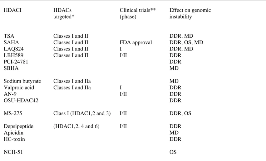
Table 1. HDAC inhibitors and their action on genomic instability.
Strutural class HDACI HDACs Clinical trials** Effect on genomic
targeted* (phase) instability
Hydroxamic acid- TSA Classes I and II DDR, MD
Derived compounds SAHA Classes I and II FDA approval DDR, OS, MD
LAQ824 Classes I and II I DDR, MD
LBH589 Classes I and II I/II DDR
PCI-24781 DDR
SBHA MD
Short-chain fatty acids Sodium butyrate Classes I and IIa MD
Valproic acid Classes I and IIa I DDR
AN-9 I/II DDR
OSU-HDAC42 DDR
Benzamides MS-275 Class I (HDAC1,2 and 3) I/II DDR, OS
Cyclic peptides Depsipeptide (HDAC1,2, 4 and 6) I/II DDR
Apicidin MD
HC-toxin DDR
Thiolate NCH-51 OS
*Data from {Kim, 2006 #456}{Rasheed, 2007 #453}; **Data from {Rasheed, 2008 #452; Rasheed, 2007 #453}. DDR : DNA damage and repair; OS: Oxidative stress; MD: Mitosis defects.