Electron microscopic analysis reveals that replication factor C is sequestered by single-stranded DNA
5
0
0
Texte intégral
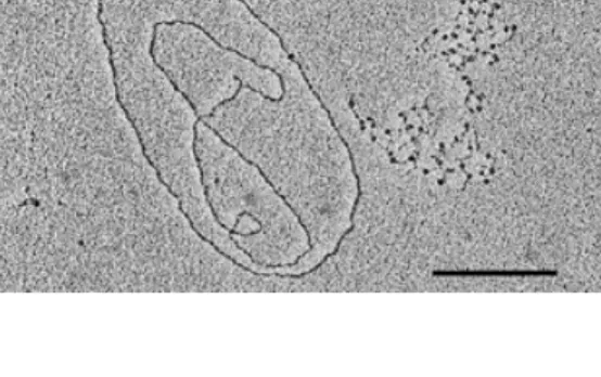
(2) 3434 Nucleic Acids Research, 1999, Vol. 27, No. 17. DNA heteroduplexes Single-stranded and replicative form I (RF I) M13mp18 DNA was obtained from New England BioLabs, Inc. (NEB; Beverly, MA, USA) The M13mp18∆5000 heteroduplex DNA was made as follows: a 2455 bp DNA fragment was amplified from ssM13mp18 in 35 PCR rounds with primers U4591 (5'-TGT TCC GCA AAA TGA TAA TG-3') and R7046 (5'-CAA TGC CTG AGT AAT GTG TA-3') (Microsynth, Balgach, Switzerland) (Fig. 2) at an annealing temperature of 52°C. After separation over a 0.8% agarose gel and extraction with AgarACE (Promega, Catalys AG, Zürich, Switzerland) the amplified fragment was hybridised to ssM13mp18 at a ratio of 1:2 (w/w) in annealing buffer (10 mM Tris–HCl, pH 8, 10 mM EDTA, 100 mM NaCl). The M13mp18∆1000 heteroduplex DNA was made as follows: 1 µg RF I M13mp18 was double digested with 15 U each of AvaII and BglII (Boehringer Mannheim, Rotkreuz, Switzerland) in buffer A [33 mM Tris–acetate, pH 7.9, 10 mM magnesium acetate, 66 mM potassium acetate, 0.5 mM dithiothreitol (DTT)] for 2 h at 37°C. The 6229 bp fragment was gel purified and hybridised to ssM13mp18 as described above. In order to eliminate the salt the buffer was exchanged for 1 mM triethanolamine–HCl (TEA–HCl), pH 7.4, over a Quickspin Sephadex G-50 column (Boehringer Mannheim). Electron microscopy Reactions of 200 ng DNA with 1.5 µg of the DNA-binding region of dRF-Cp140 were incubated for 1 h at 37°C in a total volume of 5 µl in buffer 4 (20 mM Tris–acetate, pH 7.9, 50 mM potassium acetate, 10 mM magnesium acetate, 1 mM DTT; NEB). Five units of EcoRI (Boehringer Mannheim) were added to the sample after 30 min of incubation to linearise M13mp18∆5000. Samples were fixed and spread as described below. Reactions of 200 ng M13mp18∆5000 or 100 ng M13mp18∆1000 with 0.6 mU human heteropentameric RF-C were incubated for 17 min at 37°C in a total volume of 5.5 µl in TEAD buffer (40 mM TEA–HCl, pH 7.5, 10 mM MgCl2, 1 mM DTT) in the presence of 1 mM ATP. Five units of EcoRI (Boehringer Mannheim) or BspHI (NEB) were added after 2 min of incubation to linearise the DNA. Spun-down samples were fixed in 3% (v/v) formaldehyde buffered in 15 mM TEA (pH not adjusted) for 15 min at room temperature. A prior fixation with 0.1% glutaraldehyde did not result in any significant change in the yield of protein–DNA complexes and was therefore deemed superfluous. In order to enhance the unfolding of the single-stranded DNA without disrupting the duplex DNA regions an incubation for 15 min at 37°C was followed by a brief chilling in ice water. Benzyldimethylalkylammonium chloride [BAC; Bayer, AG, Leverkusen, Germany; 0.2% (w/v)] in 100% formamide was diluted 10-fold in water and 0.1 vol was added to the sample before spreading on a hypophase of bi-distilled water. The surface of carbon-coated grids (Stork Veco B.V., The Netherlands) was treated on water droplets containing 33 µg/ml ethidium bromide for 10 min at room temperature, dried and briefly brought into contact with the monolayer at the water–air interface formed with the spreading solution containing the protein–DNA complexes. Subsequently the grids were positively stained in 1 mM uranyl acetate, 1 mM HCl in 90% ethanol, rinsed in undiluted ethanol, and air dried. The grids were rotary shadowed at an angle of 1.5° to the vertical. Figure 1. Sequestration of the DNA-binding region of the large subunit of RFC from D.melanogaster by single-stranded M13mp18. An aliquot of 1.5 µg of the DNA-binding region of dRF-Cp140 was incubated with 100 ng singlestranded and 100 ng replicative form I M13mp18 DNA and spread as described in Materials and Methods. The bar corresponds to 1 kb.. with 900 Hz carbon-platinum as measured on a quartz thin crystal monitor from Balzers. Electron micrographs were taken at magnifications of 13 500× and 18 500× on a Philips EM420 at an accelerating voltage of 100 kV. For DNA length measurements electron micrographs were enlarged 3-fold and dots on DNA filaments were mapped with computerised tracing software (developed by David Sargent, ETH Zürich). RESULTS The DNA-binding region of the large subunit of RF-C from D.melanogaster is sequestered by single-stranded DNA The DNA-binding region of dRF-Cp140 was incubated with equal amounts of single-stranded and RF I M13mp18 DNA. Due to the fused maltose binding protein which increased the molecular weight to ~60 kDa the DNA-binding region of dRFCp140 could be clearly visualised under the electron microscope. Circular RF I M13mp18 molecules were mainly free of dots whereas the single-stranded DNA was slightly collapsed and coated with numerous, distinct dots (Fig. 1). Similar results were obtained with human RF-C (data not shown) suggesting that the DNA-binding region of dRF-Cp140 and human heteropentameric RF-C preferentially bind to singlestranded DNA. The DNA-binding region of the large subunit of RF-C from D.melanogaster preferentially binds to a single-stranded gap We next examined the binding affinity in DNA molecules containing both single- and double-stranded DNA regions. These heteroduplexes also inherently contained 3' and 5' transition points from single- to double-stranded DNA which allowed the simultaneous detection of specific binding to primer/template DNA structures. In earlier experiments RF-C and the DNA-binding region of dRF-Cp140 had been shown to bind to single-stranded DNA regions of down to 30 nt and 420 nt length respectively (data not shown). However, due to the small gap size it was not possible to reliably determine the exact position of the protein–DNA complex. Thus, we designed a DNA heteroduplex with a larger gap of 5000 nt.
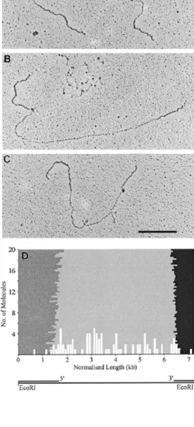
(3) Nucleic Acids Research, 1999, Vol. 27, No. 17 3435. Figure 2. The DNA-binding region of the large subunit of RF-C from D.melanogaster preferentially binds single-stranded DNA. (A) Schematic diagram of DNA heteroduplex M13mp18∆5000. (B) An aliquot of 200 ng circular M13mp18∆5000 was incubated with 1.5 µg of the DNA-binding region of dRF-Cp140 and spread as described in Materials and Methods. (C) As in (B) but in addition the DNA was asymmetrically linearised with EcoRI (see schematic below), subsequently fixed in 3% formaldehyde and spread as described in Materials and Methods. The bar corresponds to 0.5 kb.. through PCR amplification of a 2.4 kb M13mp18 fragment which was subsequently annealed to single-stranded M13mp18 (Fig. 2A) as described in Materials and Methods. In order to orient the 3' and 5' transition points between singleand double-stranded DNA M13mp18∆5000 was asymmetrically linearised with EcoRI in the polylinker region (Fig. 2C, bottom). Upon incubation of this heteroduplex with the DNA-binding region of dRF-Cp140 the resulting dots appeared in the singlestranded region (Fig. 2B) leaving the double-stranded region devoid of dots. When M13mp18∆5000 was linearised with EcoRI during the incubation with the DNA-binding region of dRF-Cp140 and subsequently fixed, the dots remained in the single-stranded region and did not dissociate from the template (Fig. 2C). Due to the partial collapse of the single-stranded DNA a mapping of the DNA-binding region of dRF-Cp140 in the gap was very difficult. Visual inspection of the protein–DNA complexes, however, did not reveal any preferential binding of the DNA-binding region of dRF-Cp140 to the 3' or 5' transition points. Human heteropentameric RF-C binds to single-stranded regions of M13mp18∆ ∆5000 and does not show preference for the 3' or 5' transition points from single- to double-stranded DNA As the DNA-binding region of dRF-Cp140 may not have sufficed for DNA structure recognition we expanded the experiments to examine the binding properties of human heteropentameric RF-C. When incubated with M13mp18∆5000 preferential binding to the single-stranded gap in the heteroduplex DNA molecule was evident (Fig. 3A–C).. Figure 3. Human heteropentameric RF-C randomly binds in the 5 kb singlestranded gap. An aliquot of 200 ng M13mp18∆5000 was linearised with EcoRI and incubated with human RF-C (0.6 mU) as described in Materials and Methods. One or more dots are located within the single-stranded gap (A), at the 5' transition point (B) or at the 3' transition point (C) between single- and doublestranded DNA. In addition, single-stranded DNA can be seen with numerous, distinct dots (B, top). (D) The histogram has been normalised to show the relative position of the dots on their respective DNA molecules. Human heteropentameric RF-C (white columns) preferentially binds to the single-stranded gap (light grey area). The double-stranded DNA with the 5' transition point at R7046 (dark grey area) and the 3' transition point at U4591 (black area) are situated to the left and right of the gap respectively (see schematic below). The bar corresponds to 1 kb.. Occasionally, more than one dot could be detected in the gap (Fig. 3A). In addition complexes could be found located at either the 3' or the 5' transition points (Fig. 3B and C). Occasionally, single-stranded DNA, which was not eliminated from the samples after heteroduplex formation, was observed with.
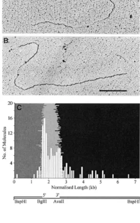
(4) 3436 Nucleic Acids Research, 1999, Vol. 27, No. 17. numerous, distinct dots (Fig. 3B, top), similar to the results obtained with the DNA-binding region of dRF-Cp140 (Fig. 1). When the protein : DNA ratio was increased 3-fold compared to Figure 3A–C the single-stranded region collapsed resulting in a protein aggregate with two or more protruding doublestranded DNA filaments corresponding to the non-collapsed double-stranded DNA (data not shown). In order to detect a preference for either transition point the concentration of human RF-C was reduced. Although the yield of protein–DNA complexes was lower under these conditions, the unfolding of the single-stranded region was sufficient for mapping the position of the resulting dots. The histogram in Figure 3D does not show any preferential binding to either transition point. It is, however, evident that human RF-C predominantly binds to the single-stranded region of the DNA heteroduplex. Human heteropentameric RF-C binds to single-stranded regions of M13mp18∆ ∆1000 and does not show preference for the 3' or 5' transition points from single- to double-stranded DNA As it is conceivable that the binding to single-stranded DNA was due to its higher ratio over double-stranded DNA, we repeated the experiment using a second template with a reduced gap size of 1 kb. Human RF-C was predominantly located in the single-stranded gap. Dots were located at either transition point or randomly distributed throughout the singlestranded gap (Fig. 4A and B). In Figure 4C the histogram reveals a slightly higher frequency of human RF-C at the 5' transition point. DISCUSSION Biochemical studies on RF-C and its subunits with a variety of DNA templates have been performed over the past decade (2,3,10–18). As the observed binding of RF-C and its large subunit to DNA substrates is varied, and it has been hypothesised that RF-C is sequestered by single-stranded DNA in plasmid templates (18,20), we decided to determine whether RF-C preferentially bound single- or double-stranded DNA in large DNA molecules. Furthermore, we tried to clarify whether a preference for either the 3' hydroxyl or the 5' phosphate group was discernible. Incubation of the DNA-binding region of dRF-Cp140 with single- and double-stranded M13mp18 at a 1:1 ratio clearly showed an almost exclusive binding to single-stranded DNA (Fig. 1). In initial experiments this binding invariably led to the collapse of the single-stranded DNA resulting in amorphous aggregates (data not shown). Upon adjustment of the protein concentration these dots were resolved into aggregates with single-stranded DNA protrusions. As protein-free single- and double-stranded DNA molecules were completely unfolded, the collapse was most likely due to the bound protein aggregates. It is interesting to note that despite an absence of primer/ template structures the DNA-binding region of dRF-Cp140 was able to bind single-stranded DNA under the conditions used (Fig. 1). Having shown that the DNA-binding region of dRF-Cp140 binds to single-stranded DNA molecules, we wanted to examine its binding affinity to heteroduplex molecules containing both single- and double-stranded DNA regions. Furthermore, we wanted to see which transition point it would preferentially. Figure 4. Human heteropentameric RF-C randomly binds in the 1 kb singlestranded gap. An aliquot of 100 ng M13mp18∆1000 was linearised with BspHI and incubated with human RF-C (0.6 mU) as described in Materials and Methods. One or more dots are located within the gap (A) or towards the 5' transition point between single- and double-stranded DNA (B). (C) The histogram shows preferential binding of human RF-C (white columns) to the single-stranded gap (light grey area), with the 5' transition point at the BglII (6935) (dark grey area) and the 3' transition point at the AvaII (5914) (black area) restriction sites to the left and right respectively (see schematic below). The bar corresponds to 1 kb.. bind to, when given a choice between a 3' hydroxyl and a 5' phosphate group. In a M13mp18 heteroduplex with a large singlestranded gap of ~5 kb it was virtually impossible to sufficiently unfold the single-stranded region in order to accurately map the position of the protein on the DNA filament. Most dots, however, were located within the single-stranded gap and, curiously, not over either transition point (Fig. 2A and B). This was not entirely unexpected, as it has been shown that the DNA-binding region of RF-Cp140 binds with less specificity and is not required for RF-C binding and subsequent PCNA loading (16,18). Therefore, its function could be to recognise and bind any single-stranded DNA encountered, further actions being determined by the presence or absence of primer/ template junctions, which may be detected in concert with the smaller subunits. Thus, we next decided to examine the affinity of human heteropentameric RF-C for the DNA heteroduplex. In the presence of ATP human RF-C randomly bound along the entire length of the single-stranded gap showing no preference for either transition point (Fig. 3C). To exclude the possibility of.
(5) Nucleic Acids Research, 1999, Vol. 27, No. 17 3437. human RF-C binding to single-stranded DNA due to its higher availability over double-stranded regions we designed a further M13mp18 heteroduplex with a smaller single-stranded gap of ~1 kb. Despite a 6-fold increase in double- over singlestranded DNA human RF-C still preferentially bound in the single-stranded gap (Fig. 4C). The slightly higher frequency of molecules bound at the 5' transition point supports the findings of Allen et al. (17) and Mossi et al. (R.Mossi, R.C.Keller and U.Hübscher, manuscript in preparation) but cannot be considered significant. Whether human RF-C directly binds to and remains immobile in the gap, or binds to a transition point and then tracks along the single-stranded DNA could not be elucidated as human RF-C binding after 0.5, 1, 2 and 10 min revealed no significant shift in position in the single-stranded gap (data not shown). The interesting question arises of what effect PCNA may have on the binding of human RF-C to specific DNA structures. Despite an unavoidable dilution of the sample caused by its dispersion on the water hypophase, the proteinfree BAC spreading technique applied for the work presented here is the most adequate to study this problem since it facilitates the spreading of large single-stranded DNA regions with bound protein complexes. However, an additionally added protein would necessitate gel filtration in order to reduce the background. This would result in a substantial primary dilution of the samples which was sufficiently compensated in the micareplica assay described by Podust et al. (11) by a subsequent concentration of the sample at the air–water interface of the water droplets before transfer to the mica. Furthermore, it would be desirable to identify and confirm the presence of protein complexes bound to DNA by immuno-gold tagging. The effects of replication accessory proteins on the binding affinity of RF-C for specific DNA structures will form the basis for future binding studies when the assay has been suitably improved. In conclusion, we have taken advantage of the electron microscope to examine the influence of primer/template structures on a DNA-binding protein in large single-stranded DNA gaps of 1–5 kb. We have demonstrated that the DNA-binding region of dRF-Cp140 and human heteropentameric RF-C are sequestered by single-stranded DNA in M13mp18 DNA molecules and do not bind to double-stranded DNA. Furthermore, no preferential affinity for 3' hydroxyl or 5' phosphate groups in the absence of PCNA were apparent. RF-C may therefore be strongly attracted by single-stranded DNA and upon PCNA binding specifically seek a template/primer junction to load the clamp in order for processive DNA synthesis by pol δ or ε to commence.. ACKNOWLEDGEMENTS We thank Heide Mayer-Rosa for her technical assistance. This work was supported by the Swiss Cancer League (grant no. KFS 164-9-1995 to R.C.K., U.H. and J.M.S.), by the UBS ‘im Auftrag eines Kunden’ to R.M., by the TMR (grant no. ERBMRXCT 970125 to G.M. and U.H.), by the Swiss National Science Foundation (grant no. 31–52246.97 to J.M.S.) and by the Kanton of Zürich. REFERENCES 1. Fairman,M.P., Prelich,G. and Stillman,B. (1987) Phil. Trans. R. Soc. Lond. B Biol. Sci., 317, 495–505. 2. Tsurimoto,T. and Stillman,B. (1991) J. Biol. Chem., 266, 1950–1960. 3. Tsurimoto,T. and Stillman,B. (1991) J. Biol. Chem., 266, 1961–1968. 4. Tsurimoto,T. and Stillman,B. (1989) Mol. Cell. Biol., 9, 609–619. 5. Mossi,R. and Hübscher,U. (1998) Eur. J. Biochem., 254, 209–216. 6. Prelich,G., Kostura,M., Marshak,D.R., Mathews,M.B. and Stillman,B. (1987) Nature, 326, 471–475. 7. Hindges,R. and Hübscher,U. (1997) J. Biol. Chem., 378, 345–362. 8. Hübscher,U. and Thommes,P. (1992) Trends Biochem. Sci., 17, 55–58. 9. Podust,V.N., Georgaki,A., Strack,B. and Hübscher,U. (1992) Nucleic Acids Res., 20, 4159–4165. 10. Podust,L.M., Podust,V.N., Floth,C. and Hübscher,U. (1994) Nucleic Acids Res., 22, 2970–2975. 11. Podust,L.M., Podust,V.N., Sogo,J.M. and Hübscher,U. (1995) Mol. Cell. Biol., 15, 3072–3081. 12. Cai,J., Uhlmann,F., Gibbs,E., Flores-Rozas,H., Lee,C.G., Phillips,B., Finkelstein,J., Yao,N., O’Donnell,M. and Hurwitz,J. (1996) Proc. Natl Acad. Sci. USA, 93, 12896–12901. 13. Fotedar,R., Mossi,R., Fitzgerald,P., Rousselle,T., Maga,G., Brickner,H., Messier,H., Kasibhatla,S., Hübscher,U. and Fotedar,A. (1996) EMBO J., 15, 4423–4433. 14. Yao,N., Turner,J., Kelman,Z., Stukenberg,P.T., Dean,F., Shechter,D., Pan,Z.Q., Hurwitz,J. and O’Donnell,M. (1996) Genes Cells, 1, 101–113. 15. Cai,J., Gibbs,E., Uhlmann,F., Phillips,B., Yao,N., O’Donnell,M. and Hurwitz,J. (1997) J. Biol. Chem., 272, 18974–18981. 16. Uhlmann,F., Cai,J., Gibbs,E., O’Donnell,M. and Hurwitz,J. (1997) J. Biol. Chem., 272, 10058–10064. 17. Allen,B.L., Uhlmann,F., Gaur,L.K., Mulder,B.A., Posey,K.L., Jones,L.B. and Hardin,S.H. (1998) Nucleic Acids Res., 26, 3877–3882. 18. Podust,V.N., Tiwari,N., Stephan,S. and Fanning,E. (1998) J. Biol. Chem., 273, 31992–31999. 19. Tsurimoto,T. and Stillman,B. (1990) Proc. Natl Acad. Sci. USA, 87, 1023–1027. 20. Tinker,R.L., Kassavetis,G.A. and Geiduschek,E.P. (1994) EMBO J., 13, 5330–5337. 21. Hübscher,U., Mossi,R., Ferrari,E., Stucki,M. and Jonsson,Z.O. (1999) In Cotterill,S. (ed.), Eukaryotic DNA Replication: A Practical Approach. Oxford University Press, Oxford, pp. 119–137..
(6)
Figure
Documents relatifs