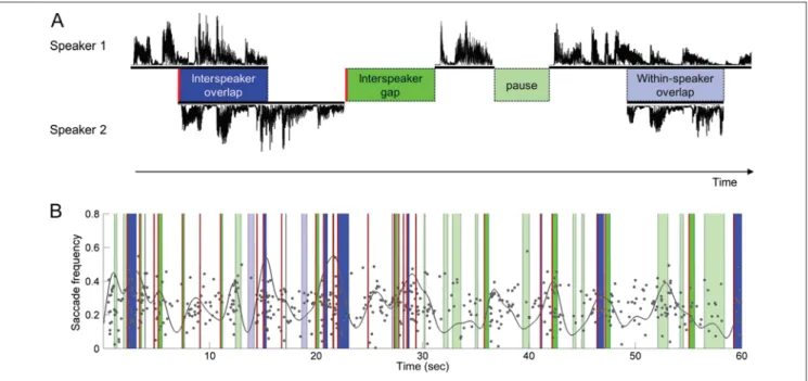
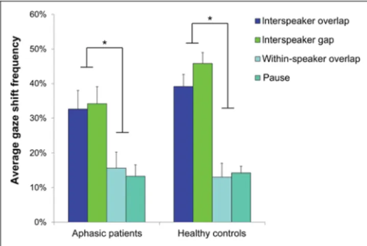
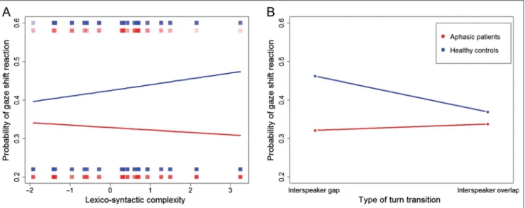
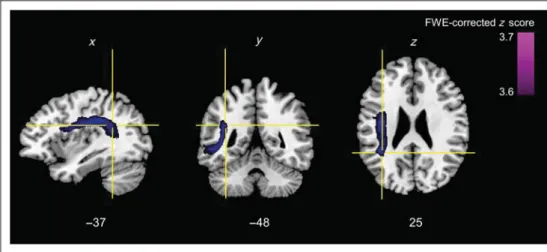
Eye gaze behavior at turn transition: how aphasic patients process speakers’ turns during video observation
Texte intégral
Figure
Documents relatifs
“translation” of Bortkiewicz’s article is very free, summing up in three lines what was developped over three pages in the original, and, inversely, dedicating a lot of print to
The epilogue to the book offers concluding remarks on how abstract concepts such as regionalism and regional identity construction materialised, or not, in works of
Fondé sur une nouvelle fantastique d’Henry James datant de la toute fin du XIX e siècle, l’opéra de Britten est dirigé par Alexander Briger, familier de ce répertoire (il
The right panel depicts a boundary pinching scenario, where around the bound- ary pinching point, a near-boundary three-arm event (with alternating pattern) occurs. We show in the
The paper highlights contributions on the position of Brussels in the European urban network, on urban hierarchy in Belgium, on the dynamics of Belgian city regions, on the
Keywords: transport geography, network analysis, modelling techniques, road transport externalities, transport and development, cruise shipping. Mots-clés: géographie des
O livro estrutura-se em prefácio, prólogo e nove capítulos, ao longo dos quais são desenvolvidos os três eixos temáticos que Bernstein se pro- põe a criticamente analisar:
Other members of the International Conference Committee: Barbara Bokus (Warsaw, Poland), Diana Boxer (Gainesville, USA), Frank Brisard (Antwerp), Winnie Cheng (Hong Kong),
