Short-Term Administration of Rifampin in the Prevention or Eradication
of Infection Due to Foreign Bodies
K. Tshefu,
w.
Zimmerli, and F. A. Waldvogel From the Infectious Disease Division, Department of Medicine, University Hospital, Geneva, SwitzerlandShort-term administration of rifampin was evaluated as a means of preventing or eradi-cating infection due to foreign bodies. Tissue cages were implanted into guinea pigs and subsequently infected with 103colony-forming units ofStaphylococcus aureusWood 46. Infection developed in all tissue cages. Rifampin was administered thereafter in-traperitoneally at a dosage of 7.5 mg/kg every 12 hr for 48 hr, and the tissue-cage fluid was monitored for possible development of infection by quantitative bacteriologic methods for 15 days. In all cases rifampin prevented or eradicated tissue-cage infection if treatment was initiated either 3 hr before or~12hr after inoculation of microorga-nisms but was ineffective if initiated >12 hr after inoculation. In cases of failure of treatment, rifampin-resistant variants could be demonstrated. Rifampin seems to pre-vent or eradicate tissue-cage infection only if given early after bacterial inoculation.
The development of an infection in the vicinity of a foreign body or prosthetic material remains a major problem in orthopedic, cardiac, and general surgery [1-3] as well as in internal medicine [4]. Conceptually, three major groups of pathogenic factors could contribute, either singly or in com-bination, to the development of such infections. Altered virulence and/or surface modifications of the infecting organisms could protect them from host-dependent antibacterial systems or from anti-microbial agents; alternatively, the foreign or prosthetic surface per se could favor, either direct-ly or indirectdirect-ly, the propagation of the offending microorganism; finally, the host's natural defense mechanisms against pyogenic organisms might be altered in the vicinity of a foreign body. Prelim-inary experimental data that favor the latter two mechanisms have recently been presented [5, 6]. Little is known about the microbiologic deter-minants of prosthetic infections; however, the efficacy of preventive, short-term antibiotic
thera-This work was supported in part by grant no. 3.836.79 from the Swiss Research Foundation.
Dr. K. Tshefu is the holder of a fellowship from the World Health Organization, Geneva, Switzerland. Dr. W. Zimmerli is the holder of a fellowship from the Swiss Research Foun-dation, Bern, Switzerland.
The authors thank Mrs. Anneliese Kahr and Mrs. Elisabeth Huggler for technical assistance and Miss Francoise Michaud for editing the manuscript.
Please address requests for reprints to Dr. F. A. Waldvogel, Infectious Disease Division, University Hospital, 1211 Geneva 4, Switzerland.
py [7, 8], in contrast to the inefficacy of antibiotic therapy initiated after infection has developed, suggests a change in bacterial behavior during the establishment of foreign-body infection.
Since it is generally accepted that the con-sequences of infection in the vicinity of foreign material are disastrous, interest is growing in the establishment of guidelines for regimens of pre-ventive antibiotic treatment either during insertion of prosthetic devices or during later septic manip-ulations. Because of the lack of an animal model, few solid data are available about optimal dosage, timing, and duration of such prevention programs and the choice of antibiotics.
In the present study we used an experimental model of foreign-body infection recently de-veloped in our laboratory [6] to address these questions. In particular, we evaluated the efficacy of rifampin, an antibiotic with high antistaphy-lococcal activity and good cell membrane penetra-tion [9-11], in preventing or eradicating foreign-body infection.
Materials and Methods
Experimental model. Sterile polytetrafluor-ethylene (Teflon'") tissue cages constructed as pre-viously described [6], with external and inter-nal diameters of 10 mm and 8 mm, respectively, and perforated by 130 regularly spaced holes were implanted under strictly aseptic conditions into the flanks of guinea pigs weighing 500-600 g.
Efficacy of Short- Term Rifampin Therapy
Animals were used for experimental infections after the incision had healed completely and the metal clips had been removed, i.e., two or more weeks after surgery.
Collection and storage of tissue-cage fluid. Samples of tissue-cage fluid were obtained by percutaneous aspiration, which has been described previously for a similar model [12]. Thereafter, samples either were used directly for bacteriologic quantitation or were stored at -70 C for further determinations.
Microbiologic characteristics of the infecting strain. Aliquots of the same initial culture of Staphylococcus aureus Wood 46, stored at -70 C, were used for all experiments. This strain was shown by the Kirby-Bauer method [13] to be sensitive to all common antibacterial agents tested, including penicillin, methicillin, erythro-mycin, tetracycline, and rifampin [14]. When sub-mitted to a standard scheme for staphylococcal identification, the strain gave positive reactions for catalase production, plasma coagulase reaction, DNase activity, and mannitol fermentation [14]. Before inoculation into tissue cages, S. aureus Wood 46 was preincubated overnight in Mueller-Hinton broth, centrifuged, washed, and resus-pended in 1.0 ml of 0.85070 saline; 0.2 ml of this diluted suspension was injected into each tissue cage, resulting in an inoculum of 1'\)103organisms.
Clumping of microorganisms was kept at a mini-mum under these preincubation conditions and was monitored regularly by microscopic evaluation. Microorganisms isolated from infected tissue cages were checked for their identity with the initial, inoculated strain by the above-mentioned biochemical tests. Appropriate antibiotic suscepti-bility tests also were performed for confirmation of identity between the inoculated and the isolated microorganisms.
Microbiologic techniques. Rifampin assays were performed with a microbiologic method in which Sarcina lutea was used as the indicator strain and DST (diagnostic sensitivity) Oxoid, (Oxoid Ltd., Beckingham, England) with 3 ml of KH2P04(IM)/100 ml was used as the medium
[15]. The assay was linear for concentrations of rifampin of 0.12 JAg/ml-1.0 ug/ml. Pure rifampin (Ciba-Geigy, Basel, Switzerland) was dissolved in N, N-dimethylformamide (Sigma, St. Louis, Mo.) at a concentration of I mg/ml. Further dilutions were performed in PBS (Dulbecco's
phosphate-S475
buffered saline; Gibco-Bio-Cult., Glasgow, Scot-land). This solution was used for the standard curves, which were established for each assay. Identical standard curves were obtained when PBS was partially substituted with 25% serum or 750/0 tissue-cage fluid.
Quantitative cultures of tissue-cage fluid were performed by plating 0.1 ml on Mueller-Hinton agar, after making appropriate dilutions in sterile water, and incubating cultures for 48 hr before performing colony counts. Absence of spon-taneous contamination of tissue-cage fluids was confirmed before starting each experiment by cul-turing undiluted fluid on Mueller-Hinton agar. Finally, in some experiments tissue cages were excised under aseptic conditions and checked for the presence of microorganisms by rolling the cages over Mueller-Hinton agar plates and cultur-ing the plates subsequently in Mueller-Hinton broth.
Results
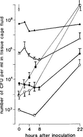
Choice of initial inoculum and of rifampin regi-men. In a first series of experiments, the rate of development of infections was quantitated by inoculating various numbers of S. aureusWood 46 into the tissue cages and by evaluating bacterial multiplication as a function of time. As shown in figure I, infection of the tissue-cage fluid was achieved with initial inocula as small as 102 cfu
and was readily demonstrable bacteriologically by 20 hr. With an inoculum of~103cfu, quantitation
of bacterial counts at 22 hr showed the presence of
1'\)105-108 organisms/ml, most of them identified
as single cocci by microscopic examination. When bacterial counts were measured after the first 24 hr, they fluctuated considerably - probably be-cause of bacterial clumping. Finally, control curves showed that bacterial growth in Mueller-Hinton broth or under in vitro conditions in tissue-cage fluid was more rapid than in the in vivo system. These results considered together sug-gested that 103 cfu of S. aureusWood 46 was an
adequate initial inoculum, that an observation period of 20 hr was sufficient for the demonstra-tion of an infecdemonstra-tion in the absence of antibiotics, and that fluid accumulating in tissue cages did not provide particularly favorable growth conditions for S. aureusWood 46.
ex-"0
10
8 ::::J-
Q)en
ro
o
Q) ::::J f/)10
6 f/)-
c
E ~ Q)a.
::> 10
4 U.o
-
0 I I Io
4
8
22
hours after inoculation
Figure 1. Growth curves of Staphylococcus aureusWood46in tissue cages in vivo and control values of bac-terial growth in either 100070tissue-cage fluid (0; n
=
3) or Mueller-Hinton broth (_;n = 3) in vitro. Indicated inocula of S.aureuswere injected into tissue cages at time zero (arrow). Values are reported as the mean ± SEMof
the cfu at the indicated time intervals after experimental infection with the following initial inoculum: rv102cfu
(0----0; n
=
8), rv104 cfu (e e;n=
4), rvl06cfu (~; n = 4), rv108 (A.----A); n = 2).
perimental tissue-cage infections, an antibiotic regimen had to be chosen that gave concentrations of antibiotic in tissue-cage fluid exceeding the MIC and MBC for S. aureusWood 46. The MIC at 18 hr in Mueller-Hinton broth was 0.005 ug/ml with an initial inoculum of 105 cfu. The MBC,
which was defined as the antibiotic concentration leading to a killing of 99.9070 at 24 hr, was 0.046 JJg/ml; no skip-tube phenomenon was observed. When tests were made in Mueller-Hinton medium supplemented with 25% pooled guinea pig serum, MIC and MBC values were 0.023 JJg/ml and 0.046 JJg/ml, respectively. Several antibiotic regimens were examined for the purpose of obtaining levels of antibiotic in tissue-cage fluid exceeding the MIC and MBC during the entire treatment period. Figure 2 shows that with a regimen of 7.5 mg of rifampin/kg given ip every 12 hr, trough levels of rifampin at 24 hr and 48 hr after initation of ther-apy were 0.115 ± 0.035 and 0.15 ± 0.027 ug/ml, respectively, values exceeding the MIC for S.aureus
Wood 46 by factors of 5 and 6.5, respectively.
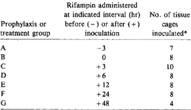
Partial prevention or eradication of infections of tissue-cage fluid with rifampin: emergence of rifampin-resistant mutants. Seven different treat-ment schedules (A-G; table 1) were evaluated; they differed by one parameter only-i.e., the time lag between the inoculation of the micro-organisms and the initiation of therapy. This time lag varied from - 3 hr (for treatment group A; antibiotic regimen started 3 hr before inoculation) to 48 hr (for group G; antibiotic regimen started 48 hr after inoculation). Tissue-cage fluids of groups A-F were aspirated and cultured 24 hr, 48 hr, six days, and 15 days after initiation of therapy.
In group G the establishment of infection was confirmed 24 hr before and at the beginning of
rif-Administration of Rifampin
Figure 2. Trough level of rifampin (J.lg/ml) in tissue-cage fluid 24hr and
48hr after administration of7.5mg of rifampin/kg ip every 12 hr for 48
hr. The solid line represents the MIC and the dotted line, the MBC of the infecting strain, Staphylococcus
au-reus Wood 46.
Time after Initiation of chemoprophylaxis ( hours) n=12 36 48
I
n=23 12 24I
I
: _._._._._.- _._._._._.---MBClof 5.aureus Wood 46 -MIC I oI
o0,2]
Rifampin ( J.l9/ml ) 0.1Efficacy of Short-Term Rifampin Therapy S477
Table 1. Treatment regimens for guinea pigs with foreign-body infections.
NOTE. Tissue-cage fluid was sampled for quantitative cultures at 24 hr, 48 hr, six days, and 15 days after the onset of the antibiotic prophylaxis or therapy.
* Tissue cages were inoculated with 103 cfu of Staphylo-coccus aureus Wood 46. Dosage of rifampin was 7.5 mg/kg ip
every 12 hr for 48 hr.
ampin treatment. Further cultures of tissue-cage fluid were performed at 24 hr, six days, nine days, and 15 days after initiation of therapy. Results of these experiments are described in tables 1 and 2. When rifampin was administered either before, during, or 12 hr after inoculation of S. aureus Wood 46, tissue cages were invariably sterile when tested six days or 15 days after initiation of the 48-hr treatment regimen. Eradication of infection was achieved at the end of therapy, even in cases for which tissue-cage fluids were culture positive at 48 hr, i.e., contained up to 103cfu/ml. If
initia-tion of therapy was delayed beyond 12 hr after inoculation, infection could not be eradicated;
Prophylaxis or treatment group A B C D E F G Rifampin administered
at indicated interval (hr) No. of tissue before ( - ) or after ( + ) cages
inoculation inoculated* -3 7
o
8 +3 10 +6 8 +12 8 +24 8 +48 4despite the fact that tissue-cage fluids were nega-tive six days after initiation of the 48-hr therapy, eight of 11 were positive at 15 days, with bacterial counts ranging from 102 to 108cfu/ml. Itis of
in-terest that after administration of antibiotic infec-tion in the tissue-cage fluid recurred slowly, re-quiring observation periods of up to 15 days for confirmation of all relapses. These results were in striking contrast with those obtained in the ab-sence of the antibiotic, for which bacterial growth could consistently be demonstrated in tissue-cage fluid 12 hr after inoculation.
Because of the delay in recurrence of infection, we investigated whether negative cultures actually reflected the inadequacy of the technique of aspir-ating tissue-cage fluid for collecting bacteria close-ly associated with the foreign body. Tissue cages with sterile fluid cultures were therefore excised 15 days after initiation of effective rifampin treat-ment and cultured according to the technique de-scribed in Materials and Methods; all cages were free of organisms. In one case of ineffective treat-ment - rifampin was started 48 hr after inocula-tion of S.aureus(group G)-tissue-cage fluid was negative at 15 days, whereas the excised tissue cage showed growth with both techniques (table 2 and figure 3).
The time course of the recurrence of infection is depicted in more detail in figure 3 for four tissue cages in animals in which rifampin treatment was started 48 hr after inoculation of bacteria. Bac-terial counts decreased from initial values of 2.5
x 104 - 1 . 6 x 10⁶ 4 8hr after inoculation to <10 cfu
Table 2. Effect of time of initiation of rifampin regimen on protection against or eradication of foreign-body infection.
Treatment group, no. of hr before (-) or after (+) inoculation that rifampin regimen initiated*
A, -3 B,O C, +3 D, +6 E, +12 F, +24 G, +48
No. of tissue-cage fluids positive for infecting organism on indicated day after initiation of rifampin/no. inoculatedf
2 6 15 0/7 1/7 (101-102) 0/7 ND 0/4 0/4 0/4 Ol4t 0110 0110 0110 ND 4/8 (101-103) 4/8 (101-103) 0/8 ND 6/8 (101-103 ) 0/16 0/16 0/8t 5/8 (102-104 ) 11/15 (101-108 ) 0/15 4/7 (108) 4/4 (103-105) ND 0/4 4/4(101-l06)§
* Tissue cages were inoculated with 103cfu ofStaphylococcus aureus Wood 46. Dosage of rifampin was 7.5 mg/kg ip every
12 hr for 48 hr.
tNumbers in parentheses are cfu/ml of tissue-cage fluid. ND = not done. t All excised tissue cages also were culture negative.
CULTURE OF ASPIRATED TISSUE CAGE (TC) FLUID
cfu
Io
/.':;•
o•
24 h GUINEA PIG 1 GUINEA PIG 2 GUINEA PIG 3 GUINEA PIG 4 RIFAMPIN-resistant RIFAMPIN-sensitive ) I I I I I/.
r
cfu
r:107 I.RS
l10
6 6.JPOS
If10
5LJ.
I I•
I I I 104 103 ~RS 102-RR
/ 101 rJNEG
1--0 <10'. I T ' I 15 d 15 days CULTURE OF EXCISED TCFigure 3. Number of cfu ofStaphylococcus aureusWood 46 in tissue-cage fluid in four guinea pigs treated according to treatment regimen G and susceptibility of isolates to rifampin at end of observation period. Time zero indicates initiation of therapy with 7.5 mg of rifampin/kg ip every 12 hr for 48 hr. Tissue cages were excised 15 days after initiation of therapy and cultured in Mueller-Hinton broth (for details see Materials and Methods); all four cages were positive for S. aureusWood 46 (at right).
six days after initiation of the 48-hr therapy regi-men. Bacterial counts in tissue-cage fluid gradual-ly increased at nine days for two of four cages and at 15 days for three of four cages. The fourth tissue-cage fluid was negative throughout the ob-servation period, whereas all excised tissue cages were positive. Finally, one of four cultures ob-tained at the end of the observation period showed two types of S. aureus, one that remained sensitive to 0.023Ilgof rifampin/rnl, the other that was re-sistant to >31 lAg of rifampin/ml. Both types of S. aureus colonies were identical in all other bio-chemical characteristics and antibiotic sensitivities.
Discussion
Despite the abundant use of antibacterial agents for the prevention of postsurgical infections, there is no general consensus as to their indications, choice, timing, and duration of administration
[16]. This lack of adequate guidelines is of even greater concern for those infections that occur after insertion of prosthetic material, since such infections are associated with a particularly poor prognosis. Prosthetic valve endocarditis and osteomyelitis following reconstructive orthopedic surgery usually require the removal of the foreign material for cure[1,17].Ittherefore seems partic-ularly desirable to establish appropriate antibiotic regimens for the prevention or early eradication of these infections, which most often are due to staphylococci.
The paucity of clinical data regarding prosthetic infections and their prevention probably reflects the difficulty in setting up adequate controlled studies, which require unambiguous criteria for the assessment of infection, accurate determina-tions of antibiotic concentradetermina-tions in the neighbor-hood of the prosthesis, and well-defined end points for both duration of antibiotic therapy and
Efficacy of Short-Term Rifampin Therapy
the consecutive observation periods. We believe that the present experimental model, which has previously been used for pharmacokinetic stud-ies [12, 18] and for establishment of successful growth of fastidious microorganisms [19], can be applied to answer some of these questions.
Our results show that administration of rifam-pin at a dosage regimen that achieves bactericidal concentrations in tissue-cage fluid for at least 48 hr prevented the development of foreign-body in-fection, provided that antibacterial treatment was started ~12 hr after inoculation of a rifampin-sensitive strain of S.
aureus.
Ifthe time lag be-tween the inoculation of the pathogen and the initiation of therapy was>12 hr, however, infec-tion invariably developed despite treatment with rifampin for 48 hr, and occasionally rifampin-resistant variants emerged. Finally, whereas tis-sue-cage infections developed in <20 hr in the absence of rifampin administration, more than six days were required for them to be detected micro-biologically after unsuccessful rifampin therapy. This observation could be explained by a pro-longed postantibiotic effect, as described for other antibacterial agents [20]. Alternatively, persistent bacteria may have remained adherent to the for-eign body and protected from the antibiotic effect, contaminating the tissue-cage fluid at a later stage. Finally, some rifampin-resistant mutants of S.au-reus
Wood 46 produce less catalase and are less pathogenic than the wild strain in mice [21]. In our experience, however, both the resistant and the sensitive variants were equally susceptible to the bactericidal action of peritoneal granulocytes. Present work in our laboratory is aimed at the further evaluation of these possibilities.The experimental system presented in this com-munication requires two comments. First, despite others' claims to having encountered difficulty in performing bactericidal assays with S.
aureus
Wood 46 [22], we did not encounter any such problems; the MIC and MBC were in the range of those given by Mandell for the same organism [10] and by others for various strains of S.
aureus
[9,23]. Second, we occasionally observed delayed in-fection of tissue-cage fluid or inin-fection limited to the tissue cage after ineffective rifampin treat-ment. These observations could suggest that in the case of "effective" rifampin treatment cultures of tissue-cage fluid were falsely negative because either the observation period was too short or
8479
microorganisms persisted on the Teflon'" surface. These possibilities were ruled out by control ex-periments involving the excision of tissue cages with culture-negative fluid and the demonstration of the sterility of the cages by adequate culture techniques.
The incomplete protection or eradication of infection demonstrated with short-term adminis-tration of rifampin in our experimental model is in keeping with similar data obtained with experi-mental endocarditis [24], osteomyelitis [25], and peritonitis [26]. In all four conditions, high num-bers of bacteria were exposed to rifampin, either because of a prolonged delay between inoculation and initiation of antibacterial therapy (our study and [24, 25]) or because of a high initial inoculum [26].Itis therefore not surprising that in all four experimental conditions, emergence of rifampin-resistant variants was demonstrated. This observa-tion obtained in different experimental models might well set a limit on the use of rifampin as a single therapeutic agent - but not necessarily as a prophylactic drug - since initial inocula probably are low in clinical postsurgical infections.
References
1. Waldvogel FA, Vasey H. Osteomyelitis: the past decade. N Engl J Med 1980;303:360-70
2. Kloster FE. Complications of artificial heart valves. JAMA 1979;241:2201-3
3. Georgiade NG, King EH, Harris WA, Tenery JH, Schlech BA. Effect of three proteinaceous foreign materials on infected and subinfected wound models. Surgery 1975; 77:569-76
4. Waldvogel FA. Infections et protheses. In: Bastin R, ed. Les infections bacteriennes d'actualite. Masson, Paris: 1977:103-20
5. Peters G, Locci R, Pu1verer G. Microbial colonization of prosthetic devices. II. Scanning electron microscopy of naturally infected intravenous catheters. Zentralbl Bak-teriol [B) 1981;173:293-9
6. Zimmerli W, Waldvogel FA, Vaudaux P, Nydegger VE. Pathogenesis of foreign body infection: description and characteristics of an animal model. J Infect Dis 1982; 146:487-97
7. Pollard JP, Hughes SPF, Scott JE, Evans MJ, Benson MKD. Antibiotic prophylaxis in total hip replacement. Br Med J 1979;1:707-9
8. Kaiser AB, Clayson KR, Mulherin JL Jr, Roach AC, Allen TR, Edwards WH, Dale WA. Antibiotic prophylaxis in vascular surgery. Ann Surg 1978;188:283-9
9. Sabath LD, Garner C, Wilcox C, Finland M. Susceptibility ofStaphylococcus aureusandStaphylococcus epidermi-dis to 65 antibiotics. Antimicrob Agents Chemother 1976;9:962-9
10. Mandell GL, Vest TK. Killing of intraleukocytic
Staphy-lococcus aureusby rifampin: in vitro and in vivo studies. J Infect Dis 1972;125:486-90
11. Solberg CO, HelIum KB. Protection of phagocytosed bac-teria against antimicrobial agents. Scand J Infect Dis [Suppl] 1978;14:246-50
12. Chisholm GD. The tissue cage model in the distribution of antibacterial agents. Scand J Infect Dis [Suppl] 1978; 14:118-24
13. Bauer AW, Kirby WMM, Sherris JC, Turck M. Antibiotic susceptibility testing by a standardized disk method. Am J Clin Pathol 1966;45:493-6
14. Lennette EH, Spaulding EH, Truant JP, eds. Manual of clinical microbiology. 2nd ed. Washington DC: Ameri-can Society for Microbiology, 1974
15. Williams JD, Leung T. Rifamycins. In: Reeves DS, Phillips I, Williams JD, Wise R, eds. Laboratory methods in antimicrobial chemotherapy. Edinburgh: Churchill Livingstone, 1978:239-43
16. Shapiro M, Townsend TR, Rosner B, Kass EH. Use of antimicrobial drugs in general hospitals. Patterns of prophylaxis N Engl J Med 1979;301:351-5
17. Karchmer AW, Stinson EB. The role of surgery in infec-tive endocarditis. In: Remington JS, Swartz MN, eds. Current clinical topics in infectious diseases. New York; McGraw-Hill, 1980:124-57
18. Kaye D, Parsons IN, Carrizosa J, Kobasa WD. Treatment of experimental Staphylococcus aureus abscesses: com-parison of cefazolin, cephalothin, cefoxitin, and cefa-mandole. Antimicrob Agents Chemother 1979;15:200-3
19. Tight RR, Perkins RL. Treponema pallidum infection in subcutaneous polyethylene chambers in rabbits. Infect Immun 1976;13:1606-12
20. Bundtzen RW, Gerber AU, Cohn DL, Craig WA. Post-antibiotic suppression of bacterial growth. Rev Infect Dis 1981;3:28-37
21. Mandell GL. Catalase, superoxide dismutase, and viru-lence of Staphylococcus aureus. In vitro and in vivo studies with emphasis on staphylococcal-leukocyte inter-action. J Clin Invest 1975;55:561-6
22. Zinner SH, Husson M, Klastersky J. Effect of mixing on rifampin bactericidal activity against staphylococci. Antimicrob Agents Chemother 1981 ;20:267-9 23. Tuazon CU, Lin MYC, Sheagren IN. In vitro activity of
rifampin alone and in combination with nafcillin and vancomycin against pathogenic strains of
Staphylo-coccus aureus.Antimicrob Agents Chemother 1978;13: 759-61
24. Sande MA, Johnson ML. Antimicrobial therapy of experi-mental endocarditis caused by Staphylococcus aureus. J Infect Dis 1975;131:367-75
25. Norden CW, Experimental osteomyelitis. IV. Therapeutic trials with rifampin alone and in combination with gentamicin, sisomicin, and cephalothin. J Infect Dis 1975;132:493-9
26. Mandell GL, Moorman DR. Treatment of experimental staphylococcal infections: effect of rifampin alone and in combination on development of rifampin resistance. Antimicrob Agents Chemother 1980;17:658-62