40. Chen CN, Hsieh FJ, Cheng YM et al.. The significance of placenta growth factor in angiogenesis and clinical outcome of human gastric cancer. Cancer Lett 2004; 213: 73–82.
41. Jain RK, Duda DG, Willett CG et al.. Biomarkers of response and resistance to antiangiogenic therapy. Nat Rev Clin Oncol 2009; 6: 327–338.
42. Okugawa Y, Miki C, Toiyama Y et al.. Serum soluble VCAM-1 as a valuable prognostic marker in colorectal carcinoma [abstract]. Presented at: American
Society of Clinical Oncology Gastrointestinal Cancers Symposium, San Francisco, CA, 2008.
43. Pasieka Z, Kuzdak K, Czyz W et al.. Soluble intracellular adhesion molecules (sICAM-1, sVCAM-1) in peripheral blood of patients with thyroid cancer. Neoplasma 2004; 51: 34–37.
44. Silva HC, Garcao F, Coutinho EC et al.. Soluble VCAM-1 and E-selectin in breast cancer: relationship with staging and with the detection of circulating cancer cells. Neoplasma 2006; 53: 538–543.
Annals of Oncology 24: 718–725, 2013 doi:10.1093/annonc/mds519 Published online 8 November 2012
Neoadjuvant chemoradiotherapy with or without
panitumumab in patients with wild-type
KRAS, locally
advanced rectal cancer (LARC): a randomized,
multicenter, phase II trial SAKK 41/07
D. Helbling
1, G. Bodoky
2, O. Gautschi
3, H. Sun
4, F. Bosman
5, B. Gloor
6, R. Burkhard
7,
R. Winterhalder
8, A. Madlung
9, D. Rauch
10, P. Saletti
11, L. Widmer
7, M. Borner
12, D. Baertschi
4,
P. Yan
5, J. Benhattar
5, E. O. Leibundgut
13, S. Bougel
5& D. Koeberle
141
Department of Medical Oncology, Gastrointestinal Tumorcenter Zurich, Zurich, Switzerland;2
Department of Oncology, St. Laszlo Hospital, Budapest, Hungary;
3
Department of Medical Oncology, Luzerner Kantonsspital, Lucerne;4
Swiss Group for Clinical Cancer Research, Coordinating Center, Bern;5
Department of Pathology, Lausanne;6
Department of Visceral Surgery and Medicine, University Hospital Bern, Bern;7
Onkozentrum Hirslanden, Hirslanden Hospital Zurich, Zurich;8
Department of Medicine, Medical Oncology, Kantonsspital, Luzern;9
Department of Radiation Oncology with Division of Medical Radiation Physics, University Hospital, Bern;
10
Departement of Medicine, Medical Oncology, Regionalspital Thun, Thun;11
Ospedale San Giovanni, Oncology Institute of Southern Switzerland, Bellinzona;
12
Department of Medicine, Medical Oncology, Spitalzentrum, Biel;13
Central Haematology Laboratory, University Hospital Bern, Bern;14
Division of Oncology, Kantonsspital St. Gallen, St. Gallen, Switzerland
Received 15 July 2012; revised 19 August 2012; accepted 30 August 2012
Background:
We conducted a randomized, phase II, multicenter study to evaluate the anti-epidermal growth factor receptor (EGFR) mAb panitumumab (P) in combination with chemoradiotherapy (CRT) with standard-dose capecitabine as neoadjuvant treatment for wild-type KRAS locally advanced rectal cancer (LARC).Patients and methods:
Patients with wild-type KRAS, T3-4 and/or N+ LARC were randomly assigned to receive CRT with or without P (6 mg/kg). The primary end-point was pathological near-complete or complete tumor response ( pNC/CR), defined as grade 3 (pNCR) or 4 (pCR) histological regression by Dworak classification (DC).Results:
Forty of 68 patients were randomly assigned to P + CRT and 28 to CRT. pNC/CR was achieved in 21 patients (53%) treated with P + CRT [95% confidence interval (CI) 36%–69%] versus 9 patients (32%) treated with CRT alone (95% CI: 16%–52%). pCR was achieved in 4 (10%) and 5 (18%) patients, and pNCR in 17 (43%) and 4 (14%) patients. In immunohistochemical analysis, most DC 3 cells were not apoptotic. The most common grade≥3 toxic effects in the P + CRT/CRT arm were diarrhea (10%/6%) and anastomotic leakage (15%/4%).Conclusions:
The addition of panitumumab to neoadjuvant CRT in patients with KRAS wild-type LARC resulted in a high pNC/CR rate, mostly grade 3 DC. The results of both treatment arms exceeded prespecified thresholds. The addition of panitumumab increased toxicity.Key words: capecitabine, chemoradiotherapy, panitumumab, radiotherapy, rectal cancer, wild-type KRAS
*Correspondence to: Dr D. Helbling, Department of Medical Oncology, Gastrointestinal Tumorcenter Zurich, Seestrasse 259, Zurich 8038, Switzerland. Tel: +41-43-344-33-33; E-mail: daniel.helbling@gitz.ch
© The Author 2012. Published by Oxford University Press on behalf of the European Society for Medical Oncology. All rights reserved. For permissions, please email: journals.permissions@oup.com.
introduction
In patients with locally advanced rectal cancer (LARC),
complete surgical resection is a prerequisite for cure.
Neoadjuvant chemoradiotherapy (CRT) is frequently used to
downstage LARC tumors and to enable surgery to be more
effective and provide locoregional control [
1
–
3
]. The standard
neoadjuvant regimen is 5-
fluorouracil (5-FU) and external
radiotherapy (RT). Neoadjuvant CRT with capecitabine has
been shown to be tolerated with comparable activity to 5-FU
regimens [
4
,
5
].
The epidermal growth factor receptor (EGFR)-targeted
monoclonal antibodies (mAbs) cetuximab and panitumumab
have shown efficacy as monotherapy in phase III studies in
patients with chemotherapy-refractory metastatic colorectal
cancer [
6
–
8
] and also improved outcomes when added to
standard chemotherapy in the
first-line and subsequent lines of
therapy [
7
,
9
,
10
]. Preliminary data suggest that EGFR-targeted
agents in combination with RT may be synergistic, as RT
increases EGFR expression within tumor cells, while EGFR
blockade sensitizes the cells to the effects of RT [
11
,
12
]. In the
setting of locally advanced head and neck cancer, addition of
cetuximab to RT enhanced locoregional control and survival
[
13
,
14
].
Intensive research investigating the response according to
molecular tumor markers in order to allow better patient
selection, has shown that KRAS-activating mutations predict
resistance to cetuximab and panitumumab [
15
,
16
]. Mutations
in KRAS codons 12 or 13 occur in
∼35%–45% of colorectal
cancers and lead to constitutive signaling via impaired ability
of GTPase-activating proteins to hydrolyze KRAS-bound
guanosine triphosphate (GTP) [
17
]. Other molecular markers
that have been linked to response/nonresponse to
EGFR-targeted mAbs in the colorectal cancer setting include
mutations of BRAF [
18
,
19
] or PIK3CA [
19
], EGFR gene copy
number [
20
] and levels of EGFR ligands such as amphiregulin
(AREG) and epiregulin (EREG) [
21
].
We conducted a randomized, phase II, multicenter study to
evaluate the ef
ficacy and safety of panitumumab in
combination with capecitabine and external beam RT as a
neoadjuvant regimen for patients with advanced wild-type
KRAS LARC.
patients and methods
Patients with centrally tested wild-type KRAS LARC were recruited at centers in Switzerland and from one center in Budapest, Hungary. The study was conducted according to the Declaration of Helsinki. The protocol was approved by the independent ethics committee at participating centers. Patients provided written informed consent before randomization. Separate informed consent was obtained for KRAS testing, biobanking and translational research.
inclusion criteria
The eligibility criteria included: previously untreated histologically confirmed locally advanced adenocarcinoma of the rectum with or without nodal involvement and requiring surgery; stage cT3/4 and/or cN1/2 cM0 and wild-type KRAS gene status, as assessed by a central pathology; World Health
Organization (WHO) performance status 0–1; age ≥18 years and adequate organ function. Patients with distant metastases were excluded from the study.
pretreatment evaluation
Baseline evaluation included computed tomography of the abdomen and thorax, and magnetic resonance imaging of the pelvis.
KRAS mutation status and EGFR gene copy number analyses were conducted at a central laboratory (Institute of Pathology, University of Lausanne, CHUV, Switzerland), on formalin-fixed paraffin-embedded tissues (5μm paraffin sections, microdissected to contain >70% tumor tissues) prepared from diagnostic tumor biopsy samples collected before the trial. The KRAS mutation status was analyzed by pyrosequencing of a PCR-amplified exon 2 sequence containing codons 12 and 13. EGFR copy number analysis was carried out by quantitative PCR of tumor DNA. Serum samples were collected for KRAS mutational screening and biomarker testing and were assessed at the laboratory of Molecular Diagnostics, University Hospital Bern.
study conduct
After screening, the eligible patients were randomly assigned to receive either CRT alone or panitumumab + CRT (P + CRT) as neoadjuvant treatment. Randomization was stratified by: T-stage (T3 versus T4); tumor localization (<10 cm versus≥10 cm); institution and EGFR gene copy number (<2.9 versus≥2.9 copies per nucleus).
treatment
Panitumumab (6 mg/kg every 2 weeks for 8 weeks) was administered i.v. over∼60 min.
Capecitabine (825 mg/m2) was taken twice daily orally throughout RT.
RT was applied with a total dose of 45 Gy in 25 fractions of 1.8 Gy over 5 weeks, starting from 7 days after thefirst panitumumab administration (P + CRT arm) (supplementary Figure S1, available at Annals of Oncology online). Patients were irradiated in the prone position using a linear accelerator (3D-conformal or, preferably intensity-modulated, RT).
surgery and histopathology
Surgery was planned 6 weeks (±1 week) after completion of CRT. Total mesorectal excision with sphincter preservation was to be carried out whenever feasible, according to the standardized techniques, using a laparoscopic or open approach. Afterfixation and processing, resected specimens were microscopically examined for the presence of tumor cells, fully evaluated and staged according to the American Joint Committee on Cancer TNM system [22]. Tumor regression was graded by the local pathologists according to the regression grading scale proposed by Dworak et al. [grade 0 = none; 1 = minimal; 2 = moderate ( predominantlyfibrotic changes with few tumor cells); 3 = good (very few tumor cells); 4 = complete (no tumor cells)] [23]. A central pathology review (by F. Bosman, P. Yun) was carried out for samples classified as Dworak grade 2 or 3, as this cut-off was decisive regarding the primary end-point. Radical resection (R0) was defined as microscopic and macroscopic tumor-free resection with clear circumferential resection margins (invasion front ≥2 mm from the lateral resection margin).
dose modi
fications
Chemotherapy and RT were interrupted if grade≥3 toxicity developed (capecitabine and panitumumab were also interrupted in case of diarrhea and mucositis grade 2). Study treatment was restarted when toxicity had resolved to grade≤1. Dose reductions were required after grade ≥3
toxicity. If the treatment was delayed for≥2 weeks, the patient was withdrawn from the study treatment.
adjuvant chemotherapy and follow-up evaluations
It was recommended that patients received adjuvant therapy for 4–6 months according to local practice. All patients with R0 resection were followed (every 3 months during thefirst year and every 6 months thereafter) until disease progression or for 3 years after surgery.concomitant therapy
All patients randomly assigned to panitumumab received a pre-emptive supportive treatment regimen, consisting of a skin moisturizer, sunscreen and doxycycline 100 mg orally twice daily.
study end-points
The primary end-point of the study was pathological near-complete or complete tumor response ( pNC/CR), prospectively defined as grade 3 (near-complete) or 4 (complete) histological regression using the Dworak classification (DC) [23].
The early secondary end-points were R0 or R1 resection, sphincter preservation, downstaging of primary tumor and/or lymph nodes (comparison between cT/N and ypT/N) and postoperative complications (within 8 weeks after surgery) defined as insufficiency of anastomosis; fistula; severe local infection requiring antibiotics; bladder or erectile dysfunction or need for additional interventions/operation. The late secondary end-points (in the R0 population) were time (from randomization) to local relapse, time to distant relapse and disease-free survival.
translational studies
The following markers were evaluated in baseline serum (for full methodological details, see supplementary information, available at Annals of Oncology online): circulating tumor KRAS mutations by real-time quantitative PCR and concentrations of EGFR and the related growth factors AREG, betacellulin, epidermal growth factor (EGF), EGFR, EREG, fibroblast growth factor-basic, heparin-binding EGF-like growth factor (HB-EGF), platelet-derived growth factorβ polypeptide, placental growth factor, tenascin C, transforming growth factorα and vascular endothelial growth factor by using antibody suspension bead arrays and suspension array system.
Pretreatment tumor specimens were examined for mutations in KRAS exon 3 (codons 59 and 61), KRAS exon 4 (codons 117 and 146), BRAF exon 15 (codon 600), NRAS exon 2 (codons 12 and 13), PIK3CA exon 9 (codons 542, 545 and 546) and PIK3CA exon 20 (codons 1043 and 1047) (by pyrosequencing after PCR amplification).
To assess the viability of the remaining tumor cells following neoadjuvant treatment, the available post-surgery tumor specimens were tested for the Ki-67 proliferation marker MIB-1 and the early apoptosis marker M30 (by immunohistochemistry). Quantitative methylation analysis of RASSF2A and EGFR promoter regions was also carried out (by pyrosequencing after bisulfite modification of genomic DNA and PCR amplification).
statistical considerations
The required sample size was calculated separately for each treatment arm using the Fleming’s single-stage design [24] with 80% power and 5% significance level. For the P + CRT arm, 40 patients were required to test the null hypothesis of the pNC/CR rate≤10% versus the alternative of ≥25%. If there were ≤7 patients with pNC/CR, the treatment would not be
recommended for further investigation. Twenty-seven patients would be needed in the CRT arm to test the null hypothesis of the pNC/CR rate ≤5% versus the alternative of ≥20%. Analyses of the primary end-point, as well as pathological tumor response by the Dworak regression grade, were based on the intention-to-treat analysis set (all randomly assigned patients). The remaining early secondary end-points were based on the resection population (all patients who underwent surgical resection). Statistical comparisons between the treatment arms were not planned.
results
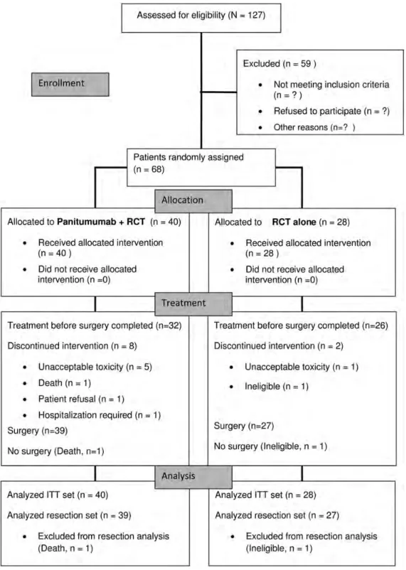
Between March 2009 and May 2010, a total of 68 patients (53
men and 15 women, aged 31
–80 years) at 19 centers were
randomly assigned, 40 to P + CRT and 28 to CRT (Figure
1
).
Most patients (n = 65) had WHO performance status 0. The
most common T-stage was cT3. The majority of patients had
tumor localization <10 cm from the anal margin, lymph node
involvement and <2.9 EGFR gene copies (Table
1
).
treatment exposure
The median total CRT doses were similar in the P + CRT and
the CRT arms: RT 45 and 45 Gy; capecitabine 98% and 97% of
target dose; and panitumumab 88% of target dose. There were
four panitumumab dose delays (for diarrhea, and/or hand-foot
syndrome, n = 3; skin toxicity, n = 1) and 12 reduced/omitted
doses (mainly due to diarrhea or proctitis/ulceration, n = 6 or
skin toxicity/dry skin/erythema multiforme, n = 4).
surgery and outcomes
One patient (P + CRT) died of unknown causes and another
patient (CRT) was withdrawn from the study because of
incorrect diagnosis. A further eight patients stopped treatment
because of adverse events (P + CRT, n = 5; CRT, n = 1) or other
reasons, including patient
’s wish or renal failure requiring
hospitalization (P + CRT, one patient each). A total of 66
patients underwent surgery (P + CRT, n = 39; CRT, n = 27).
A median of 15 lymph nodes per patient was harvested in both
the treatment arms. Tumor regression grade is shown in
Table
2
and achievement of the primary and early secondary
end-points is summarized in Table
3
.
Complete regression by the Dworak criteria (grade 4) was
achieved in four patients (10%) and near-complete regression
(grade 3) in 17 patients (43%) in the P + CRT arm. In the CRT
arm,
five patients (18%) and four patients (14%), respectively,
achieved complete and near-complete regression (Table
2
).
A central pathology review for grade 2 or 3 regression was
completed for all but two patients (both in the P + CRT arm).
The primary end-point, pNC/CR, was achieved in 21 patients
(53%; 95% CI 36%–69%) in the P + CRT arm and 9 patients
(32%; 95% CI 16%–52%) in the CRT arm.
R0 resection was achieved in 33 patients (85%) in the
P + CRT arm and in 25 patients (93%) in the CRT arm.
Downstaging of the primary tumor and/or lymph nodes was
achieved in 34 patients (87%) and 23 patients (85%), with
sphincter preservation being achieved in 27 (69%) and 19
(70%) patients.
postoperative complications
Postoperative complications are listed in Table
4
. The most
common complications were anastomosis leakage, local
infection and requirement for further intervention/operation
[n = 7 patients each (18%) in the P + CRT arm versus 2, 4 and
4 patients (7%, 15% and 15%) in the CRT arm].
follow-up and deaths
The median follow-up time for 68 assessable patients was 9.1
months. So far only four patients died (two in each arm). Both
the patients in the P + CRT arm died perioperatively, whereas
the patients in the CRT arm died after tumor recurrences
during the follow-up phase. Both of the latter patients had R0
resection, but the circumferential resection margin was <2 mm
in one patient.
translational studies
The median EGFR gene copy number was 1.0. The EGFR copy
number did not associate with pNC/CR (data not shown).
Mutational analysis in tissue sections revealed a total of 23
Figure 1. Consort diagram.mutations in 18 tumors (26%), including 12 mutations in
KRAS exons 3 and 4, 7 mutations in PIK3CA, 2 mutations in
NRAS and 1 mutation in BRAF. Thirteen tumors harbored a
single mutation and
five tumors had two concomitant
mutations in PIK3CA and KRAS (exons 3 and 4) (n = 4) and
NRAS (n = 1). There was no signi
ficant association between
additional mutations and with pNC/CR (data not shown).
The RASSF2A promoter was hypermethylated in 75% of the
rectal tumors, but no signi
ficant association was found with
pNC/CR (data not shown). In contrast, the EFGR promoter
was hypomethylated in all tumors.
The remaining tumor cells of the available specimens with
regression Dworak grade 3 showed no difference in MIB-1 and
M30 expression compared with those with regression Dworak
grades 0–2 for both the treatment arms (supplementary
Table S1, available at Annals of Oncology online).
No mutation of KRAS exon 2 (codon 12 and 13) was
detected in serum. None of the 12 serum biomarkers
analyzed were significantly associated with pNC/CR
(supplementary Tables S2 and S3, available at Annals of
Oncology online).
Table 4. Number and percentage of patients with postoperative complications within 8 weeks after surgery
Complication Panitumumab + chemoradiotherapy (CRT; n = 39) CRT (n = 27) Total (n = 66) Anastomotic leakage 7 (18%) 2 (7%) 9 (14%) Fistula 2 (5%) 2 (7%) 4 (6%) Local infection 7 (18%) 4 (15%) 11 (17%) Bladder dysfunction 3 (8%) 2 (7%) 5 (8%) Erectile dysfunction 3 (8%) 3 (11%) 6 (9%) Interventions/operation 7 (18%) 4 (15%) 11 (17%) Other 6 (15%) 8 (30%) 14 (21%)
Table 2. Number and percentage of patients achieving each Dworak regression grade (intent-to-treat analysis)
Dworak regression grade Panitumumab + chemoradiotherapy (CRT, n = 40) CRT (n = 28) All patients (n = 68) None (grade 0) 0 (0%) 1 (4%) 4 (6%) Minimal (grade 1) 9 (23%) 11 (40%) 20 (29%) Moderate (grade 2) 7 (18%) 6 (21%) 13 (19%) Good (grade 3) 17 (43%) 4 (14%) 21 (31%) Total (grade 4) 4 (10%) 5 (18%) 9 (13%)
Table 1. Baseline characteristics of patients Panitumumab + chemoradiotherapy (CRT) (n = 40) CRT (n = 28) All patients (n = 68) Age (median/range) 62/31–80 60/35–77 61/31–80 Sex Male 33 (82%) 20 (71%) 53 (78%) Female 7 (18%) 8 (29%) 15 (22%)
World Health Organization (WHO) performance status
0 38 (95%) 27 (96%) 65 (96%) 1 2 (5%) 1 (4%) 3 (4%) N stage cN0 8 (20%) 7 (25%) 15 (22%) cN1 24 (60%) 13 (46%) 37 (54%) cN2 8 (20%) 8 (29%) 16 (24%) T stageb cT2 4 (10%) 2 (7%) 6 (9%) cT3 34 (85%) 26 (93%) 60 (88%) cT4 2 (5%) 0 (0%) 2 (3%) Histological grade G1 0 (0%) 2 (7%) 2 (3%) G2 35 (88%) 23 (82%) 58 (85%) G3 3 (8%) 3 (11%) 6 (9%) Gx 2 (5%) 0 (0%) 2 (3%)
Tumor localizationa,b(cm)
<10 32 (80%) 22 (79%) 54 (79%)
≥10 8 (20%) 6 (21%) 14 (21%)
EGFR gene copy numberb
<2.9 38 (95%) 26 (93%) 64 (94%)
≥2.9 2 (5%) 2 (7%) 4 (6%)
Categorical parameters shown as frequency and percentage.
aIn centimeters from the anal verge. bStratification factor.
Table 3. Number and proportion of patients achieving primary and key secondary end-points
No. of patients
Percenta 95% CI
(exact) Pathological near-complete or complete tumor response (pNC/CR)
Panitumumab + chemoradiotherapy (CRT) 21/40 53 (36, 69)% CRT 09/28 32 (16, 52)% Total 30/68 44 (32, 57)% R0 resectionb Panitumumab + CRT 33/39 85 (70, 94)% CRT 25/27 93 (76, 99)% Total 58/66 89 (78, 95)% Sphincter preservation Panitumumab + CRT 27/39 69 (52, 83)% CRT 19/27 70 (50, 86)% Total 46/66 70 (57, 80)%
Downstaging of primary tumor or lymph nodes
Panitumumab + CRT 34/39 87 (73, 96)%
CRT 23/27 85 (66, 96)%
Total 57/66 86 (76, 94)%
Downstaging of primary tumor and lymph nodes
Panitumumab + CRT 16/39 41 (26, 58)%
CRT 08/27 30 (14, 50)%
Total 24/66 36 (25, 49)%
CI, confidence intervals.
a
Based on the intent-to-treat population for pCR and the operated population for the remaining end-points.
b
Only one patient (from arm A) had R1 resection.
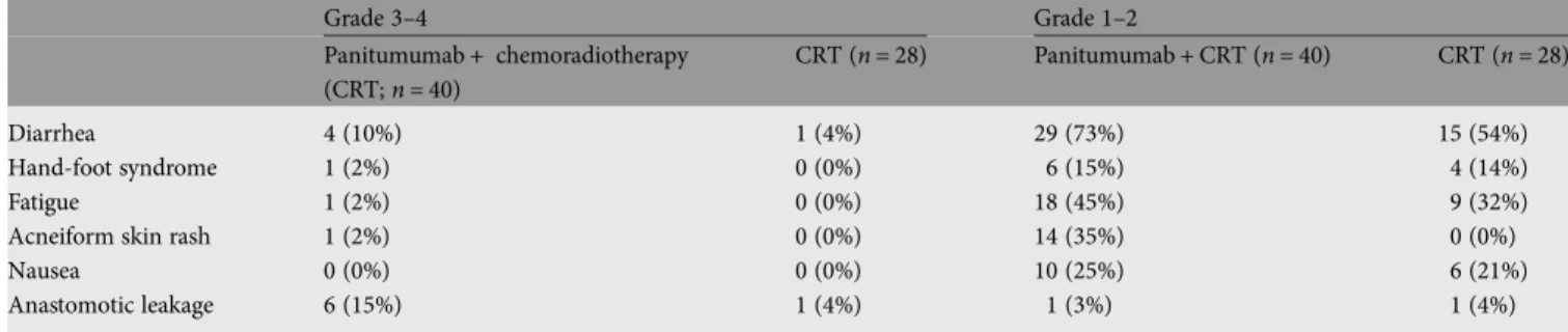
tolerability
All 68 patients were assessable for safety. The most common
CTC grade
≥3 toxic effects in the P + CRT and the CRT arm
were: diarrhea (10% and 4% of patients), hand-foot syndrome
(2% and 0%), fatigue (2% and 0%), acneiform skin rash (2%
and 0%) and leakage (15% and 4%) (Table
5
; grade 1
–2 toxic
effects also shown).
Adverse events led to treatment discontinuation in
five
patients in the P + CRT arm (diarrhea, enterocolitis, ulceration/
proctitis, dry skin, erythema multiforme/pruritus /itching) and
one patient in the CRT arm (cardiac arrest). Seventeen patients
had 18 serious adverse events [12 patients (30%) in the
P + CRT arm,
five patients (18%) in the CRT arm]. Of these
events, three in the P + CRT arm and two in the CRT arm
were classi
fied as clearly related to treatment. There were two
deaths during the safety monitoring period, both in the
P + CRT arm: from acute cardiac failure and sepsis resulting
from anastomatic leakage, respectively.
discussion
In patients with LARC, the rates of pCR have generally been
low (<10%) with neoadjuvant regimens consisting of CRT and
cetuximab [
25
–
30
]. It is possible that this might re
flect issues
such as the type of chemotherapy used, dosage and scheduling.
Moreover, these studies did not preselect patients with
wild-type KRAS tumors. However, two studies using combination
chemotherapies with EGFR-targeted Mab achieved pCR rates
of >20% [
31
,
32
].
Our study investigated the addition of panitumumab to
neoadjuvant capecitabine and external RT. Using a prospective
approach we excluded patients with tumors harboring KRAS
mutations. Although KRAS testing is now integrated into
clinical practice before the initiation of cetuximab or
panitumumab treatment in patients with metastatic colorectal
cancer [
33
,
34
], the role of KRAS mutations in determining the
response to EGFR-targeted mAbs in the neoadjuvant setting is
less clear. In a number of phase II studies in LARC, which did
not select patients by KRAS status, including the StarPan/
STAR-02 study, KRAS and/or BRAF mutations did not
correlate with pCR after neoadjuvant treatment containing
cetuximab or panitumumab, probably because of low numbers
of patients in these studies [
31
,
32
,
35
–
38
].
The primary end-point of our study was pNC/CR, de
fined
as Dworak grade 3, as well as grade 4 tumor regression. This is
based on the previous data showing that Dworak grade 3 in
this setting may confer a similar clinical prognosis to complete
response [
39
,
40
]. In the P + CRT arm, there were 4 grade 4
(10%) and 17 grade 3 regressions (43%), compared with only 5
grade 4 (18%) and 4 grade 3 (14%) regressions in the CRT
arm, for pNC/CR rates of 53% and 32%. Central pathology
review was mandatory for all tumors with Dworak grade 2 or 3
regression. Our M30/Ki-67 results indicate that most of the
remaining tumor cells are not apoptotic and show proliferation
rates in the range of untreated tumors. This
fits with the cancer
stem cell hypothesis, in that eradication of responsive cancer
cells leaves behind a cancer cell population with clonogenic
potential. Although this was true for both the treatment arms,
it was more pronounced in the P + CRT arm in which the high
pNC/CR rate was mainly achieved by Dworak grade 3
(Table
2
). In the light of the ongoing debate regarding the
validity of pCR as a surrogate end-point in rectal cancer trials,
our results point out the fact that pCR (defined as Dvorak
grade 4) only re
flects a part of the possible destruction
following neoadjuvant treatment schedules. This might at least
partly explain why in the EXPERT-C trial the pCR was not
affected by the addition of cetuximab, whereas the response
rate and overall survival were changed significantly [
38
].
As this was a phase II trial with two parallel treatment arms,
formal statistical comparisons between the two treatment arms
were not conducted. We included a standard arm in this trial
for calibration purposes and to avoid biases due to patient
selection and heterogeneous medical care.
The StarPan/STAR-02 study achieved a 21% pCR rate (12/57
patients; 95% CI 10%–32%) based on complete tumor
regression (ypT0N0) with a neoadjuvant regimen of
panitumumab, 5-FU and oxaliplatin-RT [
32
]. Grade 3
–4
diarrhea occurred in 39% of the patients with one toxic death,
and grade 3/4 skin toxicity in 19% of the patients [
32
]. In our
study, all the patients randomly assigned to P + CRT received a
pre-emptive supportive treatment including oral doxycycline.
Such measures have been shown to markedly reduce the
incidence of skin toxicity and diarrhea associated with
panitumumab [
41
]. Grade 3
–4 diarrhea occurred in 10% of
our patients, with grade 3
–4 acneiform rash in only 2%.
Postoperative complications were relatively common in the
P + CRT arm. The rate of postoperative anastomotic leakage
that we encountered (18% of patients) with one patient
developing fatal sepsis is in the upper range [
42
–
44
] of the
reported literature. In our study, panitumumab was continued
until 2 to 3 weeks before surgery and this may have delayed the
Table 5. Number and percentage of patients with most common grade 3–4 or 1–2 toxicities by common terminology criteria for adverse eventsGrade 3–4 Grade 1–2 Panitumumab + chemoradiotherapy (CRT; n = 40) CRT (n = 28) Panitumumab + CRT (n = 40) CRT (n = 28) Diarrhea 4 (10%) 1 (4%) 29 (73%) 15 (54%) Hand-foot syndrome 1 (2%) 0 (0%) 6 (15%) 4 (14%) Fatigue 1 (2%) 0 (0%) 18 (45%) 9 (32%)
Acneiform skin rash 1 (2%) 0 (0%) 14 (35%) 0 (0%)
Nausea 0 (0%) 0 (0%) 10 (25%) 6 (21%)
Anastomotic leakage 6 (15%) 1 (4%) 1 (3%) 1 (4%)
healing process, thus contributing to additional perioperative
toxicity.
None of the mutations in the genes belonging to the EGFR
pathway were associated with the response to panitumumab.
The rate of BRAF mutations in our series of rectal cancers
(1.5%) as in the EXPERT-C trial [
38
] is signi
ficantly lower
than the rates reported in colon cancers (5%
–12%) [
19
,
45
].
This was expected, since BRAF mutations are generally
associated with tumors with microsatellite instability, which are
mainly localized in the proximal colon.
In conclusion, this study showed that the addition of
panitumumab to neoadjuvant CRT in patients with KRAS
wild-type LARC resulted in a high pNC/CR rate, mostly grade
3 DC. The results of both the treatments exceeded prespeci
fied
thresholds and are promising for the experimental arm. The
addition of panitumumab was well tolerated but increased
toxicity. More vigorous validation of these results is required in
a phase III trial.
Acknowledgements
Julia Balfour, a medical writer, Dundee, Scotland, assisted with
writing of this manuscript. Julia Balfour was paid by the SAKK
with a grant from Amgen, Switzerland. We also thank Barbara
Hügli, a laboratory technician, Bern, for excellent technical
assistance.
funding
This work was
financially supported by Amgen and Swiss State
Secretariat for Education and Research (SER).
disclosure
The authors have declared no conflicts of interest.
references
1. Bosset JF, Collette L, Calais G et al.. Chemotherapy with preoperative radiotherapy in rectal cancer. N Engl J Med 2006; 355: 1114–1123. 2. Klaassen RA, Nieuwenhuijzen GA, Martijn H et al.. Treatment of locally advanced
rectal cancer. Surg Oncol 2004; 13: 137–147.
3. Sauer R, Becker H, Hohenberger W et al.. Preoperative versus postoperative chemoradiotherapy for rectal cancer. N Engl J Med 2004; 351: 1731–1740. 4. Gerard JP, Azria D, Gourgou-Bourgade S et al.. Comparison of two neoadjuvant
chemoradiotherapy regimens for locally advanced rectal cancer: results of the phase III trial ACCORD 12/0405-Prodige 2. J Clin Oncol 2010; 28: 1638–1644. 5. Hofheinz R, Wenz FK, Post S et al. Capecitabine (Cape) versus 5-fluorouracil
(5-FU)-based (neo) adjuvant chemoradiotherapy (CRT) for locally advanced rectal cancer (LARC): long-term results of a randomized, phase III trial. J Clin Oncol 2011; 29: (suppl; abstr 3504) 2011.
6. Cunningham D, Humblet Y, Siena S et al.. Cetuximab monotherapy and cetuximab plus irinotecan in irinotecan-refractory metastatic colorectal cancer. N Engl J Med 2004; 351: 337–345.
7. Douillard JY, Siena S, Cassidy J et al.. Randomized, phase III trial of panitumumab with infusionalfluorouracil, leucovorin, and oxaliplatin (FOLFOX4) versus FOLFOX4 alone asfirst-line treatment in patients with previously untreated metastatic colorectal cancer: the PRIME study. J Clin Oncol 2010; 28: 4697–4705. 8. Jonker DJ, O’Callaghan CJ, Karapetis CS et al.. Cetuximab for the treatment of
colorectal cancer. N Engl J Med 2007; 357: 2040–2048.
9. Peeters M, Price TJ, Cervantes A et al.. Randomized phase III study of panitumumab withfluorouracil, leucovorin, and irinotecan (FOLFIRI) compared
with FOLFIRI alone as second-line treatment in patients with metastatic colorectal cancer. J Clin Oncol 2010; 28: 4706–4713.
10. Bokemeyer C, Bondarenko I, Makhson A et al.. Fluorouracil, leucovorin, and oxaliplatin with and without cetuximab in thefirst-line treatment of metastatic colorectal cancer. J Clin Oncol 2009; 27: 663–671.
11. Bonner JA, Maihle NJ, Folven BR et al.. The interaction of epidermal growth factor and radiation in human head and neck squamous cell carcinoma cell lines with vastly different radiosensitivities. Int J Radiat Oncol Biol Phys 1994; 29: 243–247.
12. Liang K, Ang KK, Milas L et al.. The epidermal growth factor receptor mediates radioresistance. Int J Radiat Oncol Biol Phys 2003; 57: 246–254.
13. Bonner JA, Harari PM, Giralt J et al.. Radiotherapy plus cetuximab for squamous-cell carcinoma of the head and neck. N Engl J Med 2006; 354: 567–578.
14. Bonner JA, Harari PM, Giralt J et al.. Radiotherapy plus cetuximab for locoregionally advanced head and neck cancer: 5-year survival data from a phase 3 randomised trial, and relation between cetuximab-induced rash and survival. Lancet Oncol 2010; 11: 21–28.
15. Amado RG, Wolf M, Peeters M et al.. Wild-type KRAS is required for panitumumab efficacy in patients with metastatic colorectal cancer. J Clin Oncol 2008; 26: 1626–1634.
16. Karapetis CS, Khambata-Ford S, Jonker DJ et al.. K-ras mutations and benefit from cetuximab in advanced colorectal cancer. N Engl J Med 2008; 359: 1757–1765.
17. Siena S, Sartore-Bianchi A, Di Nicolantonio F et al.. Biomarkers predicting clinical outcome of epidermal growth factor receptor-targeted therapy in metastatic colorectal cancer. J Natl Cancer Inst 2009; 101: 1308–1324.
18. Di Nicolantonio F, Martini M, Molinari F et al.. Wild-type BRAF is required for response to panitumumab or cetuximab in metastatic colorectal cancer. J Clin Oncol 2008; 26: 5705–5712.
19. De Roock W, Claes B, Bernasconi D et al.. Effects of KRAS, BRAF, NRAS, and PIK3CA mutations on the efficacy of cetuximab plus chemotherapy in chemotherapy-refractory metastatic colorectal cancer: a retrospective consortium analysis. Lancet Oncol 2010; 11: 753–762.
20. Sartore-Bianchi A, Moroni M, Veronese S et al.. Epidermal growth factor receptor gene copy number and clinical outcome of metastatic colorectal cancer treated with panitumumab. J Clin Oncol 2007; 25: 3238–3245.
21. Khambata-Ford S, Garrett CR, Meropol NJ et al.. Expression of epiregulin and amphiregulin and K-ras mutation status predict disease control in metastatic colorectal cancer patients treated with cetuximab. J Clin Oncol 2007; 25: 3230–3237.
22. Greene LP, Balch CM, Fleming ID et al.. AJCC Cancer Staging Manual, 6th edition. New York: Springer 2002.
23. Dworak O, Keilholz L, Hoffmann A. Pathological features of rectal cancer after preoperative radiochemotherapy. Int J Colorectal Dis 1997; 12: 19–23. 24. Fleming TR. One-sample multiple testing procedure for phase II clinical trials.
Biometrics 1982; 38: 143–151.
25. Bertolini F, Chiara S, Bengala C et al.. Neoadjuvant treatment with single-agent cetuximab followed by 5-FU, cetuximab, and pelvic radiotherapy: a phase II study in locally advanced rectal cancer. Int J Radiat Oncol Biol Phys 2009; 73: 466–472.
26. Eisterer W, De Vries A, Oefner D et al.. Neoadjuvant chemoradiation therapy with capecitabine (X) plus cetuximab (C), and external beam radiotherapy (RT) in locally advanced rectal cancer (LARC): ABCSG trial R03. J Clin Oncol 2009; 27:15S; Proc ASCO, A4109.
27. Horisberger K, Treschl A, Mai S et al.. Cetuximab in combination with capecitabine, irinotecan, and radiotherapy for patients with locally advanced rectal cancer: results of a Phase II MARGIT trial. Int J Radiat Oncol Biol Phys 2009; 74: 1487–1493.
28. Machiels JP, Sempoux C, Scalliet P et al.. Phase I/II study of preoperative cetuximab, capecitabine, and external beam radiotherapy in patients with rectal cancer. Ann Oncol 2007; 18: 738–744.
29. Rodel C, Arnold D, Hipp M et al.. Phase I-II trial of cetuximab, capecitabine, oxaliplatin, and radiotherapy as preoperative treatment in rectal cancer. Int J Radiat Oncol Biol Phys 2008; 70: 1081–1086.
30. Velenik V, Ocvirk J, Oblak I et al. A phase II study of cetuximab, capecitabine and radiotherapy in neoadjuvant treatment of patients with locally advanced resectable rectal cancer. Eur J Surg Oncol 2010; 36: 244–250.
31. Kim SY, Hong YS, Kim DY et al.. Preoperative chemoradiation with cetuximab, irinotecan, and capecitabine in patients with locally advanced resectable rectal cancer: a multicenter phase II study. Int J Radiat Oncol Biol Phys 2010; 81(3): 677–683.
32. Pinto C, Di Fabio F, Maiello E et al.. Phase II study of panitumumab, oxaliplatin, 5-fluorouracil, and concurrent radiotherapy as preoperative treatment in high-risk locally advanced rectal cancer patients (StarPan/STAR-02 Study). Ann Oncol 2011; 22(11): 2424–2430.
33. National Comprehensive Cancer Network. NCCN Clinical Practice Guidelines in Oncology. Colon Cancer (version 3. 2011).www.nccn.org. Last accessed 29 March 2011.
34. National Comprehensive Cancer Network. NCCN Clinical Practice Guidelines in Oncology. Rectal Cancer (version 4. 2011).www.nccn.org. Last accessed 29 March 2011.
35. Bengala C, Bettelli S, Bertolini F et al.. Epidermal growth factor receptor gene copy number, K-ras mutation and pathological response to preoperative cetuximab, 5-FU and radiation therapy in locally advanced rectal cancer. Ann Oncol 2009; 20: 469–474.
36. Debucquoy A, Haustermans K, Daemen A et al.. Molecular response to cetuximab and efficacy of preoperative cetuximab-based chemoradiation in rectal cancer. J Clin Oncol 2009; 27: 2751–2757.
37. Gaedcke J, Grade M, Jung K et al.. KRAS and BRAF mutations in patients with rectal cancer treated with preoperative chemoradiotherapy. Radiother Oncol 2010; 94: 76–81.
38. Dewdney A, Cunningham D, Tabernero J et al.. Multicenter randomized phase II clinical trial comparing neoadjuvant oxaliplatin, capecitabine, and preoperative radiotherapy with or without cetuximab followed by total mesorectal excision in patients with high-risk rectal cancer (EXPERT-C). J Clin Oncol 2012; 30(14): 1620–1627.
39. Beddy D, Hyland JM, Winter DC et al.. A simplified tumor regression grade correlates with survival in locally advanced rectal carcinoma treated with neoadjuvant chemoradiotherapy. Ann Surg Oncol 2008; 15: 3471–3477. 40. Guillem JG, Chessin DB, Cohen AM et al.. Long-term oncologic outcome
following preoperative combined modality therapy and total mesorectal excision of locally advanced rectal cancer. Ann Surg 2005; 241: 829–836; discussion 836–828.
41. Lacouture ME, Mitchell EP, Piperdi B et al.. Skin toxicity evaluation protocol with panitumumab (STEPP), a phase II, open-label, randomized trial evaluating the impact of a pre-emptive skin treatment regimen on skin toxicities and quality of life in patients with metastatic colorectal cancer. J Clin Oncol 2010; 28: 1351–1357. 42. Law WI, Chu KW, Ho JW et al. Risk factors for anastomotic leakage after low
anterior resection with total mesorectal excision. Am J Surg 2000; 179: 92–96. 43. Nesbakken A, Nygaard K, Lunde OC. Outcome and late functional results after
anastomotic leakage following mesorectal excision for rectal cancer. Br J Surg 2001; 88: 400–404.
44. Rullier E, Laurent C, Garrelon JL et al.. Risk factors for anastomotic leakage after resection of rectal cancer. Br J Surg 1998; 85: 355–358.
45. Kalady MF, Dejulius KL, Sanchez JA et al.. BRAF mutations in colorectal cancer are associated with distinct clinical characteristics and worse prognosis. Dis Colon Rectum 2012; 55: 128–133.
Annals of Oncology 24: 725–733, 2013 doi:10.1093/annonc/mds528 Published online 8 November 2012
Her2/neu testing in gastric cancer: evaluating the risk
of sampling errors
V. S. Warneke
1,†, H.-M. Behrens
1,2,†, C. Böger
1, T. Becker
3, F. Lordick
4, M. P. A. Ebert
5&
C. Röcken
1*
1
Department of Pathology, Christian-Albrechts University, Kiel;2
Department of Pathology, Charité University Hospital, Berlin;3
Department of General Surgery and Thoracic Surgery, Christian-Albrechts University, Kiel;4
University Cancer Centre Leipzig, University of Leipzig, Leipzig;5
Department of Medicine II, Faculty of Clinical Medicine Mannheim, University of Heidelberg, Mannheim, Germany
Received 11 April 2012; revised 2 September 2012; accepted 4 September 2012
Background:
We evaluated the risk of sampling errors in specimens of biopsy size, which may be caused by heterogeneous overexpression of Her2/neu in gastric cancer (GC).Patients and methods:
The study cohort comprised 454 gastrectomy patients with adenocarcinoma of the stomach or esophago-gastric junction. Tissue micro-arrays (TMAs) served as‘biopsy procedure’ and were generated from formalin-fixed and paraffin-embedded tissue: five tissue cylinders were collected randomly from each tumor, rendering 2230 core cylinders. These were compared with 454 whole tissue sections obtained from the same paraffin blocks. Her2/neu expression and gene amplification were analyzed by immunohistochemistry and in situ hybridization. The Her2/neu status was determined according to GC scoring system by two independent observers.†VSW and H-MB contributed equally to this work.
*Correspondence to: Dr C. Röcken, Department of Pathology, Christian-Albrechts University, Arnold-Heller-Str. 3, Haus 14, D-24105 Kiel, Germany.
Tel: +49-431-597-3401; Fax: +49-431-597-3462; E-mail: christoph.roecken@uk-sh.de
© The Author 2012. Published by Oxford University Press on behalf of the European Society for Medical Oncology.
This is an Open Access article distributed under the terms of the Creative Commons Attribution License (http://creativecommons.org/licenses/by-nc/3.0/), which permits non-commercial reuse, distribution, and reproduction in any medium, provided the original work is properly cited. For commercial re-use, please contact journals. permissions@oup.com.