Human Reproduction vol.1 no.l p p . 3 - 6 , 1986
Trophoblastic and decidual response to RU486: effects on human
chorionic gonadotrophin, human placental lactogen, prolactin and
pregnancy-associated plasma protein-A production in vitro
P.Bischof1, M.T.Sizonenko and W.L.Herrmann
Division of Biology of Growth and Reproduction, Department of Obstetrics and Gynecology, University of Geneva, Switzerland
'Present address: Laboratoire d'hormonologie, Maternity, CH-1211 Geneve 4. Switzerland
RU486 [17/3-hydroxy-ll/3-(4-dimethylaminophenyl)-17a-(prop-l-ynyl)-oestra-4,9-dien-3-one] a potent progesterone antagonist, was shown to induce abortions in humans. Human chorionic gonadotrophin (HCG) and pregnancy-associated plasma protein-A (PAPP-A) decreased after RU486 ad-ministration, but it was not clear whether these effects were due to RU486 or secondary to trophoblast damage. To answer this question we tested the in-vitro effects of RU486 on short-term cultures of trophoblastic and decidual explants. It was observed that RU486 induced a significant inhibition of the trophoblastic production rate of /SHCG and PAPP-A but not human placental lactogen. This effect could be overcome by addition of progesterone (for PAPP-A and /SHCG) or cor-tisol (for /SHCG). Decidual prolactin (Prl) or PAPP-A secre-tions were also inhibited by RU486. Progesterone antagonized these effects, whereas cortisol was ineffective. These results suggest that PAPP-A is a progesteroneAjependent protein and that the abortifacknt effect of RU486 in humans could at least partially be due to an inhibition of the production of HCG and/or PAPP-A.
Introduction
RU486 [17/S-hydroxy-l l/3-(4-dimethylaminophenyl)-17a-(prop-l-ynyl)-oestra-4,9-dien-3-one] is a potent progesterone antagonist with no agonistic activity (Philibert et al., 1982), but with demon-strable anti-glucocorticoid activity (Gaillard et al., 1984). RU486 induces menses in primates (Healy et al., 1983) and was tested as an abortifacient in humans (Herrmann et al., 1982). In this last study nine out of 11 women aborted after RU486 treatment (200 mg daily for 4 days). Gonadotrophin and steroid hormone levels (Herrmann et al., 1985) as well as pregnancy-associated plasma protein-A (PAPP-A) concentrations (Bischof etal., 1985) were studied in these women. It was observed that maternal levels of chorionic gonadotrophin (/3HCG) and PAPP-A decreased in RU486-induced abortions, but it could not be determined if this decrease was induced by RU486 or if it was secondary to tropho-blast damage. In order to find an answer to this question and to gain some insight into the mechanism whereby RU486 induces abortions, we investigated the in-vitro effects of this progesterone antagonist on trophoblastic and decidual explant cultures. Materials and methods
RU486 was a gift from Roussel-Uclaf (France), progesterone and cortisol from Sigma (Calbiochem, Lucerne, Switzerland). Steroid solutions were prepared in dimethyl sulphoxide (DMSO), stored at 4°C and added to the culture media after final sterilization. Blood was obtained from third trimester pregnant volunteers. After clotting, the sera were pooled, stored at -20°C and added to the culture media (10%) when required [referred to hereafter as pregnancy serum (PS)].
Trophoblast and decidua were obtained from 10 patients undergoing surgical termination of pregnancy (7—12 weeks) Tissues were brought to the laboratory within 5 min after dilatation and curettage, prepared and cultured as reported previously (Bischof et al., 1984a). Trophoblast and decidua were cultured in duplicates for 8 h (with one change after 4 h) in medium 199 (Gibco, Basel, Switzerland) supplemented with 5% fetal calf serum. After these initial 8 h, the culture media were changed: one group of tissues was left to incubate in medium 199 alone, whereas five other groups were incubated in medium 199 supplemented, respectively, with RU486 (100 ng/ml), progesterone (500 ng/rril), cortisol (500 ng/ml), PS (10%) and a combination of these. Media were changed once again after 24 h of culture. At the end of the culture period (48 h), tissues were harvested, weighed and homogenized in 2 ml of culture media.
Prolactin (Prl), human placental lactogen (HPL) and /3HCG were measured in media and tissue homogenates using commercially available kits (CIS-IRE, Medipro, Teufen, Switzerland). PAPP-A was measured in the same samples by a solid phase second antibody radioimmunoassay as described elsewhere (Bischof etal, 1981).
The results were expressed as production rates and calculated as follows: con-centrations of each protein as measured in the medium after 24 and 48 h of culture were added and divided by the wet weight of the tissue and by the number of hours the tissue was in contact with the specific medium (^g/g/40 h). Statistical evaluation was done by the t-test for unpaired variables.
Results Trophoblast
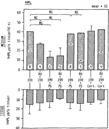
HPL. Irrespective of the composition of the incubation medium, neither the tissue concentration of HPL nor its production rate
HPL mean * SD Q. I RU I RU I RU 199 199 199 199 199 199 199 199 + + Cort. Cort fij
Fig. 1. Trophoblastic production rate and tissue concentration of HPL, RU
(RU486), PG (progesterone), PS (pooled maternal pregnancy serum), Cort (cortisol), 199 (medium 199 from Gibco). Numbers within the bars indicate the number of experiments run in duplicate.
P.Biscbof, M.T.Sizonenko and W.L.Heirmann Decidual Prl flHCG mean ± SD o RU 199 199 199 199 199 199 199 199 PG PG PS PS Cort. Cort. en en 0.5 1 -1.5 mean ± SD US p<0,005 P<0,03 p<0.03
Fig. 2. Trophoblastic production rate and tissue concentration of (3HCG
(abbreviations as Figure 1). Trophoblastic PAPP-A mean ± SD 0 -, 100-199 CVxNN • 1 NS 199 199 PG 199 PG 199 PS 199 PS 199 Cort. 199 Cort.
Fig. 4. Decidua] production rate and tissue concentration of Prl
(abbreviations as Figure 1). Decidual PAPP-A 125 100 75 50 25 -0 0 25 5 0 -75 mean ± SD p<0.005 p<0.03 ^3 F7^ K^l ^ ^ ^
ll
J10: Rv;^ 199 199 199 PG PG 199 + PS RU 199 199 199 PS Cort. Cort. i \ \ \ \ ] 1\\\M K\\\1 P< 0.005 p<0.003Fig. 3. Trophoblastic production rate and tissue concentration of PAPP-A
(abbreviations as Figure 1).
Fig. 5. Decidual production rate and tissue concentration of PAPP-A
In-vitro effects of RU486
were significantly changed (Figure 1).
&HCG. RU486 induced a significant (p <0.05) decrease in the production rate and in the tissue concentration (p <0.005) of /3HCG as compared with control media (Figure 2). These ef-fects could be reversed by the addition of 500 ng/ml progesterone and cortisol whereas neither progesterone nor cortisol alone had a significant effect. The addition of PS to the incubation medium significantly increased the production rate of j3HCG over control medium (p <0.03). The addition of RU486 (100 ng/ml) to PS-supplemented media induced a significant (p < 0.03) decrease of /3HCG as compared with PS alone (Figure 2).
PAPP-A. The tissue concentration of PAPP-A did not change significantly irrespective of the composition of the incubation media. The secretion rate of PAPP-A, however, was significantly (p <0.05) inhibited by RU486 (100 ng/ml) as compared with anti-progesterone-free media (Figure 3). Progesterone (500 ng/ml) but not cortisol (500 ng/ml) did reverse this in-hibitory effect whereas progesterone or cortisol alone did not alter PAPP-A production. As for /3HCG, media supplemented with PS significantly (p <0.03) increased the production rate of PAPP-A as compared with control media, whereas RU486 significantly (p <0.05) antagonized this effect (Figure 3). Decidua
Prl. The production rate of Prl was similar in the different incu-bation media, but the tissue concentration of Prl was significantly (p <0.05) increased over controls by RU486 (100 ng/ml). This effect could be reversed by the addition of progesterone (Figure 4). PS-supplemented media induced a significant increase in Prl tissue concentration when compared with controls (p <0.03), but RU486 could not reverse this effect (Figure 4).
PAPP-A. PS-supplemented media significantly increased the pro-duction rate (p < 0.005) and the tissue concentration (p <0.005) of PAPP-A. Both effects could be reversed by RU486 (Figure 5). In all other media, the production rate of PAPP-A remained similar to controls.
Discussion
It is well-documented that the decidua produces Prl (Riddick and Kusmik, 1977; de Ziegler and Gurpide, 1982) and that this pro-duction can be stimulated by progesterone (Daly et al., 1983). The present data show that RU486 increases the concentration of Prl in decidual tissue, suggesting that this potent progesterone antagonist interferes with the secretion rather than the produc-tion of Prl. However, under our experimental condiproduc-tions, pro-gesterone did not stimulate the production of Prl, whereas it did antagonize the effects of RU486. An obvious explanation for this apparent discrepancy is that progesterone receptors in the decidua were probably saturated with endogenous progesterone, but that RU486 could displace bound progesterone from its receptor because it has greater affinity for the receptor than progesterone itself. This could be overcome only by increasing the concentra-tion of progesterone.
As shown previously, PAPP-A is produced by trophoblastic as well as decidual explants. PS but not serum from non-pregnant women stimulates the production of PAPP-A from both tissues (Bischof et al., 1984a). The factor which stimulates the pro-duction of PAPP-A could well be progesterone since in both tissues the secretion of PAPP-A was inhibited by RU486 and since progesterone, but not cortisol, was able to reverse these effects. These results are in good agreement with previous obser-vations, implying indirectly that progesterone was a trigger of
endometrial PAPP-A production (Bischof et al., 1982, 1984b; Sjoberg et al., 1984).
Finally, the inhibition of trophoblastic /3HCG production seems to be specific since RU486 did not modify the production of HPL. This is in good agreement with the in-vivo effects of RU486. It is not clear, however, if this effect was due to a metabolite of RU486 or to the anti-gestagenic or anti-glucorticoid properties of RU486 since both progesterone and cortisol were able to over-come the effects of RU486. The trophoblastic production of HCG has long been considered as independent of any feedback mech-anism. Recently, however, it was demonstrated that in long-term cultures of trophoblastic explants, progesterone inhibited the pro-duction of HCG (Wilson et al., 1984). These results are in con-trast to the present observations. It remains to be determined if long-term cultures of trophoblast behave differently from short-term cultures and if cortisol and not progesterone is the main regulator of placental HCG production.
Taken together, these results also suggest that the trophoblast of first trimester pregnancies contains an active progesterone receptor, a notion which is still controversial (Coulam and Spels-berg, 1983; Younes et al., 1981).
In conclusion, the present results tend to suggest that the aborti-facient effect of RU486 in humans could at least partially be due to an inhibition of the production of HCG and/or PAPP-A.
Acknowledgements
The authors wish to thank Mrs C Gruffat and M.Mendez for their skilful assistance and Mrs D.Roiron for typing the manuscript. This work was supported by grant No. 3 909.082 from the Swiss National Fund for Scientific Research.
References
Bischof.P., Haenggeli.L., Sizonenko,M.T., Herrmann.W.L. and Sizonenko.P.C. (1981), A radioimmunoassay for the measurement of pregnancy-associated plasma protein-A (PAPP-A) in humans. Biol. Reprod , 24, 1076. Bischof.P., DuBerg.S., Schindler.A.M., Obradovich.D., Weil.A., Faigaux.R.,
Herrmann.W.L. and Sizonenko.P.C. (1982), Endometrial and plasma con-centrations of pregnancy-associated plasma protein-A (PAPP-A). fir. J. Obstct. Gynaecol, 89, 701.
Bischof.P., DuBerg.S., Sizonenko.M.T., Schindler.A.M., Beguin.F., Herrmann, W.L. and Sizonenko.P.C. (1984a), ln-vitro production of pregnancy-associated plasma protein-A (PAPP-A) by human decidua and trophoblast. Am. J. Obstet. GynecoL, 148, 13.
Bischof.P., Schindler.A.M., Umer.F., Mensi.N., Herrmann.W.L. and Sizonenko, P.C. (1984b), Pregnancy-associated plasma protein-A (PAPP-A) concentra-tion in uterine fluid and unmunohistochemical localizaconcentra-tion in the endometrium. Br. J. Obstet. Gynaecol, 91, 863.
Bischof.P., Schindier.A.M., Wyss.R., Herrmann.W.L. and Sizonenko.P.C. (1985), Progesterone dependence and extratrophoblastic origin of pregnancy-associated plasma protein-A (PAPP-A) in early pregnancy. Arch. Gynecol, m press.
Coulam,C.B. and Spelsberg.T.C. (1983), The placenta as a target tissue for steroids. Troph. Res., 1, 249.
Daly.D.C, Maslar.I.A. and Riddick,D.H. (1983), Term decidua response to estradiol and progesterone. Am. J. Obstet. Gynecol., 145, 679.
de Ziegler.D. and Gurpide,E. (1982), Production of Prl by cultures of cells from human decidua. J. Qin. Endocrinol. Metab., 55, 511.
Gaillard.R.D., Riondel.A., Muller.M.F., Herrmann.W. and Baulieu.E.E. (1984), RU486: a steroid with antiglucosteroid activity that only disinhibits the human pituitary-adrenal system at a specific time of day. Proc. Nail. Acad Sci. USA,
81, 3879.
Healy.D.L., Baulieu.E.E. and Hodgen.G.D. (1983), Induction of menstruation by an antiprogesterone steroid (RU486) in primates: site of action, dose-response relationships and hormonal effects. Fertil. Steril., 45, 253.
Herrmann.W.L., Wyss.R., Riondel.A., Philibert.D., Teutsch.G., Sakiz.E. and Baulieu.E.E. (1982), The effect of an antiprogesterone steroid in women: in-terruption of the menstrual cycle and early pregnancy. C.R. Acad. Sci. (Ill),
294, 933.
Herrmann.W.L., Schindler.A.M., Wyss.R. and Bischof.P. (1985), Effects of the antiprogesterone RU486 in early pregnancy and during the menstrual cycle. Proceedings of an International Symposium on Future Aspects in Contraception,
P.Bischof, M.T.Sizonenko and W.L.Hemnann
Runnebaum.B., RabeJ., Kiesel.L. (eds.) (1985), MTP Press Ltd., pp.243-27O. Philibert.D., Deraedt.R., Teutsch.G., Tournemine.C. and Sakiz.E. (1982), RU486 a new lead for steroid antihormones. Program of the 64th Annual Meeting of The Endocrine Society San Francisco, California (Abstract 668). Riddick.D.H. and Kusmik.W.F. (1977), Decidua: a possible source of amniotic
fluid prolactin. Am. J. Obstet. Gynecol., 127, 187.
SjobergJ., WahlstromJ. and Seppala,M. (1984), Pregnancy-associated protein-A in the human endometrium is dependent on the effect of progesterone. J. Clin. Endocrinol. Metab., 58, 359.
Wilson,E.A., Jaward.M.J. and Powell,D.E. (1984), Effect of estradiol and pro-gesterone on human chorionic gonadotropin secretion in vitro. Am J. Obstet. Gynecol., 149, 143.
Younes.M.A., Besch.N.F. and Besch,P.K. (1981), Estradiol and progesterone binding in human term placental cytosol. Am. J. Obstet. Gynecol., 141, 170. Received on 27 August 1985; accepted on 2 October 1985
![Fig. 4. Decidua] production rate and tissue concentration of Prl (abbreviations as Figure 1)](https://thumb-eu.123doks.com/thumbv2/123doknet/14885527.646603/2.915.63.426.94.552/fig-decidua-production-rate-tissue-concentration-abbreviations-figure.webp)
